Collagen IV immunohistochemistry and autoimmune bullous diseases: defining the location of the subepidermal bullae
Dear Editor,
Bullous pemphigoid (BP) is the most common autoimmune blistering disease (ABD).1 Compatible clinical, histopathologic, and immunological criteria are necessary to differentiate among ABD.1,2 Particularly, among subepidermal types, the level of cleavage of the blister and the exact target of the pathogenic autoantibodies (pointed out by deposition of the immunoreactions) are necessary to make a correct diagnosis.1
BP and epidermolysis bullosa acquisita (EBA) may resemble clinical and histopathologically. Further studies, such as Indirect Immunofluorescence (IIF) and/or Direct Immunofluorescence (DIF) in a patient’s salt-split skin, are needed to distinguish between these two entities.1,2 However, techniques require fresh frozen tissue and sophisticated laboratory equipment.3
Immunohistochemistry (IHC) has also been used to study the location of basal membrane components and the presence of immunoreactions in ABD, particularly in BP.3-5 Collagen IV is a main component of the dermal-epidermal junction, mostly found at the lamina densa.5 Performing collagen IV IHC on formalin-fixed paraffin-embedded skin biopsies offers an inexpensive means to distinguish among subepidermal ABD.3-6
We aimed to evaluate the usefulness of collagen IV IHC in BP cases attended at our center. Therefore, a retrospective, observational study that included all BP patients diagnosed between January 2000 and June 2020 was conducted.
The diagnosis of BP cases had been based on the combination of criteria encompassing the presence of compatible clinical features, typical histopathological findings, and the presence of a compatible DIF pattern (C3, IgG, IgM, and/or IgA linear deposits along the dermo-epidermal junction) and/or positive IIF/Enzyme-Linked Immunosorbent Assay (ELISA) (detection of circulating IgG anti-epidermal basement membrane antibodies by IIF microscopy studies using NaCl-separated normal human skin and/or detection of anti- BP180 and/or anti-BP230 IgG antibodies by ELISA [MESACUP anti-skin profile test]).
All cases with a confirmed diagnosis were included if, at least, three out of the four criteria were present and compatible: clinical, histopathological, serological, including IIF and/or ELISA and DIF. Skin biopsy with H&E stain and DIF was performed in all patients included in the study and results of at least one immunological study (IIF and/or ELISA) had to be present.
To assess the level of the blister and to differentiate BP from other subepidermal bullous diseases, type IV collagen immunohistochemical staining was performed in formalin-fixed paraffin-embedded tissue sections (Diagnostic Biosystems Mob229/PDM276, Mouse AntiHuman Collagen IV, purified, polyclonal antibody, IgG1, dilution 1/10-1/40). Staining at the dermal portion of the blister (floor) was considered to be compatible with BP.
From a total of 257 BP cases collected, 194 patients,in which collagen IV IHC was performed, were included (Table 1); in the remaining 63, this technique was not yet available at our center.
Epidemiological and clinicopathological characteristics of included collagen IV IHC BP cases.
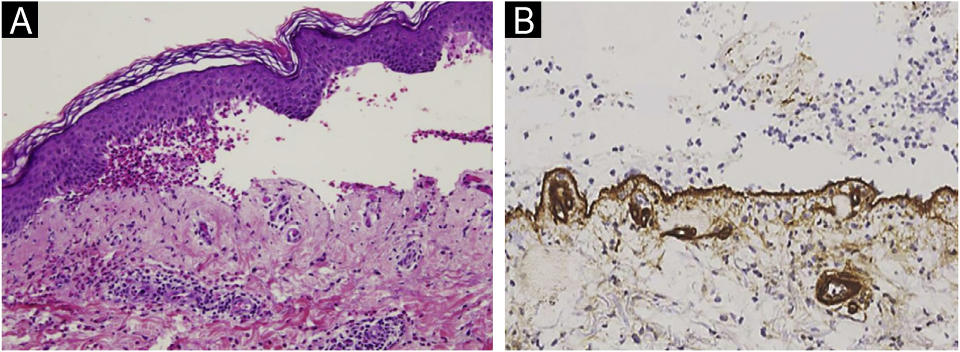
Subepidermal blistering accompanied by the characteristic inflammatory infiltrate was present in all patients. In 67.5% the intensity of the eosinophilic infiltrate was very intense (>21 eosinophils/ × 40 high power field) (Fig. 1A). 93.7% presented linear deposits of IgG and/or C3 at the basal membrane in DIF. In 193 of them, collagen IV was detected on the floor of the blister (Fig. 1B).

(A) Histopathological image of the blister from a BP patient (Hematoxylin & eosin, ×10): subepidermal blistering with an eosinophilic inflammatory infiltrate. (B) Collagen IV immunohistochemistry in BP (×20). Note the location of collagen IV (brown line) on the floor of the blister.
When comparing with EBA cases (confirmed by the presence of autoantibodies against collagen VII detected by ELISA [MESACUP anti-skin profile test]) attended in the same study period (n = 5), collagen IV was stained at the roof of the blister (Fig. 2). Salt-split skin DIF, probably due to technical limitations, lead to distinguish between BP and EBA in only 1 case.

Collagen IV immunohistochemistry performed in an EBA patient (×10). Positivity is observed in the roof of the blister and in dermal structures that contain collagen IV, such as vessel walls.
In comparison with salt-split DIF, IHC is a simple and reproducible technique. It can be performed at the formalinfixed paraffin-embedded tissue sections of the blister, without the need for additional processing.6-8 By demonstrating the location of collagen IV by IHC, the level of cleavage of blister may be established and, therefore, it can be helpful in the diagnosis of subepidermal ABD.7,8
Collagen IV is located at the lamina densa. All the structures related to the hemidesmosome and lamina lucida [including BP Antigen 1 (BPAG1/BP230) and BP Antigen 2 (BPAG2/BP180), laminin 1 y, laminin 332, cx4[36 integrin] are located above lamina densa; collagen VII, however, is placed inferior to collagen IV.8 Therefore, when performing collagen IV IHQ in subepidermal ABD, except for EBA and bullous systemic lupus erythematous, the stain will be present on the floor of the blister (Table 2).9,10
This technique has the limitation that only the location of collagen IV is being marked and the presence of immunoreactants or deposits cannot be evaluated. In the salt-split skin technique, the level of cleavage is obtained at the lamina lucida and, through DIF, the presence of immunodeposits can be assessed.
This study presents several limitations due to the lack of a control group, the scant number of EBA cases andthe absence of other pemphigoid group and bullous disease patients.
Although widely known,3-5,7,8 after reviewing the published literature and through our observations, we consider that collagen IV IHC should gain visibility and presence in the diagnostic process of ABD.9,10 We highlight this stain as a perfect first approach in the study of a blister, in general, and, especially, in the suspicion of ABD. Furthermore, gold standard techniques (DIF and/or salt-split skin DIF/IIF) results may be better interpreted along with collagen IV IHC findings.3-5,7,8,10
References
- 1 Borradori L, Ven Beek N, Feliciani C, Tedbirt B, Antiga E, Bergman R, et al. Updated S2k guidelines for the management of bullous pemphigoid initiated by the European Academy of Dermatology and Venereology (EADV). J Eur Acad Dermatol Venereol. 2022;36:1689-704.
- 2 Meijer JM, Diercks GFH, de Lang EWG, Pas HH, Jonkman MF. Assessment of diagnostic strategy for early recognition of bullous and nonbullous variants of pemphigoid. JAMA Dermatol. 2019;155:158-65.
- 3 Yeol Lee H, Ham SP, Choi YW, Park HJ. The value of Type IV collagen immunohistochemical staining in the differential diagnosis of autoimmune subepidermal bullous diseases. Acta Dermatovenerol Croat. 2018;26:133-8.
- 4 Pardo RJ, Penneys NS. Location of basement membrane type IV collagen beneath subepidermal bullous diseases. J Cutan Pathol. 1990;17:366-441.
- 5 Sasaki T, Amano S, Nishiyama T, Nakayama Y, Burgeson RE, Nakajima H. Expression of laminin 5, and collagen IV and VII in bullous pemphigoid skin. Arch Dermatol Res. 1998;290:283-5.
- 6 Abreu-Velez AM, Howard MS. Collagen IV in normal skin and in pathological processes. N Am J Med Sci. 2012;4:1-8.
- 7 García-Díez I, Martínez-Escala ME, Ishii N, Hashimoto T, Mascaró-Galy JM, Pujol RM, et al. Usefulness of a simple immunohistochemical staining technique to differentiate antip200 pemphigoid from other autoimmune blistering diseases: a report of 2 cases. Actas Dermosifiliogr. 2017;108:e1-5.
- 8 Lau I, Goletz S, Holtsche MM, Zillikens D, Fechner K, Schmidt E. Anti-p200 pemphigoid is the most common pemphigoid disease with serum antibodies against the dermal side by indirect immunofluorescence microscopy on human salt-split skin. J Am Acad Dermatol. 2019;81:1195-7.
- 9 El-Domyati M, Abdel-Wahab H, Ahmad H. Immunohistochemical localization of basement membrane laminin 5 and collagen IV in adult linear IgA disease. Int J Dermatol. 2015;54:922-8.
- 10 Halagovec A, Héjj F, Baranová Z. Fibronectin, interstitial collagens and type IV collagen in dermatitis herpetiformis. Cas Lek Cesk. 1996;135:273-6.