PHACE syndrome: report of five cases
Dear Editor,
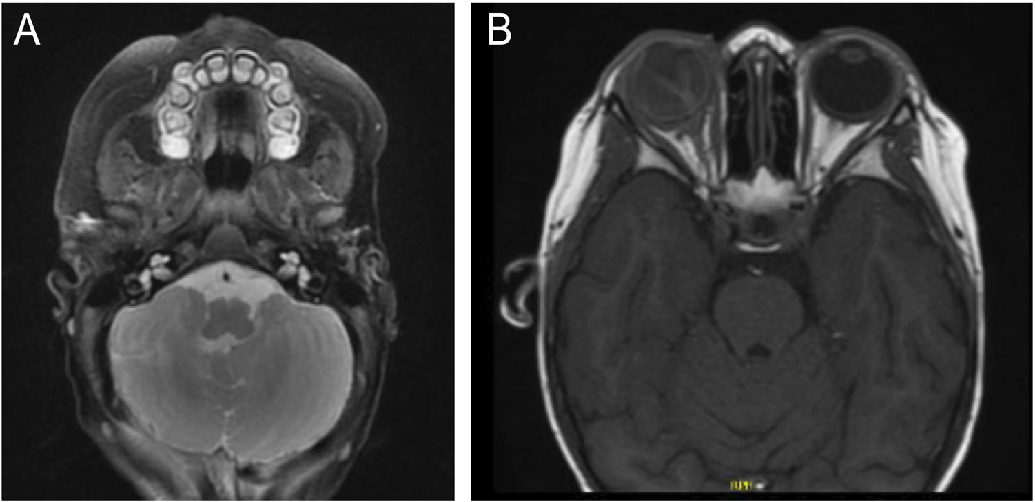
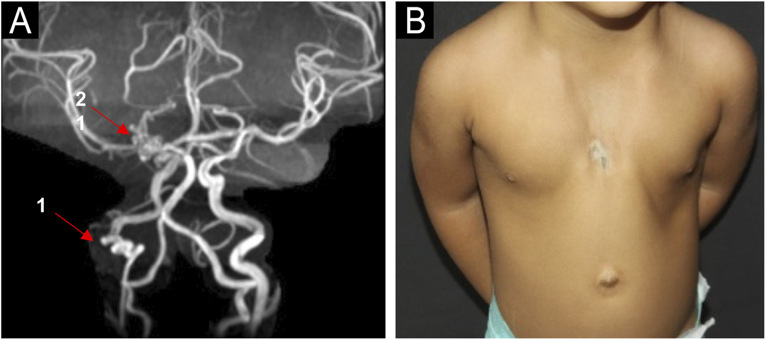
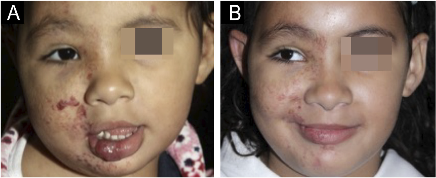
Infantile hemangioma (IH) is the most frequent benign tumor in the pediatric population, occurring in up to 10% of children less than 1-year and usually involving the head and neck region, with female predominance.1,2 PHACE syndrome (Posterior fossa malformations, Hemangiomas, Arterial anomalies, Coarctation of the aorta/Cardiac defects, and Eye abnormalities) is a rare neurocutaneous syndrome characterized by the association of infantile hemangioma larger than 5 cm on the face, scalp or cervical region and structural anomalies of the brain, cerebral vasculature, eyes, sternum, heart, and/or aorta.1,3 A modified acronym, PHACES, has been suggested to include Sternal abnormalities.1,3 The pathogenesis of PHACE syndrome remains unknown, and there is no evidence of genetic abnormalities contributing to its development. PHACE syndrome also seems to have female predominance, and it occurs in 2%‒3% of all IH cases and 20% of patients with large or segmental facial hemangiomas.1,4 Five patients with PHACE syndrome were followed at the Pediatric Dermatology Outpatient Clinic of Hospital das Clínicas, Faculdade de Medicina, Universidade de São Paulo, for an average follow-up period of five years. All the patients were female and had a large facial infantile hemangioma that developed at birth or soon thereafter. Prematurity was observed in one patient. Family history of PHACE syndrome or parents’ consanguinity was not observed. Four of five patients presented with brain and cardiac anomalies. Ocular and sternal abnormality was identified in two and one patient, respectively. All patients were treated with oral and/or topical beta-blockers. Patients' features and clinical findings are described in Table 1. Magnetic Resonance Imaging (MRI) and Magnetic Resonance Angiography (MRA) findings are presented in Figs. 1 and 2A, respectively. Fig. 2B illustrates sternal aplasia and thoracic cutis aplasia. The evolution of the lesions after oral propranolol treatment is shown in Figs. 3 and 4.
(A) MRI showing hypoplasia of the right cerebellum (case 1). (B) MRI showing right retinoblastoma (case 2).

(A) MRA showing tortuous: 1) Right internal carotid artery and 2) right posterior cerebral artery and (B) Sternal aplasia and thoracic cutis aplasia (case 3).

Female patient with right hemiface infantile hemangioma and PHACE syndrome (case 4). (A) At 2-years and 4-months of age after 17-months of treatment with oral propranolol (2 mg/kg/day). (B) At 7-years and 1-month of age, showing good response of the infantile hemangioma after 17-months of treatment with oral propranolol (2 mg/kg/day) and 2-years and 4-months of topical timolol.
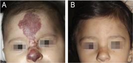
Female patient with frontonasal infantile hemangioma and PHACE syndrome (case 5). (A) At 1-month and 29-day of age. (B) At 1-year and 6-months of age, showing good response of the infantile hemangioma after 16-months of treatment with oral propranolol (2 mg/kg/day).
The five cases described in this report illustrate the spectrum of features seen in PHACE syndrome, highlighting the heterogeneity of malformations associated with the condition.
The most common extracutaneous involvement is structural and vascular in the brain, followed by cardiac defects.1,3,5 The combined incidence of structural cerebral and cerebrovascular anomalies is greater than 50%.2 A variety of structural cardiac abnormalities have been reported in approximately one-third of PHACE patients.2 In our series, the majority of patients had brain and cardiac anomalies, as observed in the literature.5
Ocular anomalies are relatively rare in PHACE syndrome,4 however, we had 2 patients with ophthalmological abnormalities. One of these patients presented with a retinoblastoma, a rare malignant ocular tumor, which has been previously described in association with PHACE syndrome in two case reports.4,6
One of our patients presented with a vestibular schwannoma. The occurrence of this tumor in PHACE syndrome patients is a rare finding, with scarce data in the literature.7
Speech delay is a common finding1,8 and we identified in one of our patients.
Current literature suggests propranolol is effective in treating IH associated with PHACE syndrome.3,9 Although the resolution of IH is well-documented during treatment with oral propranolol, its use in PHACE syndrome in patients who have cardiac and vascular malformations is controversial due to the risk of stroke.1,3 Four of our five patients were treated with oral propranolol, and none experienced cerebrovascular complications. Careful examination and management by a multidisciplinary team are crucial to providing better care for these patients.9 The multidisciplinary group comprises neuroradiology, neurosurgery, neurology, cardiology, cardiothoracic surgery, dermatology, otorhinolaryngology, hematology-oncology, and plastic surgery.1 The initial physical examination of all suspected cases should include an assessment for ocular and sternal midline abnormalities, in addition to neurological, cardiac, and vascular evaluations.1 A screening echocardiogram should be executed and if abnormalities are identified, cardiac MRI and MRA are recommended.1,10 Gadolinium MRI and MRA of the brain, neck, and aortic arch should also be implemented during follow-up for all suspected cases of PHACE syndrome. MRI is insufficient to detect all arterial abnormalities, requiring an MRA.1
We emphasize the importance of early recognition of the syndrome and evaluation of associated abnormalities, enabling effective interventions to prevent future complications. Further research into the long-term outcomes and better treatment strategies for this rare condition is necessary to improve both the quality of care and quality of life for affected children.
☆
Financial support
Research data availability
Does not apply.
References
- 1 Rotter, A, Samorano, LP, Rivitti-Machado, MC, Oliveira, ZNP, Gontijo, B. PHACE syndrome: clinical manifestations, diagnostic criteria, and management. An Bras Dermatol. 2018; 93:405-11.
- 2 Roganović, J, Adams, D. PHACES syndrome - case report and literature review. Coll Antropol. 2009;33:311-14.
- 3 Bellaud, G, Puzenat, E, Billon-Grand, NC, Humbert, P, Aubin, F. PHACE syndrome, a series of six patients: clinical and morphological manifestations, propranolol efficacy, and safety. Int J Dermatol. 2015;54:102-7.
- 4 Valdivielso-Ramos M, Torrelo A, Martin-Santiago A, Campos M, Conde E, de la Cueva P, et al. Infantile hemangioma with minimal or arrested growth as the skin manifestation of PHACE syndrome. Pediatr Dermatol. 2018;35:622-27.
- 5 Haggstrom AN, Garzon MC, Baselga E, Chamlin SL, Frieden IJ, Holland K et al. Risk for PHACE syndrome in infants with large facial hemangiomas. Pediatrics.2010;126:e418-26.
- 6 Fernández-Ibieta, M, López-Gutiérrez, JC. Lymphatic malformation, retinoblastoma, or facial cleft: atypical presentations of PHACE syndrome. Case Rep Dermatol Med. 2015;2015:487562.
- 7 Viera Plasencia, A, Amador, L, Biehler, J, Kantor, J. Manifestation of a vestibular schwannoma in a patient with PHACE syndrome. Cureus. 2025;17:78946.
- 8 Martin KL, Arvedson JC, Bayer ML, Drolet BA, Chun R, Siegel DH. Risk of dysphagia and speech and language delay in PHACE syndrome. Pediatr Dermatol. 2015;32:64-9.
- 9 Winter PR, Itinteang T, Leadbitter P, Tan ST. PHACE syndrome - clinical features, aetiology and management. Acta Paediatr. 2016;105:145-53.
- 10 Garzon MC, Epstein LG, Heyer GL, Frommelt PC, Orbach DB, Baylis AL et al. PHACE syndrome: consensus-derived diagnosis and care recommendations. J Pediatr. 2016;178:24-33.