Prosthetic Valve Endocarditis: A Multidisciplinary Challenge
Keywords
Endocarditis; Multimodal Imaging; Heart Valve Prosthes
Palavras-chave
Endocardite; Imagem Multimodal; Próteses Valvulares Cardíacas
Keywords
Endocarditis; Multimodal Imaging; Heart Valve Prosthes
Palavras-chave
Endocardite; Imagem Multimodal; Próteses Valvulares Cardíacas
Clinical case
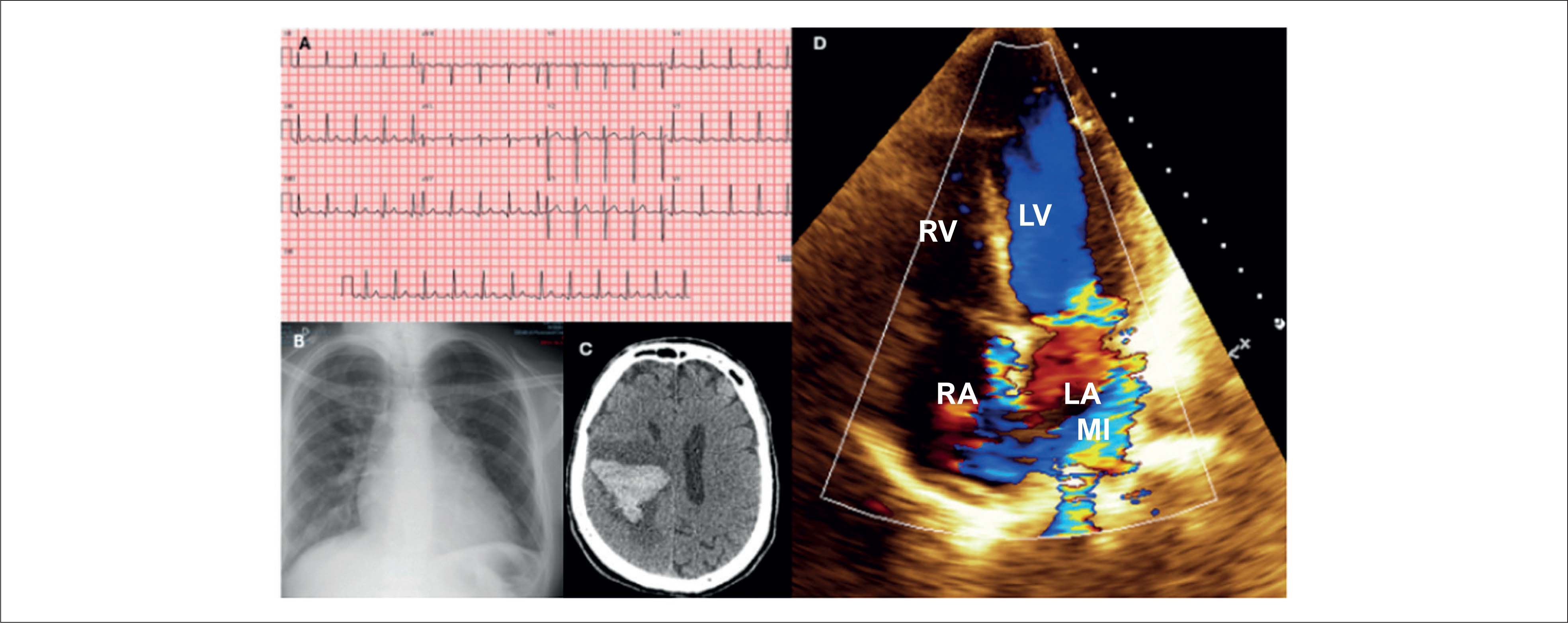
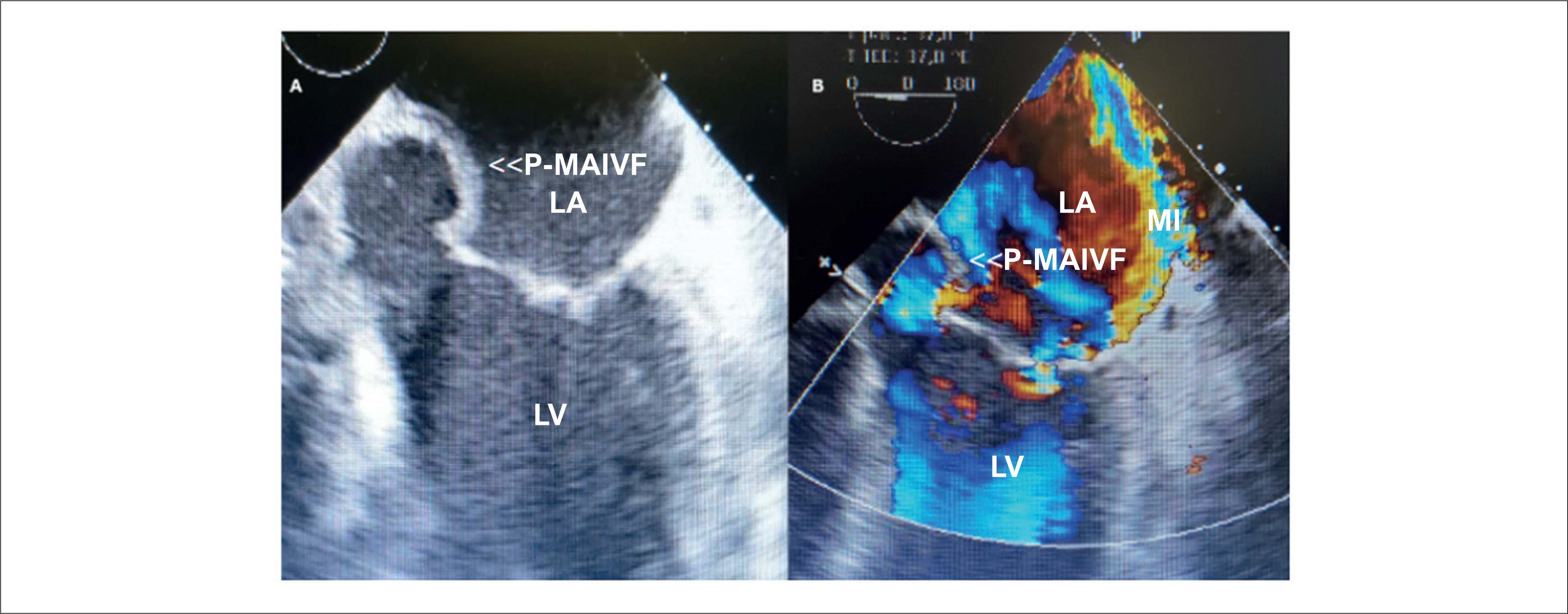
We report the case of a 55-year-old male patient who was admitted to the hospital in July 2019 with sudden left hemiplegia, mitral systolic murmur 4+, and aortic systolic and diastolic murmurs 2+. He had a history of aortic valve replacement in 2015, with a mechanical prosthetic aortic valve (PAV) due to infective endocarditis (IE). Cranial computed tomography (CT) revealed a right frontoparietal hematoma ( Figure 1C ), and blood tests revealed hyperglycemia and elevated C-reactive protein. His electrocardiogram (ECG) showed sinus rhythm with repolarization abnormalities ( Figure 1A ), and a chest X-ray showed cardiomegaly +/4 ( Figure 1B ). Transthoracic echocardiography (TTE) showed preserved function, thickening of the mobile elements of the mechanical PAV, mild double aortic lesion, pseudoaneurysm of the mitral-aortic intervalvular fibrosa, and important eccentric mitral regurgitation ( Figure 1D ). Blood cultures were requested, and ceftriaxone 4 g/d and teicoplanin 800 mg/d were started. Transesophageal echocardiography (TEE) showed vegetations on the ventricular side of the mechanical PAV leaflets and periprosthetic abscess ( Figure 2 ). Group B Streptococcus agalactiae was identified, and surgical treatment was indicated after 4 weeks due to cardiovascular stability and hemorrhagic stroke.

(A) ECG; (B) Posteroanterior chest X-ray; (C) Cranial CT; (D) Transthoracic echocardiogram. LA: left atrium; LV: left ventricle; MI: mitral insufficiency; RA: right atrium; RV: right ventricle.

Transesophageal echocardiogram. LA: left atrium; LV: left ventricle; MI: mitral insufficiency; P-MAIVF: pseudoaneurysm of the mitral-aortic intervalvular fibrosa.
On day 22 of hospitalization, the patient presented pulmonary congestion and respiratory failure. Repeated TTE revealed partial loosening of the mechanical PAV and severe periprosthetic leak. Emergency surgery was performed to clean and replace the mechanical PAV with a biological PAV, without mitral valve approach. Culture of the explanted prosthesis revealed no bacterial growth.
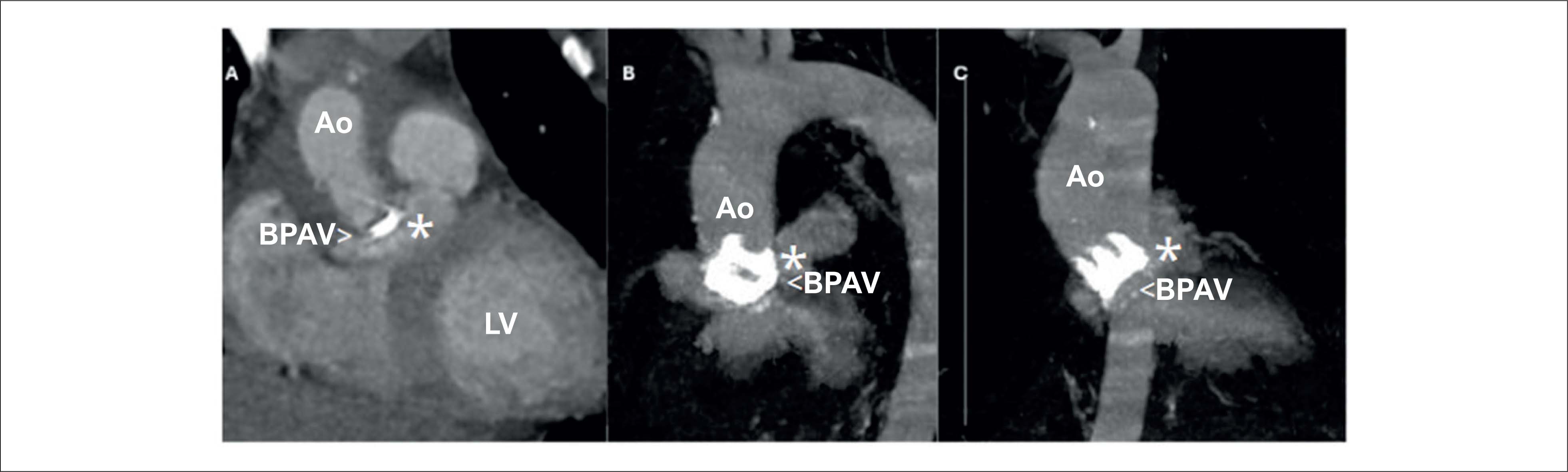
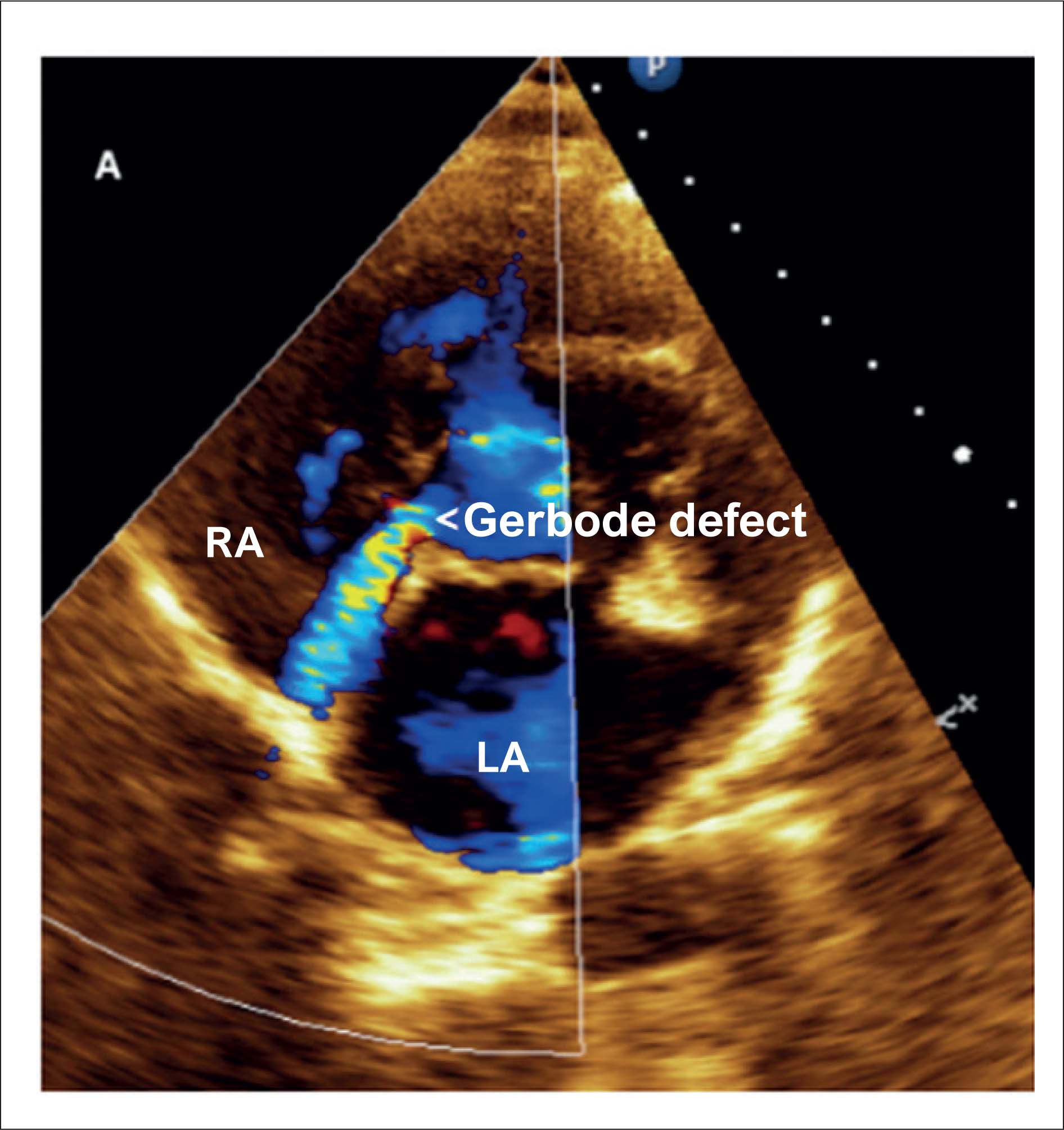
The postoperative period was complicated, with fungemia due to Candida guilliermondii . On postoperative day 14, TTE and TEE identified a periprosthetic abscess with the presence of a fistula ( Figure 3 ). CT angiography showed that the biological PAV had periaortic and perivalvular contrast extravasation ( Figure 4 ). On postoperative day 18, a diastolic murmur appeared, and a new TTE showed a fistula from the ventricular outflow tract to the right atrium, with systolic flow, that was 2 mm in diameter (Gerbode defect), as shown in Figure 5 .

(A, B, and C) Transthoracic echocardiogram; (D) Transesophageal echocardiogram. BPAV: biological prosthetic aortic valve; LA: left atrium; LV: left ventricle. Asterisk (*) indicates abscess.

T angiography of the aorta. Ao: ascending aorta; BPAV: biological prosthetic aortic valve; LV: left ventricle. Asterisk (*) indicates perivalvular leak.

The heart team contraindicated further surgery, maintaining meropenem, teicoplanin and caspofungin for 42 days. After repeat TEE without any change in progression, inflammatory tests and negative blood cultures normalized, and the patient was discharged from the hospital.
The patient was readmitted with fever 30 days later, and the previous antibiotic regimen was restarted, with no infectious focus and negative blood cultures. Positron emission tomography with computed tomography (PET/CT) with 18FDG showed focal glycolytic hypermetabolism in the biological PAV and abscess surrounding the ascending aorta ( Figure 6 ). Surgery was again contraindicated, and amoxicillin 3 g/day was chosen for an indefinite period.

18FDG PET/CT. BPAV: Biological prosthetic aortic valve. Asterisk (*) indicates glycolytic hypermetabolism with maximum standardized uptake value (SUVmax) = 11.2.
In November 2019, 18FDG PET/CT showed no uptake, suggesting resolution of prosthetic valve endocarditis (PVE). At the last evaluation, in July 2023, the patient was stable, without infectious complications. ETT showed dilation of the left cavities, periprosthetic fistula, Gerbode defect, moderate pulmonary hypertension, and biological PAV with maintained gradients.
Discussion
The incidence of PVE varies from 1% to 6% per year; it is more common in biological prostheses, and it represents 20% to 30% of IE cases.1 The diagnosis of PVE still poses challenges, leading to delays in management, which is sometimes decided on an almost individual basis.2
Asymptomatic neurological symptoms or neuroimaging findings may indicate embolism, and they can be the first manifestation of IE, associated with higher mortality and indication for early surgery.1 , 3 , 4 Blood cultures were collected and, because it was a case of late PVE (1 year after the first surgery), coverage against Gram-positive pathogens was considered.1 , 5
The reported case was diagnosed by the modified Duke criteria,1 with nonspecific ECG, but PVE can manifest with atrioventricular block. Laboratory findings assisted in risk stratification and evaluation of therapeutic response. The most frequent agents in late PVE are as follows: S. aureus , 22.9%; coagulase negative staphylococci, 17.2%; and viridans streptococci, 16%.3 S agalactiae was isolated, which rarely causes PVE and, unlike cases of late PVE, progresses rapidly with valve destruction, embolism, 20% mortality, and need for surgery in 40% to 50% of cases.6
The specificity of TTE for PVS is 96%, and the sensitivity ranges from 40% to 50%, lower than in native valve IE, most often involving the prosthetic ring (mainly in mechanical prostheses).1 , 7 The European guideline indicates TEE when TTE is inconclusive or in suspected complications, whereas the United States guideline advises that it should always be performed.1 , 8 The sensitivity of TEE ranges from 82% to 96%, and the specificity is 86%.1 , 7 , 8 On day 3 of hospitalization, TEE revealed a pseudoaneurysm of the mitral-aortic intervalvular fibrosa, which can lead to coronary compression, pericardial effusion, and fistulization. The sensitivity on TEE and TTE is 90% and 43%, respectively, with indication for surgery.9
On day 22 of hospitalization, the patient presented a mechanical complication, which is responsible for 30% of urgent surgeries, as well as congestive heart failure, abscess and persistent fever. Failure to perform surgery is a marker of worse prognosis.1 , 3 , 5
Postoperative mortality ranges from 6% to 29% in observational studies, and it is associated with age, urgent or emergency surgery, prior cardiac surgery, prosthetic valve, multivalvular involvement, perivalvular abscess, and kidney failure.10 , 11
After fungemia, TEE and CT showed a periaortic abscess with fistula and Gerbode defect, which can be found rarely in complications after cardiac surgery or IE.7 , 12 Since the prosthesis culture was negative, a new PVE was suspected, given that fungi are a cause of acute PVE.1 , 3 , 5
CT became more important in the latest revision of the Duke criteria and in the European guideline, going on to be considered a major criterion for diagnosis, with a sensitivity of 88% and a specificity of 93%, surpassing TEE in the identification of paravalvular abscess.1 TEE, on the other hand, has a specificity of 100% for paravalvular leak. Nuclear magnetic resonance imaging has limited use compared to CT, since prosthetic valves impair assessment.
In spite of the indication for reoperation, the patient improved and was discharged, being readmitted after 4 weeks. Patients with surgical indication who did not undergo surgery, usually due to clinical complications, present reinfection and recurrence of 5% during 1-year follow-up. When comparing mortality between operated and non-operated patients, recurrence is 9% and 34%, respectively.13
In cases of inconclusive diagnosis, 18FDG PET/CT can be requested. Positive findings are predictors of complications, recurrence of IE, and new embolic events. It is important to differentiate uptake in the recent postoperative period (diffuse and homogeneous) from PVE (intense, heterogeneous, focal, or multifocal), with sensitivity of 54% to 87% and specificity of 56% to 93%, reclassifying the Duke criteria on admission and increasing sensitivity to 82% to 96% without loss of specificity.1 , 14
Prolonged antibiotic therapy is recommended in cases of IE caused by Candida or as an alternative in patients with surgical indications who did not undergo surgery.1 In a study with 31 patients who did not undergo surgery and received prolonged oral antibiotic therapy, the event-free survival rate at 1 year was 74%; the overall survival rate was 84.3%; and the recurrence rate was 12% (mean after 111 days).15 The absence of uptake on the control PET/CT allows for safe suspension of treatment with a low risk of complications.14
The following are associated with worse prognosis in IE: advanced age, diabetes mellitus, S. aureus , fungi, acute PVE, congestive heart failure, stroke, abscesses, and failure to perform surgery when indicated.10 , 16 The patient was at high risk, with an estimated mortality rate of 60% in 6 months. In fungal IE, the prognosis is significantly worse, and, even with early diagnosis and appropriate treatment, mortality rates of 10% to 75% have been reported.
Despite the risk factors and various complications related to PVE, the patient remains stable to date, reinforcing the importance of multimodal imaging and a multidisciplinary approach.
-
Sources of Funding
There were no external funding sources for this study.
-
Study Association
This study is not associated with any thesis or dissertation work.
-
Ethics Approval and Consent to Participate
This study was approved by the Ethics Committee of the Hospital 9 de Julho under the protocol number 19795619.9.0000.5455. All the procedures in this study were in accordance with the 1975 Helsinki Declaration, updated in 2013. Informed consent was obtained from all participants included in the study.
References
-
1 Delgado V, Marsan NA, Waha S, Bonaros N, Brida M, Burri H, et al. 2023 ESC Guidelines for the Management of Endocarditis. Eur Heart J. 2023;44(39):3948-4042. doi: 10.1093/eurheartj/ehad193.
» https://doi.org/10.1093/eurheartj/ehad193 -
2 Cuervo G, Quintana E, Regueiro A, Perissinotti A, Vidal B, Miro JM, et al. The Clinical Challenge of Prosthetic Valve Endocarditis: JACC Focus Seminar 3/4. J Am Coll Cardiol. 2024;83(15):1418-30. doi: 10.1016/j.jacc.2024.01.037.
» https://doi.org/10.1016/j.jacc.2024.01.037 -
3 Habib G, Erba PA, Iung B, Donal E, Cosyns B, Laroche C, et al. Clinical Presentation, Aetiology and Outcome of Infective Endocarditis. Results of the ESC-EORP EURO-ENDO (European Infective Endocarditis) Registry: A Prospective Cohort Study. Eur Heart J. 2019;40(39):3222-32. doi: 10.1093/eurheartj/ehz620.
» https://doi.org/10.1093/eurheartj/ehz620 -
4 Cuervo G, Escrihuela-Vidal F, Gudiol C, Carratalà J. Current Challenges in the Management of Infective Endocarditis. Front Med. 2024;8:641243. doi: 10.3389/fmed.2021.641243.
» https://doi.org/10.3389/fmed.2021.641243 -
5 Shah ASV, McAllister DA, Gallacher P, Astengo F, Pérez JAR, Hall J, et al. Incidence, Microbiology, and Outcomes in Patients Hospitalized with Infective Endocarditis. Circulation. 2020;141(25):2067-77. doi: 10.1161/CIRCULATIONAHA.119.044913.
» https://doi.org/10.1161/CIRCULATIONAHA.119.044913 -
6 Machado C, Almeida C, Santos E, Pelicano N, Dourado R, Martins D. Endocardite a Streptococcus Agalactiae. Rev Port Cardiol. 2012;31(9):619-21. doi: 10.1016/j.repc.2012.01.017.
» https://doi.org/10.1016/j.repc.2012.01.017 -
7 Arockiam AD, Agrawal A, El Dahdah J, Honnekeri B, Kafil TS, Halablab S, et al. Contemporary Review of Multi-Modality Cardiac Imaging Evaluation of Infective Endocarditis. Life. 2023;13(3):639. doi: 10.3390/life13030639.
» https://doi.org/10.3390/life13030639 -
8 Baddour LM, Wilson WR, Bayer AS, Fowler VG, Tleyjeh IM, Rybak MJ, et al. Infective Endocarditis in Adults: Diagnosis, Antimicrobial Therapy, and Management of Complications: A Scientific Statement for Healthcare Professionals from the American Heart Association. Circulation. 2015;132(15):1435-86. doi: 10.1161/CIR.0000000000000296.
» https://doi.org/10.1161/CIR.0000000000000296 -
9 Kuroda M, Yamamoto H, Nakamura Y. Infective Endocarditis Complicated by Pseudoaneurysm of the Mitral-Aortic Intervalvular Fibrosa without Valvular Involvement. JACC Case Rep. 2020;2(8):1212-6. doi: 10.1016/j.jaccas.2020.05.071.
» https://doi.org/10.1016/j.jaccas.2020.05.071 -
10 Marques A, Cruz I, Caldeira D, Alegria S, Gomes AC, Broa AL, et al. Risk Factors for In-Hospital Mortality in Infective Endocarditis. Arq Bras Cardiol. 2020;114(1):1-8. doi: 10.36660/abc.20180194.
» https://doi.org/10.36660/abc.20180194 -
11 Sousa C, Pinto FJ. Infective Endocarditis: Still More Challenges Than Convictions. Arq Bras Cardiol. 2022;118(5):976-88. doi: 10.36660/abc.20200798.
» https://doi.org/10.36660/abc.20200798 -
12 Takagui ASM, Baggio TC, Savariz MB, Theilacker G, Baggio TE. Left Ventricular-to-Right Atrial Shunt (Gerbode Defect) Associated with Interventricular Communication in a 42-Year-Old Patient. ABC Imagem Cardiovasc. 2021;34(1):1-4. doi: 10.47593/2675-312X/20213401eabc142.
» https://doi.org/10.47593/2675-312X/20213401eabc142 -
13 Camazón NV, Cediel G, Aragón RN, Mateu L, Llibre C, Sopena N, et al. Short- and Long-Term Mortality in Patients with Left-Sided Infective Endocarditis Not Undergoing Surgery Despite Indication. Rev Esp Cardiol. 2020;73(9):734-40. doi: 10.1016/j.rec.2019.09.011.
» https://doi.org/10.1016/j.rec.2019.09.011 -
14 Lancellotti P, Go YY. The Power of Negative Metabolic Imaging: Negative FDG-PET/CT Predicts Infective Endocarditis Resolution. Eur Heart J Cardiovasc Imaging. 2023;24(11):1489-90. doi: 10.1093/ehjci/jead175.
» https://doi.org/10.1093/ehjci/jead175 -
15 Beaumont AL, Mestre F, Decaux S, Bertin C, Duval X, Iung B, et al. Long-Term Oral Suppressive Antimicrobial Therapy in Infective Endocarditis (SATIE Study): An Observational Study. Open Forum Infect Dis. 2024;11(5):ofae194. doi: 10.1093/ofid/ofae194.
» https://doi.org/10.1093/ofid/ofae194 -
16 Gatti G, Perrotti A, Obadia JF, Duval X, Iung B, Alla F, et al. Simple Scoring System to Predict In-Hospital Mortality after Surgery for Infective Endocarditis. J Am Heart Assoc. 2017;6(7):e004806. doi: 10.1161/JAHA.116.004806.
» https://doi.org/10.1161/JAHA.116.004806
Edited by
-
Editor responsible for the review:
Marcelo Tavares