Three-Dimensional Echocardiographic Assessment of the Right Ventricle: Why Should We Use It
Abstract
Right ventricular (RV) assessment using two-dimensional (2D) echocardiography has historically faced significant challenges due to the chamber's complex and unique geometry and its thoracic orientation. In this context, three-dimensional (3D) echocardiography has emerged as a promising tool to overcome and illuminate these limitations, enabling accurate quantification of volumes and ejection fraction without relying on geometric assumptions. As a result, the routine incorporation of 3D echocardiography into RV evaluation may redefine diagnostic and prognostic paradigms, fostering a more precise and personalized approach in modern cardiology. To consolidate and highlight this technique, this review article explores the technical principles of 3D echocardiography for RV assessment, discusses its advantages over conventional 2D imaging, examines its validation against cardiac magnetic resonance (CMR), and reviews key clinical applications, including pulmonary hypertension, functional tricuspid regurgitation, congenital heart disease, and right-sided heart failure. Additionally, the article outlines current limitations of the technique, future perspectives, and practical recommendations based on contemporary literature.
Keywords
Three-dimensional Echocardiography; Right Ventricle; Ejection Fraction; Cardiac Magnetic Resonance Imaging
Resumo
A análise do Ventrículo Direito (VD) pela ecocardiografia bidimensional (2D) tem historicamente enfrentado desafios devido a complexa e peculiar geometria e orientação torácica desta câmara cardíaca. Neste cenário, a ecocardiografia tridimensional (3D) emergiu como uma ferramenta promissora para superar e descortinar tais limitações, permitindo uma quantificação acurada dos volumes e fração de ejeção, sem depender de suposições geométricas. Logo, a incorporação rotineira da ecocardiografia 3D no estudo do VD poderá redefinir paradigmas diagnósticos e prognósticos, promovendo uma abordagem mais precisa e personalizada na cardiologia moderna. E para sedimentar e destacar esta ferramenta, este artigo de revisão, que aborda os fundamentos técnicos da ecocardiografia 3D no estudo do VD, discute as vantagens sobre a ecocardiografia bidimensional convencional, sua validação frente a ressonância magnética cardíaca (RMC) e revisa aplicações clínicas relevantes, incluindo hipertensão pulmonar, insuficiência tricúspide funcional, cardiopatias congênitas e insuficiência cardíaca direita. Além disso, são apresentadas as limitações atuais da técnica, perspectivas futuras e recomendações práticas baseadas na literatura atual.
Palavras-chave
Ecocardiografia Tridimensional; Ventrículo Direito; Função Ventricular Direita; Ressonância Magnética Cardíaca

Advantages and clinical implications of three-dimensional echocardiography in the evaluation of the right ventricle. RVEF: right ventricular ejection fraction.
Introduction
The shape of the right ventricle (RV) is complex, and therefore any image obtained using the Two-dimensional (2D) method cannot accurately represent it. In the apical 2D echocardiographic view, the RV appears triangular, while in the transverse view and under normal conditions, it has a crescent shape. Its architecture is composed of three main components: the inlet tract, which consists of the tricuspid valve (TV), chordae tendineae, and papillary muscle; the apical trabecular myocardium; and the infundibulum or cone, which refers to the smooth region of the ventricular myocardial outflow tract. The latter represents 25% to 30% of its volume.1,2
The three parts of the right ventricle are not in the same plane, as seen in a 3D echocardiogram of a normal individual (Figure 1). The inlet tract contracts earlier than the infundibulum, and the response of these three segments to medication, sympathetic stimulation, volume overload, and pressure may differ. For example, studies in animals and humans have suggested that the inotropic response of the infundibulum may be greater than that of the inlet tract.3

3D image showing parts of the right ventricle in different planes. The image shows the inlet tract, trabecular portion, and infundibulum in three-dimensional section, highlighting the anatomical structures.
Furthermore, myofibrils exhibit a circumferential arrangement in the subepicardial tissue and a longitudinal arrangement in the subendocardial tissue, with contraction occurring primarily in a longitudinal direction. This partly explains why longitudinal strain analysis has shown greater predictive value and why many studies on RV strain and strain rate focus on longitudinal strains, rather than radial or circumferential strains. Furthermore, longitudinal deformation of the right ventricular free wall showed a stronger correlation with right ventricular ejection fraction (RVEF), determined by magnetic resonance imaging (MRI), than with changes in Fractional Area Change (FAC) and the S' wave of the lateral tricuspid annulus, in a heterogeneous group of patients.4
In the clinical setting, accurate assessment of the right ventricle (RV), when available, is essential in several cardiovascular conditions, including pulmonary diseases, congenital heart disease, right heart failure, and after valve interventions. As previously described, due to its asymmetrical anatomy, pyramidal shape, and longitudinal peristaltic contraction pattern, its analysis is made difficult by conventional two-dimensional (2D) echocardiographic methods.5,6 Furthermore, interobserver variability and dependence on orthogonal planes limit the reproducibility and accuracy of 2D echocardiography in quantifying RV function.7
Therefore, three-dimensional (3D) echocardiography emerges as a fundamental tool in understanding this complex cardiac chamber, representing a significant advance in this context, offering direct volumetric measurements and a better characterization of its contractile mechanics. With the development of dedicated software and transducers with higher temporal and spatial resolution, it has become possible to integrate RV assessment more robustly and reliably into clinical practice (Central Illustration).1,8
Next, we present the technical principles of 3D echocardiography in Right Ventricular (RV) analysis, review its most relevant clinical applications, and discuss its limitations, according to current literature recommendations.9,10
Technical Fundamentals of 3D Right Ventricular Echocardiography
A proper 3D dataset of the right ventricle requires special attention to specific technical aspects:
-
Full-volume acquisition: ideally with breath-holding, over four or six cardiac cycles for greater temporal resolution, using matrix-array transducers, in the focused apical window of the right ventricle or in the parasternal inlet window.
-
Volume rate: a balance should be sought between high temporal resolution (>20 volumes/s) and complete anatomical coverage.
-
Optimized visualization of the tricuspid valve: it is crucial to align the planes to include the tricuspid annulus, the apex of the RV, and the entire cavity.
For three-dimensional visualization of the tricuspid valve via transesophageal imaging, images with three-dimensional zoom in the distal esophagus should be obtained, so as to position the valve more perpendicular to the emitting source, thus optimizing spatial resolution.
Modern software uses machine learning-based auto-contouring algorithms to quantify end-diastolic volume (EDV), end-systolic volume (ESV), and RVEF as seen in Figure 2.3,11,12

Real-time acquisition and three-dimensional reconstruction of the right ventricle. The top panel shows the total volume obtained from the apical window. The lower panel displays the orthogonal multiplanar reconstruction, with automatic endocardial border delineation for volume and ejection fraction calculation.
Evaluation of Right Ventricular Volumes and Ejection Fraction
The clinical validation for determining ventricular volumes and right ventricular ejection fraction (RVEF) by magnetic resonance imaging is well established.13 In three-dimensional echocardiography, experimental data in vitro and in initial clinical studies confirm good accuracy in quantifying RV volume and EF.1 However, RV volumes derived from 3D echocardiography showed consistent underestimation compared to CMR, including a mean RV EF difference that can reach −0.9%.13 Therefore, some authors recommend a cutoff point for right ventricular systolic dysfunction when the 3D right ventricular ejection fraction (RVEF) is less than 45%.14,15
When calculating right ventricular volumes and ejection fraction using 3D echocardiography, studies have shown significant differences in relation to gender: the absolute end-diastolic volume was greater in men (129 ± 25 mL vs. 102 ± 33 mL in women; P < 0.01). However, when indexing by lean body mass (but not by body surface area or height), this difference disappeared (2.1 ± 0.5 vs. 2,2 ± 0.4 mL/kg; p = NS)(8). The normal range of values for men is 87 mL/m2 for EDV; 44 mL/m2 for ESV and for women 74 mL/m2 for EDV; 36 mL/m2 for ESV.15
Advantages of 3D Echocardiography Compared to 2D in Right Ventricular Assessment
Due to the complex geometry of the right ventricle (RV), the accuracy of 2D echocardiography is limited for volume measurements, leading to underestimation of volumes and significant dependence on the orientation of the slice planes.5 On the other hand, 3D echocardiography allows complete volumetric acquisition of the RV, true anatomical reconstruction, and quantification without geometric assumptions, with excellent correlation with CMR (r ≈ 0.80–0.92) and less systematic bias compared to 2D.16
From a clinical standpoint, the superiority of 3D echocardiography over 2D echocardiography is most evident in situations of marked right ventricular remodeling (e.g., pulmonary hypertension, severe functional tricuspid regurgitation, and congenital heart disease), where anatomical distortion makes the geometric model of 2D echocardiography even less representative (Table 1).17,18 However, 3D echocardiography still requires a higher quality acoustic window and can be limited by arrhythmias and low frame rate, especially in unstable patients.14
3D Echocardiography vs. VD CMR: Accuracy, Clinical Applicability, and the Role of AI
As previously mentioned, cardiac magnetic resonance (CMR) is widely recognized as the gold standard for quantifying right ventricular volumes and ejection fraction due to its high reproducibility and independence from the acoustic window.13 However, 3D echocardiography has emerged as a promising alternative, especially in contexts where CMR is unavailable, contraindicated, or impractical.
Despite the superiority of CMR in terms of absolute accuracy, 3D echocardiography offers practical advantages that make it ideal for bedside use, in critically ill patients, and in serial assessments. In conditions such as right heart failure, pulmonary hypertension, or during follow-up of valve therapies, 3D echocardiography allows for the rapid acquisition of prognostic parameters, such as volumes and RVEF, and tricuspid annulus area in real time.19,20
Furthermore, software with artificial intelligence has been improving the accuracy of 3D echocardiography by reducing interobserver variability, shortening post-processing time, and improving the consistency of measurements, bringing its results even closer to those of CMR19 (Table 2).
Comparison between three-dimensional echocardiography and cardiac magnetic resonance (CMR) for right ventricular assessment
Prognostic comparison of RV systolic function: analysis of RVEF by 3D echocardiography, longitudinal strain of the RV free wall by 2D echocardiography, and CMR
Determining prognosis based on the assessment of right ventricular systolic function traditionally used conventional parameters derived from 2D echocardiography, such as tricuspid annular excursion (TAPSE) and area variation (FAC). However, modern techniques such as 3D echocardiography, 2D right ventricle free-wall longitudinal strain (2D-RVFWLS) and Cardiac Magnetic Resonance imaging (CMR) demonstrate greater accuracy and prognostic power.
A. Validation and Prognosis: EF 3D vs. 2D-RVFWLS vs. CMR
In patients with dilated cardiomyopathy, 3D EF showed a strong association with adverse cardiac events, surpassing the prognostic relevance of 2D-RVFWLS in multivariate analysis; 3D EF remained the only independent predictor after adjustment for clinical and echocardiographic variables (cut-off 43.4%, AUC = 0.76).21,22
Other evidence suggests that 3D EF may offer additional and incremental prognostic value over 2D strain and other conventional parameters, including in populations such as patients with severe COVID-19.21
In Heart Failure with preserved Ejection Fraction (HFpEF), longitudinal strain of the right ventricular free wall, measured using 3D speckle tracking, showed prognostic value equivalent to 3D EF and superior to 2D-RVFWLS (HR 5.73 vs. 3.17 and 3.47).21
B. CMR and Prognostic Correlation
Although CMR remains the gold standard for right ventricular volume quantification, comparative studies show that 3D EF correlates well with ejection fraction measured by CMR, with excellent reproducibility, and can be used as an alternative for prognostic determination in many clinical contexts23,24 (Table 3).
Relevant Clinical Applications of 3D Right Ventricular Echocardiography
The main clinical applications of 3D RV echocardiography are described below (Tables 4 and 5).
A. Pulmonary Hypertension
In Pulmonary Hypertension (PH), RV function is the main prognostic determinant. 3D echocardiography allows for more precise quantification of the Right Ventricular Ejection Fraction (RVEF). An RVEF < 45% for 3D is associated with a higher risk of decompensation and mortality.17,25,19
B. Functional Tricuspid Regurgitation
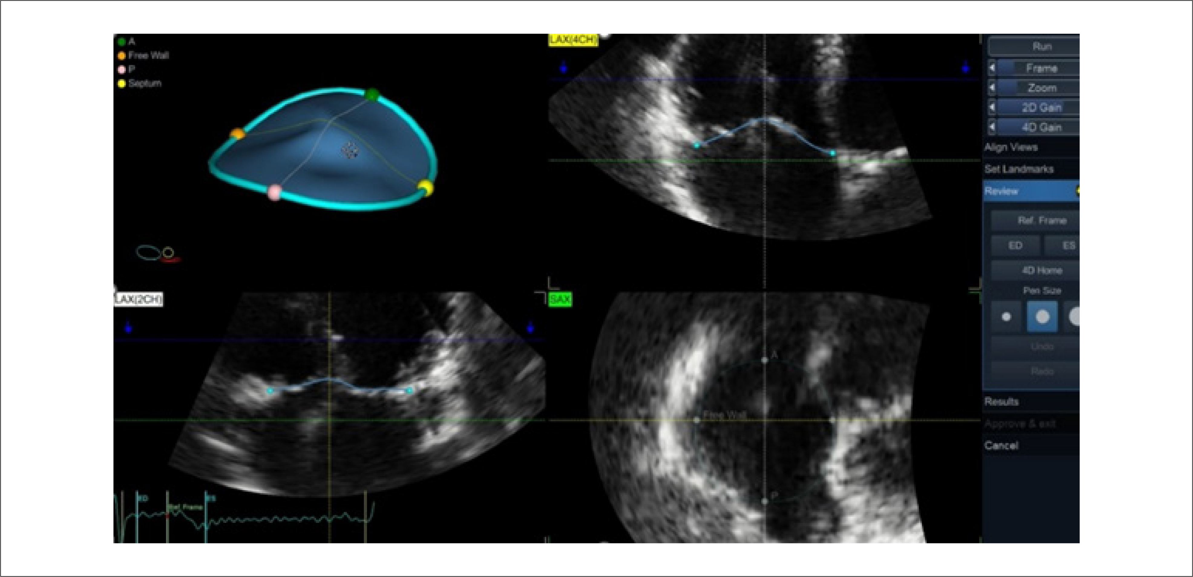
3D echocardiography allows for the evaluation of the exact mechanism of Tricuspid Regurgitation (TR), including dilation and geometry of the tricuspid annulus and papillary muscles (Figures 3 and 4). 3D reconstruction allows for more accurate measurement of the Effective Regurgitant Orifice Area (EROA) than the 2D PISA method. Data important for planning percutaneous interventions, such as the separation between the cusps, the height of the valve tenting, and electrode interference in valve function, can also be determined more accurately through three-dimensional reconstructions.19

Image showing anteroposterior dilation of the tricuspid annulus seen on two-dimensional and three-dimensional echocardiography (yellow arrows). The illustration above shows the anatomical arrangement of the annulus.

Three-dimensional multiplanar reconstruction of the tricuspid valve by 3D echocardiography, showing the spatial model of the annulus with anatomical reference points (apex, septum, free wall, and commissures). Orthogonal cuts (LAX 4-chamber, LAX 2-chamber, and SAX transverse) allow for precise contour adjustment and detailed analysis of valve geometry.
C. Congenital Heart Diseases
In congenital heart diseases such as tetralogy of Fallot, ventricular septal defect, or Ebstein's anomaly, 3D echocardiography provides accurate volumetric assessment in atypical geometries where 2D echocardiography fails. This is fundamental in surgical planning and longitudinal follow-up.26,27
D. Heart Failure with RV Dysfunction
Right ventricular dysfunction in Heart Failure with preserved Ejection Fraction (HFpEF) or reduced Ejection Fraction (HFrEF) is associated with a worse prognosis. 3D echocardiography allows for the early detection of RVEF reduction, even before significant changes in TAPSE or tricuspid annulus S’ wave velocity.28
E. Structural Interventions and Post-Procedure Monitoring
Procedures such as percutaneous pulmonary valve implantation, tricuspid clipping, and percutaneous tricuspid valve implantation, and occlusion of interatrial septal defects require pre- and post-procedure evaluation of the right ventricle, tricuspid valve, and septal defect diameters, which is performed with greater accuracy by real-time 3D echocardiography.29
Furthermore, during procedures for treating tricuspid regurgitation, an adequate anatomical demonstration of the valve, as well as the interaction between the prostheses and the valve tissue, is essential, making the use of three-dimensional transesophageal echocardiography crucial.
Technical Limitations and Future Perspectives
Despite significant advances, 3D echocardiography still faces technical challenges that limit its routine application in all clinical settings. However, the development of new technologies and artificial intelligence algorithms has driven its continuous evolution.
A. Temporal and Spatial Resolution
One of the most recognized limitations of 3D echocardiography compared to 2D is its lower temporal resolution. Multibeat acquisition is necessary to improve temporal resolution, especially when acquiring large volumes. However, this type of acquisition can introduce artifacts in uncooperative patients, those with hemodynamic instability, tachyarrhythmias, or irregular respiratory patterns.14 Furthermore, the spatial resolution is still inferior to that of magnetic resonance imaging, which may make endocardial delimitation difficult in the right ventricle with intense trabeculations or distorted anatomy.30,31
B. Dependence on the Acoustic Window
Three-dimensional echocardiography remains limited by the quality of the acoustic window. In patients with COPD, obesity, or on mechanical ventilation, the image obtained may be inadequate for accurate reconstruction of right ventricular volumes. In these cases, even with advanced software, the analysis may be unfeasible or inaccurate.7,32
C. Variability and Learning Curve
Although the accuracy of 3D echocardiography has been demonstrated in multicenter studies, significant interobserver variability still exists in centers with less experience. The learning curve for acquisition, reconstruction, and interpretation is longer than that of 2D echocardiography, requiring specific training.33
D. Processing Time and Workflow
Post-processing time, although reduced with modern software, still represents a practical barrier. In high-traffic environments such as ICUs or outpatient clinics, routine use can be hampered by the need for specific workstations and trained operators.16
E. Future Perspectives
The most promising innovations in the field include:
-
Integration with artificial intelligence (AI): The integration of AI in echocardiography has accelerated the 3D quantification of the right ventricle: Genovese et al. demonstrated that machine learning-based software automates the contouring of the right ventricle (RV), reducing interobserver variability and accelerating analysis time to 15 seconds, without manual editing, in approximately 32% of cases, with excellent reproducibility.11 A recent review showed that AI impacts all stages of the workflow — from the automatic acquisition of standardized slices to automated functional interpretation, promoting greater clinical efficiency.34
-
Portable 3D echocardiography (handheld ultrasound devices - HUDs): Although most studies with wearable devices focus on the left ventricle, the results support the feasibility of automated volumetric quantification of the right ventricle using artificial intelligence or algorithms integrated into wearable devices. This reinforces the discussion about the use of 3D echocardiography in the clinical context of emergency bedside and ICU settings.35
-
Multimodal fusion with MR and CT: 3D echocardiography has advanced beyond the isolated quantification of the right ventricle, acting as a multimodal integration platform with Cardiac Magnetic Resonance imaging (CMR) and Computed Tomography (CT), especially in complex scenarios of congenital heart disease and percutaneous interventions. A recent review highlights this emerging clinical utility, emphasizing the combination of anatomical and functional data from multiple modalities for planning and follow-up.36
-
Three-dimensional deformation (3D strain) assessment: Three-dimensional speckle tracking, a relatively recent technology in 3D echocardiography, was developed to allow the simultaneous analysis of myocardial deformation and the quantification of right ventricular (RV) volumes and ejection fraction in a single volumetric dataset.30 In addition to global quantification, 3D strain allows for the assessment of regional RV wall motion, revealing heterogeneous segmental deformation patterns—findings that may have prognostic relevance and aid in understanding ventricular mechanics in different clinical contexts. This approach, therefore, represents a potentially useful advancement to complement the functional analysis of the RV, especially in diseases that involve complex remodeling.30
-
Three-dimensional modeling of the right ventricle: technical integration and advanced: Three-dimensional (3D) modeling of the right ventricle (RV), based on 3D echocardiography, represents a technological leap in cardiac morphofunctional assessment. With segmentation and volumetric reconstruction algorithms, it is possible to build accurate anatomical models of the RV — including its inlet, body, and outlet regions — without relying on two-dimensional geometric assumptions. These reconstructions have high fidelity, with excellent accuracy for volumes and right ventricular ejection fraction (RVEF).37,16 In addition to reproducing anatomy with high precision, these models allow the generation of segmental strain maps, enabling the analysis of regional myocardial behavior and the identification of dyskinesias or areas with contractile alterations typical of congenital heart disease or valvular dysfunction.38 The use of these methods in patients with volume or pressure overload—for example, in pulmonary hypertension or valvular regurgitation—has already demonstrated utility in geometric and functional characterization, with a direct impact on risk stratification and therapeutic planning.39 In the field of medical education and surgical planning, 3D models have been integrated into augmented reality tools and 3D printing systems, enabling personalized anatomical simulation for training and to support interprofessional decision-making in complex cases.40
-
Assistance in the implantation of ventricular assist devices: In a recent publication, three-dimensional echocardiographic assessment of LV and RV volumes and shape is reported as useful for describing the impact of Left Ventricular Assist Device (LVAD) on the heart.25 RVEF and RV free wall deformation derived from three-dimensional echocardiography were associated with RV failure and long-term outcome in patients undergoing LVAD implantation. These parameters have the potential to be predictors of right heart failure in LVAD surgery.41
Conclusions
Three-dimensional (3D) echocardiography represents one of the most relevant innovations in the functional and anatomical evaluation of the right ventricle (RV) in modern clinical practice. By overcoming limitations inherent in two-dimensional echocardiography, especially those related to the geometric complexity of the RV, the method offers direct, reproducible volumetric quantification with excellent correlation to cardiac magnetic resonance imaging—the gold standard for assessing ventricular function and volume.
Its clinical application ranges from the early diagnosis of right ventricular dysfunction to the monitoring of structural therapies and risk stratification in various heart diseases, with parameters such as ejection fraction, end-systolic volume, and tricuspid annulus area demonstrating consistent prognostic value.
Despite remaining technical limitations, such as lower temporal resolution, dependence on acoustic windows, and the need for a learning curve, advances in artificial intelligence, analysis automation, and transducer miniaturization open promising prospects for expanding their use on a large scale.
Therefore, the progressive incorporation of 3D echocardiography into the routine of cardiovascular imaging laboratories is strongly recommended, especially in the evaluation of the right ventricle, as a first-line diagnostic and prognostic tool. In the coming years, technical and interpretative mastery of this method will be an important differentiating factor in the practice of the modern echocardiographer.
-
Sources of Funding
There were no external funding sources for this study.
-
Study Association
This study is not associated with any thesis or dissertation work.
-
Ethics Approval and Consent to Participate
This article does not contain any studies with human participants or animals performed by any of the authors.
-
Use of Artificial Intelligence
The authors did not use any artificial intelligence tools in the development of this work.
Acknowledgment
The authors thank the teams at the Three-Dimensional Echocardiography Laboratory and the Cardiovascular Imaging Department for their technical and scientific support during the development of this work. They also thank their colleagues who contributed suggestions and critical reviews that improved the quality of the manuscript.
This study did not receive direct financial support from public or private funding agencies.
Availability of Research Data
The underlying content of the research text is contained within the manuscript.
References
-
1 Shiota T, Jones M, Chikada M, Fleishman CE, Castellucci JB, Cotter B, et al. Real-Time Three-Dimensional Echocardiography for Determining Right Ventricular Stroke Volume in an Animal Model of Chronic Right Ventricular Volume Overload. Circulation. 1998;97(19):1897-900. doi: 10.1161/01.cir.97.19.1897.
» https://doi.org/10.1161/01.cir.97.19.1897 -
2 Nesser HJ, Tkalec W, Patel AR, Masani ND, Niel J, Markt B, et al. Quantitation of Right Ventricular Volumes and Ejection Fraction by Three-Dimensional Echocardiography in Patients: Comparison with Magnetic Resonance Imaging and Radionuclide Ventriculography. Echocardiography. 2006;23(8):666-80. doi: 10.1111/j.1540-8175.2006.00286.x.
» https://doi.org/10.1111/j.1540-8175.2006.00286.x - 3 Shiota T. 3D Echocardiography. 3rd ed. Philadelphia: Elsevier; 2021.
-
4 Focardi M, Cameli M, Carbone SF, Massoni A, De Vito R, Lisi M, et al. Traditional and Innovative Echocardiographic Parameters for the Analysis of Right Ventricular Performance in Comparison with Cardiac Magnetic Resonance. Eur Heart J Cardiovasc Imaging. 2015;16(1):47-52. doi: 10.1093/ehjci/jeu156.
» https://doi.org/10.1093/ehjci/jeu156 -
5 Rudski LG, Lai WW, Afilalo J, Hua L, Handschumacher MD, Chandrasekaran K, et al. Guidelines for the Echocardiographic Assessment of the Right Heart in Adults: A Report from the American Society of Echocardiography Endorsed by the European Association of Echocardiography, a Registered Branch of the European Society of Cardiology, and the Canadian Society of Echocardiography. J Am Soc Echocardiogr. 2010;23(7):685-713. doi: 10.1016/j.echo.2010.05.010.
» https://doi.org/10.1016/j.echo.2010.05.010 -
6 Haddad F, Hunt SA, Rosenthal DN, Murphy DJ. Right Ventricular Function in Cardiovascular Disease, Part I: Anatomy, Physiology, Aging, and Functional Assessment of the Right Ventricle. Circulation. 2008;117(11):1436-48. doi: 10.1161/CIRCULATIONAHA.107.653576.
» https://doi.org/10.1161/CIRCULATIONAHA.107.653576 -
7 Lang RM, Badano LP, Mor-Avi V, Afilalo J, Armstrong A, Ernande L, et al. Recommendations for Cardiac Chamber Quantification by Echocardiography in Adults: An Update from the American Society of Echocardiography and the European Association of Cardiovascular Imaging. Eur Heart J Cardiovasc Imaging. 2015;16(3):233-70. doi: 10.1093/ehjci/jev014.
» https://doi.org/10.1093/ehjci/jev014 -
8 Maffessanti F, Muraru D, Esposito R, Gripari P, Ermacora D, Santoro C, et al. Age-, Body Size-, and Sex-Specific Reference Values for Right Ventricular Volumes and Ejection Fraction by Three-Dimensional Echocardiography: A Multicenter Echocardiographic Study in 507 Healthy Volunteers. Circ Cardiovasc Imaging. 2013;6(5):700-10. doi: 10.1161/CIRCIMAGING.113.000706.
» https://doi.org/10.1161/CIRCIMAGING.113.000706 -
9 Mukherjee M, Rudski LG, Addetia K, Afilalo J, D’Alto M, Freed BH, et al. Guidelines for the Echocardiographic Assessment of the Right Heart in Adults and Special Considerations in Pulmonary Hypertension: Recommendations from the American Society of Echocardiography. J Am Soc Echocardiogr. 2025;38(3):141-86. doi: 10.1016/j.echo.2025.01.006.
» https://doi.org/10.1016/j.echo.2025.01.006 -
10 Soliman-Aboumarie H, Joshi SS, Cameli M, Michalski B, Manka R, Haugaa K, et al. EACVI Survey on the Multi-Modality Imaging Assessment of the Right Heart. Eur Heart J Cardiovasc Imaging. 2022;23(11):1417-22. doi: 10.1093/ehjci/jeac183.
» https://doi.org/10.1093/ehjci/jeac183 -
11 Genovese D, Rashedi N, Weinert L, Narang A, Addetia K, Patel AR, et al. Machine Learning-Based Three-Dimensional Echocardiographic Quantification of Right Ventricular Size and Function: Validation Against Cardiac Magnetic Resonance. J Am Soc Echocardiogr. 2019;32(8):969-77. doi: 10.1016/j.echo.2019.04.001.
» https://doi.org/10.1016/j.echo.2019.04.001 -
12 Tamborini G, Marsan NA, Gripari P, Maffessanti F, Brusoni D, Muratori M, et al. Reference Values for Right Ventricular Volumes and Ejection Fraction with Real-Time Three-Dimensional Echocardiography: Evaluation in a Large Series of Normal Subjects. J Am Soc Echocardiogr. 2010;23(2):109-15. doi: 10.1016/j.echo.2009.11.026.
» https://doi.org/10.1016/j.echo.2009.11.026 -
13 Maceira AM, Prasad SK, Khan M, Pennell DJ. Reference Right Ventricular Systolic and Diastolic Function Normalized to Age, Gender and Body Surface Area from Steady-State Free Precession Cardiovascular Magnetic Resonance. Eur Heart J. 2006;27(23):2879-88. doi: 10.1093/eurheartj/ehl336.
» https://doi.org/10.1093/eurheartj/ehl336 -
14 Lang RM, Badano LP, Tsang W, Adams DH, Agricola E, Buck T, et al. EAE/ASE Recommendations for Image Acquisition and Display Using Three-Dimensional Echocardiography. Eur Heart J Cardiovasc Imaging. 2012;13(1):1-46. doi: 10.1093/ehjci/jer316.
» https://doi.org/10.1093/ehjci/jer316 -
15 Wang S, Wang S, Zhu Q, Wang Y, Li G, Kong F, et al. Reference Values of Right Ventricular Volumes and Ejection Fraction by Three-Dimensional Echocardiography in Adults: A Systematic Review and Meta-Analysis. Front Cardiovasc Med. 2021;8:709863. doi: 10.3389/fcvm.2021.709863.
» https://doi.org/10.3389/fcvm.2021.709863 -
16 Muraru D, Spadotto V, Cecchetto A, Romeo G, Aruta P, Ermacora D, et al. New Speckle-Tracking Algorithm for Right Ventricular Volume Analysis from Three-Dimensional Echocardiographic Data Sets: Validation with Cardiac Magnetic Resonance and Comparison with the Previous Analysis Tool. Eur Heart J Cardiovasc Imaging. 2016;17(11):1279-89. doi: 10.1093/ehjci/jev309.
» https://doi.org/10.1093/ehjci/jev309 -
17 Grapsa J, O’Regan DP, Pavlopoulos H, Durighel G, Dawson D, Nihoyannopoulos P. Right Ventricular Remodelling in Pulmonary Arterial Hypertension with Three-Dimensional Echocardiography: Comparison with Cardiac Magnetic Resonance Imaging. Eur J Echocardiogr. 2010;11(1):64-73. doi: 10.1093/ejechocard/jep169.
» https://doi.org/10.1093/ejechocard/jep169 -
18 Jenkins C, Chan J, Bricknell K, Strudwick M, Marwick TH. Reproducibility of Right Ventricular Volumes and Ejection Fraction Using Real-Time Three-Dimensional Echocardiography: Comparison with Cardiac MRI. Chest. 2007;131(6):1844-51. doi: 10.1378/chest.06-2143.
» https://doi.org/10.1378/chest.06-2143 -
19 Prihadi EA, van der Bijl P, Dietz M, Abou R, Vollema EM, Marsan NA, et al. Prognostic Implications of Right Ventricular Free Wall Longitudinal Strain in Patients with Significant Functional Tricuspid Regurgitation. Circ Cardiovasc Imaging. 2019;12(3):e008666. doi: 10.1161/CIRCIMAGING.118.008666.
» https://doi.org/10.1161/CIRCIMAGING.118.008666 -
20 Kitano T, Kovács A, Nabeshima Y, Tokodi M, Fábián A, Lakatos BK, et al. Prognostic Value of Right Ventricular Strains Using Novel Three-Dimensional Analytical Software in Patients with Cardiac Disease. Front Cardiovasc Med. 2022;9:837584. doi: 10.3389/fcvm.2022.837584.
» https://doi.org/10.3389/fcvm.2022.837584 -
21 Meng Y, Zhu S, Xie Y, Zhang Y, Qian M, Gao L, et al. Prognostic Value of Right Ventricular 3D Speckle-Tracking Strain and Ejection Fraction in Patients with HFpEF. Front Cardiovasc Med. 2021;8:694365. doi: 10.3389/fcvm.2021.694365.
» https://doi.org/10.3389/fcvm.2021.694365 -
22 Vîjîiac A, Onciul S, Guzu C, Verinceanu V, Bătăilă V, Deaconu S, et al. The Prognostic Value of Right Ventricular Longitudinal Strain and 3D Ejection Fraction in Patients with Dilated Cardiomyopathy. Int J Cardiovasc Imaging. 2021;37(11):3233-44. doi: 10.1007/s10554-021-02322-z.
» https://doi.org/10.1007/s10554-021-02322-z -
23 Li Y, Wang T, Haines P, Li M, Wu W, Liu M, et al. Prognostic Value of Right Ventricular Two-Dimensional and Three-Dimensional Speckle-Tracking Strain in Pulmonary Arterial Hypertension: Superiority of Longitudinal Strain Over Circumferential and Radial Strain. J Am Soc Echocardiogr. 2020;33(8):985-94.e1. doi: 10.1016/j.echo.2020.03.015.
» https://doi.org/10.1016/j.echo.2020.03.015 -
24 Erley J, Tanacli R, Genovese D, Tapaskar N, Rashedi N, Bucius P, et al. Myocardial Strain Analysis of the Right Ventricle: Comparison of Different Cardiovascular Magnetic Resonance and Echocardiographic Techniques. J Cardiovasc Magn Reson. 2020;22(1):51. doi: 10.1186/s12968-020-00647-7.
» https://doi.org/10.1186/s12968-020-00647-7 -
25 Addetia K, Uriel N, Maffessanti F, Sayer G, Adatya S, Kim GH, et al. 3D Morphological Changes in LV and RV during LVAD Ramp Studies. JACC Cardiovasc Imaging. 2018;11(2 Pt 1):159-69. doi: 10.1016/j.jcmg.2016.12.019.
» https://doi.org/10.1016/j.jcmg.2016.12.019 -
26 van der Zwaan HB, Helbing WA, McGhie JS, Geleijnse ML, Luijnenburg SE, Roos-Hesselink JW, et al. Clinical Value of Real-Time Three-Dimensional Echocardiography for Right Ventricular Quantification in Congenital Heart Disease: Validation with Cardiac Magnetic Resonance Imaging. J Am Soc Echocardiogr. 2010;23(2):134-40. doi: 10.1016/j.echo.2009.12.001.
» https://doi.org/10.1016/j.echo.2009.12.001 -
27 Dragulescu A, Grosse-Wortmann L, Fackoury C, Riffle S, Waiss M, Jaeggi E, et al. Echocardiographic Assessment of Right Ventricular Volumes after Surgical Repair of Tetralogy of Fallot: Clinical Validation of a New Echocardiographic Method. J Am Soc Echocardiogr. 2011;24(11):1191-8. doi: 10.1016/j.echo.2011.08.006.
» https://doi.org/10.1016/j.echo.2011.08.006 -
28 Molnár AÁ, Sánta A, Merkely B. Echocardiography Imaging of the Right Ventricle: Focus on Three-Dimensional Echocardiography. Diagnostics. 2023;13(15):2470. doi: 10.3390/diagnostics13152470.
» https://doi.org/10.3390/diagnostics13152470 -
29 Agricola E, Asmarats L, Maisano F, Cavalcante JL, Liu S, Milla F, et al. Imaging for Tricuspid Valve Repair and Replacement. JACC Cardiovasc Imaging. 2021;14(1):61-111. doi: 10.1016/j.jcmg.2020.01.031.
» https://doi.org/10.1016/j.jcmg.2020.01.031 -
30 Ishizu T, Seo Y, Atsumi A, Tanaka YO, Yamamoto M, Machino-Ohtsuka T, et al. Global and Regional Right Ventricular Function Assessed by Novel Three-Dimensional Speckle-Tracking Echocardiography. J Am Soc Echocardiogr. 2017;30(12):1203-13. doi: 10.1016/j.echo.2017.08.007.
» https://doi.org/10.1016/j.echo.2017.08.007 -
31 Wu VC, Takeuchi M. Three-Dimensional Echocardiography: Current Status and Real-Life Applications. Acta Cardiol Sin. 2017;33(2):107-18. doi: 10.6515/acs20160818a.
» https://doi.org/10.6515/acs20160818a -
32 Herberg U, Smit F, Winkler C, Dalla-Pozza R, Breuer J, Laser KT. Real-Time 3D-Echocardiography of the Right Ventricle-Paediatric Reference Values for Right Ventricular Volumes Using Knowledge-Based Reconstruction: A Multicentre Study. Quant Imaging Med Surg. 2021;11(7):2905-17. doi: 10.21037/qims-20-1155.
» https://doi.org/10.21037/qims-20-1155 -
33 Pinedo M, Villacorta E, Tapia C, Arnold R, López J, Revilla A, et al. Inter- and Intra-Observer Variability in the Echocardiographic Evaluation of Right Ventricular Function. Rev Esp Cardiol. 2010;63(7):802-9. doi: 10.1016/s1885-5857(10)70165-1.
» https://doi.org/10.1016/s1885-5857(10)70165-1 -
34 Zhou J, Du M, Chang S, Chen Z. Artificial Intelligence in Echocardiography: Detection, Functional Evaluation, and Disease Diagnosis. Cardiovasc Ultrasound. 2021;19(1):29. doi: 10.1186/s12947-021-00261-2.
» https://doi.org/10.1186/s12947-021-00261-2 -
35 de Raat FM, van Houte J, Montenij LJ, Bouwmeester S, Felix SEA, Bingley P, et al. Evaluation of the Image Quality and Validity of Handheld Echocardiography for Stroke Volume and Left Ventricular Ejection Fraction Quantification: A Method Comparison Study. Int J Cardiovasc Imaging. 2024;40(1):15-25. doi: 10.1007/s10554-023-02942-7.
» https://doi.org/10.1007/s10554-023-02942-7 -
36 Randazzo M, Maffessanti F, Kotta A, Grapsa J, Lang RM, Addetia K. Added Value of 3D Echocardiography in the Diagnosis and Prognostication of Patients with Right Ventricular Dysfunction. Front Cardiovasc Med. 2023;10:1263864. doi: 10.3389/fcvm.2023.1263864.
» https://doi.org/10.3389/fcvm.2023.1263864 -
37 Hameed A, Condliffe R, Swift AJ, Alabed S, Kiely DG, Charalampopoulos A. Assessment of Right Ventricular Function-a State of the Art. Curr Heart Fail Rep. 2023;20(3):194-207. doi: 10.1007/s11897-023-00600-6.
» https://doi.org/10.1007/s11897-023-00600-6 -
38 Addetia K, Lang RM. Decoding the Right Ventricle in 3 Dimensions. JAMA Cardiol. 2018;3(10):910-1. doi: 10.1001/jamacardio.2018.2452.
» https://doi.org/10.1001/jamacardio.2018.2452 -
39 Otani K, Nabeshima Y, Kitano T, Takeuchi M. Accuracy of Fully Automated Right Ventricular Quantification Software with 3D Echocardiography: Direct Comparison with Cardiac Magnetic Resonance and Semi-Automated Quantification Software. Eur Heart J Cardiovasc Imaging. 2020;21(7):787-95. doi: 10.1093/ehjci/jez236.
» https://doi.org/10.1093/ehjci/jez236 -
40 Muraru D. 22nd Annual Feigenbaum Lecture: Right Heart, Right Now: The Role of Three-Dimensional Echocardiography. J Am Soc Echocardiogr. 2022;35(9):893-909. doi: 10.1016/j.echo.2022.05.011.
» https://doi.org/10.1016/j.echo.2022.05.011 -
41 Magunia H, Dietrich C, Langer HF, Schibilsky D, Schlensak C, Rosenberger P, et al. 3D Echocardiography Derived Right Ventricular Function is Associated with Right Ventricular Failure and Mid-Term Survival after Left Ventricular Assist Device Implantation. Int J Cardiol. 2018;272:348-55. doi: 10.1016/j.ijcard.2018.06.026.
» https://doi.org/10.1016/j.ijcard.2018.06.026
Edited by
-
Editor responsible for the review:
Marcelo Tavares