Coexistence of Partial Anomalous Pulmonary Venous Connection and Coronary Artery Fistulas: A Rare Case Report
Abstract
Background Dyspnoea is a common clinical symptom that frequently prompts hospital admission and is associated with significant morbidity. While it most often results from prevalent cardiopulmonary conditions, rare congenital cardiovascular anomalies can also manifest with dyspnoea. Partial Anomalous Pulmonary Venous Connection (PAPVC) and Coronary Artery Fistulas (CAFs) are uncommon congenital malformations of the cardiovascular system, and their simultaneous presence is exceedingly rare. Early recognition of such anomalies is critical to avoid progressive hemodynamic compromise and to guide appropriate management strategies.
Case Presentation We present the case of a 55-year-old man who experienced an acute onset of dyspnoea lasting approximately two hours. Initial clinical assessment and routine investigations—including physical examination, echocardiography, electrocardiography, and right and left heart catheterization—raised suspicion of an underlying cardiac abnormality, prompting further evaluation. Subsequent cardiac Magnetic Resonance Imaging (MRI) and Multi-Detector Computed Tomography (MDCT) revealed the presence of a Partial Anomalous Pulmonary Venous Connection (PAPVC) accompanied by Coronary Artery Fistulas (CAFs). Given the non-complex characteristics of the shunt in this case, a shared decision was reached with the patient to proceed with conservative management.
Discussion PAPVC and Coronary Artery Fistulas CAFs are rare entities that should be considered in the differential diagnosis. However, recommending their evaluation as initial diagnostic hypotheses may result in unnecessary investigations.
Keywords
Echocardiography; Differential Diagnosis; Coronary Vessels
Resumo
Fundamento A dispneia é um sintoma clínico comum que frequentemente leva à internação hospitalar e está associada a significativa morbidade. Embora geralmente resulte de condições cardiopulmonares prevalentes, anomalias cardiovasculares congênitas raras também podem se manifestar com dispneia. A Conexão Venosa Pulmonar Anômala Parcial (PAPVC) e as Fístulas de Artéria Coronária (FACs) são malformações congênitas incomuns do sistema cardiovascular, cuja presença simultânea é extremamente rara. O reconhecimento precoce dessas anomalias é fundamental para evitar comprometimento hemodinâmico progressivo e orientar estratégias de manejo adequadas.
Apresentação do Caso Apresentamos o caso de um homem de 55 anos que apresentou início agudo de dispneia com duração aproximada de duas horas. A avaliação clínica inicial e os exames de rotina – incluindo exame físico, ecocardiografia, eletrocardiografia e cateterismo cardíaco direito e esquerdo – levantaram suspeita de uma anomalia cardíaca subjacente, motivando investigação adicional. A subsequente Ressonância Magnética Cardíaca (RMC) e a Tomografia Computadorizada Multidetector (TCMD) revelaram a presença de uma Conexão Venosa Pulmonar Anômala Parcial acompanhada de FACs. Dadas as características não complexas do shunt neste caso, foi tomada uma decisão compartilhada com o paciente de prosseguir com tratamento conservador.
Discussão A PAPVC e as FACs são entidades raras que devem ser consideradas no diagnóstico diferencial. No entanto, recomendá-las como hipóteses diagnósticas iniciais pode resultar em investigações desnecessárias.
Palavras-chave
Ecocardiografia; Diagnóstico Diferencial; Vasos Coronários
Introduction
A normal pulmonary venous pattern with four distinct veins is observed in approximately 60–70% of the population.1 Developmental anomalies can result in Partial (PAPVC) or Total Aanomalous Pulmonary Venous Connection (TAPVC), with anomalous drainage patterns reported in up to 38% of individuals.2 PAPVC may occur in isolation, in association with an Atrial Sseptal Defect (ASD), or as part of complex congenital heart disease, and often remains undiagnosed due to mild or absent symptoms.3,4
PAPVC involves a left-to-right shunt and is usually hemodynamically insignificant. It is often discovered incidentally, for example during imaging for a central venous catheter that appears malpositioned. Despite its subtle presentation, associated anomalies can increase the risk of morbidity and mortality.5
Coronary Artery Fistulas (CAFs) are rare congenital anomalies, with acquired cases being even more uncommon, and are most often detected incidentally. Small CAFs are typically asymptomatic, whereas larger fistulas can lead to cardiac chamber dilation or ischemia if left untreated.
We report a rare case of coexisting PAPVC and CAFs – an unusual combination that presents diagnostic challenges. Although each condition is individually rare, their simultaneous occurrence is exceptionally uncommon and has been seldom documented in the literature.6,7
Case Presentation
A 55-year-old patient presented for further evaluation of dyspnoea. He reported a mild retrosternal burning sensation radiating caudally beneath the left costal arches. The chest discomfort was non-exertional. There was no history of diabetes, connective tissue disorders, other systemic anomalies, or significant family history of disease.
On physical examination, the patient was afebrile, with no tachypnoea (respiratory rate: 13/min), oxygen saturation of 93%, and blood pressure of 150/95 mmHg. The rest of the physical examination was unremarkable. On the day of admission, the electrocardiogram demonstrated sinus rhythm with a heart rate of 92 bpm. High-sensitivity cardiac troponin levels were elevated, measuring 39 ng/L at baseline and 41 ng/L at one-hour follow-up (reference <14 ng/L), indicating myocardial injury without significant dynamic change. NT-proBNP was 89 pg/mL (normal <227 pg/mL in males aged 50-65 years).
In this case, left heart catheterization was performed prior to cardiac MRI due to clinical suspicion of a left-to-right shunt and the need to exclude coronary artery anomalies. The invasive procedure enabled precise identification of a coronary artery fistula between the Left Anterior Descending Artery (LAD) and the pulmonary trunk, providing essential anatomical details for subsequent therapeutic planning.
Although MRI offers comprehensive structural evaluation, cardiac catheterization remains the gold standard for direct coronary visualization and hemodynamic assessment in such settings.
The patient remained largely asymptomatic, without evidence of cyanosis or angina. Following thorough medical consultation, he opted for conservative management with beta-blockers and diuretics. Given the absence of a significant left-to-right shunt, surgical intervention was not indicated.
Echocardiography and Electrocardiography
Echocardiography, which serves as both the gold standard and the primary diagnostic modality in the emergency department, demonstrated signs of right heart dilation. Both right and left ventricular systolic function were preserved, with normal ejection fractions (EF) — Right Ventricular Internal Diameter at Diastole (RVIDD) basal: 49 mm, RVIDd mid: 27 mm, FAC: 55%, TAPSE: 31 mm. Additionally, there was evidence of mild tricuspid regurgitation (Grade I), with pulmonary artery systolic pressure of 34 mmHg and a Qp:Qs ratio of 1.4. These findings are consistent with right heart volume overload, likely secondary to left-to-right shunting.
Electrocardiography revealed non-specific changes but supported the echocardiographic evidence of right heart volume overload.
To further clarify the differential diagnosis, the patient underwent comprehensive hemodynamic assessment, including both right and left heart catheterization.
Right and Left Heart Catheterization
On the same day, the patient underwent comprehensive cardiac catheterization. During right heart catheterization, direct evidence of a left-to-right shunt was observed. Oxygen saturation measurements revealed higher values in the superior vena cava compared with the inferior vena cava (SVC: 83%, IVC: 79%, RA: 81%, RV: 83%, PA: 85%).
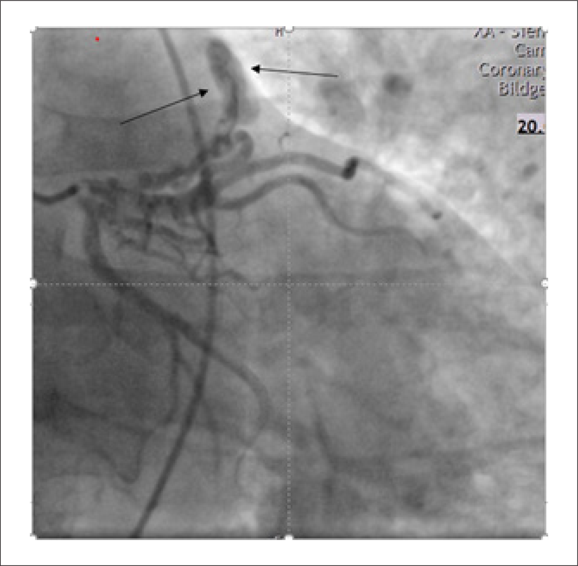
Left heart catheterization (Figure 1) identified a fistula between the LAD artery and the pulmonary trunk. No coronary artery stenosis was detected, which allowed progression to the next step of the evaluation (Figure 1).

Coronary angiography showing the left anterior descending artery with a fistulous connection to the pulmonary trunk
In light of these catheterization findings, cardiac MRI was performed to further clarify the diagnosis.
MRI (Magnetic Resonance Imaging)
To achieve better visualization of cardiac structures, cardiac MRI was performed. Left ventricular function was normal. Mild right ventricular dysfunction with right vetricular dilatation was observed, without evidence of arrhythmogenic right ventricular cardiomyopathy (Table 1). No signs of myocardial inflammation or fibrosis were detected.
Left ventricle (LV) and right ventricle (RV) Volumetry: measurements of the volume of the both ventricle, including end-diastolic volume (EDV), end-systolic volume (ESV), stroke volume (SV), and ejection fraction (EF)
A fistulous network was identified, with connections involving the proximal LAD, the conus arteriosus of the right coronary artery, and the pulmonary trunk (Figures 2 and 3).

Maximum intensity projection coronal images from a case of left-sided cardiac partial anomalous pulmonary venous connection

Coronal maximum intensity projection images demonstrating a coronary artery fistula originating from the left anterior descending artery and draining into the pulmonary trunk; the fistulous connection is indicated by the arrow
Stress imaging or physiologic assessment was not performed to evaluate for ischemia attributable to the coronary–pulmonary fistula. This decision was based on the patient's stable clinical status, the non-complex nature of the shunt, and the absence of ischemic symptoms.
MDCT (Multi-Detector Computed Tomography)
As MDCT represents the modality of choice for detailed assessment of cardiac anatomy and structural abnormalities, we elected to perform this study. MDCT revealed anomalous drainage of the upper and partially the lower left pulmonary veins into the left brachiocephalic vein. The left brachiocephalic vein was ectatic, with a maximum diameter of approximately 25 mm. The pulmonary trunk was dilated, measuring 32 mm (normal ≤29 mm in men), with associated dilatation of both pulmonary arteries. Dilatation of the right ventricle and right atrium was also noted. In addition, previously identified extensive fistulous connections between the proximal LAD and the pulmonary trunk were confirmed (Figures 4 and 5).

Two volume-rendered images in different orientation of our patient with partial anomalous pulmonary venous connection; the left inferior pulmonary vein is draining at the brachiocephalic vein

Volume-rendered computed tomography images in two different orientations depicting a coronary artery fistula originating from the left anterior descending artery and draining into the pulmonary trunk. The fistulous connection is indicated by the arrow
Discussion
PAPVC is a rare congenital defect in which one or more pulmonary veins drain into the systemic venous circulation—such as the superior vena cava or right atrium—instead of the left atrium. This results in a partial left-to-right shunt and is often detected incidentally, with a reported prevalence of 0.4–0.7%.
Both PAPVC and CAFs are rare anomalies, and their coexistence is extremely uncommon. Clinical presentation varies depending on the severity and location of the abnormal drainage.
Medium to large or symptomatic CAFs may require transcatheter closure, ideally performed in specialized centers. In cases involving large coronary aneurysms, surgical repair and anticoagulation may be necessary. Follow-up imaging is recommended to monitor for potential recanalization.
Conclusion
PAPVC and coronary artery fistulas are rare congenital cardiac anomalies, and their coexistence has been scarcely reported in the literature. Clinical presentation varies widely, ranging from incidental findings to symptoms influenced by the extent of abnormal venous drainage, the anatomical site of connection, and any associated cardiac defects.
Advanced imaging techniques such as MDCTA and MRA play a critical role in the precise delineation of these anomalies. Their ability to provide high-resolution, three-dimensional anatomical detail significantly aids clinicians in diagnosis, clinical assessment, and the development of appropriate management strategies.8,9
-
Sources of Funding
There were no external funding sources for this study.
-
Study Association
This study is not associated with any thesis or dissertation work.
-
Ethics Approval and Consent to Participate
The authors confirm that consent for submission and pub lication of this case report has been obtained from the patient in line with the COPE o guidance.
-
Use of Artificial Intelligence
The authors did not use any artificial intelligence tools in the development of this work.
Availability of Research Data
The underlying content of the research text is contained within the manuscript.
References
-
1 Lacomis JM, Goitein O, Deible C, Schwartzman D. CT of the Pulmonary Veins. J Thorac Imaging. 2007;22(1):63-76. doi: 10.1097/RTI.0b013e3180317aaf.
» https://doi.org/10.1097/RTI.0b013e3180317aaf -
2 Kato R, Lickfett L, Meininger G, Dickfeld T, Wu R, Juang G, et al. Pulmonary Vein Anatomy in Patients Undergoing Catheter Ablation of Atrial Fibrillation: Lessons Learned by Use of Magnetic Resonance Imaging. Circulation. 2003;107(15):2004-10. doi: 10.1161/01.CIR.0000061951.81767.4E.
» https://doi.org/10.1161/01.CIR.0000061951.81767.4E -
3 Kelle AM, Backer CL, Gossett JG, Kaushal S, Mavroudis C. Total Anomalous Pulmonary Venous Connection: Results of Surgical Repair of 100 Patients at a Single Institution. J Thorac Cardiovasc Surg. 2010;139(6):1387-1394.e3. doi: 10.1016/j.jtcvs.2010.02.024.
» https://doi.org/10.1016/j.jtcvs.2010.02.024 -
4 Furlanetto G, Furlanetto BH, Henriques SR, Lopes LM, Miranda ET, Porto CM, et al. Mixed Type Total Anomalous Pulmonary Venous Connection: Early Results and Surgical Techniques. World J Pediatr Congenit Heart Surg. 2015;6(1):26-32. doi: 10.1177/2150135114554660.
» https://doi.org/10.1177/2150135114554660 -
5 Hart A, Lee EY. Multidetector Computed Tomography of Pediatric Vascular Imaging with Advanced Postprocessing Techniques. Adv Clin Radiol 2020;2:191-211. doi: 10.1016/j.yacr.2020.06.003.
» https://doi.org/10.1016/j.yacr.2020.06.003 -
6 Yildiz A, Okcun B, Peker T, Arslan C, Olcay A, Vatan MB. Prevalence of Coronary Artery Anomalies in 12,457 Adult Patients who Underwent Coronary Angiography. Clin Cardiol. 2010;33(12):E60-4. doi: 10.1002/clc.20588.
» https://doi.org/10.1002/clc.20588 -
7 Gillebert C, van Hoof R, van de Werf F, Piessens J, De Geest H. Coronary Artery Fistulas in an Adult Population. Eur Heart J. 1986;7(5):437-43. doi: 10.1093/oxfordjournals.eurheartj.a062086.
» https://doi.org/10.1093/oxfordjournals.eurheartj.a062086 -
8 Dillman JR, Yarram SG, Hernandez RJ. Imaging of Pulmonary Venous Developmental Anomalies. AJR Am J Roentgenol. 2009;192(5):1272-85. doi: 10.2214/AJR.08.1526.
» https://doi.org/10.2214/AJR.08.1526 -
9 Haramati LB, Moche IE, Rivera VT, Patel PV, Heyneman L, McAdams HP, et al. Computed Tomography of Partial Anomalous Pulmonary Venous Connection in Adults. J Comput Assist Tomogr. 2003;27(5):743-9. doi: 10.1097/00004728-200309000-00011.
» https://doi.org/10.1097/00004728-200309000-00011
Edited by
-
Editor responsible for the review:
Tiago Magalhães