
 A Hidden Connection: Anomalous Left Circumflex Artery Arising From the Right Pulmonary Artery Unveiled by Cardiovascular Computed Tomography
Keywords
Coronary Vessels; Pulmonary Artery; X-Ray Computed Tomography; Cardiac Catheterization
Palavras-chave
Vasos Coronários; Artéria Pulmonar; Tomografia Computadorizada por Raios X; Cateterismo Cardíaco
Keywords
Coronary Vessels; Pulmonary Artery; X-Ray Computed Tomography; Cardiac Catheterization
Palavras-chave
Vasos Coronários; Artéria Pulmonar; Tomografia Computadorizada por Raios X; Cateterismo Cardíaco
Introduction
Congenital coronary artery anomalies are uncommon but clinically significant conditions that may present across a broad spectrum, ranging from incidental findings to myocardial ischemia, ventricular arrhythmias, heart failure (HF), and sudden cardiac death (SCD). In adults, anomalous coronary origins may be particularly challenging to recognize because symptoms are often nonspecific and may overlap with more prevalent causes of chest pain, dyspnea, or palpitations.
In rare cases, a coronary artery may originate from the pulmonary arterial circulation, resulting in myocardial perfusion that depends on collateral flow from the remaining coronary arteries arising from the aorta, thereby predisposing the myocardium to ischemia-related electrical instability. Noninvasive anatomical imaging plays a pivotal role in establishing a definitive diagnosis, particularly when conventional angiography is inconclusive.
We report an exceptionally rare case of an anomalous left circumflex artery (LCx) arising from the right pulmonary artery (RPA), identified after invasive coronary angiography failed to selectively engage the LCx. Subsequent cardiovascular computed tomography (CT) precisely delineated the anomalous origin and demonstrated a collateral-dependent perfusion pattern.
Case report
A 48-year-old man with a history of ventricular bigeminy, obstructive sleep apnea, and peripheral edema presented for evaluation of symptomatic premature ventricular contractions (PVCs). Initial ambulatory Holter monitoring demonstrated a high PVC burden (34%), rare atrial ectopy, and episodes of nonsustained supraventricular tachycardia. Transthoracic echocardiography revealed mildly reduced left ventricular (LV) systolic function, with an ejection fraction of 45%-50%. Following initiation of beta-blocker therapy, the patient developed progressive dyspnea, worsening palpitations, and functional limitation, leading to treatment discontinuation.
Coronary angiography was performed because of abnormal stress test findings and concern for underlying myocardial ischemia. During the procedure, selective engagement of the LCx was unsuccessful, raising suspicion for a congenital coronary anomaly. Subsequent cardiovascular CT clearly demonstrated an anomalous origin of the LCx from the RPA, with collateral retrograde perfusion supplied by a dominant right coronary artery (RCA), as shown in Figure 1-4.


3D reconstruction generated using Vitrea software demonstrating the course of the LCx arising from the RPA. The image also shows the aortic root with a separate origin of the LCx from the left coronary cusp.
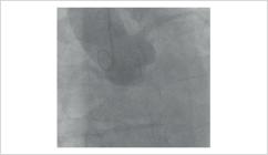
Aortography demonstrating the ostia of the left main coronary artery and RCA. No ostium of the LCx is identified.

Right anterior oblique caudal projection demonstrating the left coronary system, including the left anterior descending artery and a large first diagonal branch, without visualization of the LCx.
This case highlights the critical role of advanced noninvasive imaging in the anatomical characterization of anomalous coronary artery origins, particularly when conventional angiography is inconclusive. Cardiovascular CT provided high-resolution 3D visualization that was essential for accurate diagnosis, risk stratification, and therapeutic planning in a patient initially evaluated for ventricular arrhythmia.
Discussion
Congenital coronary artery anomalies comprise a heterogeneous group of rare but clinically significant malformations, with an overall prevalence < 2% in the general population. Among these anomalies, anomalous origin of the LCx from the RPA is exceptionally rare, with only isolated cases reported in prior research.1 Although many coronary anomalies remain asymptomatic, anomalous coronary origins may lead to myocardial ischemia, ventricular arrhythmias, HF, or SCD, particularly when myocardial perfusion depends on collateral circulation.
In the present case, the patient's high PVC burden, combined with the inability to selectively engage the LCx during invasive coronary angiography, raised suspicion for an anomalous coronary anatomy. Although invasive angiography remains the reference standard for coronary artery assessment, its diagnostic capability may be limited in cases involving anomalous vessels arising from non-aortic structures.2 In this context, cardiovascular CT offers substantial advantages, including high spatial resolution, multiplanar reconstruction, and 3D anatomical visualization, all of which are essential for accurately defining anomalous coronary pathways.
In our patient, cardiovascular CT was instrumental not only in confirming the anomalous origin of the LCx from the RPA but also in demonstrating retrograde collateral perfusion from a dominant RCA to the LCx territory. Identification of this anatomy carries important implications for risk stratification, therapeutic decision-making, and long-term clinical management.3 Furthermore, this case highlights the indispensable role of multimodality imaging in contemporary cardiovascular diagnostics, particularly when conventional techniques are inconclusive or potentially misleading.4
From a management perspective, recognition of a coronary artery originating from the pulmonary circulation is critical because treatment decisions are often guided by symptom burden, evidence of myocardial ischemia, ventricular function, arrhythmic profile, and the adequacy of collateral perfusion. Multidisciplinary assessment involving advanced cardiac imaging specialists, interventional cardiologists, electrophysiologists, and cardiothoracic surgeons may be necessary to individualize management strategies, which can range from clinical surveillance and functional evaluation to surgical or percutaneous intervention in selected high-risk cases. In this patient, advanced cardiovascular CT served as the definitive imaging modality for characterization of a rare congenital coronary anomaly initially investigated in the context of ventricular arrhythmia, underscoring the transformative role of noninvasive imaging in modern cardiovascular medicine.
Conclusion
Anomalous origin of the LCx from the RPA is an exceptionally rare but clinically relevant congenital coronary anomaly, particularly in patients presenting with ventricular arrhythmias, LV dysfunction, or suspected myocardial ischemia. When invasive coronary angiography is nondiagnostic, especially in cases in which the LCx cannot be selectively engaged, cardiovascular CT may serve as the definitive imaging modality for accurate delineation of coronary origin and collateral perfusion patterns. Precise anatomical characterization is essential for appropriate diagnosis, risk stratification, and individualized management planning.
-
Sources of Funding
There were no external funding sources for this study.
-
Study Association
This study is not associated with any thesis or dissertation work.
-
Ethics Approval and Consent to Participate
This article does not contain any studies with human participants or animals performed by any of the authors.
-
Use of Artificial Intelligence
The authors did not use any artificial intelligence tools in the development of this work.
Availability of Research Data
The underlying content of the research text is contained within the manuscript.
References
-
1 Faria B, Calvo L, Ribeiro S, Ruivo C, Lourenço A. Anomalous Origin of the Circumflex Coronary Artery from the Right Pulmonary Artery: Diagnosis Through Cardiac CT. Arq Bras Cardiol. 2021;117(1 suppl 1):8-11. doi: 10.36660/abc.20200060.
» https://doi.org/10.36660/abc.20200060 -
2 Korosoglou G, Ringwald G, Giannitsis E, Katus HA. Anomalous Origin of the Left Circumflex Coronary Artery from the Pulmonary Artery. A Very Rare Congenital Anomaly in an Adult Patient Diagnosed by Cardiovascular Magnetic Resonance. J Cardiovasc Magn Reson. 2008;10(1):4. doi: 10.1186/1532-429X-10-4.
» https://doi.org/10.1186/1532-429X-10-4 -
3 Vergara-Uzcategui CE, Urquiza RV, Salinas P, Núñez-Gil IJ. Anomalous Origin of Left Circumflex Artery from the Right Pulmonary Artery of an Adult. REC Interv Cardiol. 2021;3(1):65-72. doi: 10.24875/RECICE.M20000128.
» https://doi.org/10.24875/RECICE.M20000128 -
4 Harky A, Bashir M, Garner M, Hsia T. Anomalous Origin of the Circumflex Coronary Artery Presenting with Ventricular Fibrillation Cardiac Arrest. BMJ Case Rep. 2017;2017:bcr2016219184. doi: 10.1136/bcr-2016-219184.
» https://doi.org/10.1136/bcr-2016-219184
Edited by
-
Editor responsible for the review:
Marcelo Tavares


