Single Coronary Artery and Stress Cardiomyopathy: An Association Demonstrated by Multimodality Imaging
Keywords:
Coronary Vessels; Takotsubo Cardiomyopathy; Coronary Angiography; Echocardiography; Magnetic Resonance Imaging
Palavras-chave:
Vasos Coronários; Cardiomiopatia de Takotsubo; Angiografia Coronária; Ecocardiografia; Cinerressonância Magnética
Keywords:
Coronary Vessels; Takotsubo Cardiomyopathy; Coronary Angiography; Echocardiography; Magnetic Resonance Imaging
Palavras-chave:
Vasos Coronários; Cardiomiopatia de Takotsubo; Angiografia Coronária; Ecocardiografia; Cinerressonância Magnética
Case Report
A 58-years-old female patient was admitted with retrosternal chest pain described as a continuous, oppressive tightness that had started two days earlier, after a tooth extraction procedure performed under ineffective local anesthesia. The patient’s past medical history included diabetes, dyslipidemia, and ovarian cancer, and she was receiving aspirin, rosuvastatin, metformin, dapagliflozin, and semaglutide. Physical examination was unremarkable. The chest pain protocol was initiated, and the initial electrocardiogram showed sinus rhythm with left anterior fascicular block, early repolarization in the inferior leads, and T-wave inversion in aVL, V1, and V2 (Figure 1A). The patient received 5 mg of sublingual isosorbide dinitrate, and blood samples were collected for laboratory testing. High-sensitivity troponin T (hs-cTnT) was 171 ng/L, confirming myocardial injury. An initial diagnosis of non–ST-elevation myocardial infarction (NSTEMI) was made, and the patient underwent coronary angiography (CA) for anatomic assessment (Figure 1 / Video S1).

– Initial Electrocardiogram and Percutaneous Angiography Findings. A) Initial electrocardiogram demonstrating T-wave inversion in aVL, V1 and V2. B) CA revealed a SCA emerging from the right coronary sinus with anomalous course anterior to the right ventricle (RV) conus and providing the left branches. No coronary stenosis was identified. C) Aortography revealed no abnormalities. D-E) Left ventriculography in diastole (D) and systole (E) demonstrating mid-ventricle akinesia and balooning.
Video S1
– Percutaneous CA and Left Ventriculography. View: http://abcimaging.org/supplementary-material/2026/3902/ABCImag-2026-0023_video_S1_-_Takotsubo.mp4
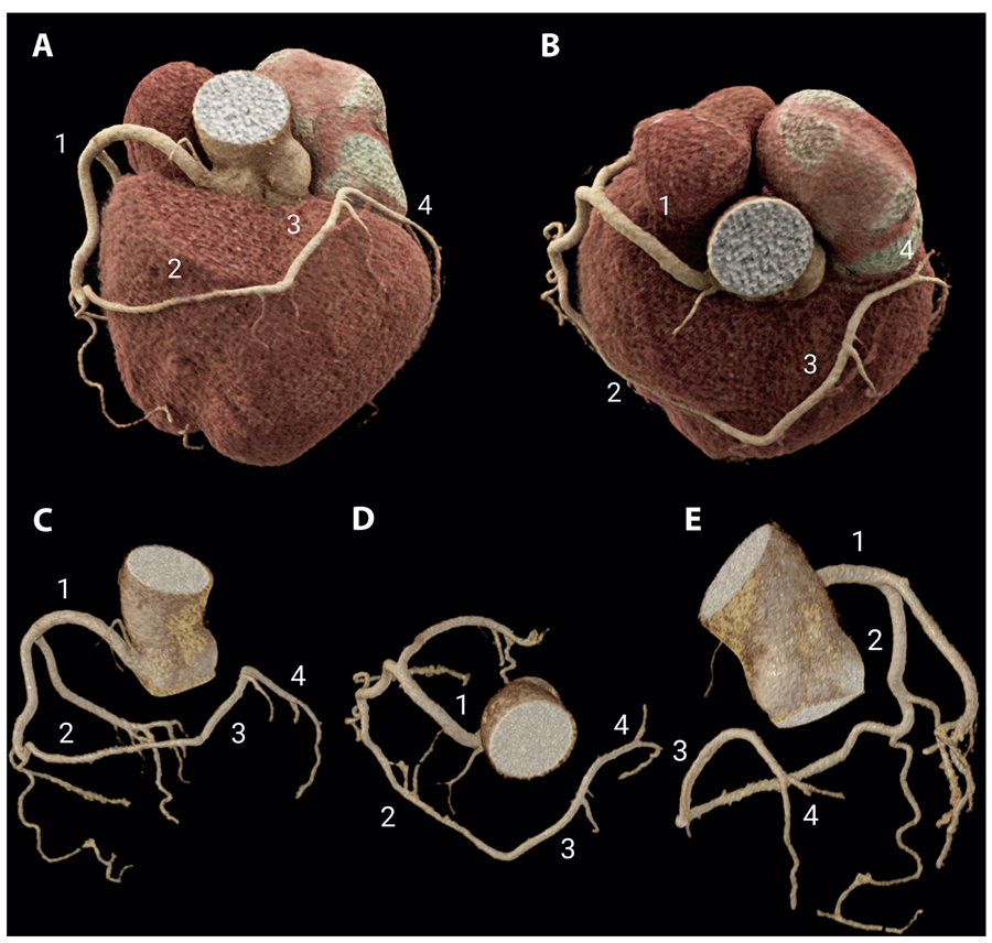
Angiography revealed a single right coronary artery (RCA), which was further evaluated by coronary computed tomography angiography (CCTA; Figure 2). The left coronary branches originated from a large right marginal branch, with an anomalous course anterior to the right ventricular conus, followed by an ascending course of the left anterior descending (LAD) artery in the interventricular sulcus and subsequent branching into the circumflex (CX) and obtuse marginal (OM) arteries. However, no evidence of plaque or stenosis was identified. Left ventriculography (Video S1) revealed regional wall motion abnormalities, with midventricular akinesia and balooning, which were further confirmed by echocardiography (Figure 3/Video S2). These findings are typically described as the midventricular phenotype of stress cardiomyopathy (SCM).

– CCTA Findings. 3D rendered cardiac (A-B) and coronary reconstruction (C-E) better depicted coronary anatomy. 1) Single RCA emerging from the right sinus with normal course through the atrioventricular sulcus. 2) Elongated right marginal artery coursing anterior to the right ventricle conus to reach the anterior interventricular sulcus. 3) Anterior interventricular branch with ascending course with septal and diagonal branches (LAD artery territory). 4) Left atrioventricular branch coursing laterally and branching into left OM branches (left CX artery territory).

– Transthoracic Echocardiogram (TTE) Findings. A-B) TTE Four-chamber view in diastole (A) and systole (B) demonstrating mid inferoseptal segment akinesia and bulging (white arrowhead). C-D) Parasternal long-axis view in diastole (C) and systole (D) demonstrating mid anteroseptal segment akinesia and bulging (yellow arrowhead). E-F) Two-chamber view in diastole (E) and systole (F) demonstrating mid inferior akinesia (red arrowhead) and mid anterior hypokinesia (blue arrowhead). TTE confirmed previous left ventriculography findings, which were suggestive of SCM.
Video S2
– Transthoracic Echocardiography. View: http://abcimaging.org/supplementary-material/2026/3902/ABCImag-2026-0023_video_S2_-_Takotsubo.mp4
The patient received supportive care, with improvement in pain and a decrease in hs-cTnT to 39 ng/L. Cardiac magnetic resonance (CMR), performed four days later, before hospital discharge, revealed a preserved ejection fraction with mild hypokinesia of the midventricular anterior and septal walls (Figure 4/Video S3). The T2-weighted short tau inversion recovery (STIR) sequence demonstrated edema in the mid-anterior and septal walls, with mild mid-wall late gadolinium enhancement, interpreted as a subacute finding of SCM. The patient was discharged after seven days, with complete pain resolution and no complications during hospitalization.

– Cardiac Magnetic Resonance Findings (CMR). Steady-state free precession (SSFP) cine images were acquired in two-chamber view in diastole (A) and systole (B) and revealed mid anterior segment hypokinesia (green arrows). Phase-sensitive inversion recovery (PSIR) late gadolinium enhancement (LGE) in the same view (C), revealed patchy mid-wall enhancement in mid anterior and mid inferior segments (green arrows). Systolic cine (D), Short-tau inversion recovery (STIR) and PSIR imagens in mid-ventricle short axis view (E and F respectively) demonstrate mid anterior and anteroseptal hypokinesia, transmural oedema (E) and patchy mid-wall LGE (F) in mid anterior, anteroseptal, inferoseptal and inferior segments (red arrows). Four-chamber view systolic cine (G), STIR (H) and PSIR (I) images demonstrated hypokinesia, oedema and patchy mid-wall LGE in mid inferoseptal segment (yellow arrows).
Video S3
– Cine Cardiac Magnetic Resonance Sequences. View: http://abcimaging.org/supplementary-material/2026/3902/ABCImag-2026-0023_video_S3_-_Takotsubo.mp4
Discussion
Isolated single coronary artery (SCA) is a rare congenital anomaly in which a single artery arises from a single coronary ostium and supplies the entire myocardium, branching in different patterns to perfuse the coronary territories.1 Although most patients remain asymptomatic, some variants, particularly those with interarterial and intramural courses, may promote exercise-induced ischemia because of extrinsic compression caused by increased pulsation of the aorta and pulmonary artery. These patients are at increased risk of sudden cardiac death during exercise, particularly younger patients under 30 years of age, with a left coronary artery following an interarterial and intramural course.2 In addition, an acute-angle takeoff and a tortuous course may result in abnormal blood flow and predispose to atherosclerotic disease because of endothelial injury.3
Recent reports have described SCM in patients with SCA, even in the absence of an interarterial course.3-6 SCM is an acute and transient myocardial injury characterized by myocardial stunning, that is, transient regional left ventricular systolic dysfunction, triggered by an emotionally or physically stressful event. The classic presentation of SCM, known as Takotsubo syndrome consists of apical and midventricular akinesia, hypokinesia, or dyskinesia associated with basal hyperkinesia, resulting in apical ballooning. However, other phenotypes have also been described, including the midventricular pattern observed in the present case.7 The mechanism of myocardial injury remains unclear. It has been hypothesized that dysregulation of the central autonomic nervous system and increased levels of stress-related neuropeptides may promote microvascular constriction, impaired perfusion, and ischemic stunning.8
Gräni et al.6 reported a case of a single RCA with a deep subpulmonary intraseptal course of the LAD/CX, presenting with the classic apical ballooning phenotype of SCM (Takotsubo syndrome). Despite the absence of an interarterial course, the transeptal path was hypothesized to contribute to vasospasm, endothelial dysfunction, and SCM. However, extrinsic compression as a potential mechanism was not observed in other reports.
Neiva et al3 described a similar case of a single RCA, with hypoplastic LAD and CX arteries arising from a posterolateral branch that coursed anterior to the right ventricular conus, presenting with classic SCM and complete remission after seven days. Salazar Marín et al.4 reported a case of a single RCA with a different branching pattern, characterized by independent origins of the LAD and CX, with prepulmonary and retroaortic courses, respectively, associated with classic SCM.
Across these reports, the anomalous origin of the LAD and CX was a common finding, though without the typical high-risk features for ischemia. However, Miura et al.5 described a left SCA coursing in the posterior sulcus, supplying the posterior descending and posterior left ventricular arteries, and presenting with the same SCM phenotype. A specific epicardial coronary artery distribution alone could not explain the occurrence of SCM in these patients, and the mechanism of myocardial injury remains unclear. All reported cases, including the present one, had a favorable prognosis with complete remission of symptoms.
Conclusion
This case is the first to describe the association between a distinct SCM phenotype, the midventricular form, and an isolated right SCA. The potential association between SCM and SCA is of increasing interest, given the growing number of reports despite the rarity of SCA. However, the mechanism predisposing patients with SCA to SCM remains unknown, and a high-risk coronary course alone was absent in most previous reports, as well as in the present case.
References
-
1 Lipton MJ, Barry WH, Obrez I, Silverman JF, Wexler L. Isolated Single Coronary Artery: Diagnosis, Angiographic Classification, and Clinical Significance. Radiology. 1979;130(1):39-47. doi: 10.1148/130.1.39.
» https://doi.org/10.1148/130.1.39 -
2 Taylor AJ, Rogan KM, Virmani R. Sudden Cardiac Death Associated with Isolated Congenital Coronary Artery Anomalies. J Am Coll Cardiol. 1992;20(3):640-7. doi: 10.1016/0735-1097(92)90019-j.
» https://doi.org/10.1016/0735-1097(92)90019-j -
3 Neiva J, Silva MP, Pires-Morais G, Dias A, Ponte M, Caeiro D, et al. Right Single Coronary Artery as an Incidental Finding in Takotsubo Syndrome and Acute Heart Failure: Case Report and Review of the Literature. Rev Port Cardiol. 2019;38(3):215-23. doi: 10.1016/j.repc.2018.06.013.
» https://doi.org/10.1016/j.repc.2018.06.013 -
4 Salazar Marín S, Hanna F, Ramírez N, Valencia JM. Takotsubo Cardiomyopathy and Single Coronary Artery: A Unique Combination. Rev Colomb Cardiol. 2016;23(4):303.e1-303.e6. doi: 10.1016/j.rccar.2015.10.001.
» https://doi.org/10.1016/j.rccar.2015.10.001 -
5 Miura S, Manabe O, Naya M, Ando A, Usami A, Miyazaki C, et al. Multimodality Evaluation of Takotsubo Cardiomyopathy in an Isolated Single Coronary Artery Anomaly. J Nucl Cardiol. 2022;29(2):874-80. doi: 10.1007/s12350-020-02312-z.
» https://doi.org/10.1007/s12350-020-02312-z -
6 Gräni C, Grunwald C, Windecker S, Siontis GCM. Coronary Artery Anomaly in Takotsubo Cardiomyopathy: Cause or Innocent Bystander? Tex Heart Inst J. 2020;47(1):44-6. doi: 10.14503/THIJ-18-6809.
» https://doi.org/10.14503/THIJ-18-6809 -
7 Singh T, Khan H, Gamble DT, Scally C, Newby DE, Dawson D. Takotsubo Syndrome: Pathophysiology, Emerging Concepts, and Clinical Implications. Circulation. 2022;145(13):1002-19. doi: 10.1161/CIRCULATIONAHA.121.055854.
» https://doi.org/10.1161/CIRCULATIONAHA.121.055854 -
8 Galiuto L, De Caterina AR, Porfidia A, Paraggio L, Barchetta S, Locorotondo G, et al. Reversible Coronary Microvascular Dysfunction: A Common Pathogenetic Mechanism in Apical Ballooning or Tako-Tsubo Syndrome. Eur Heart J. 2010;31(11):1319-27. doi: 10.1093/eurheartj/ehq039.
» https://doi.org/10.1093/eurheartj/ehq039
-
Study Association:
This study is not associated with any thesis or dissertation work.
-
Ethics Approval and Consent to Participate:
This study was approved by the Ethics Committee of the Hospital Sírio Libanês / Sociedade Beneficente de Senhoras under the protocol number 7.226.271. All the procedures in this study were in accordance with the 1975 Helsinki Declaration, updated in 2013. Informed consent was obtained from all participants included in the study.
-
Use of Artificial Intelligence:
The authors did not use any artificial intelligence tools in the development of this work.
-
Availability of Research Data:
The underlying content of the research text is contained within the manuscript.
-
Sources of Funding:
There were no external funding sources for this study.
Edited by
-
Editor responsible for the review:
Marcelo Tavares
The underlying content of the research text is contained within the manuscript.