ATTRwt Cardiac Amyloidosis and Aortic Regurgitation in a Patient with Acute Myelomonocytic Leukemia: an Unusual Combination
Amyloidosis; Aortic Valve Insufficiency; Acute Myelomonocytic Leukemia
Amiloidose; Insuficiência da Valva Aórtica; Leucemia Mielomonocítica Aguda
Amyloidosis; Aortic Valve Insufficiency; Acute Myelomonocytic Leukemia
Amiloidose; Insuficiência da Valva Aórtica; Leucemia Mielomonocítica Aguda
Introduction
Systemic amyloidosis is a relatively rare multisystem disease caused by the deposition of misfolded protein in various tissues and organs. Cardiac amyloidosis (CA) is recognized as an underlying cause of left ventricular wall thickening, heart failure, and arrhythmia with variable clinical presentation. The clinical diagnosis of cardiac involvement in amyloidosis is challenging, requiring a patient-centered diagnostic work-up that ensures an appropriate diagnosis.1 , 2
Although amyloidosis has been associated with multiple malignant disorders, its association with leukemia is rather uncommon.3 - 5 Also, there is a relationship between CA and valvular heart disease, particularly aortic stenosis (AS).6 The association of ATTRwt CA and aortic regurgitation (AR) in a patient with acute myelomonocytic leukemia (MML) is unique and has not been described so far.
Description
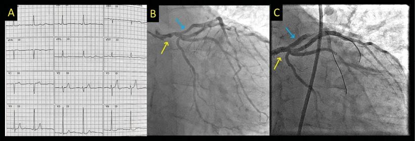
A 72-year-old hypertensive male patient diagnosed with acute AML was referred to the emergency department with a recent onset of chest discomfort at rest and signs of mild pulmonary congestion. At presentation, physical examination revealed blood pressure of 195/62 mmHg, heart rate of 96 beats per minute, respiratory rate of 26 movements per minute, and oxygen saturation of 90%. There were jugular venous distention and bilateral rales. The cardiac auscultation revealed an S3-gallop and a diastolic murmur in the aortic position. The electrocardiogram (ECG) revealed sinus rhythm, with no signs of ST segment changes or T-wave abnormalities ( Figure 1A ). The cardiac troponin was 2,025 ng/L (reference of 58 ng/L). The patient was started on treatment for non-ST elevation acute coronary syndrome (ACS). After clinical stabilization, a coronary angiogram was performed, revealing a critical lesion in the proximal aspect of the left anterior descending artery and the left trunk ( Figure 1B , Supplementary Video 1). The patient was then treated with drug-eluting stents in the left trunk and left anterior descending artery (LAD) with success ( Figure 1C , Supplementary Video 2).

– (1A): The 12-lead ECG at the presentation showed no significant ST/T abnormalities. (1B): Coronary angiogram showing severe lesion in the left coronary trunk (yellow arrow) and left anterior descending artery (blue arrow). (1C): coronary angiogram after drug-eluting stents in the left coronary trunk (yellow arrow) and left anterior descending artery (blue arrow).
The patient’s past medical history was characterized by acute MML, which had been treated with decitabine for 18 months with a good clinical response. Other medications included losartan 50 mg twice a day, hydrochlorothiazide 25 mg once a day, and rosuvastatin 10 mg once a day. During his admission, laboratory exams showed hemoglobin of 9.5 g/dL, platelets of 172,000 /mm3, and leucocyte count of 6,560/mm3 with 9% immature forms. A transthoracic echocardiogram was performed for risk stratification post-ACS, showing findings suggestive of CA: there was concentric thickening of the cardiac chambers, with grade 2 diastolic impairment function. The ejection fraction was 53%, and the global longitudinal strain (GLS) was -15.9% with strain magnitude preserved exclusively at the apex (apical sparring pattern) ( Figure 2A ). There was also aortic valvular thickening with moderate AR ( Figure 2B ).
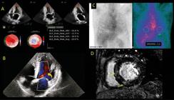
– (2A): Echocardiogram showing GLS of 15.9% with apical sparring pattern. (2B): Echocardiogram (5-chamber view) with color Doppler showing AR (orange arrow). (2C): Technetium-pyrophosphate cardiac single-photon emission computed tomography (Tc-PYP SPECT) revealing heart uptake equal to rub uptake (grade 2 visual scale) and quantitative analysis with heart-to-contralateral lung uptake ration (H/CL) of 1.3 in the third hour. (2D): Cardiac MRI reveals mild irregular and diffuse areas of late gadolinium enhancement within basal and mid segments of the LV (yellow arrows).
A work-up for amyloidosis was then started. There was no evidence of monoclonal protein. The serum free light chain (sFLC) assay revealed a normal ratio of kappa and lambda free light chain (0.77, considering a glomerular filtration rate of 52 ml/min). The serum (SIFE) and urine immunofixation electrophoresis (UIFE) revealed the absence of monoclonal protein.
A technetium-pyrophosphate cardiac single-photon emission computed tomography (Tc-PYP SPECT) was performed, revealing heart uptake equal to rub uptake (grade 2 visual scale, as described by Perugini et al.).7 The quantitative analysis revealed a heart-to-contralateral lung uptake ratio (H/CL) of 1.5 in the first hour and 1.3 in the third hour ( Figure 2C ).
A cardiac magnetic resonance imaging (MRI) revealed biventricular function within normal limits, with a left ventricular ejection fraction (LVEF) of 56% and a right ventricular ejection fraction (RVEF) of 38.5%. The was increased thickening of the LV walls, particularly in the mid septum (1.6 cm). There were mild irregular and diffuse areas of late gadolinium enhancement within basal and mid segments of LV ( Figure 2D ).
Genetic testing for transthyretin (TTR) gene mutation was negative for TTR variants known to cause CA. A diagnosis of wild-type transthyretin amyloidosis (ATTRwt) was performed. Despite this diagnosis, tafamidis was not utilized, based on the decision made by the National Committee for the Incorporation of Technologies (Conitec), an institution to assist the Brazilian Ministry of Health in decision-making (Conitec, Brazilian Ministry of Health, 2022) * .
The patient continued hematological treatment for MML and control of other cardiac risk factors with good clinical evolution. His pharmacological treatment consisted of losartan, hydrochlorothiazide, amlodipine, hydralazine, spironolactone, bisoprolol, and dapagliflozin. After two years of good cardiac evolution and no further complications, the patient experienced a hematological progression of his disease and died after an episode of respiratory sepsis.
Discussion
Systemic amyloidoses consist of protein misfolding disorders that form insoluble amyloid fibrils, which, in turn, are deposited in the tissues leading to organ damage. Two types of amyloid account for 95% of CA: light-chain amyloid (AL), due to immunoglobin light-chain deposition, and TTR cardiac amyloidosis (ATTR-CM), which can be due to hereditary mutation (ATTRh) or ATTRwt. Hereditary transthyretin cardiac amyloidosis (hATTR-CM) is caused by one of the known heritable mutations in the TTR gene. In contrast, ATTRwt is caused by age-related changes in the wild-type TTR.1 , 2 According to the most recent guidelines, AL can be excluded by obtaining a monoclonal protein screen comprising three laboratory tests: sFLC, SIFE, and UIFE. Additionally, the combined finding of positive bone radiotracer scintigraphy in patients with no evidence of detectable monoclonal protein in urine or serum was found to be 100% specific for ATTR-CM.1 , 8
Although systemic amyloidosis has been associated with multiple malignant disorders, its association with leukemia is rather uncommon. In 1970, Kyle et al. described a rapidly progressive acute MML development in a patient with systemic amyloidosis of five years’ duration who had received a prolonged course of melphalan.3 Chronic lymphocytic leukemia can also be related to AL amyloidosis involving the heart. Cases of chronic MML were described in association with primary systemic amyloidosis by Cohen et al.5 and Okuda et al.4 As far as we are concerned, this is the first report of the coexistence of acute MML and ATTRwt amyloidosis. Although a non-biopsy diagnosis of ATTRwt can be obtained when certain diagnostic criteria are fulfilled, tissue characterization still represents the gold standard for the diagnosis and typing of CA. In this case, however, financial constraints and limitations of cardiac biopsy in our health system made this procedure unsuitable.1 , 2
Among valvular heart diseases, AS is the most prevalent disease in amyloidosis, with a prevalence of 16% in patients with severe AS planned to undergo transcatheter aortic valve replacement (TAVR).9 Treibel et al. described that approximately 1 in 7 patients undergoing TAVR has occult CA.6 Cases of mitral and tricuspid regurgitation seem to be increased in patients with CA.10 The presence of AR has not been associated with CA. As far as we are concerned, this is the first description of CA in a patient with aortic insufficiency. Higher quality treatment of hematological and cardiological conditions has possibly led to an improvement in this patient’s survival, which reinforces the role of cardio-oncology specialists in regular monitoring of these patients.
Conclusion
CA is an infiltrative disease that requires a high clinical suspicion for an appropriate diagnosis. In this case, we describe a very unusual association between ATTRwt CA and AR in a patient with acute MML. The increasing knowledge of CA and the improved prognosis of oncology diseases are possible causes for such association.
References
-
1 Simões MV, Fernandes F, Marcondes-Braga FG, Scheinberg P, Correia EB, Rohde LEP, et al. Position Statement on Diagnosis and Treatment of Cardiac Amyloidosis - 2021. Arq Bras Cardiol. 2021;117(3):561-98. doi: 10.36660/abc.20210718.
» https://doi.org/10.36660/abc.20210718 -
2 Kittleson MM, Ruberg FL, Ambardekar AV, Brannagan TH, Cheng RK, Clarke JO, et al. 2023 ACC Expert Consensus Decision Pathway on Comprehensive Multidisciplinary Care for the Patient with Cardiac Amyloidosis: A Report of the American College of Cardiology Solution Set Oversight Committee. J Am Coll Cardiol. 2023;81(11):1076-126. doi: 10.1016/j.jacc.2022.11.022.
» https://doi.org/10.1016/j.jacc.2022.11.022 -
3 Kyle RA, Pierre RV, Bayrd ED. Multiple Myeloma and Acute Myelomonocytic Leukemia. N Engl J Med. 1970;283(21):1121-5. doi: 10.1056/NEJM197011192832101.
» https://doi.org/10.1056/NEJM197011192832101 - 4 Okuda T, Yumoto Y, Kamakari K, Kano T. Primary Amyloidosis Complicated with Chronic Myelomonocytic Leukemia. Rinsho Ketsueki. 1996;37(9):858-62.
-
5 Cohen AM, Mittelman M, Gal R, Halperin M, Djaldetti M. Chronic Myelomonocytic Leukemia Associated with Primary Amyloidosis. Leuk Lymphoma. 1994;16(1-2):183-7. doi: 10.3109/10428199409114157.
» https://doi.org/10.3109/10428199409114157 -
6 Treibel TA, Fontana M, Gilbertson JA, Castelletti S, White SK, Scully PR, et al. Occult Transthyretin Cardiac Amyloid in Severe Calcific Aortic Stenosis: Prevalence and Prognosis in Patients Undergoing Surgical Aortic Valve Replacement. Circ Cardiovasc Imaging. 2016;9(8):e005066. doi: 10.1161/CIRCIMAGING.116.005066.
» https://doi.org/10.1161/CIRCIMAGING.116.005066 -
7 Perugini E, Guidalotti PL, Salvi F, Cooke RM, Pettinato C, Riva L, et al. Noninvasive Etiologic Diagnosis of Cardiac Amyloidosis Using 99mTc-3,3-Diphosphono-1,2-Propanodicarboxylic Acid Scintigraphy. J Am Coll Cardiol. 2005;46(6):1076-84. doi: 10.1016/j.jacc.2005.05.073.
» https://doi.org/10.1016/j.jacc.2005.05.073 -
8 Gillmore JD, Maurer MS, Falk RH, Merlini G, Damy T, Dispenzieri A, et al. Nonbiopsy Diagnosis of Cardiac Transthyretin Amyloidosis. Circulation. 2016;133(24):2404-12. doi: 10.1161/CIRCULATIONAHA.116.021612.
» https://doi.org/10.1161/CIRCULATIONAHA.116.021612 -
9 Castaño A, Narotsky DL, Hamid N, Khalique OK, Morgenstern R, De Luca A, et al. Unveiling Transthyretin Cardiac Amyloidosis and its Predictors among Elderly Patients with Severe Aortic Stenosis Undergoing Transcatheter Aortic Valve Replacement. Eur Heart J. 2017;38(38):2879-87. doi: 10.1093/eurheartj/ehx350.
» https://doi.org/10.1093/eurheartj/ehx350 -
10 Aimo A, Camerini L, Fabiani I, Morfino P, Panichella G, Barison A, et al. Valvular Heart Disease in Patients with Cardiac Amyloidosis. Heart Fail Rev. 2024;29(1):65-77. doi: 10.1007/s10741-023-10350-1.
» https://doi.org/10.1007/s10741-023-10350-1
-
Study Association:
This article is part of the Final Paper of the Specialization in Cardio-Oncology submitted by Diego Chemello, from Sociedade Brasileira de Cardiologia, Instituto Nacional do Câncer.
-
Ethics Approval and Consent to Participate:
This study was approved by the Ethics Committee of the Universidade Federal de Santa Maria under the protocol number 83537224.0.0000.5346. All the procedures in this study were in accordance with the 1975 Helsinki Declaration, updated in 2013. Informed consent was obtained from all participants included in the study.
-
Sources of Funding:
There were no external funding sources for this study.
*Supplemental Materials
See the Supplemental Video 1, please click here .
See the Supplemental Video 2, please click here .
Edited by
-
Editor responsible for the review:
Marcelo Tavares