
 My Approach to Wilkins-Block score in rheumatic mitral stenosis
Abstract
Mitral stenosis (MS) is the main clinical expression of chronic rheumatic heart disease. In symptomatic patients with severe MS and favorable anatomy, percutaneous mitral balloon valvuloplasty (PMBV) is the first-line therapeutic strategy. For the proper selection of candidates, the Wilkins-Block echocardiographic score was developed to characterize mitral valve morphology and predict eligibility for the procedure. This score includes four structural domains: leaflet mobility, valve thickening, degree of calcification, and subvalvular apparatus involvement, grading each parameter from 1 to 4 points, resulting in an overall range of 4 to 16 points, in increasing order of anatomical severity. The Wilkins score has become a widely validated tool, showing a consistent correlation with the immediate and long-term outcomes of PMBV. Among the limitations of the score, the absence of commissural anatomy assessment stands out, a variable recognized as a relevant prognostic determinant due to its strong association with the occurrence of post-procedure mitral regurgitation, considered the most frequent and clinically significant complication of PMBV. With the accumulation of clinical and technical experience, the indications for PMBV have been progressively extended to include patients with less favorable anatomical profiles. In these contexts, selection must be particularly rigorous, incorporating not only morphological criteria derived from echocardiographic evaluation, but also clinical aspects, in order to ensure satisfactory results and minimize the risk of complications.
Keywords
Mitral Valve Stenosis; Echocardiography; Rheumatic Heart Disease
Resumo
A estenose mitral (EM) constitui a principal expressão clínica da cardiopatia reumática crônica. Em pacientes sintomáticos portadores de EM grave, e que apresentem anatomia favorável, a valvoplastia mitral percutânea com balão (VMPB) configura-se como a estratégia terapêutica de primeira escolha. Para a seleção adequada dos candidatos, o escore ecocardiográfico de Wilkins-Block foi desenvolvido com o objetivo de caracterizar a morfologia valvar mitral e predizer a elegibilidade ao procedimento. Esse escore contempla quatro domínios estruturais: mobilidade dos folhetos, espessamento valvar, grau de calcificação e envolvimento do aparato subvalvar, graduando cada parâmetro de 1 a 4 pontos, o que resulta em uma variação global de 4 a 16 pontos, em ordem crescente de gravidade anatômica. O escore de Wilkins consolidou-se como ferramenta amplamente validada, apresentando correlação consistente com os desfechos imediatos e de longo prazo da VMPB. Entre as limitações do escore, destaca-se a ausência de avaliação da anatomia comissural, variável reconhecida como determinante prognóstico relevante por sua forte associação com a ocorrência de insuficiência mitral pós-procedimento, considerada a complicação mais frequente e clinicamente significativa da VMPB. Com o acúmulo de experiência clínica e técnica, as indicações da VMPB foram progressivamente estendidas para abranger pacientes com perfis anatômicos menos favoráveis. Nesses contextos, a seleção deve ser particularmente criteriosa, incorporando não apenas os critérios morfológicos derivados da avaliação ecocardiográfica, mas também aspectos clínicos, de modo a assegurar resultados satisfatórios e minimizar o risco de complicações.
Palavras-chave
Estenose da Valva Mitral; Ecocardiografia; Cardiopatia Reumática
Introduction
Rheumatic fever is a disease resulting from pharyngotonsillitis caused by the bacterium Streptococcus pyogenes in susceptible individuals1. Chronic rheumatic heart disease (CRHD) is the most important consequence of rheumatic fever and constitutes a significant public health problem, especially in low- and middle-income countries, where it continues to be associated with high morbidity and mortality.1,2 The mitral valve (MV) is the most frequently affected in CRHD, with mitral stenosis (MS) being its main chronic manifestation.
Pathophysiology of mitral stenosis
Rheumatic mitral stenosis (RMS) results from thickening and calcification of the cusps, shortening of the chordae tendineae, and commissural fusion, progressively reducing the valve area. The consequent atrioventricular diastolic gradient leads to pulmonary venocapillary congestion, pulmonary hypertension, and right ventricular dysfunction.3 Left atrial pressure overload promotes structural remodeling, predisposing to arrhythmias, especially atrial fibrillation (AF), which precipitates symptoms and increases the risk of thromboembolic events. It is estimated that 80% of strokes in patients with rheumatic stenosis occur in the MS-AF association, with a substantial impact on quality of life.2,4
Echocardiography in the evaluation of the mitral valve
The MV is a complex anatomical structure that includes the fibromuscular ring, two cusps (anterior and posterior), chordae tendineae, and papillary muscles, and has a close structural relationship with the underlying ventricular myocardium.5 The anterior cusp is longer and semicircular, while the posterior cusp is shorter and segmented; the posterior cusp is subdivided into three segments (P1-lateral, P2-central, P3-medial), corresponding to A1, A2, and A3 of the anterior cusp.5
Echocardiographic evaluation of the mitral valve requires a multimodal approach, in which two-dimensional transthoracic echocardiography (2D TTE) represents the initial and fundamental method for morphological and functional analysis of the valve. This modality enables an accurate analysis of the thickness and mobility of the cusps, the presence and extent of calcifications, and the integrity and impairment of the subvalvular apparatus.6 Doppler ultrasound allows for the quantification of the severity of mitral valve lesions by measuring transvalvular gradients, effective orifice areas in cases of stenosis, and regurgitation parameters, such as volume and regurgitant fraction. Color Doppler analysis complements this assessment, allowing for the visualization of the regurgitant jet, estimation of the vena contracta, and application of the Proximal Isovelocity Surface Area (PISA) technique for a more accurate quantification of mitral regurgitation.6 The Transesophageal echocardiography (TEE) is recommended when greater anatomical detail is needed, particularly in the characterization of mitral regurgitation mechanisms, the evaluation of infective endocarditis, and the planning of percutaneous or surgical therapies.5 Three-dimensional (3D) echocardiography, especially via TEE, improves the spatial analysis of the valve, enabling multiplanar reconstructions with direct anatomical correlation, a more precise measurement of the valve area, a dynamic evaluation of the annulus, and the segmental characterization of the cusps.6
Percutaneous mitral balloon valvuloplasty
The PMBV. introduced in 1984 by Inoue et al., is the treatment of choice for severe mitral stenosis with favorable anatomy.7 It is an invasive procedure, with efficacy comparable to surgical commissurotomy.8 Balloon inflation promotes mitral valve opening, primarily through commissural separation of fibrotic adhesions.9 When inflated inside the valve orifice, the balloon generates radial forces that concentrate in the commissural regions, promoting tension redistribution and favoring symmetrical orifice opening.9
Prior echocardiographic evaluation is mandatory to determine if the MV anatomy is adequate for commissural opening and if it will allow for an increased valve area without inducing significant mitral regurgitation. Several scores have been developed for this purpose.10 Obtaining good results depends particularly on the appropriate selection of patients. Several outcome predictors have been described, including age, functional class, previous commissurotomy, valve area, valve anatomy, and balloon size. Among these, mitral valve morphology is the single most determinant factor, reinforcing the need for the systematic use of echocardiographic scores. Unfavorable results are frequently related to adverse morphological changes, especially cusp calcification and subvalvular involvement.11 To estimate the probability of successful dilation, the Wilkins-Block score was developed, based on the 2D assessment of valve morphology.
Wilkins-Block Score
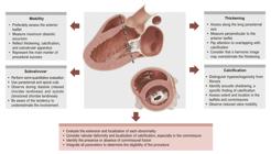
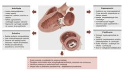
Wilkins et al., in 1988,12 proposed an echocardiographic score for the structural evaluation of RMS, which demonstrated a significant correlation with valve opening capacity after percutaneous valvuloplasty. In the original score, success was defined dichotomously by obtaining a mitral valve area > 1.0 cm2, without considering the development of mitral regurgitation. This score is obtained through a systematic and relatively simple analysis of valve morphology as viewed through a TTE, using the conventional cuts employed in the evaluation of the mitral valve. In certain situations, modified cuts may be necessary for better anatomical characterization, especially of the subvalvular apparatus (Central Figure).
The evaluation of each parameter is performed subjectively through a semi-quantitative score that covers leaflet mobility, valve thickening, degree of calcification, and involvement of the subvalvular apparatus. Each item receives a score from 1 to 4 points, resulting in a total score ranging from 4 to 16 points, in ascending order of structural severity. In general, patients with a score ≤ 8 have favorable anatomy and are the best candidates for PMBV, with high rates of immediate success and maintenance of long-term results.13 Patients with intermediate scores, between 9 and 11, may still be considered for the procedure, especially when a detailed evaluation of the commissures suggests a favorable morphology. Scores ≥ 12 reflect extensively affected and calcified valves, in which the probability of significant hemodynamic gain is reduced and the chance of complications increases, making surgical valve replacement more appropriate.
Leaflet mobility is graded according to the degree of restriction of the anterior leaflet during the diastole (Figure 1A). Increased restriction corresponds to progressively higher scores. Thus, when the limitation is restricted to the ends of the leaflets, 1 point is assigned. In cases where mobility is preserved in the medial and basal regions, the value is 2 points. Restriction of mobility limited only to the basal region corresponds to 3 points. Finally, complete immobility of the leaflets is scored with 4 points. Table 1 presents a practical checklist for the systematic application of the Wilkins score.

– Schematic representation of the Wilkins score. A: Leaflet mobility (red marking indicates progression of reduced mobility). B: Subvalvular involvement (red marking indicates progression of thickening and shortening of the chordae tendineae). C: Anterior leaflet thickening (the detailed image shows the values that determine each score). D: Valvular calcification (white dots progress in quantity and extent across the leaflets).
Subvalvular involvement refers to the involvement of the chordae tendineae and papillary muscles (Figure 1B). One point is assigned when the thickening is minimal and restricted to the region immediately below the leaflets. Thickening that extends beyond this level, but that is limited to the proximal third of the chordae tendineae, corresponds to 2 points. When there is involvement that reaches the distal third of the chordae tendineae, 3 points are assigned. Finally, diffuse thickening associated with the shortening of all chordae tendineae, extending to the level of the papillary muscles, receives 4 points.
The thickness of the leaflets is measured in the parasternal section at maximum diastole (Figure 1C). Leaflets with thickness close to normal (≈4–5 mm) receive 1 point. Thickening restricted to the extremities, preserving the middle region, with measurements between 5–8 mm, corresponds to 2 points. Diffuse thickening involving all portions of the leaflet, still within the range of 5–8 mm, receives 3 points. When there is significant thickening of the entire leaflet, with measurements greater than 8–10 mm, 4 points are assigned.
Valvular calcification is determined by the presence and extent of hyperechogenic areas (Figure 1D). The isolated presence of a small area receives 1 point. Minimal areas confined to the extremities correspond to 2 points. When calcification extends to the middle region of the leaflets, 3 points are assigned. Diffuse calcification, extending beyond the limits of the leaflets, is scored with 4 points.
Although each component of the score is evaluated individually, the parameter that best reflects the overall morphology of the valve is the sum of the scores assigned to each item, translating the degree of structural involvement. Leaflet mobility, in turn, constitutes an integrating parameter, as it reflects the combined impact of thickening, calcification, and subvalvular involvement, all contributing to the restriction of valve movement. Figure 2 illustrates a case of mitral stenosis with thin leaflets, restriction limited to the extremities, absence of calcification, and no evidence of subvalvular involvement. By contrast, Figure 3 demonstrates calcification affecting the medial and distal portions of the leaflets, with a preservation of the basal region and no involvement of the commissures, which would not contraindicate the percutaneous procedure.

– Rheumatic mitral stenosis (RMS) with favorable anatomy. 2D transthoracic echocardiogram (TTE): parasternal long-axis view (A) and apical four-chamber view (B), showing mitral valve with thin, mobile leaflets without calcification, consistent with anatomy favorable to PMBV.

– Valvular calcification. Echocardiographic images show the calcification of the mitral valve leaflets, affecting their medial and distal portions, with preservation of the base, demonstrated in the parasternal long-axis view (A) and in the apical four-chamber view (B). It is important to note that, in this case, the calcification is restricted to the leaflets, without commissure involvement, which does not constitute a contraindication to PMBV.
Experience accumulated over the years
Since its introduction, the Wilkins score continues to be the most widely used tool for selecting suitable candidates for PMBV, due to its simplicity and practicality.14 The main valvular heart disease guidelines of the AHA/ACC and ESC7,15 use the Wilkins score as an eligibility criterion for patients with rheumatic MS for the percutaneous procedure.
In addition to its usefulness in morphological characterization, the Wilkins score is an important predictor of long-term outcomes after PMBV.16 However, this score does not include the assessment of commissural anatomy, currently recognized as a relevant prognostic determinant due to its association with the development of post-procedure mitral regurgitation (MR).17Figure 4 illustrates the wide spectrum of commissural involvement patterns in RMS. In patients with marked commissural asymmetry, balloon inflation tends to exert disproportionate force on the contralateral commissure, predisposing to the onset of MR. Similarly, when there is significant commissural calcification, the balloon’s inability to adequately separate the commissures results in excessive force transmission to the valve tissue, favoring rupture or laceration of the leaflets.18,19

– Patterns of commissural involvement (parasternal short axis view). A: symmetrical commissural involvement. B/C: commissural asymmetry with predominance of thickening and calcification of the posteromedial commissure. D: large valvular deformity and commissural asymmetry with predominance of thickening and calcification of the anterolateral commissure.
With the advancement of experience, the indications for PMBV have been progressively expanded to include less favorable conditions, demonstrating that it is a safe and effective procedure in certain patients with an intermediate Wilkins score.16 The selection of these cases, however, must be judicious and individualized, considering not only echocardiographic parameters, but also clinical and hemodynamic aspects, such as age, history of previous commissurotomy, NYHA functional class, presence of atrial fibrillation and pulmonary hypertension.14 Among the echocardiographic criteria, intense calcification of the leaflets, severe or asymmetric commissural calcification or fusion, and the presence of extensive subvalvular disease stand out as major risk factors for complications.20
Limitations of the Wilkins-Block score
Despite its widespread use in clinical practice, the Wilkins score has important limitations. The analysis of the components is semi-quantitative and subject to interobserver variability, in addition to assigning identical weight to variables that have different predictive capacities for the post-PMBV outcome. The score also does not assess commissural anatomy, a fundamental aspect in predicting post-PMBV MR, and frequently underestimates subvalvular involvement. Added to this are the difficulty in distinguishing nodular fibrosis from calcification, the disregard for the heterogeneous distribution of lesions, and the lack of integration of information provided by transesophageal or 3D echocardiography.10,11,17
In view of these limitations, new models, such as the Nunes score,17 have been proposed. Unlike the Wilkins score, the Nunes score assigns different weights to variables, incorporates commissural parameters, and allows one to predict the risk of post-procedure MR, in addition to identifying good candidates for PMBV even among patients with borderline Wilkins scores (9 to 11 points), traditionally considered to be at a higher risk.17Table 2 presents the main studies that described new models for evaluating MV morphology, when compared to the Wilkins score. With the evolution of PMBV, several studies have also identified additional valve morphology variables associated with post-PMBV outcomes, highlighting the inherent limitations of the Wilkins score.
Conclusions and future perspectives
The Wilkins score has become an international benchmark for selecting patients for PMBV, due to its simplicity and broad clinical applicability. However, the evidence accumulated over the last few decades demonstrates that its limitations reduce the ability to predict post-procedure outcomes in certain scenarios, especially due to the lack of assessment of commissural anatomy and the uniform weighting assigned to variables with unequal prognostic impact. Other scores, including the Nunes score, emerge as promising alternatives, incorporating additional parameters and a greater ability to discriminate outcomes, especially in the subgroup of patients with intermediate Wilkins scores. These advances contribute to reducing complications, such as post-procedure mitral regurgitation, and increasing the safety of PMBV recommendations within more heterogeneous populations.
In the future, the integration of new imaging technologies, including 3D echocardiography, TEE, and quantitative morphological analysis techniques, should further refine the structural assessment of the MV. Furthermore, algorithms based on artificial intelligence and machine learning can offer more robust predictive models, capable of accurately combining anatomical, clinical, and hemodynamic parameters. Such strategies would not only be able to improve the selection of candidates for PMBV, but they could also guide therapeutic decisions in cases of unfavorable anatomy, expanding the role of echocardiography in the contemporary management of RMS.
References
- 1 Cunningham MW, Kirvan CA. Post-Streptococcal Autoimmune Sequelae, Rheumatic Fever and Beyond: A New Perspective. In: Ferretti JJ, Stevens DL, Fischetti VA, editors. Streptococcus pyogenes: Basic Biology to Clinical Manifestations. 2nd ed. Oklahoma City: University of Oklahoma Health Sciences Center; 2022.
-
2 Figueiredo FA, Esteves WAM, Hung J, Gomes NFA, Taconeli CA, Pantaleão AN, et al. Left Atrial Function in Patients with Rheumatic Mitral Stenosis: Addressing Prognostic Insights Beyond Atrial Fibrillation Prediction. Eur Heart J Imaging Methods Pract. 2024;2(2):qyae067. doi: 10.1093/ehjimp/qyae067.
» https://doi.org/10.1093/ehjimp/qyae067 -
3 Jain S, Mankad SV. Echocardiographic Assessment of Mitral Stenosis: Echocardiographic Features of Rheumatic Mitral Stenosis. Cardiol Clin. 2013;31(2):177-91. doi: 10.1016/j.ccl.2013.03.006.
» https://doi.org/10.1016/j.ccl.2013.03.006 -
4 Vasconcelos M, Vasconcelos L, Ribeiro V, Campos C, Di-Flora F, Abreu L, et al. Incidence and Predictors of Stroke in Patients with Rheumatic Heart Disease. Heart. 2021;107(9):748-54. doi: 10.1136/heartjnl-2020-318054.
» https://doi.org/10.1136/heartjnl-2020-318054 -
5 Robinson S, Ring L, Augustine DX, Rekhraj S, Oxborough D, Harkness A, et al. The Assessment of Mitral Valve Disease: A Guideline from the British Society of Echocardiography. Echo Res Pract. 2021;8(1):G87-G136. doi: 10.1530/ERP-20-0034.
» https://doi.org/10.1530/ERP-20-0034 -
6 Nishimura RA, Otto CM, Bonow RO, Carabello BA, Erwin JP 3rd, Fleisher LA, et al. 2017 AHA/ACC Focused Update of the 2014 AHA/ACC Guideline for the Management of Patients with Valvular Heart Disease: A Report of the American College of Cardiology/American Heart Association Task Force on Clinical Practice Guidelines. Circulation. 2017;135(25):e1159-95. doi: 10.1161/CIR.0000000000000503.
» https://doi.org/10.1161/CIR.0000000000000503 -
7 Otto CM, Nishimura RA, Bonow RO, Carabello BA, Erwin JP 3rd, Gentile F, et al. 2020 ACC/AHA Guideline for the Management of Patients with Valvular Heart Disease: Executive Summary: A Report of the American College of Cardiology/American Heart Association Joint Committee on Clinical Practice Guidelines. J Am Coll Cardiol. 2021;77(4):450-500. doi: 10.1016/j.jacc.2020.11.035.
» https://doi.org/10.1016/j.jacc.2020.11.035 -
8 Turi ZG, Reyes VP, Raju BS, Raju AR, Kumar DN, Rajagopal P, et al. Percutaneous Balloon versus Surgical Closed Commissurotomy for Mitral Stenosis. A Prospective, Randomized Trial. Circulation. 1991;83(4):1179-85. doi: 10.1161/01.cir.83.4.1179.
» https://doi.org/10.1161/01.cir.83.4.1179 -
9 Palacios I, Block PC, Brandi S, Blanco P, Casal H, Pulido JI, et al. Percutaneous Balloon Valvotomy for Patients with Severe Mitral Stenosis. Circulation. 1987;75(4):778-84. doi: 10.1161/01.cir.75.4.778.
» https://doi.org/10.1161/01.cir.75.4.778 -
10 Silbiger JJ. Advances in Rheumatic Mitral Stenosis: Echocardiographic, Pathophysiologic, and Hemodynamic Considerations. J Am Soc Echocardiogr. 2021;34(7):709-722.e1. doi: 10.1016/j.echo.2021.02.015.
» https://doi.org/10.1016/j.echo.2021.02.015 -
11 Soliman OI, Anwar AM, Metawei AK, McGhie JS, Geleijnse ML, Ten Cate FJ. New Scores for the Assessment of Mitral Stenosis Using Real-Time Three-Dimensional Echocardiography. Curr Cardiovasc Imaging Rep. 2011;4(5):370-7. doi: 10.1007/s12410-011-9099-z.
» https://doi.org/10.1007/s12410-011-9099-z -
12 Wilkins GT, Weyman AE, Abascal VM, Block PC, Palacios IF. Percutaneous Balloon Dilatation of the Mitral Valve: An Analysis of Echocardiographic Variables Related to Outcome and the Mechanism of Dilatation. Br Heart J. 1988;60(4):299-308. doi: 10.1136/hrt.60.4.299.
» https://doi.org/10.1136/hrt.60.4.299 -
13 Palacios IF. Percutaneous Mitral Balloon Valvuloplasty - State of the Art. Mini-Invasive Surg. 2020;4:73. doi: 10.20517/2574-1225.2020.72.
» https://doi.org/10.20517/2574-1225.2020.72 -
14 Prota-Filho LEB, Meneguz-Moreno RA, Queiroz CCV, Wohnrath FC, Carboni FAC, Silva GRC, et al. Novel Prognostic Score for Immediate and Late Success after Percutaneous Mitral Balloon Commissurotomy in Patients with Mitral Stenosis. J Invasive Cardiol. 2020;32(6):211-7. doi: 10.25270/jic/19.00363.
» https://doi.org/10.25270/jic/19.00363 -
15 Vahanian A, Beyersdorf F, Praz F, Milojevic M, Baldus S, Bauersachs J, et al. 2021 ESC/EACTS Guidelines for the Management of Valvular Heart Disease: Developed by the Task Force for the Management of Valvular Heart Disease of the European Society of Cardiology (ESC) and the European Association for Cardio-Thoracic Surgery (EACTS). Rev Esp Cardiol. 2022;75(6):524. doi: 10.1016/j.rec.2022.05.006.
» https://doi.org/10.1016/j.rec.2022.05.006 -
16 Paiva M, Correia AS, Lopes R, Gonçalves A, Almeida R, Almeida PB, et al. Selection of Patients for Percutaneous Balloon Mitral Valvotomy: Is there a Definitive Limit for the Wilkins Score? Rev Port Cardiol. 2013;32(11):873-8. doi: 10.1016/j.repc.2013.02.017.
» https://doi.org/10.1016/j.repc.2013.02.017 -
17 Nunes MC, Tan TC, Elmariah S, Lago R, Margey R, Cruz-Gonzalez I, et al. The Echo Score Revisited: Impact of Incorporating Commissural Morphology and Leaflet Displacement to the Prediction of Outcome for Patients Undergoing Percutaneous Mitral Valvuloplasty. Circulation. 2014;129(8):886-95. doi: 10.1161/CIRCULATIONAHA.113.001252.
» https://doi.org/10.1161/CIRCULATIONAHA.113.001252 -
18 Nunes MCP, Levine RA, Braulio R, Pascoal-Xavier MA, Elmariah S, Gomes NFA, et al. Mitral Regurgitation after Percutaneous Mitral Valvuloplasty: Insights into Mechanisms and Impact on Clinical Outcomes. JACC Cardiovasc Imaging. 2020;13(12):2513-26. doi: 10.1016/j.jcmg.2020.07.020.
» https://doi.org/10.1016/j.jcmg.2020.07.020 -
19 Passos LSA, Becker-Greene D, Braulio R, Le TD, Gelape CL, Almeida LFR, et al. Proinflammatory Matrix Metalloproteinase-1 Associates with Mitral Valve Leaflet Disruption Following Percutaneous Mitral Valvuloplasty. Front Cardiovasc Med. 2022;8:804111. doi: 10.3389/fcvm.2021.804111.
» https://doi.org/10.3389/fcvm.2021.804111 -
20 Abdelghani M, Nunes MCP, Anwar AM, Prendergast B. Assessment of Suitability for Percutaneous Mitral Commissurotomy: A Contemporary Review of Key Anatomical Criteria and Predictive Models. Eur Heart J Cardiovasc Imaging. 2024;25(6):739-53. doi: 10.1093/ehjci/jeae052.
» https://doi.org/10.1093/ehjci/jeae052 -
21 Cannan CR, Nishimura RA, Reeder GS, Ilstrup DR, Larson DR, Holmes DR, et al. Echocardiographic Assessment of Commissural Calcium: A Simple Predictor of Outcome after Percutaneous Mitral Balloon Valvotomy. J Am Coll Cardiol. 1997;29(1):175-80. doi: 10.1016/s0735-1097(96)00422-6.
» https://doi.org/10.1016/s0735-1097(96)00422-6 -
22 Padial LR, Abascal VM, Moreno PR, Weyman AE, Levine RA, Palacios IF. Echocardiography can Predict the Development of Severe Mitral Regurgitation after Percutaneous Mitral Valvuloplasty by the Inoue Technique. Am J Cardiol. 1999;83(8):1210-3. doi: 10.1016/s0002-9149(99)00061-2.
» https://doi.org/10.1016/s0002-9149(99)00061-2 -
23 Palacios IF, Sanchez PL, Harrell LC, Weyman AE, Block PC. Which Patients Benefit from Percutaneous Mitral Balloon Valvuloplasty? Prevalvuloplasty and Postvalvuloplasty Variables that Predict Long-Term Outcome. Circulation. 2002;105(12):1465-71. doi: 10.1161/01.cir.0000012143.27196.f4.
» https://doi.org/10.1161/01.cir.0000012143.27196.f4 -
24 Cruz-Gonzalez I, Sanchez-Ledesma M, Sanchez PL, Martin-Moreiras J, Jneid H, Rengifo-Moreno P, et al. Predicting Success and Long-Term Outcomes of Percutaneous Mitral Valvuloplasty: A Multifactorial Score. Am J Med. 2009;122(6):581.e11-9. doi: 10.1016/j.amjmed.2008.10.038.
» https://doi.org/10.1016/j.amjmed.2008.10.038 -
25 Rifaie O, Esmat I, Abdel-Rahman M, Nammas W. Can a Novel Echocardiographic Score Better Predict Outcome after Percutaneous Balloon Mitral Valvuloplasty? Echocardiography. 2009;26(2):119-27. doi: 10.1111/j.1540-8175.2008.00774.x.
» https://doi.org/10.1111/j.1540-8175.2008.00774.x -
26 Anwar AM, Attia WM, Nosir YF, Soliman OI, Mosad MA, Othman M, et al. Validation of a New Score for the Assessment of Mitral Stenosis Using Real-Time Three-Dimensional Echocardiography. J Am Soc Echocardiogr. 2010;23(1):13-22. doi: 10.1016/j.echo.2009.09.022.
» https://doi.org/10.1016/j.echo.2009.09.022 -
27 Babu DS, Ranganayakulu KP, Rajasekhar D, Vanajakshamma V, Kumar TP. Assessment of Mitral Valve Commissural Morphology by Transoesophageal Echocardiography Predicts Outcome after Balloon Mitral Valvotomy. Indian Heart J. 2013;65(3):269-75. doi: 10.1016/j.ihj.2013.04.022.
» https://doi.org/10.1016/j.ihj.2013.04.022
-
Study association:
The authors of this article are affiliated with the Postgraduate Program in Health Sciences: Infectious Diseases and Tropical Medicine at UFMG.
-
Ethics approval and consent to participate:
This article does not contain any studies with human participants or animals performed by any of the authors.
-
Use of Artificial Intelligence:
During the preparation of this work, the author(s) used Chat GPT 5.2 for grammatical correction of the manuscript. After using this tool, the author(s) reviewed and edited the content as needed and take full responsibility for the content of the published article.
-
Data Availability Statement:
The underlying content of the research text is contained within the manuscript.
-
Sources of funding:
Maria Carmo Pereira Nunes receives a research grant from CNPq, in the PQ-1A category.
Edited by
-
Editor responsible for the review:
Marcelo Tavares
The underlying content of the research text is contained within the manuscript.

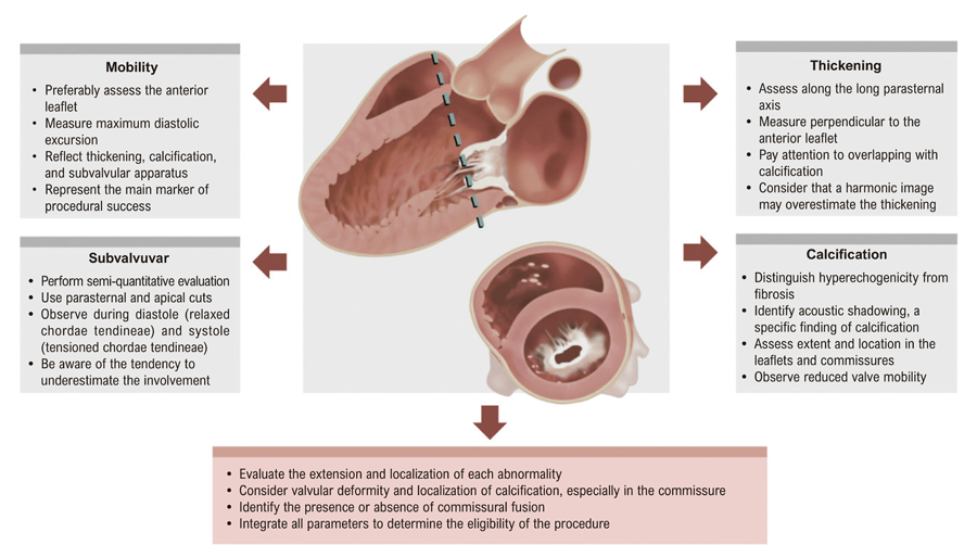 Practical recommendations for echocardiographic evaluation of rheumatic mitral stenosis (RMS) in the selection of candidates for percutaneous mitral balloon valvuloplasty (PMBV).


