Dilated Cardiomyopathy as a Rare Initial Manifestation of ANCA-positive Microscopic Polyangiitis: Case Report
Keywords
Microscopic Polyangiitis; Anti-Neutrophil Cytoplasmic Antibody-Associated Vasculitis; Dilated Cardiomyopathy; Echocardiography
Palavras-chave
Poliangiite Microscópica; Vasculite Associada a Anticorpo Anticitoplasma de Neutrófilos; Cardiomiopatia Dilatada; Ecocardiografia
Keywords
Microscopic Polyangiitis; Anti-Neutrophil Cytoplasmic Antibody-Associated Vasculitis; Dilated Cardiomyopathy; Echocardiography
Palavras-chave
Poliangiite Microscópica; Vasculite Associada a Anticorpo Anticitoplasma de Neutrófilos; Cardiomiopatia Dilatada; Ecocardiografia
Introduction
Antineutrophil cytoplasmic antibody (ANCA)-associated vasculitis constitute a group of systemic small-vessel inflammatory diseases characterized by pauci-immune necrotizing vasculitis and multisystem involvement, including granulomatosis with polyangiitis (GPA), eosinophilic granulomatosis with polyangiitis (EGPA), and microscopic polyangiitis (MPA).1-4 MPA is classically associated with rapidly progressive glomerulonephritis and alveolar hemorrhage, conditions that carry high morbidity and mortality when not promptly recognized and treated.1,4
In addition to predominant renal and pulmonary involvement, there is growing evidence that patients with ANCA-associated vasculitis have an increased cardiovascular risk, related not only to traditional factors but also to disease-specific mechanisms such as persistent inflammation, endothelial dysfunction, and accelerated atherosclerosis.5-8 Studies suggest a higher incidence of major cardiovascular events (myocardial infarction, stroke, and heart failure) compared with the general population.6
Cardiac involvement, although more frequently described in EGPA and GPA, can also occur in MPA, manifesting as myocarditis, pericarditis, coronary or microvascular vasculitis, and ventricular dysfunction.9,10 Imaging modalities, including echocardiography with myocardial deformation analysis (global longitudinal strain – GLS), can detect subclinical abnormalities and contribute to prognostic stratification, reinforcing the importance of systematic cardiologic evaluation in these patients.11 Despite recognition of the increased cardiovascular risk, myocardial involvement in MPA remains underdiagnosed and poorly characterized, particularly regarding GLS-detectable deformation patterns and their clinical implications.
In this context, the aim of this report is to describe a case of dilated cardiomyopathy with severe systolic dysfunction in a young patient with MPA, highlighting the finding of an apical sparing GLS pattern and its diagnostic and follow-up implications.
This case report was approved on March 7, 2026, by the Institutional Research Ethics Committee under Opinion No. 8,265,839 and CAAE 94929525.6.0000.5558, with written informed consent obtained from the patient.
Case Report
A 28-year-old male patient, previously healthy, developed an acute respiratory syndrome in June 2024, initially treated in the outpatient setting as community-acquired pneumonia. In the following weeks, he progressed with cough, hemoptysis, worsening dyspnea, exercise intolerance, lower-limb edema, foamy urine, and hematuria.
Upon hospital admission, he presented with severe anemia (hemoglobin 4.4 g/dL) and acute kidney injury requiring renal replacement therapy (creatinine 8.63 mg/dL; urea 244 mg/dL; potassium 7.1 mEq/L), in addition to hematuria and proteinuria. Chest radiography showed diffuse pulmonary infiltrates and cardiomegaly. Hemodialysis was initiated.
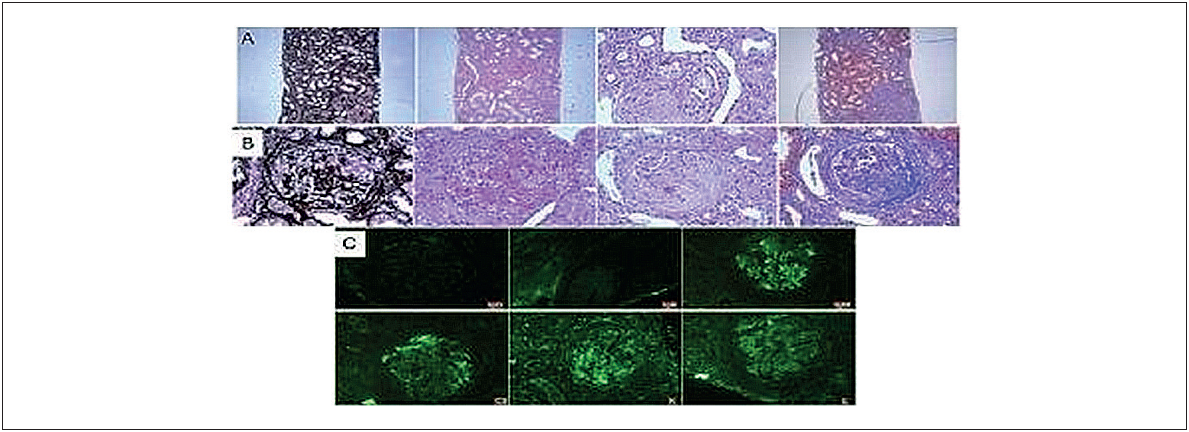
Etiologic investigation revealed positive p-ANCA and a renal biopsy consistent with pauci-immune crescentic glomerulonephritis, confirming the diagnosis of MPA (Figure 1). Pulse therapy with methylprednisolone was administered, followed by three cycles of cyclophosphamide, with clinical improvement.
Renal biopsy: (A) Tubulointerstitial compartment showing interstitial fibrosis, tubular atrophy, and monocyte infiltration; (B) Sclerotic glomeruli and/or glomeruli with proliferative/necrotizing crescentic lesions; (C) Negative immunofluorescence for light chains and immunoglobulins.
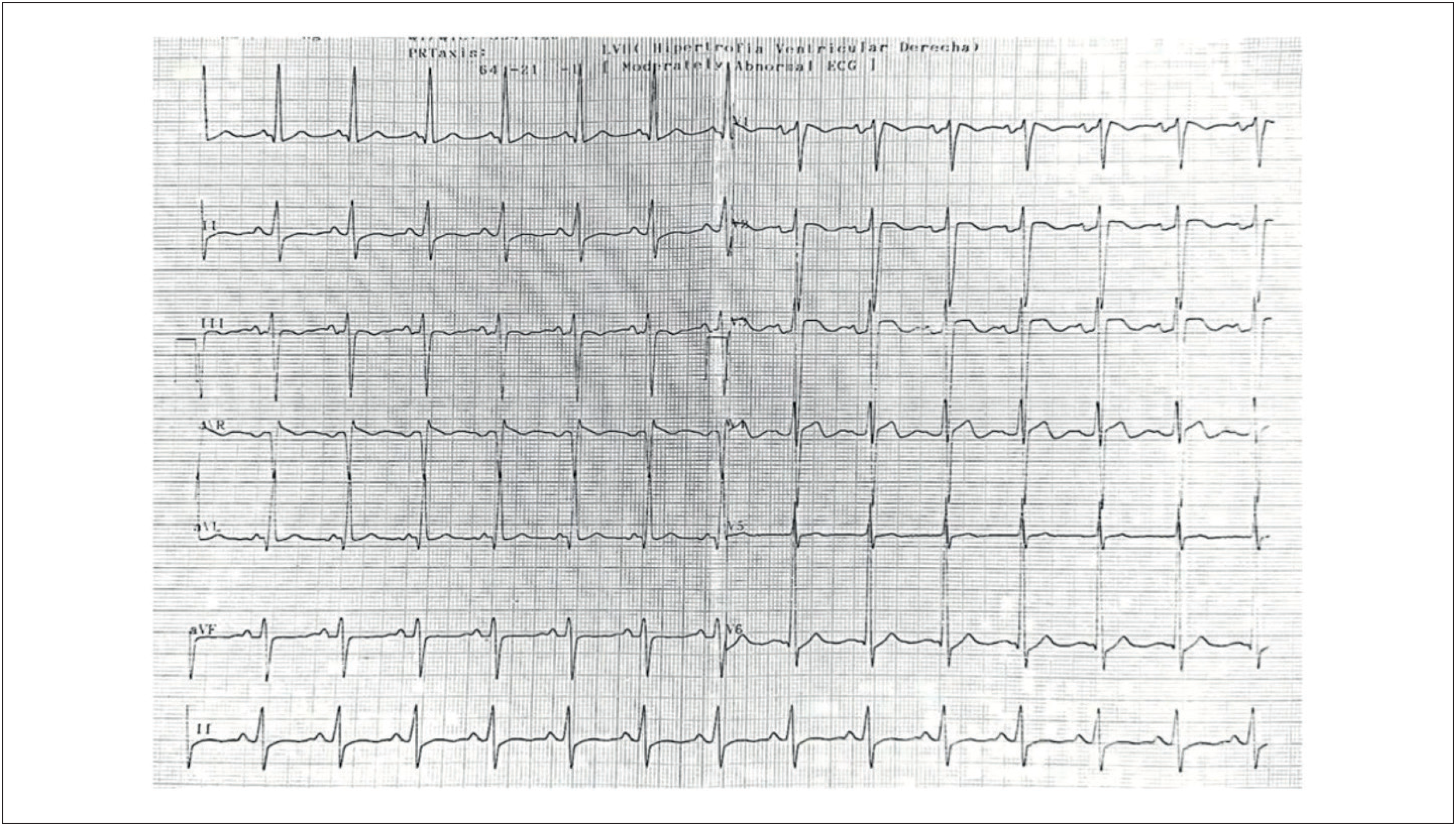
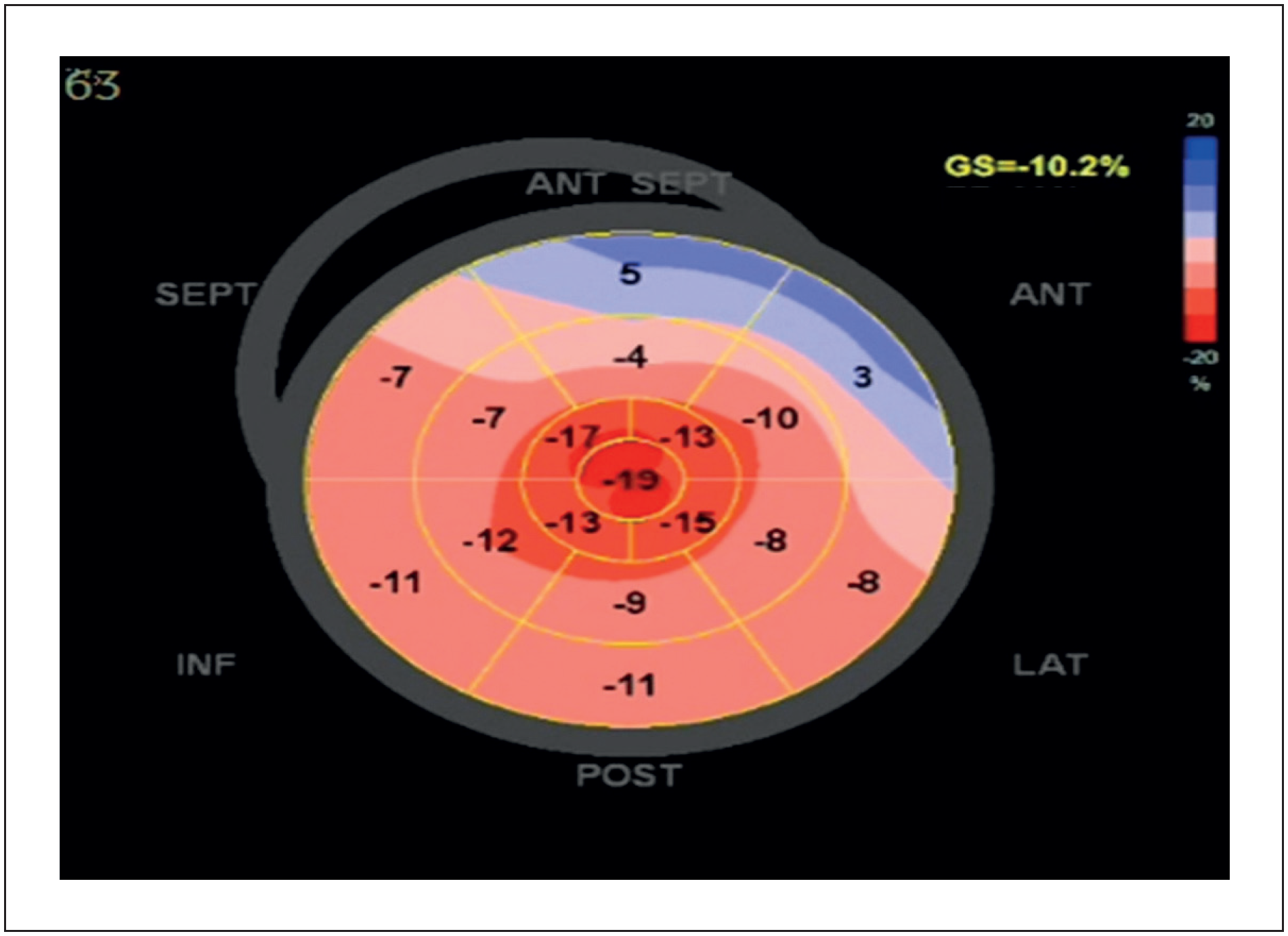
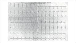
In November 2024, transthoracic echocardiography (TTE) demonstrated dilated cardiomyopathy with diffuse hypokinesia and severe systolic dysfunction (ejection fraction 26%), with no other evident etiologies. In February and April 2025, he continued to present systolic dysfunction during hospitalizations for infection. The electrocardiogram showed left-sided chamber overload (Figure 2). GLS analysis revealed an apical sparing pattern (Figure 3). Additional findings included moderate functional mitral regurgitation and a small pericardial effusion, along with right ventricular dysfunction (Video 1). The patient is currently under outpatient follow-up in the Cardiology Department of the University Hospital of Brasília, receiving optimized treatment for heart failure with reduced ejection fraction, remaining in functional class II (NYHA), with ongoing clinical and serial echocardiographic monitoring. The case timeline is summarized in Table 1.
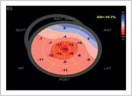
Transthoracic echocardiogram: polar map of left ventricular GLS showing reduced strain with an apical sparing pattern.
Video 1
Transthoracic echocardiogram showing severe left ventricular systolic dysfunction, moderate right ventricular dysfunction, functional mitral regurgitation, and GLS with an apical. View: http://abcimaging.org/supplementary-material/2026/3902/ABCImag-2026-0038_RC_Vídeo_MPA.mp4
Discussion
Cardiac involvement in ANCA-associated vasculitis is heterogeneous and has historically been more frequently recognized in EGPA, followed by GPA, and considered rare in MPA.4,9,10 However, its true frequency may be underestimated, as myocardial manifestations can be asymptomatic or attributed to comorbidities, renal dysfunction, or metabolic effects of corticosteroid therapy.7,8 Recent studies highlight the prognostic impact of cardiovascular involvement and support the need for a proactive approach to screening and follow-up.5,6
From a pathophysiological standpoint, ANCA-mediated neutrophil activation promotes diffuse endothelial injury, microvascular inflammation, and possible direct myocardial involvement, favoring functional microvascular ischemia, myocarditis, and progressive ventricular remodeling.2,12 In MPA, the dilated cardiomyopathy described in case reports and small series has been attributed predominantly to diffuse microvascular inflammation and/or subclinical myocarditis, which may coexist with hypertension and volume overload in patients with renal dysfunction.9,11
In the present case, the identification of severe systolic dysfunction in a young patient, without evidence of coronary artery disease, viral infectious etiology, or drug toxicity, reinforces the plausibility of a causal relationship with MPA.9 Echocardiography demonstrated not only dilation and diffuse hypokinesia but also a marked reduction in GLS, with an apical sparing pattern characterized by a basal-to-apical deformation gradient, in which the basal segments show greater strain reduction compared with the apical segments, resulting in relative preservation of apical deformation.
The incorporation of GLS as a complementary tool to ejection fraction is supported by the Position Statement of the Department of Cardiovascular Imaging, which recommends its use for early detection of myocardial dysfunction and for serial follow-up, highlighting its incremental value in clinical practice. In alignment with this, international consensus documents also endorse the clinical relevance of GLS. Although this pattern is classically associated with cardiac amyloidosis, it is not pathognomonic and must be interpreted in the context of the clinical presentation and other imaging findings.11,13-15
In terms of differential diagnosis, the apical sparing pattern on GLS should be understood as a suggestive but nonspecific sign, most commonly observed in cardiac amyloidosis, but also described in other conditions (e.g., ventricular hypertrophy, pressure-overload cardiomyopathies, chronic kidney disease, and some forms of myocarditis).11,16 Thus, in the absence of structural findings typical of infiltrative cardiomyopathy, interpretation should integrate conventional echocardiographic parameters (wall thickness, filling pattern, chamber dimensions, right ventricular function, and valvular disease), biomarkers, and – when available – cardiac magnetic resonance (CMR) for assessment of edema and fibrosis (LGE/T1/ECV), thereby reducing the risk of false positives and guiding follow-up.11,17
The practical message of this case highlights the importance of a cardiovascular imaging–based approach. In the setting of ANCA-associated vasculitis with possible myocardial involvement, TTE is recommended to assess cardiac structure and function, including GLS for detecting subclinical dysfunction and enabling serial comparison. In situations of unexplained decline in left ventricular ejection fraction (LVEF)/GLS, disproportionate symptoms, or discordance between clinical status and echocardiographic findings, CMR should be considered for tissue characterization and evaluation of myocarditis or fibrosis. Additionally, periodic reassessment is advisable at intervals determined by disease activity, functional class, and hemodynamic stability.8,11
Thus, this case illustrates two important points: (i) the need for systematic cardiovascular surveillance in ANCA-associated vasculitis — including MPA — with serial TTE and, when available, GLS and/or CMR for more detailed characterization; and (ii) the importance of clinical–imaging correlation when faced with suggestive echocardiographic patterns, avoiding isolated conclusions. Multidisciplinary management involving nephrology, rheumatology, and cardiology remains essential to optimize outcomes and guide timely interventions.8,11
One limitation that should be mentioned in this report is the absence of CMR for tissue characterization. The exam was not performed for two reasons: (1) unavailability within the public health system (SUS) during the evaluation period, and (2) the presence of severe renal dysfunction requiring hemodialysis, a situation in which the administration of gadolinium-based contrast may be associated with the risk of nephrogenic systemic fibrosis — a rare but potentially severe and difficult-to-manage event. Thus, interpretation of the imaging findings was based on clinical–echocardiographic correlation, including GLS analysis.
Conclusion
We report a rare cardiac manifestation of MPA in a young patient, presenting with dilated cardiomyopathy and severe systolic dysfunction, associated with an apical sparing pattern on GLS. This case reinforces the importance of systematic and serial cardiovascular evaluation in patients with ANCA-associated vasculitis, aiming for early diagnosis and improved prognostic stratification.
-
Sources of Funding
There were no external funding sources for this study.
-
Study Association
This article is part of the Medical Residency by Karoline Gonzaga Costa conducted at Hospital Universitário de Brasília.
-
Ethics Approval and Consent to Participate
This study was approved by the Ethics Committee of the Faculdade de Medicina of Universidade de Brasília (UNB) under the protocol number 94929525.6.0000.5558. All the procedures in this study were in accordance with the 1975 Helsinki Declaration, updated in 2013. Informed consent was obtained from all participants included in the study.
-
Use of Artificial Intelligence
The authors did not use any artificial intelligence tools in the development of this work.
Availability of Research Data
The underlying content of the research text is contained within the manuscript.
References
-
1 Nguyen Y, Pagnoux C, Karras A, Quéméneur T, Maurier F, Hamidou M, et al. Microscopic Polyangiitis: Clinical Characteristics and Long-Term Outcomes of 378 Patients from the French Vasculitis Study Group Registry. J Autoimmun. 2020;112:102467. doi: 10.1016/j.jaut.2020.102467.
» https://doi.org/10.1016/j.jaut.2020.102467 -
2 Jennette JC, Falk RJ. Small-Vessel Vasculitis. N Engl J Med. 1997;337(21):1512-23. doi: 10.1056/NEJM199711203372106.
» https://doi.org/10.1056/NEJM199711203372106 -
3 Hellmich B, Sanchez-Alamo B, Schirmer JH, Berti A, Blockmans D, Cid MC, et al. EULAR Recommendations for the Management of ANCA-Associated Vasculitis: 2022 Update. Ann Rheum Dis. 2024;83(1):30-47. doi: 10.1136/ard-2022-223764.
» https://doi.org/10.1136/ard-2022-223764 -
4 Yates M, Watts R. ANCA-Associated Vasculitis. Clin Med. 2017;17(1):60-4. doi: 10.7861/clinmedicine.17-1-60.
» https://doi.org/10.7861/clinmedicine.17-1-60 -
5 Moiseev S, Bulanov N, Crnogorac M, Direskeneli H, Galesic K, Gazel U, et al. Traditional and Disease Specific Risk Factors for Cardiovascular Events in ANCA-Associated Vasculitis: A Multinational Retrospective Study. J Rheumatol. 2023;50(8):1094. doi: 10.3899/jrheum.220851.C1.
» https://doi.org/10.3899/jrheum.220851.C1 -
6 Xie W, Xiao S, Li X, Huang J, Zhang Z. Cardiovascular and Cerebrovascular Outcomes in Anti-Neutrophil Cytoplasmic Antibody-Associated Vasculitis: A Systematic Review with Meta-Analysis. Autoimmun Rev. 2024;23(6):103587. doi: 10.1016/j.autrev.2024.103587.
» https://doi.org/10.1016/j.autrev.2024.103587 -
7 Kronbichler A, Leierer J, Gauckler P, Shin JI. Comorbidities in ANCA-Associated Vasculitis. Rheumatology. 2020;59(Suppl 3):iii79-iii83. doi: 10.1093/rheumatology/kez617.
» https://doi.org/10.1093/rheumatology/kez617 -
8 Kronbichler A, Bajema IM, Bruchfeld A, Kirsztajn GM, Stone JH. Diagnosis and Management of ANCA-Associated Vasculitis. Lancet. 2024;403(10427):683-98. doi: 10.1016/S0140-6736(23)01736-1.
» https://doi.org/10.1016/S0140-6736(23)01736-1 -
9 Sarmad A, Ghozloujeh ZG, Hanna PE, Kronbichler A, Geetha D, Heidari-Bateni G, et al. Cardiovascular Involvement in ANCA-Associated Vasculitis. Nephrol Dial Transplant. 2025;40(12):2256-67. doi: 10.1093/ndt/gfaf070.
» https://doi.org/10.1093/ndt/gfaf070 -
10 Thompson GE, Bourne MH Jr, Moura MC, Baqir M, Cartin-Ceba R, Makol A, et al. Pleuritis and Pericarditis in Antineutrophil Cytoplasmic Autoantibody-Associated Vasculitis. Chest. 2021;160(2):572-81. doi: 10.1016/j.chest.2021.02.049.
» https://doi.org/10.1016/j.chest.2021.02.049 -
11 Sarmad A, Ghozloujeh ZG, Hanna PE, Kronbichler A, Geetha D, Heidari-Bateni G, et al. Cardiovascular Involvement in ANCA-Associated Vasculitis. Nephrol Dial Transplant. 2025;40(12):2256-67. doi: 10.1093/ndt/gfaf070.
» https://doi.org/10.1093/ndt/gfaf070 -
12 Nakazawa D, Masuda S, Tomaru U, Ishizu A. Pathogenesis and Therapeutic Interventions for ANCA-Associated Vasculitis. Nat Rev Rheumatol. 2019;15(2):91-101. doi: 10.1038/s41584-018-0145-y.
» https://doi.org/10.1038/s41584-018-0145-y -
13 Almeida ALC, Melo MDT, Bihan DCSL, Vieira MLC, Pena JLB, Del Castillo JM, et al. Position Statement on the Use of Myocardial Strain in Cardiology Routines by the Brazilian Society of Cardiology's Department Of Cardiovascular Imaging - 2023. Arq Bras Cardiol. 2023;120(12):e20230646. doi: 10.36660/abc.20230646.
» https://doi.org/10.36660/abc.20230646 -
14 Thomas JD, Edvardsen T, Abraham T, Appadurai V, Badano L, Banchs J, et al. Clinical Applications of Strain Echocardiography: A Clinical Consensus Statement from the American Society of Echocardiography Developed in Collaboration with the European Association of Cardiovascular Imaging of the European Society of Cardiology. J Am Soc Echocardiogr. 2025;38(11):985-1020. doi: 10.1016/j.echo.2025.07.007.
» https://doi.org/10.1016/j.echo.2025.07.007 -
15 Kittleson MM, Ruberg FL, Ambardekar AV, Brannagan TH, Cheng RK, Clarke JO, et al. 2023 ACC Expert Consensus Decision Pathway on Comprehensive Multidisciplinary Care for the Patient with Cardiac Amyloidosis: A Report of the American College of Cardiology Solution Set Oversight Committee. J Am Coll Cardiol. 2023;81(11):1076-126. doi: 10.1016/j.jacc.2022.11.022.
» https://doi.org/10.1016/j.jacc.2022.11.022 -
16 Kottam A, Hanneman K, Schenone A, Daubert MA, Sidhu GD, Gropler RJ, et al. State-of-the-Art Imaging of Infiltrative Cardiomyopathies: A Scientific Statement from the American Heart Association. Circ Cardiovasc Imaging. 2023;16(11):e000081. doi: 10.1161/HCI.0000000000000081.
» https://doi.org/10.1161/HCI.0000000000000081 -
17 Arbelo E, Protonotarios A, Gimeno JR, Arbustini E, Barriales-Villa R, Basso C, et al. 2023 ESC Guidelines for the Management of Cardiomyopathies. Eur Heart J. 2023;44(37):3503-626. doi: 10.1093/eurheartj/ehad194.
» https://doi.org/10.1093/eurheartj/ehad194
Edited by
-
Editor responsible for the review:
Marcelo Tavares