Acute Thrombosis During Ductus Arteriosus Stenting Successfully Treated with Balloon Angioplasty and In Situ Alteplase Infusion: Case Report
Keywords
Congenital Heart Defects; Tricuspid Atresia; Ductus Arteriosus; Percutaneous Coronary Intervention; Thrombosis
Palavras-chave
Cardiopatias Congênitas; Atresia Tricúspide; Canal Arterial; Intervenção Coronária Percutânea; Trombose
Keywords
Congenital Heart Defects; Tricuspid Atresia; Ductus Arteriosus; Percutaneous Coronary Intervention; Thrombosis
Palavras-chave
Cardiopatias Congênitas; Atresia Tricúspide; Canal Arterial; Intervenção Coronária Percutânea; Trombose
Introduction
Tricuspid atresia with pulmonary atresia is a rare congenital heart defect, representing about 1% to 3% of all congenital heart diseases.1 Tricuspid atresia is subdivided into type I (normal position of the great arteries) and type II (transposition of the great arteries), with type 1A characterized by the presence of associated pulmonary atresia or stenosis.2 Pulmonary blood flow in these patients is dependent on the patency of the ductus arteriosus, making its maintenance essential in the neonatal period.
The treatment of tricuspid atresia follows a staged surgical approach, aiming for a Fontan circulation as the final goal. In the neonatal period, the focus is to ensure adequate pulmonary blood flow through hemodynamic or surgical interventions, including systemic-pulmonary shunts or ductal interventions.3
This case report describes a newborn with type 1A tricuspid atresia who underwent a hemodynamic intervention with stent implantation in the ductus arteriosus, discussing technical aspects of the procedure, complications, and clinical evolution.
Case report
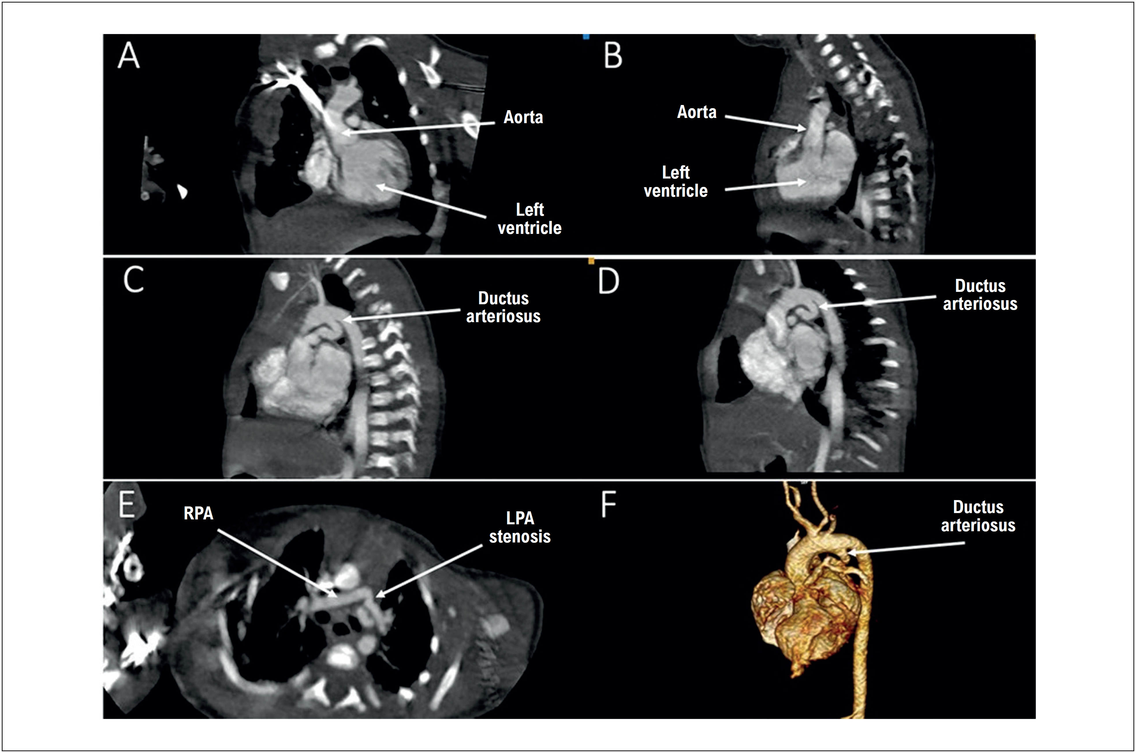
A term male newborn weighing 2,920 g was transferred at 6 days of life to a cardiac intensive care unit. The patient was hemodynamically stable, with oxygen saturation of 85% in room air, and was receiving a continuous intravenous infusion of prostaglandin at 0.01 mcg/kg/min. Transthoracic echocardiography demonstrated tricuspid valve atresia, pulmonary valve atresia, a hypoplastic right ventricle, the aorta arising from the left ventricle, and a tortuous ductus arteriosus (type 1A tricuspid atresia) (Figure 1). Chest computed tomography angiography showed a tortuous ductus arteriosus and stenosis at the origin of the left pulmonary artery (Figure 2). The patient also had a multicystic dysplastic left kidney. After multidisciplinary discussion, ductal stenting was chosen as a palliative procedure in the neonatal period.

Transthoracic echocardiography. A and B) Patent foramen ovale and atretic tricuspid valve. C) Left ventricle connected to the aorta. D) Tortuous patent ductus arteriosus. LA: left atrium; RA: right atrium.

Aortic computed tomography angiography. A and B) Left ventricle connected to the aorta. C and D) Markedly tortuous patent ductus arteriosus. E) Stenosis at the origin of the left pulmonary artery. E) Three-dimensional reconstruction showing the anatomy of the ductus arteriosus. LPA: left pulmonary artery; RPA: right pulmonary artery.
Interventional procedure
Due to the angle between the aortic end of the ductus arteriosus and the aortic arch, the right carotid artery was chosen as the access route. To improve catheter and guidewire manipulation and ergonomics, the patient was positioned in reverse orientation on the hemodynamic table (Figure 3).
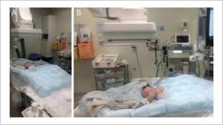
A and B) Image showing the patient positioned in reverse orientation on the catheterization table (with the cranial portion away from the radiation source).
The right carotid artery was punctured under ultrasound guidance, and a 5F slender transradial introducer was placed. Intravenous heparin (100 IU/kg) and prophylactic cefazolin (50 mg/kg) were administered. Left heart catheterization and cineangiography confirmed the tortuous ductal anatomy and the stenosis at the origin of the left pulmonary artery (proximal to the ductal insertion). A 0.014″ Balance Heavy Weight (BHW) guidewire was placed in the left pulmonary artery with microcatheter support. A Mini Trek 2 × 8 mm balloon catheter was positioned in the ductal trajectory for radiopaque landmark-based measurements. At this point, the patient developed decreased end-tidal carbon dioxide (ETCO2) and oxygen saturation, with significant ductal spasm and reduced effective pulmonary flow. Continuous adrenaline infusion was started. The BHW guidewire was replaced with an exchange-length 0.014″ wire. With guidewire support, an Inspiron 4 × 19 mm stent was implanted in the ductus arteriosus (Figure 4).

Percutaneous intervention. A, B, and C) Angiograms obtained via carotid access showing a tortuous ductus arteriosus and stenosis at the origin of the left pulmonary artery. D) With the support of a 0.014″ BHW guidewire positioned in the left pulmonary artery, a Mini Trek 2 × 8 mm balloon catheter was placed along the ductal trajectory; at this moment, ductal spasm was observed. E) An Inspiron 4 × 19 mm stent was implanted in the ductus arteriosus. F) Angiogram showing preserved flow through the ductal stent but with worsening stenosis at the origin of the left pulmonary artery. LPA: left pulmonary artery; RPA: right pulmonary artery.
The patient showed transient improvement in saturation and ETCO2, but a few minutes later developed significant hypoxemia, decreased ETCO2, and bradycardia. Acute stent thrombosis was identified, and in situ alteplase was administered as a 0.05 mg/kg bolus, followed by continuous infusion at 0.5 mg/kg/h. Sequential balloon angioplasty of the entire stent with a Trek 4 × 12 mm balloon catheter was performed. These measures restored ductal flow and partially improved heart rate, oxygen saturation, and ETCO2.
Stenosis at the origin of the left pulmonary artery significantly restricted flow to the pulmonary trunk and right pulmonary artery. Proximal optimization technique (POT) was used with a 4 × 8 mm balloon (inflated to burst pressure). With the support of a JR 4F catheter, a 0.014″ guidewire was placed in the right pulmonary artery through the lateral mesh of the prior stent. Balloon angioplasty of the right pulmonary artery origin was then performed with a Trek 3 × 12 mm balloon (opening the lateral mesh of the previous stent) (Figure 5). After balloon angioplasty, the guidewire was withdrawn from the right pulmonary artery, but the stenosis at the origin of the left pulmonary artery persisted.

Percutaneous intervention. A) Angiography showing acute thrombosis of the ductal stent. B) Angioplasty with a 4 × 12 mm Trek balloon catheter along the entire length of the stent. C) Angiography demonstrating effective recanalization of the stent. D) Guidewire positioned in the right pulmonary artery through the lateral struts of the stent. E) Angioplasty with a 3 × 12 mm Trek balloon catheter (opening the lateral struts of the previous stent). F) Guidewire withdrawn from the right pulmonary artery, showing persistence of stenosis at the origin of the left pulmonary artery. RPA: right pulmonary artery; LPA: left pulmonary artery.
The 0.014″ guidewire was then repositioned in the right pulmonary artery, and an Inspiron 3.5 × 9 mm stent was implanted through the lateral mesh of the previous stent, directed toward the origin of the left pulmonary artery and the pulmonary trunk (Figure 6). The introducer was removed, and manual hemostatic compression and compressive occlusive dressing were applied.
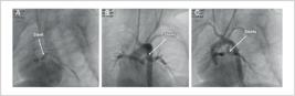
Percutaneous intervention. A) Implantation of an Inspiron 3.5 × 9 mm stent through the lateral mesh of the previous stent. B and C) Angiograms showing adequate flow through the stent assembly from the ductus arteriosus to both pulmonary arteries.
The patient was transferred to the intensive care unit on mechanical ventilation and continuous intravenous adrenaline (0.1 mcg/kg/min). Alteplase continued for another 3 hours but discontinued due to bleeding at the puncture and central venous access sites. Grade I left intracranial hemorrhage was detected on post-procedure transfontanellar ultrasound, which normalized on follow-up 3 months later. The patient had infectious complications such as pneumonia and sepsis with positive blood cultures for Pseudomonas aeruginosa, treated with broad-spectrum antibiotics. He also experienced extubation failure and prolonged mechanical ventilation (total of 49 days). Hospitalization was prolonged, with discharge 3 months after the procedure. Transthoracic echocardiography showed patent stents with adequate flow through the ductus arteriosus and pulmonary branches. Clinically, the patient remains hemodynamically stable, with oxygen saturation of 84% in room air, and is being followed for planned Glenn surgery.
Discussion
Tricuspid atresia with pulmonary atresia is one of the most complex challenges in interventional pediatric cardiology, especially because of the absolute dependence on ductal flow for pulmonary perfusion in the neonatal period.4,5 This case illustrates the multiple layers of anatomic and technical complexity involved in the management of these patients, highlighting both the therapeutic possibilities and the inherent limitations and complications of interventional procedures in high-risk neonates.
Ductal stenting has emerged as a viable and often preferable alternative to surgical shunts in selected neonates, offering significant advantages in terms of perioperative morbidity and mortality.6,7 Recent comparative studies have shown similar survival rates between the two therapeutic modalities, but with a lower incidence of immediate complications in the percutaneous intervention group.8,9 However, appropriate patient selection remains critical, considering factors such as neonatal weight, ductal anatomy, associated pulmonary stenoses, and pre-procedure hemodynamic stability.10,11
The angle formed between the aortic end of the ductus arteriosus and the aortic arch, as seen in this case, is an anatomical limitation that requires significant technical adaptations, including changes in patient positioning and careful selection of the vascular access route.12-14 The extreme ductal tortuosity15 combined with the short length of the pulmonary branches in a newborn complicates coronary guidewire navigation and the support it provides for stent delivery. The use of specific materials such as a microcatheter and more than one 0.014″ guidewire positioned simultaneously makes the procedure feasible. Pre-existing pulmonary branch stenosis prior to stent implantation is a risk factor for worsening stenosis and total occlusion of a pulmonary branch.16 Techniques such as positioning a 0.014″ guidewire in each pulmonary branch during stent implantation can increase procedural safety by maintaining patency of the stenotic pulmonary branch and facilitating the opening of the stent's lateral mesh. In this case, this technique was not possible because the patient developed acute clinical deterioration during guidewire manipulation within the ductus, causing ductal spasm.
The occurrence of acute stent thrombosis is one of the most feared complications in neonatal procedures, with an incidence of 2% to 3%. Predisposing factors include elevated hematocrit, prolonged procedure time, endothelial trauma during manipulation, and activation of the coagulation cascade secondary to the prosthetic material. Immediate treatment with local fibrinolytics, as demonstrated in this case with alteplase, has been effective, allowing successful recanalization with low risk of systemic bleeding. Sequential mechanical angioplasty of the stent also restores adequate flow quickly and sustainably.10,17
The technique of stent implantation through the lateral mesh of a previously placed stent ("stent-in-stent") used to address stenosis at the origin of the left pulmonary artery represents a significant technical innovation in pediatric interventional cardiology. The literature documents growing experience with this technique in various anatomical situations, demonstrating medium-term safety and efficacy.18,19
The complications observed in this case, including grade I intracranial hemorrhage and hospital-acquired infections, reflect the fragility of neonates undergoing complex and prolonged interventional procedures. Intracranial hemorrhage, possibly related to the use of fibrinolytics, is a known but relatively rare complication, requiring strict neurological monitoring in the post-procedural period.20,21 The development of nosocomial infections in critically ill patients on prolonged mechanical ventilation is an additional challenge in post-intervention management, demanding rigorous prevention and early treatment protocols. The favorable medium-term outcome, with maintained stent patency and hemodynamic stability, supports the efficacy of the chosen therapeutic strategy and the use of this approach as an effective bridge to subsequent surgical staging in selected patients.22,23
Conclusion
This case demonstrates the feasibility and efficacy of hemodynamic intervention in neonates with type 1A tricuspid atresia. Ductal stenting allowed for adequate pulmonary blood flow and clinical stabilization. The observed complications, including acute stent thrombosis, worsening of left pulmonary artery origin stenosis after ductal stenting, and hemodynamic instability, reflect the complexity of neonatal procedures and the need for rapid and effective medical (fibrinolytic) and interventional (balloon angioplasty of the thrombosed stent, POT technique, balloon and stent angioplasty through the lateral stent mesh) strategies. The favorable medium-term outcome, with preserved pulmonary flow and ventricular function, supports the use of this technique as a bridge to future surgical staging in selected patients.
-
Sources of Funding
There were no external fundingsources for this study.
-
Study Association
This study is not associated with any thesis or dissertation work.
-
Ethics Approval and Consent to Participate
This study was approved by the Ethics Committee of the CEP do Hospital de Urgëncias de Goiás under the protocol number 85497418.2.0000.0033. All the procedures in this study were in accordance with the 1975 Helsinki Declaration, updated in 2013. Informed consent was obtained from all participants included in the study.
-
Use of Artificial Intelligence
The authors did not use any artificial intelligence tools in the development of this work.
Availability of Research Data
The data cannot be made publicly available due to legal considerations related to Brazil's General Data Protection Law, as the data could allow the identification of the patient in the case in question.
References
- 1 Minocha PK, Horenstein MS, Phoon C. Tricuspid Atresia. Treasure Island: StatPearls Publishing; 2024.
-
2 Karamlou T, Ashburn DA, Caldarone CA, Blackstone EH, Jonas RA, Jacobs ML, et al. Matching Procedure to Morphology Improves Outcomes in Neonates with Tricuspid Atresia. J Thorac Cardiovasc Surg. 2005;130(6):1503-10. doi: 10.1016/j.jtcvs.2005.07.024.
» https://doi.org/10.1016/j.jtcvs.2005.07.024 -
3 Downing TE, Boucek DM, Glatz AC, Qureshi AM, Zampi JD, Petit CJ, et al. Stenting the Ductus Arteriosus in Neonates with Ductal-Dependent Pulmonary Blood Flow: Technical and Anatomic Considerations. Pediatr Cardiol. 2025. doi: 10.1007/s00246-025-03834-4.
» https://doi.org/10.1007/s00246-025-03834-4 -
4 Schranz D, Michel-Behnke I, Heyer R, Vogel M, Bauer J, Valeske K, et al. Stent Implantation of the Arterial Duct in Newborns with a Truly Duct-Dependent Pulmonary Circulation: A Single-Center Experience with Emphasis on Aspects of the Interventional Technique. J Interv Cardiol. 2010;23(6):581-8. doi: 10.1111/j.1540-8183.2010.00576.x.
» https://doi.org/10.1111/j.1540-8183.2010.00576.x -
5 Mini N, Schneider MBE, Asfour B, Mikus M, Zartner PA. Duct Stenting vs. Modified Blalock-Taussig Shunt: New Insights Learned from High-Risk Patients with Duct-Dependent Pulmonary Circulation. Front Cardiovasc Med. 2022;9:933959. doi: 10.3389/fcvm.2022.933959.
» https://doi.org/10.3389/fcvm.2022.933959 -
6 Bentham JR, Zava NK, Harrison WJ, Shauq A, Kalantre A, Derrick G, et al. Duct Stenting versus Modified Blalock-Taussig Shunt in Neonates with Duct-Dependent Pulmonary Blood Flow: Associations with Clinical Outcomes in a Multicenter National Study. Circulation. 2018;137(6):581-8. doi: 10.1161/CIRCULATIONAHA.117.028972.
» https://doi.org/10.1161/CIRCULATIONAHA.117.028972 -
7 Glatz AC, Petit CJ, Goldstein BH, Kelleman MS, McCracken CE, McDonnell A, et al. Comparison between Patent Ductus Arteriosus Stent and Modified Blalock-Taussig Shunt as Palliation for Infants with Ductal-Dependent Pulmonary Blood Flow: Insights from the Congenital Catheterization Research Collaborative. Circulation. 2018;137(6):589-601. doi: 10.1161/CIRCULATIONAHA.117.029987.
» https://doi.org/10.1161/CIRCULATIONAHA.117.029987 -
8 Boucek DM, Qureshi AM, Goldstein BH, Petit CJ, Glatz AC. Blalock-Taussig Shunt versus Patent Ductus Arteriosus Stent as First Palliation for Ductal-Dependent Pulmonary Circulation Lesions: A Review of the Literature. Congenit Heart Dis. 2019;14(1):105-9. doi: 10.1111/chd.12707.
» https://doi.org/10.1111/chd.12707 -
9 Li D, Zhou X, Li M. Arterial Duct Stent versus Surgical Shunt for Patients with Duct-Dependent Pulmonary Circulation: A Meta-Analysis. BMC Cardiovasc Disord. 2021;21(1):9. doi: 10.1186/s12872-020-01817-2.
» https://doi.org/10.1186/s12872-020-01817-2 -
10 Alwi M. Stenting the Ductus Arteriosus: Case Selection, Technique and Possible Complications. Ann Pediatr Cardiol. 2008;1(1):38-45. doi: 10.4103/0974-2069.41054.
» https://doi.org/10.4103/0974-2069.41054 -
11 Bahaidarah S, Al-Ata J, Alkhushi N, Azhar A, Zaher Z, Alnahdi B, et al. Outcome of Ductus Arteriosus Stenting Including Vertical Tubular and Convoluted Tortuous Ducts with Emphasis on Technical Considerations. Egypt Heart J. 2021;73(1):83. doi: 10.1186/s43044-021-00210-4.
» https://doi.org/10.1186/s43044-021-00210-4 -
12 Choudhry S, Balzer D, Murphy J, Nicolas R, Shahanavaz S. Percutaneous Carotid Artery Access in Infants < 3 Months of Age. Catheter Cardiovasc Interv. 2016;87(4):757-61. doi: 10.1002/ccd.26310.
» https://doi.org/10.1002/ccd.26310 -
13 Bauser-Heaton H, Qureshi AM, Goldstein BH, Glatz AC, Nicholson GT, Meadows JJ, et al. Use of Carotid and Axillary Artery Approach for Stenting the Patent Ductus Arteriosus in Infants with Ductal-Dependent Pulmonary Blood Flow: A Multicenter Study from the Congenital Catheterization Research Collaborative. Catheter Cardiovasc Interv. 2020;95(4):726-33. doi: 10.1002/ccd.28631.
» https://doi.org/10.1002/ccd.28631 -
14 Bauser-Heaton H, Qureshi AM, Goldstein BH, Glatz AC, Petit CJ. Use of Novel "Flip Technique" Aids in Percutaneous Carotid Artery Approach in Neonates. JACC Cardiovasc Interv. 2019;12(16):1630-1. doi: 10.1016/j.jcin.2019.04.053.
» https://doi.org/10.1016/j.jcin.2019.04.053 -
15 Qureshi AM, Goldstein BH, Glatz AC, Agrawal H, Aggarwal V, Ligon RA, et al. Classification Scheme for Ductal Morphology in Cyanotic Patients with Ductal Dependent Pulmonary Blood Flow and Association with Outcomes of Patent Ductus Arteriosus Stenting. Catheter Cardiovasc Interv. 2019;93(5):933-43. doi: 10.1002/ccd.28125.
» https://doi.org/10.1002/ccd.28125 -
16 Koneti NR, Bakhru S, Dhulipudi B, Rajan S, Sreeram N. Stent Strut Dilation in Branch Pulmonary Artery Stenosis Following Stenting of Arterial Duct in Duct-Dependent Pulmonary Circulation. Pediatr Cardiol. 2025;46(1):53-60. doi: 10.1007/s00246-023-03319-2.
» https://doi.org/10.1007/s00246-023-03319-2 -
17 Bauser-Heaton H, Price K, Weber R, El-Said H. Stenting of the Patent Ductus Arteriosus: A Meta-Analysis and Literature Review. J Soc Cardiovasc Angiogr Interv. 2022;1(6):100392. doi: 10.1016/j.jscai.2022.100392.
» https://doi.org/10.1016/j.jscai.2022.100392 -
18 Wu H, Li M, Lin C. Influence of Balloon Location during Proximal Optimization Technique (POT): A Finite Element Analysis. J Biomech. 2021;127:110703. doi: 10.1016/j.jbiomech.2021.110703.
» https://doi.org/10.1016/j.jbiomech.2021.110703 -
19 Darremont O, Leymarie JL, Lefèvre T, Albiero R, Mortier P, Louvard Y. Technical Aspects of the Provisional Side Branch Stenting Strategy. EuroIntervention. 2015;11(Suppl V):V86-90. doi: 10.4244/EIJV11SVA19.
» https://doi.org/10.4244/EIJV11SVA19 -
20 Wang M, Hays T, Balasa V, Bagatell R, Gruppo R, Grabowski EF, et al. Low-Dose Tissue Plasminogen Activator Thrombolysis in Children. J Pediatr Hematol Oncol. 2003;25(5):379-86. doi: 10.1097/00043426-200305000-00006.
» https://doi.org/10.1097/00043426-200305000-00006 -
21 Will A. Neonatal Haemostasis and the Management of Neonatal Thrombosis. Br J Haematol. 2015;169(3):324-32. doi: 10.1111/bjh.13301.
» https://doi.org/10.1111/bjh.13301 -
22 Jayaram N, Spertus JA, Kennedy KF, Vincent R, Martin GR, Curtis JP, et al. Modeling Major Adverse Outcomes of Pediatric and Adult Patients with Congenital Heart Disease Undergoing Cardiac Catheterization: Observations from the NCDR IMPACT Registry (National Cardiovascular Data Registry Improving Pediatric and Adult Congenital Treatment). Circulation. 2017;136(21):2009-19. doi: 10.1161/CIRCULATIONAHA.117.027714.
» https://doi.org/10.1161/CIRCULATIONAHA.117.027714 -
23 Wang X, Li S, Huo D, Zhu Z, Wang W, He H, et al. Nosocomial Infections after Pediatric Congenital Heart Disease Surgery: Data from National Center for Cardiovascular Diseases in China. Infect Drug Resist. 2024;17:1615-23. doi: 10.2147/IDR.S457991.
» https://doi.org/10.2147/IDR.S457991
Edited by
-
Editor responsible for the review:
Márcio Brito