Percutaneous Closure of the Aortopulmonary Window in an Infant Using an Amplatzer Duct Occluder II Prosthesis
Cardiovascular Surgical Procedures; Aortopulmonary Septal Defect; Infant
Procedimentos Cirúrgicos Cardiovasculares; Defeito do Septo Aortopulmonar; lactente
Cardiovascular Surgical Procedures; Aortopulmonary Septal Defect; Infant
Procedimentos Cirúrgicos Cardiovasculares; Defeito do Septo Aortopulmonar; lactente
Introduction
The Aortopulmonary Window (APW) is a rare congenital malformation, representing approximately 0.2% of all congenital heart diseases. It is typically nonrestrictive and requires early surgical intervention to prevent complications such as pulmonary congestion, respiratory infections, and obstructive pulmonary vascular disease. In cases like ours, where the surgical risk is exceedingly high, percutaneous closure may present a viable alternative.
Case Report
A 1.5-month-old male infant, born with a weight of 2180 g, presented with multiple comorbidities, including esophageal atresia, congenital chylothorax, and Meckel’s diverticulum. The patient underwent several surgeries within the first few days of life. He also experienced recurrent infections and required prolonged mechanical ventilation. During evaluation, cardiomegaly was noted alongside a continuous murmur at the upper left sternal border. The echocardiogram revealed a 5.0 mm APW ( Figures 1 and 2 ). The echocardiogram revealed a large left-to-right shunt, resulting in left chamber enlargement. Following a multidisciplinary discussion with the cardiology team and given the high surgical risk, percutaneous closure of the APW was chosen as the best approach. The procedure was performed after informed consent was obtained.
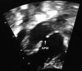

– Echocardiogram with Color Doppler revealed a significant left-to-right shunt. APW: Aortopulmonary Window
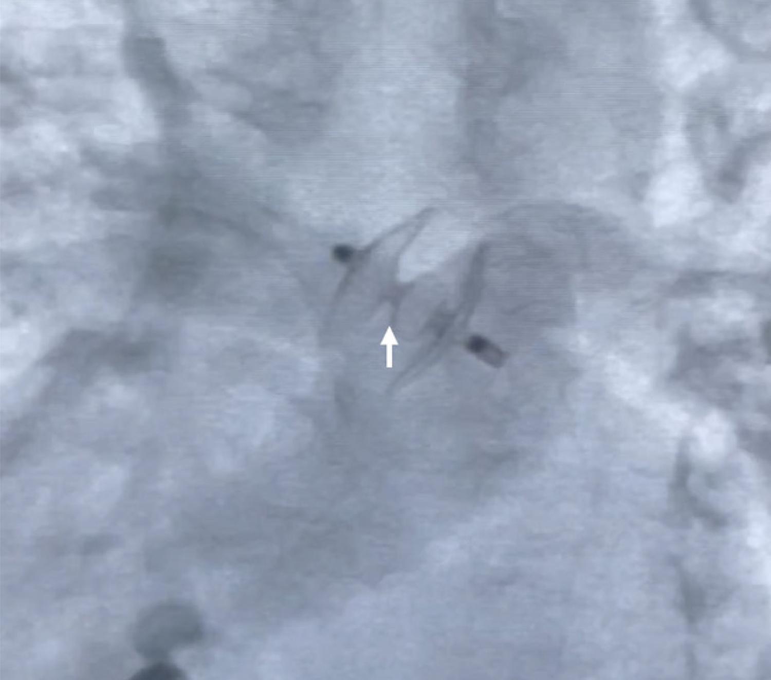
Cardiac catheterization was performed under general anesthesia using ultrasound-guided puncture of the right carotid artery and right femoral vein, with a 4F sheath in the artery and a 5F sheath in the vein. Heparin was administered at 100 IU/kg. Mean aortic and pulmonary pressures were measured at 44 and 29 mmHg, respectively. The ascending aortogram confirmed a 5.0 mm APW located distant from the left aortic cusp ( Figure 3 ). The defect was crossed via the aorta using a 4F Judkins catheter with a 3.5 bend and a 0.0035 Terumo guidewire. The guidewire was captured and exteriorized through the femoral vein, forming a venoarterial loop. A long 5F sheath and a delivery system were then advanced from the femoral vein, through the APW, and into the descending aorta. Initially, an Amplatzer Duct Occluder II (ADOII 6/6) prosthesis (St. Jude Medical, St. Paul, MN, USA) was advanced; the aortic disc was delivered followed by the pulmonary disc, which did not fix properly. The device was then replaced by an ADO II 4/4. After confirming proper device positioning using transthoracic echocardiography and fluoroscopy, ensuring no interference with aortic and pulmonary flows or proximity to the coronary arteries, the device was successfully implanted ( Fig. 4 ). Two weeks post-procedure, the patient showed no signs of heart failure, and the echocardiogram confirmed the device was well-positioned, with no significant vessel obstruction or residual shunt. The patient remained in the ICU due to other comorbidities.

– Confirmation of the position by fluoroscopy, without interfering in the aorta and pulmonary flow (white arrow).
Discussion
APW is a defect resulting from incomplete separation of the aorta and pulmonary trunk walls at the conotruncal septum during early embryogenesis. APW is classified into three morphological types. Type I defects (60-70%) are located proximally, in the aorta above the sinus of Valsalva. Type II defects (20-25%) are distal, situated in the upper ascending aorta before the emergence of the aortic branches. Type III defects, which are less common, are extensive and combine features of types I and II, involving most of the ascending aorta, the pulmonary trunk, and the right pulmonary artery. The Type II defect reported in this case accounts for 20% of APW cases and is particularly amenable to percutaneous correction due to its location. Surgery is the gold standard treatment and has been performed for many years. However, when a patient’s clinical condition is critical, with a prohibitively high surgical risk, as the case presented. In these cases, percutaneous closure becomes a valuable alternative when the defect allows for it. Reports in the literature on transcatheter closure of APW are limited, especially for patients weighing less than 3 kg, and they describe various vascular access methods and devices.1 - 13 Table 1 presents a summary of the literature review. The defect’s position was conducive to percutaneous intervention, while the infant’s multiple comorbidities made surgery exceedingly high-risk. The optimal device choice remains unclear. Device selection depends on the hemodynamicist’s experience with different types of devices designed for closing cardiac and extracardiac shunts. Given the patient’s low weight, arterial access via the internal carotid artery was selected, which is our preferred approach for infants under 3.0 kg. In this specific case, we anticipated that crossing the window would be relatively straightforward, as it ultimately proved to be.
Conclusion
Although cardiac surgery is the gold standard for APW closure, high-risk patients with anatomy favorable to percutaneous closure should be considered for this approach, as a viable and effective alternative in the management of such cases.
References
-
1 Stamato T, Benson LN, Smallhorn JF, Freedom RM. Transcatheter Closure of an Aortopulmonary Window with a Modified Double Umbrella Occluder System. Cathet Cardiovasc Diagn. 1995;35(2):165-7. doi: 10.1002/ccd.1810350218.
» https://doi.org/10.1002/ccd.1810350218 -
2 Sivakumar K, Francis E. Transcatheter Closure of Distal Aortopulmonary Window Using Amplatzer Device. Congenit Heart Dis. 2006;1(6):321-3. doi: 10.1111/j.1747-0803.2006.00055.x.
» https://doi.org/10.1111/j.1747-0803.2006.00055.x -
3 Trehan V, Nigam A, Tyagi S. Percutaneous Closure of Nonrestrictive Aortopulmonary Window in Three Infants. Catheter Cardiovasc Interv. 2008;71(3):405-11. doi: 10.1002/ccd.21366.
» https://doi.org/10.1002/ccd.21366 -
4 Kosmac B, Eicken A, Kühn A, Heinrich M, Ewert P. Percutaneous Device Closure of an Aortopulmonary Window in a Small Infant. Int J Cardiol. 2013;168(3):e102-3. doi: 10.1016/j.ijcard.2013.07.218.
» https://doi.org/10.1016/j.ijcard.2013.07.218 - 5 Zhao-Feng X, Zhi-Wei Z, Yu-Me X, Jun-Jie L. Transcatheter Closure of Aortopulmonary Window in Children: Case Reports and Literature Review. CAJ. 2011;12(2):131-5.
-
6 Fiszer R, Zbronski K, Szkutnik M. Percutaneous Closure of an Aortopulmonary Window Using Amplatzer Duct Occluder II: Additional Sizes: The First Reported Case. Cardiol Young. 2017;27(4):812-5. doi: 10.1017/S1047951116002134.
» https://doi.org/10.1017/S1047951116002134 -
7 Sabnis GR, Shah HC, Lanjewar CP, Malik S, Kerkar PG. Transcatheter Closure of Large Aortopulmonary Window in a Neonate. Ann Pediatr Cardiol. 2018;11(2):228-30. doi: 10.4103/apc.APC_158_17.
» https://doi.org/10.4103/apc.APC_158_17 -
8 Campos-Quintero A, García-Montes JA, Zabal-Cerdeira C, Cervantes-Salazar JL, Calderón-Colmenero J, Sandoval JP. Transcatheter Device Closure of Aortopulmonary Window. Is There a Need for an Alternative Strategy to Surgery? Rev Esp Cardiol (Engl Ed). 2019;72(4):349-51. doi: 10.1016/j.rec.2018.03.020.
» https://doi.org/10.1016/j.rec.2018.03.020 -
9 Uçar T, Karagözlü S, Ramoglu MG, Tutar E. Transcatheter Closure of Aortopulmonary Window with Amplatzer Duct Occluder II: Additional Size. Cardiol Young. 2020;30(3):424-6. doi: 10.1017/S1047951119003342.
» https://doi.org/10.1017/S1047951119003342 -
10 Giordano M, Butera G. Percutaneous Closure of an Aortopulmonary Window in a Young Adult Patient: A Case Report of Transcatheter Closure with an Occluder Device. Eur Heart J Case Rep. 2020;4(3):1-4. doi: 10.1093/ehjcr/ytaa059.
» https://doi.org/10.1093/ehjcr/ytaa059 -
11 Guzeltas A, Ugan Atik S, Tanidir IC. Transcatheter Closure of Aortopulmonary Window in Infants with Amplatzer Duct Occluder-I. Acta Cardiol Sin. 2021;37(3):305-8. doi: 10.6515/ACS.202105_37(3).20201019A.
» https://doi.org/10.6515/ACS.202105_37(3).20201019A -
12 Yildirim A, Erdem A, Karaagaç AT. Transcatheter Closure of the Aortopulmonary Window in a Three-Month-Old Infant with a Symmetric Membranous Ventricular Septal Defect Occluder Device. Turk Gogus Kalp Damar Cerrahisi Derg. 2021;29(1):101-4. doi: 10.5606/tgkdc.dergisi.2021.20988.
» https://doi.org/10.5606/tgkdc.dergisi.2021.20988 -
13 Abdelrazek Ali Y, Nour A, Rashad M, Sayed Tawfik A, Habachi S, Roushdy AM. Transcatheter Closure of a Large Aortopulmonary Window with the Novel Device Multifunctional Occluder (Konar) Under TEE Guidance (A Case Report). J Cardiol Cases. 2022;25(6):370-2. doi: 10.1016/j.jccase.2021.12.014.
» https://doi.org/10.1016/j.jccase.2021.12.014
-
Study Association:
This study is not associated with any thesis or dissertation work.
-
Ethics Approval and Consent to Participate:
This article does not contain any studies with human participants or animals performed by any of the authors.
-
Sources of Funding:
There were no external funding sources for this study.
Edited by
-
Editor responsible for the review:
Márcio Brito