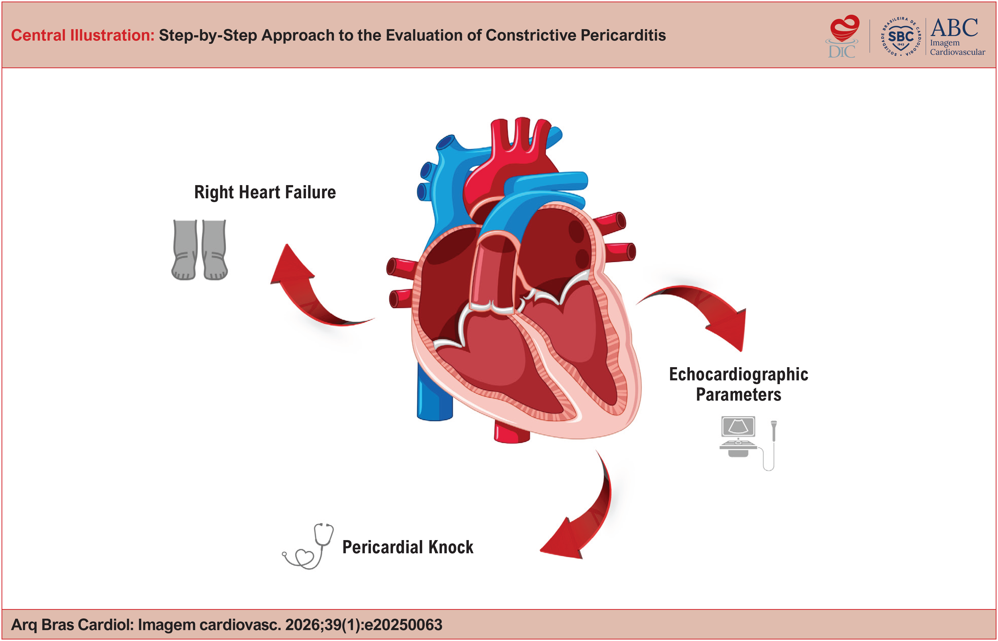
Step-by-Step Approach to the Evaluation of Constrictive Pericarditis
Abstract
Constrictive Pericarditis (CP) is an uncommon but potentially curable condition, typically presenting with right-sided heart failure and impaired ventricular filling. Diagnosis can be challenging and requires a structured approach that integrates various cardiovascular imaging modalities. This article offers a practical guide, based on evidence and clinical experience, for the step-by-step recognition of Constrictive Pericarditis, highlighting echocardiographic findings.
Keywords
Pericarditis; Echocardiography; Doppler Echocardiography
Resumo
A Pericardite Constritiva (PC) é uma condição rara, mas potencialmente curável, que se manifesta por sintomas de insuficiência cardíaca direita e restrição ao enchimento ventricular. O diagnóstico pode ser desafiador e requer uma abordagem estruturada que integra diversas modalidades de imagem cardiovascular. Este artigo oferece um guia prático, baseado em evidências e na experiência clínica, para o reconhecimento passo a passo da Pericardite Constritiva, com destaque aos achados ecocardiográficos.
Palavras-chave
Pericardite; Ecocardiografia; Ecocardiografia Doppler
Introduction
Constrictive pericarditis is a condition resulting from chronic inflammation of the pericardium, which culminates in fibrosis, thickening, and calcification, leading to marked restriction of cardiac chamber filling. Although it may develop as a complication of acute pericarditis, progression to the constrictive form usually occurs over months to years.
Etiologies include systemic infections, prior cardiac surgery, malignancy, radiotherapy, particularly mediastinal irradiation, autoimmune diseases, and idiopathic causes. In endemic countries, tuberculosis is a major cause.1,2
Clinical suspicion most often arises in patients with right-sided heart failure, characterized by systemic congestion (hepatic congestion, anasarca, cardiac cirrhosis) and features of low cardiac output, such as fatigue, cardiac cachexia, and muscle weakness. Additional findings may include a pericardial knock on physical examination and electrocardiographic abnormalities, such as low QRS voltage and nonspecific ST- and T-wave changes. In this setting, cardiovascular imaging plays a central role in establishing the diagnosis and guiding management (Center Figure).
This article aims to present a practical, evidence-based diagnostic flowchart for the evaluation of CP, with emphasis on the sequential use of imaging modalities.
Pathophysiology of CP
Following pericardial injury, an inflammatory cascade is triggered, involving innate immune mechanisms with activation of the NLRP3 inflammasome and release of proinflammatory cytokines, particularly those of the IL-1 family. This inflammatory response promotes cellular infiltration, amplifying and sustaining the autoinflammatory process. Over time, persistent inflammation leads to fibroblast proliferation, granulation tissue formation, pathological neovascularization, and progressive pericardial thickening. This process may progress to fibrosis and calcification, ultimately leading to CP.3,4
Under physiological conditions, the pericardium has sufficient elasticity to accommodate changes in cardiac volume. In CP, pericardial thickening restricts ventricular expansion during diastole.
From a hemodynamic standpoint, two key mechanisms stand out:
-
Dissociation between intrathoracic and intracardiac pressures, caused by the rigid pericardium, which prevents normal transmission of respiratory variations;
-
Marked ventricular interdependence, in which increased venous return to the right chambers during inspiration results in reduced filling of the left chambers, due to interventricular septal shift toward the Left Ventricle (LV), secondary to the inability of the Right Ventricle (RV) free wall to expand as a result of the restriction imposed by the thickened pericardium.5
Thus, ventricular filling during diastole is initially rapid but is abruptly interrupted by pericardial constraint, leading to increased filling pressures in the right chambers, reduced preload in the left chambers, and decreased cardiac output.
Finally, inflammatory and hemodynamic alterations also contribute to sodium and water retention through activation of the sympathetic nervous system and the renin–angiotensin–aldosterone system, perpetuating symptoms and complicating clinical management.
Early recognition of constrictive pericarditis allows appropriate indication of pericardiectomy, which may be curative and restore normal diastolic function in many patients.
Clinical presentation and diagnosis
Loss of normal pericardial compliance restricts diastolic ventricular filling, resulting in a clinical syndrome dominated by right-sided heart failure. In advanced stages, this presentation is frequently misinterpreted as primary hepatic or renal disease.6
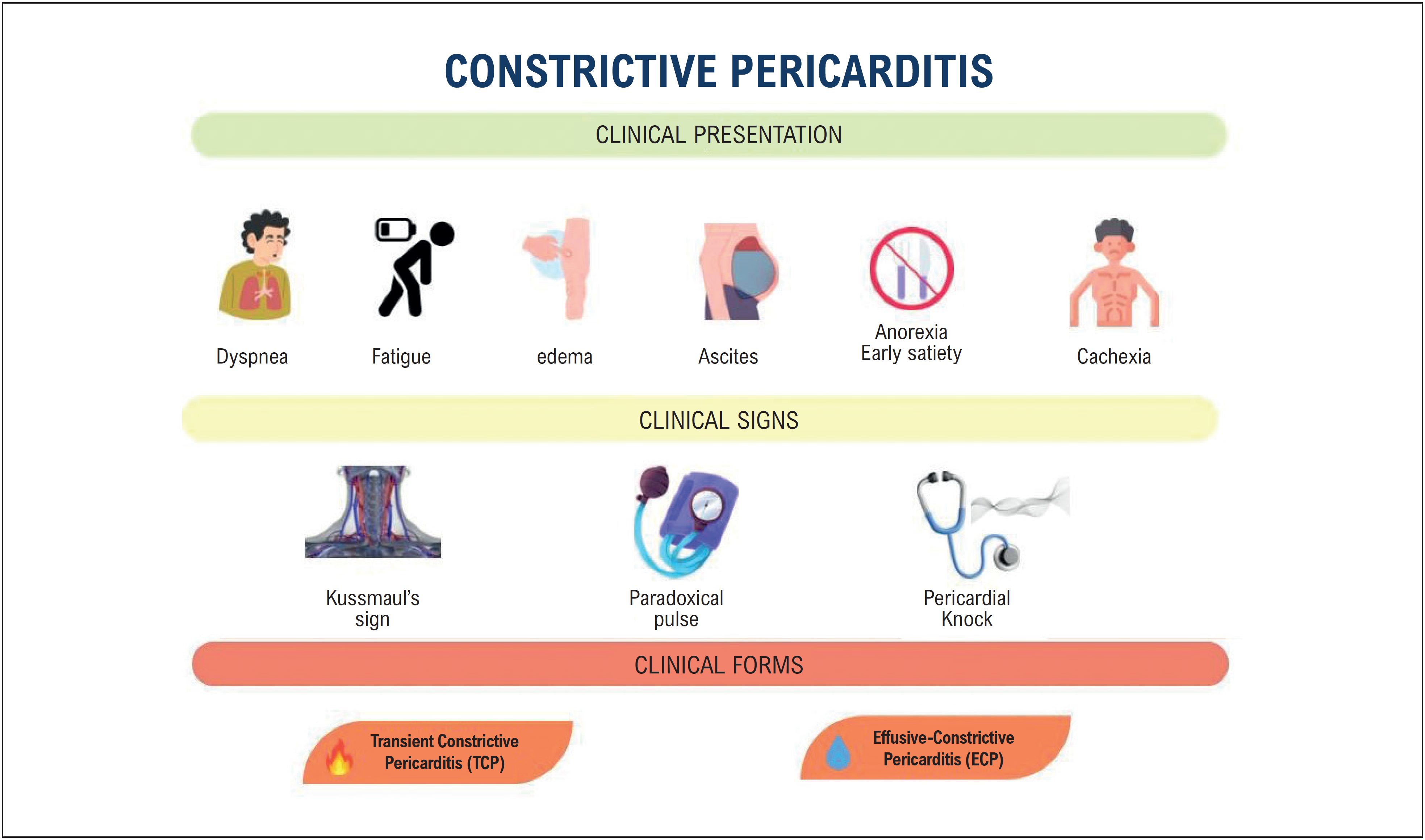
Exertional dyspnea is among the most common symptoms and often develops insidiously, reflecting elevated pulmonary venous pressures and reduced cardiac output. Fatigue is another frequent complaint and is directly related to decreased peripheral perfusion. Patients commonly present with bilateral, ascending peripheral edema as a sign of systemic congestion. Ascites, in turn, occurs in approximately half of cases and may be disproportionate to the degree of edema, misleadingly suggesting primary liver disease.7
Other findings include abdominal fullness, anorexia, and early satiety, frequently related to hepatoesplenic congestion. In advanced cases, cachexia may be present.
Physical examination should focus on characteristic findings such as Kussmaul's sign (paradoxical inspiratory jugular venous distension), pulsus paradoxus (a > 10 mmHg inspiratory decline in systolic blood pressure), and the classic pericardial knock, an early diastolic sound best heard at the mitral or tricuspid area, indicating abrupt cessation of ventricular filling.
When present together, these findings should strongly raise suspicion for CP and prompt comprehensive evaluation using complementary imaging modalities (Figure 1).

CP encompasses subtypes with important diagnostic and therapeutic implications.
-
Transient Constrictive Pericarditis (TCP): associated with active inflammation and reversible pericardial thickening, characterized by spontaneous resolution or resolution after anti-inflammatory therapy (NSAIDs, colchicine, or corticosteroids); early identification is important to avoid unnecessary pericardiectomy.6,7
-
Effusive-Constrictive Pericarditis (ECP): defined by persistence of constrictive physiology after drainage of a significant pericardial effusion (PEff). The classic hemodynamic finding is the persistence of elevated right atrial pressure after pericardiocentesis. Currently, ECP can be identified by echocardiography through the presence of constrictive features after drainage.6,7
CP's main differential diagnosis is restrictive cardiomyopathy, particularly infiltrative diseases such as amyloidosis and sarcoidosis. The distinction is based on clinical presentation, hemodynamic assessment, and imaging studies.
Among imaging diagnostic modalities, the following can be highlighted:
-
Transthoracic Echocardiography (TTE): first-line examination for anatomical and hemodynamic assessment of CP. Transesophageal and stress echocardiography are generally unnecessary for establishing the diagnosis. Limitations: inability to identify active inflammation or fibrosis.
-
Computed Tomography (CT): gold standard for the identification of pericardial calcifications and for surgical planning, allowing detailed visualization of the relationship with adjacent structures.
-
Cardiac Magnetic Resonance (CMR): a complementary modality that assesses pericardial thickening (>3 mm), edema (T2-STIR), inflammation (LGE), and therapeutic response. It is also useful for surgical planning and follow-up of reversible forms.
-
Cardiac Catheterization (CC): used when noninvasive tests are inconclusive. It demonstrates equalization of diastolic pressures, the square root sign, and respiratory discordance between RV and LV systolic pressures. Although invasive, it remains a reference standard for confirmation in equivocal cases.
Table 1 summarizes the main indications for each diagnostic method.
Echocardiographic evaluation: step by step
During echocardiographic evaluation, techniques such as M-mode and Doppler are mandatory. For optimal echocardiographic assessment, a respirometer should always be used, particularly for evaluation of parameters such as septal motion, variation of atrioventricular flows, hepatic vein Doppler, and superior vena cava Doppler.
The main echocardiographic criteria include:
-
Pericardial thickening: although not always present, identification of a thickened or calcified pericardium is suggestive, in addition to assessment of pericardial effusion.
-
Characteristic hemodynamic changes:
-
Respiratory-related septal bounce: abrupt motion of the interventricular septum during diastole, with anterior displacement during inspiration and posterior displacement during expiration (Figure 2), reflecting exaggerated ventricular interdependence.
-
Respiratory variation of mitral and tricuspid inflow velocities: inspiratory variation of mitral inflow (E wave) greater than 25% and tricuspid inflow (E wave) greater than 40%.
-
Restrictive diastolic filling pattern: E/A ratio greater than 2 and mitral E-wave deceleration time shorter than 140 ms.
-
End-diastolic expiratory reversal velocity in the hepatic vein: a ratio between end-diastolic reversed and forward flow velocity ≥ 0.8 is highly specific.
-
-
Tissue Doppler findings:
-
Preserved or increased medial mitral annular e′ velocity: medial e′ velocity ≥ 9 cm/s is highly specific for CP.
-
"Annulus reversus": medial e′ velocity exceeds lateral e′ velocity, due to the effect of pericardial restriction.
-
-
Dilation and lack of collapse of the inferior vena cava and hepatic veins: indicative of elevated right atrial pressure.

The combination of these findings increases diagnostic accuracy; particularly, the presence of respiratory septal shift associated with medial e′ velocity ≥ 9 cm/s or expiratory diastolic reversal of hepatic venous flow shows high sensitivity and specificity for the diagnosis of CP.7
A landmark Mayo Clinic study evaluated 130 patients with surgically confirmed CP and identified five key echocardiographic variables derived from prior investigations: interventricular septal motion abnormalities (including respirophasic septal shift and septal bounce), respiratory variation in mitral inflow E-wave velocity, septal mitral annular e′ velocity, the septal-to-lateral e′ ratio, and expiratory diastolic reversal of hepatic venous flow.8
In addition to these variables, the use of global longitudinal strain and three-dimensional echocardiography may provide additional information.
The integrated use of anatomical and functional criteria is essential to differentiate constrictive pericarditis from restrictive cardiomyopathy (Table 2) and other causes of diastolic heart failure.
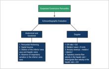
Figure 3 demonstrates a practical flowchart for the echocardiographic diagnostic approach in suspected constrictive pericarditis.
Differential Diagnosis: Constrictive Pericarditis vs. Restrictive Cardiomyopathy
Constrictive pericarditis and restrictive cardiomyopathy share symptoms of diastolic dysfunction, but there are marked differences, as summarized in Table 3.
Conclusion
Constrictive pericarditis (CP) is a complex but potentially treatable condition; its diagnosis requires a systematic approach that combines clinical signs and imaging tools. While echocardiography remains the cornerstone of initial evaluation, multimodality imaging is essential for identifying active inflammation and fibrosis and guiding therapeutic decision-making. Adoption of a structured diagnostic protocol facilitates early recognition, avoids unnecessary interventions, and ultimately improves patient outcomes.
-
Sources of Funding
There were no external funding sources for this study.
-
Study Association
This study is not associated with any thesis or dissertation work.
-
Ethics Approval and Consent to Participate
This article does not contain any studies with human participants or animals performed by any of the authors.
-
Use of Artificial Intelligence
During the preparation of this work, the author(s) used Canva for creation of the central figure of the manuscript. After using this tool/service, the author(s) reviewed and edited the content as needed and take full responsibility for the content of the published article.
Availability of Research Data
The underlying content of the research text is contained within the manuscript.
References
-
1 Tzani A, Doulamis IP, Tzoumas A, Avgerinos DV, Koudoumas D, Siasos G, et al. Meta-Analysis of Population Characteristics and Outcomes of Patients Undergoing Pericardiectomy for Constrictive Pericarditis. Am J Cardiol. 2021;146:120-7. doi: 10.1016/j.amjcard.2021.01.033.
» https://doi.org/10.1016/j.amjcard.2021.01.033 -
2 Cremer PC, Klein AL, Imazio M. Diagnosis, Risk Stratification, and Treatment of Pericarditis: A Review. JAMA. 2024;332(13):1090-100. doi: 10.1001/jama.2024.12935.
» https://doi.org/10.1001/jama.2024.12935 -
3 Del Buono MG, Bonaventura A, Vecchié A, Moroni F, Golino M, Bressi E, et al. Pathogenic Pathways and Therapeutic Targets of Inflammation in Heart Diseases: A Focus on Interleukin-1. Eur J Clin Invest. 2024;54(2):e14110. doi: 10.1111/eci.14110.
» https://doi.org/10.1111/eci.14110 -
4 Bonaventura A, Montecucco F. Inflammation and Pericarditis: Are Neutrophils Actors Behind the Scenes? J Cell Physiol. 2019;234(5):5390-5398. doi: 10.1002/jcp.27436.
» https://doi.org/10.1002/jcp.27436 -
5 Marta L, Alves M, Peres M, Ferreira R, Ferreira H, Leal M, et al. Effusive-Constrictive Pericarditis as the Manifestation of an Unexpected Diagnosis. Rev Port Cardiol. 2015;34(1):69.e1-6. doi: 10.1016/j.repc.2014.08.013.
» https://doi.org/10.1016/j.repc.2014.08.013 -
6 Klein AL, Wang TKM, Cremer PC, Abbate A, Adler Y, Asher C, et al. Pericardial Diseases: International Position Statement on New Concepts and Advances in Multimodality Cardiac Imaging. JACC Cardiovasc Imaging. 2024;17(8):937-88. doi: 10.1016/j.jcmg.2024.04.010.
» https://doi.org/10.1016/j.jcmg.2024.04.010 -
7 Wang TKM, Klein AL, Cremer PC, Imazio M, Kohnstamm S, Luis SA, et al. 2025 Concise Clinical Guidance: An ACC Expert Consensus Statement on the Diagnosis and Management of Pericarditis: A Report of the American College of Cardiology Solution Set Oversight Committee. J Am Coll Cardiol. 2025;86(25):2691-719. doi: 10.1016/j.jacc.2025.05.023.
» https://doi.org/10.1016/j.jacc.2025.05.023 -
8 Welch TD, Ling LH, Espinosa RE, Anavekar NS, Wiste HJ, Lahr BD, et al. Echocardiographic Diagnosis of Constrictive Pericarditis: Mayo Clinic criteria. Circ Cardiovasc Imaging. 2014;7(3):526-34. doi: 10.1161/CIRCIMAGING.113.001613.
» https://doi.org/10.1161/CIRCIMAGING.113.001613 -
9 Welch TD. Constrictive Pericarditis: Diagnosis, Management and Clinical Outcomes. Heart. 2018;104(9):725-31. doi: 10.1136/heartjnl-2017-311683.
» https://doi.org/10.1136/heartjnl-2017-311683 -
10 Nishimura RA. Constrictive Pericarditis in the Modern Era: A Diagnostic Dilemma. Heart. 2001;86(6):619-23. doi: 10.1136/heart.86.6.619.
» https://doi.org/10.1136/heart.86.6.619 -
11 Bogaert J, Francone M. Cardiovascular Magnetic Resonance in Pericardial Diseases. J Cardiovasc Magn Reson. 2009;11(1):14. doi: 10.1186/1532-429X-11-14.
» https://doi.org/10.1186/1532-429X-11-14 - 12 Bogaert J, Dymarkowski S. Clinical Cardiac MRI. Berlim: Springer; 2020.
-
13 Talreja DR, Nishimura RA, Oh JK, Holmes DR. Constrictive Pericarditis in the Modern Era: Novel Criteria for Diagnosis in the Cardiac Catheterization Laboratory. J Am Coll Cardiol. 2008;51(3):315-9. doi: 10.1016/j.jacc.2007.09.039.
» https://doi.org/10.1016/j.jacc.2007.09.039 -
14 Syed FF, Schaff HV, Oh JK. Constrictive Pericarditis--A Curable Diastolic Heart Failure. Nat Rev Cardiol. 2014;11(9):530-44. doi: 10.1038/nrcardio.2014.100.
» https://doi.org/10.1038/nrcardio.2014.100 -
15 Welch TD, Oh JK. Constrictive Pericarditis: Old Disease, New Approaches. Curr Cardiol Rep. 2015;17(4):20. doi: 10.1007/s11886-015-0576-x.
» https://doi.org/10.1007/s11886-015-0576-x -
16 Miranda WR, Oh JK. Constrictive Pericarditis: A Practical Clinical Approach. Prog Cardiovasc Dis. 2017;59(4):369-79. doi: 10.1016/j.pcad.2016.12.008.
» https://doi.org/10.1016/j.pcad.2016.12.008 -
17 Ha JW, Ommen SR, Tajik AJ, Barnes ME, Ammash NM, Gertz MA, et al. Differentiation of Constrictive Pericarditis from Restrictive Cardiomyopathy Using Mitral Annular Velocity by Tissue Doppler Echocardiography. Am J Cardiol. 2004;94(3):316-9. doi: 10.1016/j.amjcard.2004.04.026.
» https://doi.org/10.1016/j.amjcard.2004.04.026 -
18 Hurrell DG, Nishimura RA, Higano ST, Appleton CP, Danielson GK, Holmes DR Jr, et al. Value of Dynamic Respiratory Changes in Left and Right Ventricular Pressures for the Diagnosis of Constrictive Pericarditis. Circulation. 1996;93(11):2007-13. doi: 10.1161/01.cir.93.11.2007.
» https://doi.org/10.1161/01.cir.93.11.2007 -
19 Lloyd JW, Anavekar NS, Oh JK, Miranda WR. Multimodality Imaging in Differentiating Constrictive Pericarditis From Restrictive Cardiomyopathy: A Comprehensive Overview for Clinicians and Imagers. J Am Soc Echocardiogr. 2023;36(12):1254-65. doi: 10.1016/j.echo.2023.08.016.
» https://doi.org/10.1016/j.echo.2023.08.016
Edited by
-
Editor responsible for the review:
Marcelo Tavares