Anabolic-Androgenic Steroids and Acute Myocardial Infarction in Young Adults: A Literature Review Based on a Case Series
Abstract
Background: The use of anabolic-androgenic steroids (AAS) has increased substantially, especially among young adults seeking aesthetic enhancement and improved physical performance. Scientific evidence demonstrates a significant association between the abuse of these substances and severe cardiovascular events, including acute myocardial infarction (AMI), often occurring in individuals without traditional cardiovascular risk factors.
Objective: To describe the adverse effects of AAS on the cardiovascular system and the main pathophysiological mechanisms involved in the development of AMI through the analysis of a clinical case series combined with a review of medical literature.
Methods: A systematic literature review was conducted using the PubMed and SciELO databases, complemented by the analysis of three clinical cases. Demographic variables and characteristics related to AAS use, including duration of exposure and route of administration, were evaluated, with emphasis on the pathophysiological mechanisms associated with AMI.
Results: Analysis of the clinical cases identified different mechanisms related to AMI, including coronary thrombosis, atherosclerosis with plaque rupture, and spontaneous coronary artery dissection. The literature review also identified other relevant mechanisms, such as coronary vasospasm and toxic myocarditis. A predominance of male patients was observed, with the highest incidence occurring among individuals aged 20-40 years, and testosterone esters were the most frequently used AAS.
Conclusions: AAS abuse represents a major threat to cardiovascular health and is associated with AMI through multiple pathophysiological mechanisms. These findings reinforce the need for public awareness as well as the development of preventive strategies and clinical guidelines aimed at managing this emerging condition.
Keywords:
Anabolic Androgenic Steroids; Myocardial Infarction; Atherosclerosis; Myocarditis

Resumo
Fundamento: O uso de esteroides anabolizantes androgênicos (EAA) tem aumentado de forma expressiva, especialmente entre adultos jovens com objetivos estéticos e de melhora do desempenho físico. Evidências científicas demonstram associação significativa entre o uso abusivo dessas substâncias e eventos cardiovasculares graves, incluindo infarto agudo do miocárdio (IAM), frequentemente em indivíduos sem fatores de risco cardiovasculares tradicionais.
Objetivo: Descrever os efeitos adversos dos EAA sobre o sistema cardiovascular e os principais mecanismos fisiopatológicos envolvidos no desenvolvimento do IAM, por meio da análise de uma série de casos clínicos associada à revisão da literatura médica.
Métodos: Foi realizada uma revisão sistemática da literatura nas bases de dados PubMed e SciELO, complementada pela análise de três casos clínicos. Foram avaliadas variáveis demográficas e características relacionadas ao uso dos EAA, incluindo tempo de exposição e via de administração, com ênfase nos mecanismos fisiopatológicos associados ao IAM.
Resultados: A análise dos casos clínicos identificou diferentes mecanismos relacionados ao IAM, como trombose coronariana, aterosclerose com ruptura de placa e dissecção espontânea da artéria coronária (DEAC). A revisão da literatura evidenciou ainda outros mecanismos relevantes, como vasoespasmo coronariano e miocardite tóxica. Observou-se predominância do sexo masculino, com maior incidência entre indivíduos de 20-40 anos, sendo os ésteres de testosterona os EAA mais frequentemente utilizados.
Conclusões: O abuso de EAA representa importante ameaça à saúde cardiovascular, estando associado ao IAM por múltiplos mecanismos fisiopatológicos. Os achados reforçam a necessidade de conscientização da população, bem como do desenvolvimento de estratégias preventivas e diretrizes clínicas voltadas ao manejo dessa condição emergente.
Palavras-chave:
Esteróides Androgênicos Anabolizantes; Infarto do Miocárdio; Aterosclerose; Miocardite

Introduction
Through Resolution No. 2,333/2023, the Brazilian Federal Council of Medicine (CFM for short, in Portuguese) prohibited the prescription of anabolic-androgenic steroids (AAS) for aesthetic purposes, muscle mass gain, and physical performance enhancement. In Brazil, however, the prevalence of AAS use may reach 31.6% in specific groups, such as physical education students and gym instructors.1 Worldwide, the prevalence of nonmedical use of such substances is estimated at 4%-5% among men.2
The use of AAS is associated with muscle hypertrophy, increased energy reserves, and virilizing effects, promoting aesthetic improvement and enhanced physical performance; thus, they are widely used by high-performance athletes.1 However, excessive use is associated with important cardiovascular adverse effects, contributing to increased morbidity and mortality. The main reported complications include dyslipidemia, hypertension, coagulopathies, cardiomyopathies, arrhythmias, and acute myocardial infarction (AMI), whose occurrence may be explained by different pathophysiological mechanisms (Central Illustration).
Over the last decade, there has been an increase in the number of young AAS users without traditional cardiovascular risk factors who developed acute coronary syndrome (ACS) as the main clinical outcome.4 This scenario reinforces the hypothesis that AAS may act as triggering substances for coronary events in differents pathways (Figure 1), representing a potential public health problem.

Cardiovascular adverse effects of anabolic-androgenic steroids. Source: Adapted from Fadah et al.12 ACS: acute coronary syndrome; BP: blood pressure; CAD: coronary artery disease; HDL: high-density lipoprotein; LDL: low-density lipoprotein; Lp(a): lipoprotein(a); RAAS: renin-angiotensin-aldosterone system; SCAD: spontaneous coronary artery dissection.
Understanding this topic is essential to reduce the underreporting of AMI cases associated with the use of AAS and to enable more appropriate diagnostic, therapeutic, and preventive strategies. Therefore, the aim of this study was to review the cardiovascular alterations associated with inappropriate AAS use, with emphasis on the different pathophysiological mechanisms involved in AMI and other cardiac complications, based on a literature review and analysis of a clinical case series.
Methods
For this study, data were collected from the PubMed and SciELO databases. Articles published between 1990 and 2024, in English and Portuguese, involving studies conducted in humans, were included. The search strategy used the following keywords: "myocardial infarction," "anabolic steroids," "atherosclerosis," "atherosclerotic plaque erosion," "coronary vasospasm," "MINOCA," and "toxic myocarditis." Duplicate articles or those that did not contribute in a relevant and up-to-date manner to the objectives of the study were excluded.
In addition to the literature review, three clinical cases were selected through the analysis of hospital electronic medical records. Inclusion criteria comprised patients without previous comorbidities, with a history of current use of AAS, and who presented with an acute myocardial event requiring coronary angiography during hospitalization. Patients with preexisting cardiovascular comorbidities and those older than 50 years were excluded.
The selected patients signed an informed consent form provided after invitation to participate in the study. Confidentiality of the collected information was ensured by the researchers; data were used exclusively to fulfill the objectives of the present study, in accordance with the ethical principles established by Resolution No. 466/12 of the Brazilian National Health Council, linked to the Ministry of Health.
Results
AAS are synthetic substances structurally similar to testosterone and may be administered orally, topically, or by injection.5 Testosterone exerts androgenic functions related to the development and maintenance of male sexual characteristics as well as anabolic functions, such as skeletal muscle and bone tissue growth.6
Excessive use of these compounds is associated with a prothrombotic state, hypertension, left ventricular hypertrophy, alterations in lipid metabolism, increased visceral fat, dyslipidemia, premature atherosclerosis, coronary vasospasm, and endothelial dysfunction, increasing the risk of myocardial ischemia (Figure 2).4,7,8

Pathophysiological mechanisms of AAS associated with AMI. Source: Author's personal archive. AAS: anabolic-androgenic steroids; AMI: acute myocardial infarction; SCAD: spontaneous coronary artery dissection.
Despite the associated risks, users frequently administer doses 10-100 times higher than therapeutic levels and often combine different types of AAS simultaneously or cyclically to enhance aesthetic and physical performance effects.9
Evidence-based clinical cases: mechanisms of acute myocardial infarction in patients using anabolic-androgenic steroids
Anabolic-Androgenic Steroids use and Acute myocardial infarction associated with coronary thrombotic events
Case 1: Acute myocardial infarction caused by a coronary thrombotic event in a young patient using anabolic-androgenic steroids
A 32-year-old male patient without previous comorbidities, who had been using injectable Deca-Durabolin® for the previous 3 months, presented to the emergency department with typical chest pain associated with ST-segment elevation (STE). Coronary angiography demonstrated a negative image in the middle third of the right coronary artery (RCA), with a high thrombotic burden and embolization to the right posterior descending branch and the right posterior ventricular branch (Figure 3, Panel A).

A) Coronary angiography in the right anterior oblique projection demonstrating a negative image suggestive of thrombus in the right posterior ventricular artery. B) Intravascular ultrasound performed at the site of the thrombotic image after 5 days of antithrombotic therapy, demonstrating intact endothelium, absence of atherosclerotic plaque, and adequate luminal area. Source: Author's personal archive.
Thrombus aspiration was performed, with removal of a small amount of thrombus, followed by balloon angioplasty of the right posterior ventricular artery. Distal flow (TIMI I) and residual thrombus image persisted. Intracoronary tirofiban administration was selected, along with oral dual antiplatelet therapy using aspirin and prasugrel, in addition to full anticoagulation with heparin. Subsequently, repeat evaluation with intravascular imaging using intravascular ultrasound (IVUS) was scheduled after 5 days of clinical therapy to determine the mechanism of AMI.
After clinical treatment, a significant reduction in thrombotic burden was observed, associated with improvement in distal flow (TIMI III). Intravascular IVUS assessment (Figure 3, Panel B) demonstrated the absence of atherosclerotic plaque and residual thrombus as well as an adequate luminal area. The patient was discharged 2 days after the second cardiac catheterization. Transthoracic echocardiography (TTE) showed a left ventricular (LV) ejection fraction (LVEF) of 50% and inferior wall hypokinesia. Anticoagulant therapy with rivaroxaban was prescribed at hospital discharge.
The relationship between AAS use and increased thrombotic risk has been investigated since 1988, when the first report of sudden death in a young user of these substances was described.10 During the use of AAS, alterations occur in the primary, secondary, and tertiary phases of hemostasis, which favors thrombus formation. The prothrombotic state observed in these patients is related to increased platelet adhesion and aggregation resulting from enzymatic and glycoprotein imbalances involved in the coagulation cascade, potentially leading to AMI, stroke, and pulmonary embolism.11
Chang et al.10 demonstrated increased levels of coagulation factors II, V, VIII, IX, X, and XII, associated with greater prothrombin production and mild alterations in fibrinogen levels. Factors VIII and IX participate in the formation of the tenase complex, whereas factors V and X compose the prothrombinase complex, both essential for thrombin generation and fibrin formation. Conversely, some studies also observed increased levels of coagulation inhibitors, such as antithrombin, protein C, protein S, and tissue factor pathway inhibitor, which reduce thrombus formation.
Regarding fibrinolysis, a reduction in plasminogen activator inhibitor-1 has been described, associated with increased tissue plasminogen activator and plasminogen levels, favoring fibrin clot degradation.8,10
Other prothrombotic factors have also been described, including elevated serum homocysteine levels, increased thromboxane A2 (a potent platelet aggregator), accelerated erythropoiesis with consequent increased blood viscosity, and reduced prostacyclin levels, an important inhibitor of platelet aggregation.12
In the HAARLEM study, involving 100 men using AAS, increased levels of factors II, IX, and XI were observed, in addition to increased levels of protein S and D-dimer, which suggests maintenance of coagulation pathway activity.8 Therefore, the exact effects of AAS on the hemostatic system remain controversial, which reinforces the need for more robust studies and better clarification of the involved pathophysiological mechanisms.
Anabolic-Androgenic Steroids use and Acute myocardial infarction associated with accelerated atherosclerosis
Case 2: Anabolic-androgenic steroids and accelerated atherosclerosis
A 38-year-old male patient without previous comorbidities, who had been using Deca-Durabolin® for the previous 6 months, presented to the emergency department with typical chest pain, electrocardiogram (ECG) showing STE, and nonsustained ventricular tachycardia. Coronary angiography demonstrated total occlusion in the proximal third of the circumflex artery (Cx), involving bifurcation with the left marginal artery (LMA) with an acute appearance, in addition to total occlusion of the proximal third of the RCA with a chronic appearance, 80% obstruction in the middle third of the left anterior descending artery (LAD), and a 70% lesion in the third diagonal branch (Figures 4 and 5).

A) Coronary angiography in the left anterior oblique caudal projection demonstrating total occlusion in the middle third of the Cx. B) Coronary angiography in the right anterior oblique cranial projection demonstrating total occlusion of the Cx associated with significant lesions in the middle third of the LAD and in the third diagonal branch. Cx: circumflex artery; LAD: left anterior descending artery. Source: Author's personal archive.

Coronary angiography in the right cranial posteroanterior projection demonstrating contrast subtraction involving the middle and distal thirds of the LAD, compatible with type 1 angiographic pattern of spontaneous coronary artery dissection. LAD: left anterior descending artery. Source: Author's personal archive.
Percutaneous coronary intervention with drug-eluting stent implantation at the Cx/LMA bifurcation was performed. Dual antiplatelet therapy with aspirin and clopidogrel was initiated, in addition to high-intensity statin therapy and heart failure treatment, considering that TTE demonstrated LV inferior wall akinesia, anterolateral and lateral LV wall hypokinesia, and diastolic dysfunction with an ejection fraction of 39%. Before hospital discharge, angioplasty of the residual LAD lesion with drug-eluting stent implantation was performed.
Atherosclerosis is a chronic cardiometabolic disease characterized by lipid accumulation within the vascular wall, promoting endothelial inflammation. Its pathophysiological process begins with oxidation of low-density lipoprotein (LDL) by macrophages within the vascular intima, resulting in foam cell formation, fatty streaks, and subsequently atheromatous plaques. This process triggers oxidative imbalance, with excessive production of free radicals and activation of inflammatory cytokines responsible for the progression of atheromatosis.11
The biochemical mechanism through which AAS contribute to the development of atherosclerosis remains controversial. According to Baggish et al., doses greater than 1,000 mg/week increase levels of apolipoprotein B, the main component of LDL. In addition, AAS increase the expression of endothelial adhesion molecules, facilitating LDL migration into the vascular intima.13
An increased LDL/high-density lipoprotein (HDL) ratio is also observed due to enhanced HDL catabolism mediated by hepatic lipase, whose levels are elevated in AAS users.14 These lipid alterations are related not only to isolated substance use but mainly to duration of use, administered dose, and route of administration. Parenterally administered AAS, because they bypass first-pass hepatic metabolism, tend to have less adverse impact on the lipid profile.15,16
Additionally, reduced levels of apolipoprotein A1, a molecule involved in reverse cholesterol transport and lipid removal from the vascular wall, have been described.14
Interestingly, AAS appear to reduce the levels of lipoprotein(a) [Lp(a)], a cardiovascular risk marker with a strong genetic component. Users of danazol demonstrated decreased serum levels of Lp(a), which suggests possible distinct effects of AAS on lipid metabolism and require further clarification.17
The CRISP CT study evaluated coronary inflammation through the perivascular fat attenuation index (FAI), even in the absence of atherosclerotic plaques. AAS users presented higher perivascular FAI values, which suggests coronary perivascular inflammation even in individuals with lower body fat percentage. This finding may be related to blockade of mature adipocyte differentiation induced by AAS, characterizing these individuals as a risk group for atherosclerotic events regardless of body composition control.18,19
Anabolic-Androgenic Steroids use and Acute myocardial infarction associated with nonobstructive coronary arteries
Case 3: Myocardial infarction with nonobstructive coronary arteries caused by spontaneous coronary artery dissection
A 34-year-old male patient without previous comorbidities, who had been using Durateston® for the previous 4 months, presented to the emergency department with complete right hemiplegia and aphasia. Noncontrast cranial computed tomography demonstrated hypodensity in the territory of the middle cerebral artery (MCA), a finding compatible with ischemic stroke.
During etiological investigation of the stroke, cranial and cervical vessel computed tomography angiography demonstrated occlusion of the left MCA. TTE revealed apical LV dyskinesia associated with the presence of a mobile intracavitary thrombus measuring 41 mm × 23 mm. Therefore, the stroke was attributed to a cardioembolic mechanism.
However, due to the presence of ventricular dyskinesia, coronary angiography was performed and demonstrated contrast subtraction involving the middle and distal thirds of the LAD. The coronary arteries did not present significant obstructive lesions, and findings compatible with type 1 spontaneous coronary artery dissection (SCAD) affecting the middle and distal LAD segments were identified. Complementary intravascular imaging was not performed. The patient was discharged on clopidogrel and apixaban. TTE demonstrated an LVEF of 50%, associated with apical LV dyskinesia.
In 2020, the updated European Society of Cardiology guidelines redefined myocardial infarction with nonobstructive coronary arteries (MINOCA) as myocardial infarction of ischemic etiology in the absence of coronary stenosis greater than 50% caused by obstructive atherosclerotic disease on angiography, therefore excluding nonischemic causes previously included in the concept.20
Thus, patients presenting with clinical findings suggestive of AMI, alterations in biomarkers of acute myocardial injury, ECG abnormalities with or without STE, and echocardiographic findings compatible with myocardial ischemia, but without significant obstructive coronary disease on angiography, should be investigated for MINOCA.
Among the pathophysiological mechanisms associated with MINOCA, SCAD, which is frequently underdiagnosed, stands out. SCAD is defined as a nontraumatic, noniatrogenic, nonatherosclerotic separation of the layers of the coronary artery, resulting in false lumen formation.21
Two main pathophysiological mechanisms have been proposed: rupture of the intimal layer with communication between the subintimal space and the true lumen, and formation of intramural hematoma secondary to rupture of microvessels within the medial layer, leading to arterial compression, reduced coronary flow, ischemia, and AMI.22
The etiology of SCAD has not yet been fully clarified, but it is known to involve genetic predisposition associated with precipitating factors, such as physical or emotional stress, illicit drug use, stimulants, and hormonal alterations. Cases associated with AAS use are rare; however, cardiocirculatory stress induced by these substances (e.g., hypertension, atherosclerosis, and coronary vasospasm), associated with intense physical exercise, may favor the occurrence of SCAD.23
SCAD predominantly affects young or middle-aged women, generally between 45-53 years of age, frequently in the absence of classic atherosclerotic risk factors. It may occur in nulliparous, pregnant, postpartum, and postmenopausal women.24 Evidence suggests that cyclic hormonal alterations exert greater influence on SCAD than absolute serum levels of estrogen and progesterone.25 However, conclusive studies regarding the direct role of AAS in this context are still lacking.
Within the pathophysiological spectrum of ACS associated with use of AAS, coronary vasospasm also deserves attention. Inappropriate use of such substances promotes sympathetic hyperactivation, vasoconstriction, and increased blood pressure (BP). Coronary vasospasm is directly related to vascular smooth muscle hyperreactivity, resulting in abnormal contraction of smooth muscle cells and disturbance of coronary vasomotor tone. Vasospasm is defined as intense vasoconstriction (> 90%) of an epicardial coronary artery, with significant impairment of blood flow and potential development of myocardial ischemia.25,26
Vasospasm may occur spontaneously or due to vascular hyperreactivity in response to endogenous and exogenous substances. Testosterone is known to induce abnormal vascular response to norepinephrine by inhibiting its reuptake and favoring coronary vasospasm.27
Thus, AAS contribute to loss of coronary vasodilatory mechanisms and promote increased levels of vasoconstrictive substances, such as endothelin-1, norepinephrine, thromboxane, and angiotensin II.7,11,15
Associated with this process, AAS act as precursors of endothelial injury through alterations in the lipid profile, chronic vascular inflammation, and acceleration of atherosclerosis. This mechanism represents an important pathway of direct injury to the coronary endothelium, creating a favorable substrate for coronary spasm associated with sympathetic hyperreactivity. Consequently, vascular hypercontractility occurs due to imbalance between vasodilatory and vasoconstrictive substances, culminating in acute myocardial injury.15
In the cardiac catheterization laboratory, diagnosis of coronary vasospasm may be challenging, since the spasm may have resolved spontaneously or after nitrate administration in the emergency department. Provocative testing with intracoronary acetylcholine has diagnostic value; however, its use in clinical practice is limited due to low availability and the risk of ventricular arrhythmias associated with the procedure.
Anabolic-androgenic steroids and myocarditis
A systematic review with meta-analysis demonstrated that approximately 34.5% of MINOCA cases may present an associated diagnosis of myocarditis.27 Myocarditis is defined as an inflammatory disease of myocardium, with endomyocardial biopsy considered the diagnostic gold standard. Toxic myocarditis corresponds to a subgroup of secondary etiologies related to exposure to heavy metals, radiation, and drugs, including alcohol, amphetamines, and AAS.
AAS promote alterations in cardiac size, mass, geometry, and function.9 These modifications may mimic hypertrophic cardiomyopathy, with increased interventricular septal and LV posterior wall thickness.28 Cardiac hypertrophy represents a multifactorial response resulting from direct effects on cardiomyocytes associated with hemodynamic and metabolic alterations.28
Montisci et al.29 conducted an autopsy study involving four athletes using AAS and identified myocardial fibrosis, myofibrillar destruction, and eosinophilic infiltration within cardiac tissue. AAS induce pathological cardiac hypertrophy through modulation of gene transcription, acting directly on RNA and regulating protein synthesis via androgen receptors located in the nuclei of cardiomyocytes.12 In addition, alterations involving enzymes, ionic flow, and the myocardial interstitial matrix may also occur.
In an experimental study involving rats exposed to AAS associated with physical exercise, Carmo et al.30 demonstrated increased type III collagen production related to interstitial alterations and myocardial fibrosis, associated with greater activation of the renin-angiotensin-aldosterone system (RAAS).
Angiotensin II is the main biologically active component of the RAAS and plays an important role in regulating BP, plasma volume, and sympathetic activity.31 Studies demonstrate that cardiac angiotensin II production may occur independently of the systemic endocrine system.31 This substance promotes cardiomyocyte hypertrophy and fibroblast proliferation, stimulating collagen and fibronectin synthesis while reducing the activity of enzymes responsible for collagen degradation.30 Angiotensin II AT1 receptors show markedly increased expression in AAS users.12
Another relevant aspect is the structural similarity between AAS and aldosterone, a mineralocorticoid hormone produced in the adrenal cortex. Aldosterone also contributes to increased collagen deposition within the cardiovascular matrix, promoting the development of myocardial fibrosis.12,32
In addition to these mechanisms, alterations in enzymatic reactions, intracellular ion transport (especially calcium), excessive free radical production, and release of proinflammatory cytokines may occur.33 These phenomena favor cellular apoptosis and mitochondrial dysfunction, leading to loss of structural integrity of cardiomyocytes and modification of contractile proteins. Associated with disruption of calcium homeostasis, these alterations contribute to the development of myocardial fibrosis and cardiac hypertrophy.12
Activation of the renin-angiotensin-aldosterone axis through the direct action of angiotensin II and aldosterone promotes increased blood volume.12 This effect, associated with sympathetic hyperactivity and maintenance of increased levels of norepinephrine, favors elevation of mean BP and increases the risk of hypertension and hemodynamic overload.33
Several studies demonstrate that AAS users present increased left ventricular mass index, reduced LVEF, impaired left ventricular diastolic function, and elevated BP levels. Abdullah et al.35 demonstrated, through echocardiographic evaluation of current and former AAS users, the presence of biventricular cardiomyopathy associated with reduced right ventricular function.12
Structural alterations in cardiomyocytes also promote modifications in cardiac action potential, creating a substrate for arrhythmias and increasing the risk of sudden death in this patient population.
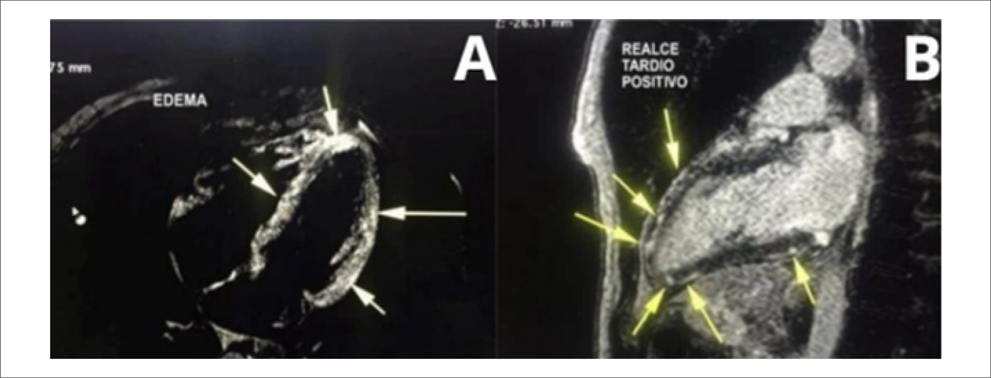
Sobreira Filho et al.36 reported a case of toxic myocarditis initially mimicking non-ST-segment elevation ACS in a 30-year-old patient using testosterone enanthate, trenbolone acetate, and boldenone. Coronary angiography did not demonstrate obstructive coronary lesions; however, ventriculography revealed severe and diffuse hypokinesia of the inferior, apical, and septal walls, a finding later confirmed by TTE, associated with reduced LVEF to 43%. Cardiac magnetic resonance imaging was essential to differentiate the nonischemic fibrosis pattern and establish a more accurate diagnosis (Figure 6, Panels A and B).36

A) Triple IR T2-weighted T2 sequence in the four-chamber view demonstrating hyperintense areas suggestive of myocardial edema. B) Late gadolinium enhancement sequence in the two-chamber view demonstrating hyperintense areas with a mesoepicardial nonischemic pattern suggestive of myocardial fibrosis and/or necrosis. Source: Author's personal archive.
Toxic myocarditis involves multiple pathophysiological mechanisms, including autoimmune reactions, exposure to cardiotoxic agents, and acute infectious processes.37 Among the associated chemical agents, AAS stand out because of increased production of proinflammatory mediators induced by the testosterone present in many of these compounds.38
According to Cooper,38 exposure to cardiotoxic agents such as AAS may induce alterations in cellular metabolism, excessive production of reactive oxygen species, and mitochondrial dysfunction, culminating in cellular necrosis or apoptosis. In addition, an immune-mediated inflammatory response characterized by infiltration of T lymphocytes and macrophages into myocardial tissue may occur, associated with release of proinflammatory cytokines (e.g., interleukin-1, tumor necrosis factor alpha, and interleukin-6), which further potentiates cardiac muscle damage.39
Discussion
Chronic use of AAS at supraphysiological doses is associated with several severe adverse effects capable of significantly compromising users’ cardiovascular health.
In the present case series, all patients were young men between 20-40 years of age, without previous comorbidities and with a history of AAS use. Each case illustrates different pathophysiological mechanisms related to the cardiovascular toxicity of these substances, including coronary thrombosis, accelerated atherosclerosis with plaque rupture, SCAD, and toxic myocarditis. Predominant use of injectable testosterone esters was observed.
Although the analyzed sample was exclusively male, the progressive increase in AAS use among young women should also be highlighted, mainly motivated by the pursuit of improved athletic performance and body aesthetics. Studies demonstrate prevalence rates of up to 16.8% among female bodybuilders, 4.4% among athletes or resistance-training practitioners, and 1.4% in the general female population.40
Despite the cardiovascular deleterious effects already widely described in the literature, increasing abusive and indiscriminate use of these substances has been observed among recreational users, frequently without adequate medical supervision and without full awareness of the associated potential risks. The medical community should remain attentive to the possible cardiovascular repercussions associated with AAS, seeking to expand knowledge on this topic in order to improve diagnostic, therapeutic, and preventive strategies.
In addition, discontinuation of AAS frequently requires a multidisciplinary approach, considering the occurrence of rebound effects and the association with psychiatric comorbidities, such as anxiety disorder and body dysmorphic disorder, often aggravated by social pressure related to pursuit of the ideal body.
Over recent decades, a marked increase has been observed in reports of AMI among young patients using AAS. However, additional studies are still needed to strengthen the causal association between use of these substances and the different pathophysiological mechanisms involved in the development of ACS, considering the possible influence of concomitant predisposing factors.
Furthermore, studies evaluating specific substances individually are needed, since concomitant use of multiple AAS makes individualized analysis of the cardiovascular effects of each compound difficult.
Conclusion
As briefly summarized in Central Illustration, it was possible to review the main mechanisms related to AMI in young patients using AAS, including coronary thrombotic events, accelerated atherosclerosis, MINOCA, and toxic myocarditis.
These findings reinforce that indiscriminate use of these substances represents an important public health problem, especially among young adults without traditional cardiovascular risk factors. Therefore, despite the prohibition established by the CFM, strengthening awareness and prevention strategies involving healthcare professionals, the general population, and media outlets remains essential in order to reduce the cardiovascular impacts associated with abusive use of AAS.
-
Sources of Funding
There were no external funding sources for this study.
-
Study Association
This article is part of the thesis of master submitted by Fabiana Rocha Botelho de Oliveira, from Hospital de Messejana.
-
Ethics Approval and Consent to Participate
This article does not contain any studies with human participants or animals performed by any of the authors.
-
Use of Artificial Intelligence
During the preparation of this work, the authors used ChatGPT to improve the grammar and semantics of the text and Open Evidence to facilitate the search for articles related to the proposed topic, assisting in the development of the final manuscript.
Availability of Research Data
The underlying content of the research text is contained within the manuscript.
References
-
1 Abrahin OSC, Souza NSF, Sousa EC, Moreira JKR, Nascimento VC. Prevalence of the Use of Anabolic Androgenic Steroids by Physical Education Students and Teachers Who Work in Health Clubs. Rev Bras Med Esporte. 2013;19(1):27-30. doi: 10.1590/S1517-86922013000100005.
» https://doi.org/10.1590/S1517-86922013000100005 -
2 Smit DL, Hon O, Venhuis BJ, den Heijer M, Ronde W. Baseline Characteristics of the HAARLEM Study: 100 Male Amateur Athletes Using Anabolic Androgenic Steroids. Scand J Med Sci Sports. 2020;30(3):531-9. doi: 10.1111/sms.13592.
» https://doi.org/10.1111/sms.13592 -
3 Perry JC, Schuetz TM, Memon MD, Faiz S, Cancarevic I. Anabolic Steroids and Cardiovascular Outcomes: The Controversy. Cureus. 2020;12(7):e9333. doi: 10.7759/cureus.9333.
» https://doi.org/10.7759/cureus.9333 -
4 Baggish AL, Weiner RB, Kanayama G, Hudson JI, Lu MT, Hoffmann U, et al. Cardiovascular Toxicity of Illicit Anabolic-Androgenic Steroid Use. Circulation. 2017;135(21):1991-2002. doi: 10.1161/CIRCULATIONAHA.116.026945.
» https://doi.org/10.1161/CIRCULATIONAHA.116.026945 -
5 Kicman AT. Pharmacology of Anabolic Steroids. Br J Pharmacol. 2008;154(3):502-21. doi: 10.1038/bjp.2008.165.
» https://doi.org/10.1038/bjp.2008.165 -
6 Corona G, Rastrelli G, Vignozzi L, Maggi M. Emerging Medication for the Treatment of Male Hypogonadism. Expert Opin Emerg Drugs. 2012;17(2):239-59. doi: 10.1517/14728214.2012.683411.
» https://doi.org/10.1517/14728214.2012.683411 -
7 Beltrame JF, Crea F, Kaski JC, Ogawa H, Ong P, Sechtem U, et al. The Who, What, Why, When, How and Where of Vasospastic Angina. Circ J. 2016;80(2):289-98. doi: 10.1253/circj.CJ-15-1202.
» https://doi.org/10.1253/circj.CJ-15-1202 -
8 Camilleri E, Smit DL, van Rein N, Le Cessie S, de Hon O, den Heijer M, et al. Coagulation Profiles during and after Anabolic Androgenic Steroid Use: Data from the HAARLEM Study. Res Pract Thromb Haemost. 2023;7(7):102215. doi: 10.1016/j.rpth.2023.102215.
» https://doi.org/10.1016/j.rpth.2023.102215 -
9 Carmo EC, Fernandes T, Oliveira EM. Anabolic Steroids: From the Athlete to Cardiacapthy Patient. Rev Educ Fís/UEM. 2012;23(2)307-18. doi: 10.4025/reveducfis.v23i2.12462.
» https://doi.org/10.4025/reveducfis.v23i2.12462 -
10 Chang S, Münster AB, Gram J, Sidelmann JJ. Anabolic Androgenic Steroid Abuse: The Effects on Thrombosis Risk, Coagulation, and Fibrinolysis. Semin Thromb Hemost. 2018;44(8):734-46. doi: 10.1055/s-0038-1670639.
» https://doi.org/10.1055/s-0038-1670639 -
11 Christou GA, Christou KA, Nikas DN, Goudevenos JA. Acute Myocardial Infarction in a Young Bodybuilder Taking Anabolic Androgenic Steroids: A Case Report and Critical Review of the Literature. Eur J Prev Cardiol. 2016;23(16):1785-96. doi: 10.1177/2047487316651341.
» https://doi.org/10.1177/2047487316651341 -
12 Fadah K, Gopi G, Lingireddy A, Blumer V, Dewald T, Mentz RJ. Anabolic Androgenic Steroids and Cardiomyopathy: An Update. Front Cardiovasc Med. 2023;10:1214374. doi: 10.3389/fcvm.2023.1214374.
» https://doi.org/10.3389/fcvm.2023.1214374 -
13 Death AK, McGrath KC, Sader MA, Nakhla S, Jessup W, Handelsman DJ, et al. Dihydrotestosterone Promotes Vascular Cell Adhesion Molecule-1 Expression in Male Human Endothelial Cells Via a Nuclear Factor-kappaB-Dependent Pathway. Endocrinology. 2004;145(4):1889-97. doi: 10.1210/en.2003-0789.
» https://doi.org/10.1210/en.2003-0789 -
14 Hartgens F, Rietjens G, Keizer HA, Kuipers H, Wolffenbuttel BH. Effects of Androgenic-Anabolic Steroids on Apolipoproteins and Lipoprotein (a). Br J Sports Med. 2004;38(3):253-9. doi: 10.1136/bjsm.2003.000199.
» https://doi.org/10.1136/bjsm.2003.000199 -
15 Severo CB, Ribeiro JP, Umpierre D, Da Silveira AD, Padilha MC, De Aquino FR Neto, et al. Increased Atherothrombotic Markers and Endothelial Dysfunction in Steroid Users. Eur J Prev Cardiol. 2013;20(2):195-201. doi: 10.1177/2047487312437062.
» https://doi.org/10.1177/2047487312437062 -
16 Dukewich M, Stolz AA. Anabolic Steroid-Associated Liver Injury. Clin Liver Dis. 2024;23(1):e0196. doi: 10.1097/CLD.0000000000000196.
» https://doi.org/10.1097/CLD.0000000000000196 -
17 Crook D, Sidhu M, Seed M, O’Donnell M, Stevenson JC. Lipoprotein Lp(a) Levels are Reduced by Danazol, an Anabolic Steroid. Atherosclerosis. 1992;92(1):41-7. doi: 10.1016/0021-9150(92)90008-5.
» https://doi.org/10.1016/0021-9150(92)90008-5 -
18 Oikonomou EK, Marwan M, Desai MY, Mancio J, Alashi A, Centeno EH, et al. Non-Invasive Detection of Coronary Inflammation Using Computed Tomography and Prediction of Residual Cardiovascular Risk (the CRISP CT Study): A Post-Hoc Analysis of Prospective Outcome Data. Lancet. 2018;392(10151):929-39. doi: 10.1016/S0140-6736(18)31114-0.
» https://doi.org/10.1016/S0140-6736(18)31114-0 -
19 Souza FR, Rochitte CE, Silva DC, Sampaio B, Passarelli M, Santos MRD, et al. Coronary Inflammation by Computed Tomography Pericoronary Fat Attenuation and Increased Cytokines in Young Male Anabolic Androgenic Steroid Users. Arq Bras Cardiol. 2023;120(11):e20220822. doi: 10.36660/abc.20220822.
» https://doi.org/10.36660/abc.20220822 -
20 Occhipinti G, Bucciarelli-Ducci C, Capodanno D. Diagnostic Pathways in Myocardial Infarction with Non-Obstructive Coronary Artery Disease (MINOCA). Eur Heart J Acute Cardiovasc Care. 2021;10(7):813-22. doi: 10.1093/ehjacc/zuab049.
» https://doi.org/10.1093/ehjacc/zuab049 -
21 Basso C, Morgagni GL, Thiene G. Spontaneous Coronary Artery Dissection: A Neglected Cause of Acute Myocardial Ischaemia and Sudden Death. Heart. 1996;75(5):451-4. doi: 10.1136/hrt.75.5.451.
» https://doi.org/10.1136/hrt.75.5.451 -
22 Heidari A, Sabzi F, Faraji R. Spontaneous Coronary Artery Dissection in Anabolic Steroid Misuse. Ann Card Anaesth. 2018;21(1):103-4. doi: 10.4103/aca.ACA_161_17.
» https://doi.org/10.4103/aca.ACA_161_17 -
23 Hayes SN, Tweet MS, Adlam D, Kim ESH, Gulati R, Price JE, et al. Spontaneous Coronary Artery Dissection: JACC State-of-the-Art Review. J Am Coll Cardiol. 2020;76(8):961-84. doi: 10.1016/j.jacc.2020.05.084.
» https://doi.org/10.1016/j.jacc.2020.05.084 -
24 Kim ESH. Spontaneous Coronary-Artery Dissection. N Engl J Med. 2020;383(24):2358-70. doi: 10.1056/NEJMra2001524.
» https://doi.org/10.1056/NEJMra2001524 -
25 Tamis-Holland JE, Jneid H, Reynolds HR, Agewall S, Brilakis ES, Brown TM, et al. Contemporary Diagnosis and Management of Patients with Myocardial Infarction in the Absence of Obstructive Coronary Artery Disease: A Scientific Statement from the American Heart Association. Circulation. 2019;139(18):e891-e908. doi: 10.1161/CIR.0000000000000670.
» https://doi.org/10.1161/CIR.0000000000000670 -
26 Carbone A, D’Andrea A, Riegler L, Scarafile R, Pezzullo E, Martone F, et al. Cardiac Damage in Athlete's Heart: When the "Supernormal" Heart Fails! World J Cardiol. 2017;9(6):470-80. doi: 10.4330/wjc.v9.i6.470.
» https://doi.org/10.4330/wjc.v9.i6.470 -
27 Hausvater A, Smilowitz NR, Li B, Redel-Traub G, Quien M, Qian Y, et al. Myocarditis in Relation to Angiographic Findings in Patients with Provisional Diagnoses of MINOCA. JACC Cardiovasc Imaging. 2020;13(9):1906-13. doi: 10.1016/j.jcmg.2020.02.037.
» https://doi.org/10.1016/j.jcmg.2020.02.037 -
28 Adami PE, Koutlianos N, Baggish A, Bermon S, Cavarretta E, Deligiannis A, et al. Cardiovascular Effects of Doping Substances, Commonly Prescribed Medications and Ergogenic Aids in Relation to Sports: A Position Statement of the Sport Cardiology and Exercise Nucleus of the European Association of Preventive Cardiology. Eur J Prev Cardiol. 2022;29(3):559-75. doi: 10.1093/eurjpc/zwab198.
» https://doi.org/10.1093/eurjpc/zwab198 -
29 Montisci M, El Mazloum R, Cecchetto G, Terranova C, Ferrara SD, Thiene G, et al. Anabolic Androgenic Steroids Abuse and Cardiac Death in Athletes: Morphological and Toxicological Findings in Four Fatal Cases. Forensic Sci Int. 2012;217(1-3):e13-8. doi: 10.1016/j.forsciint.2011.10.032.
» https://doi.org/10.1016/j.forsciint.2011.10.032 -
30 Carmo EC, Rosa KT, Koike DC, Fernandes T, Silva ND Jr, Mattos KC, et al. Association Between Anabolic Steroids and Aerobic Physical Training Leads to Cardiac Morphological Alterations and Loss of Ventricular Function in Rats. Rev Bras Med Esporte. 2011;17(2):137-41. doi: 10.1590/S1517-86922011000200014 .
» https://doi.org/10.1590/S1517-86922011000200014 - 31 Oigman W, Neves MFT. Sistema Renina-Angiotensina e Hipertrofia Ventricular Esquerda. Rev Bras Hipertens. 2000;7(3):261-7.
-
32 Santos MA, Oliveira CV, Silva AS. Adverse Cardiovascular Effects from the Use of Anabolic-Androgenic Steroids as Ergogenic Resources. Subst Use Misuse. 2014;49(9):1132-7. doi: 10.3109/10826084.2014.903751.
» https://doi.org/10.3109/10826084.2014.903751 - 33 Pereira BVM, Nascimento BR. Miocardiopatias tóxicas: álcool, anfetaminas e anabolizantes. Rev Med Minas Gerais. 2013;23(3):358-66.
-
34 Beutel A, Bergamaschi CT, Campos RR. Effects of Chronic Anabolic Steroid Treatment on Tonic and Reflex Cardiovascular Control in Male Rats. J Steroid Biochem Mol Biol. 2005;93(1):43-8. doi: 10.1016/j.jsbmb.2004.11.003.
» https://doi.org/10.1016/j.jsbmb.2004.11.003 -
35 Abdullah R, Bjørnebekk A, Hauger LE, Hullstein IR, Edvardsen T, Haugaa KH, et al. Severe Biventricular Cardiomyopathy in Both Current and Former Long-Term Users of Anabolic-Androgenic Steroids. Eur J Prev Cardiol. 2024;31(5):599-608. doi: 10.1093/eurjpc/zwad362.
» https://doi.org/10.1093/eurjpc/zwad362 -
36 Sobreira FM Filho, Lino DOC, Belém LS, Rocha RPS, Lima CJM, Alcântra ACB. Acute Myocarditis in User of Anabolic Hormones Diagnosed by Magnetic Resonance Imaging: A Case Report. ABC Imagem Cardiovasc. 2018;31(3):207-10. doi: 10.5935/2318-8219.20180031.
» https://doi.org/10.5935/2318-8219.20180031 -
37 Tschöpe C, Ammirati E, Bozkurt B, Caforio ALP, Cooper LT, Felix SB, et al. Myocarditis and Inflammatory Cardiomyopathy: Current Evidence and Future Directions. Nat Rev Cardiol. 2021;18(3):169-93. doi: 10.1038/s41569-020-00435-x.
» https://doi.org/10.1038/s41569-020-00435-x -
38 Cooper LT Jr. Myocarditis. N Engl J Med. 2009;360(15):1526-38. doi: 10.1056/NEJMra0800028.
» https://doi.org/10.1056/NEJMra0800028 -
39 Caforio AL, Pankuweit S, Arbustini E, Basso C, Gimeno-Blanes J, Felix SB, et al. Current State of Knowledge on Aetiology, Diagnosis, Management, and Therapy of Myocarditis: A Position Statement of the European Society of Cardiology Working Group on Myocardial and Pericardial Diseases. Eur Heart J. 2013;34(33):2636-48. doi: 10.1093/eurheartj/eht210.
» https://doi.org/10.1093/eurheartj/eht210 -
40 Piatkowski T, Whiteside B, Robertson J, Henning A, Lau EHY, Dunn M. What is the Prevalence of Anabolic-Androgenic Steroid Use among Women? A Systematic Review. Addiction. 2024;119(12):2088-100. doi: 10.1111/add.16643.
» https://doi.org/10.1111/add.16643
Edited by
-
Editor responsible for the review:
Marcelo Tavares