The Use of Artificial Intelligence in the Diagnosis of Cardiac Amyloidosis: Integrative Review
Abstract
Fundamento: Cardiac amyloidosis is a rare form of infiltrative cardiomyopathy characterized by the deposition of proteins in the myocardium, resulting in increased wall thickness, impaired ventricular function, and possible progression to heart failure. Diagnosis is challenging due to the low prevalence of the disease and the nonspecific nature of its clinical manifestations. The application of artificial intelligence (AI) to the analysis of medical tests emerges as a promising strategy for early detection, more accurate diagnosis, and timely initiation of treatment.
Methods: An integrative literature review was conducted on the use of AI in the diagnosis of cardiac amyloidosis. Articles published between 2019 and 2024 were searched in the PubMed, Scopus, Web of Science, Embase, and Cochrane Library databases.
Results: Of the 420 articles initially identified, 21 met the eligibility criteria and were included in the final analysis. A predominance of retrospective observational studies applying machine learning models was observed. Among the diagnostic modalities evaluated in association with AI, electrocardiography and echocardiography were the most frequently studied tests.
Conclusion: AI demonstrates high potential to improve the screening and diagnosis of cardiac amyloidosis when applied to the analysis of clinical and imaging tests. The findings of this review indicate that AI may accelerate the diagnostic process, reduce the need for invasive procedures, and optimize the use of health care resources. However, to expand its integration into clinical practice and enhance its generalizability, further model refinement and validation in more diverse populations are required.
Keywords:
Amyloidosis; Artificial Intelligence; Diagnosis
Resumo
Fundamento: A amiloidose cardíaca é uma forma rara de cardiomiopatia infiltrativa caracterizada pela deposição de proteínas no miocárdio, resultando em aumento da espessura parietal, comprometimento da função ventricular e possível progressão para insuficiência cardíaca. O diagnóstico é desafiador devido à baixa prevalência da doença e à inespecificidade das manifestações clínicas. Nesse cenário, a aplicação da inteligência artificial (IA) à análise de exames médicos surge como estratégia promissora para detecção precoce, diagnóstico mais preciso e início oportuno do tratamento.
Métodos: Realizou-se revisão integrativa da literatura sobre o uso da IA no diagnóstico da amiloidose cardíaca. Foram pesquisados artigos publicados entre 2019 e 2024 nas bases de dados PubMed, Scopus, Web of Science, Embase e Cochrane Library.
Resultados: Dos 420 artigos inicialmente identificados, 21 atenderam aos critérios de elegibilidade e foram incluídos na análise final. Observou-se predomínio de estudos observacionais retrospectivos com aplicação de modelos de aprendizado de máquina. Entre as modalidades diagnósticas avaliadas em associação com IA, o eletrocardiograma e o ecocardiograma foram os exames mais frequentemente estudados.
Conclusão: A IA demonstra elevado potencial para aprimorar o rastreio e o diagnóstico da amiloidose cardíaca quando aplicada à análise de exames clínicos e de imagem. Os achados desta revisão indicam que a IA pode acelerar o processo diagnóstico, reduzir a necessidade de procedimentos invasivos e otimizar o uso de recursos em saúde. Entretanto, para ampliar sua integração à prática clínica e sua capacidade de generalização, são necessários aprimoramentos adicionais nos modelos e validações em populações mais diversas.
Palavras-chave:
Amiloidose; Inteligência Artificial; Diagnóstico
Introduction
Amyloidosis is a generic term used to describe the extracellular deposition of fibrils formed by low-molecular-weight protein subunits derived from different precursor proteins. Amyloid deposits may result in a wide variety of clinical manifestations, which vary according to the type of protein involved, the amount deposited, and the tissue location. In the genesis of these deposits, initially soluble peptides undergo conformational changes, predominantly acquiring an antiparallel beta-pleated sheet structure, which favors their stacking into twisted fibrils.1
There are dozens of systemic and localized forms of amyloidosis. Among them, four precursor proteins may give rise to both localized and systemic deposits. The main systemic forms are immunoglobulin light-chain (AL) amyloidosis and (ATTR) transthyretin amyloidosis. These forms are named according to the precursor protein of the amyloid deposit (AL or ATTR) and account for approximately 95% of cases of cardiac amyloidosis. The remaining forms correspond to other subtypes of amyloidosis, which are also clinically relevant.2,3
ATTR amyloidosis is characterized by the misfolding and subsequent deposition of transthyretin, a protein responsible for the transport of thyroid hormone and vitamin A. It may present in wild-type or hereditary form.2-4 Similarly, AL amyloidosis results from the accumulation of misfolded immunoglobulin light chains produced by plasma cells associated with dyscrasias.2,3
In cardiac amyloidosis, it is a rare form of progressive cardiomyopathy whose population prevalence has not yet been well established.5 The disease is caused by myocardial deposition of misfolded amyloid proteins, resulting in restrictive cardiomyopathy, with possible progression to heart failure, conduction system disorders, and cardiac death.3,5,6 It may present with cardiovascular signs and symptoms or be diagnosed during the investigation of extracardiac manifestations of the disease.3,6,7 Due to its heterogeneous clinical phenotype and frequently nonspecific manifestations, diagnosis and management tend to occur late.3,6

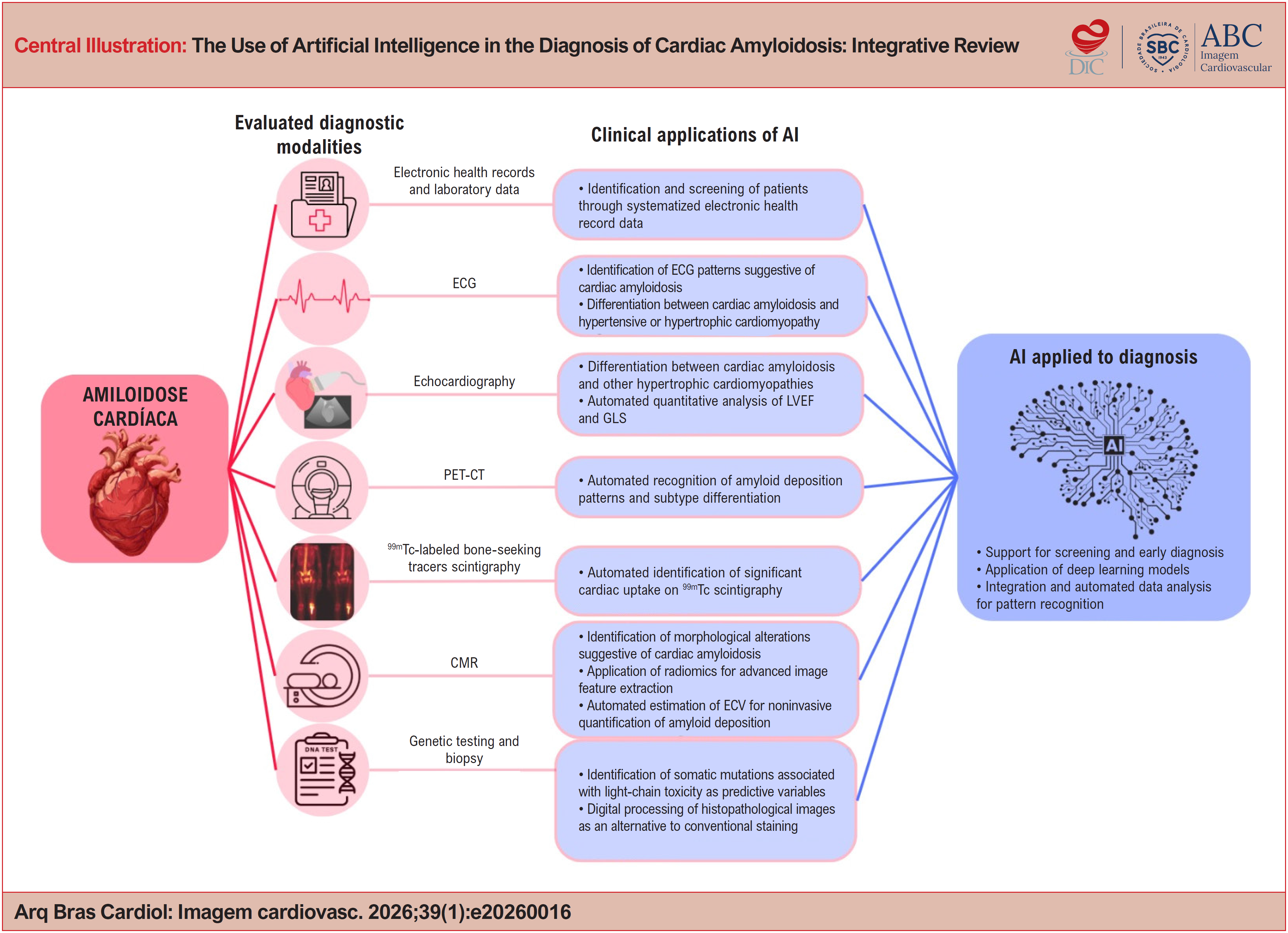
With regard to the diagnostic approach to cardiac amyloidosis, it is essential to recognize clinical scenarios and abnormalities in complementary tests that indicate the need for investigation. However, diagnosis is challenging, especially because it is often an indolent disease whose symptoms may overlap with those of more prevalent heart diseases.3,5 Depending on the clinical context and resource availability, various tools may be employed, including electrocardiography (ECG), echocardiography, cardiac magnetic resonance (CMR), bone-seeking tracers scintigraphy, monoclonal protein screening by immunofixation, and biopsy of the affected tissue. Each method has its own characteristics and different levels of diagnostic accuracy (Central Illustration).
Some findings may increase clinical suspicion of cardiac amyloidosis, such as discordance between increased left ventricular (LV) wall thickness and low QRS voltage, unexplained LV hypertrophy, low-flow, low-gradient aortic stenosis, relative apical sparing of longitudinal strain, a diffuse circumferential subendocardial late gadolinium enhancement pattern of the LV on CMR, and myocardial uptake of bone tracers on bone-seeking tracers scintigraphy.
Despite the diversity of available diagnostic methods, cardiac amyloidosis remains underdiagnosed,3,5-7 which has important repercussions for patients’ quality of life. At the same time, the development of therapies capable of improving clinical outcomes has driven the search for strategies to increase diagnostic rates. These interventions may reduce or stabilize protein deposition, with a consequent reduction in the relative risk of hospitalizations, morbidity, and mortality associated with the disease.
Because of the need for early detection of cardiac amyloidosis, the development of mechanisms that optimize screening and diagnosis with lower costs and risks is essential.3 This review aims to present the main advances in disease detection, with emphasis on promising technological tools in diagnostic medicine, especially artificial intelligence (AI).
AI is a branch of computer science dedicated to the development of systems capable of performing tasks that simulate human cognitive functions, such as decision-making and complex reasoning. In the field of medical diagnosis, these systems are trained using machine learning techniques, in which large volumes of data, often images, are used for pattern recognition. Among the most commonly employed approaches are convolutional neural networks (CNNs), which consist of multiple layers that extract progressively more complex features from the analyzed data, enabling automated identification of patterns in images and other types of information. In general, the larger and more representative the dataset used for training, the greater the model's accuracy tends to be.
Incorporating AI into medical practice enables the more effective screening of rare diseases and improves diagnostic accuracy. AI appears particularly promising for rare diseases that are often underrecognized in clinical practice. Automated systems can integrate multiple signs, symptoms, and complementary findings, helping to guide clinical reasoning. Thus, core skills in medicine, such as pattern recognition, are increasingly being incorporated into computational models with the aim of enhancing diagnostic reliability and supporting decision-making in everyday medical practice.
Several studies have investigated the applicability of AI in the early diagnosis of cardiac amyloidosis. Among the most recent approaches are the integration of AI with imaging methods, such as positron emission tomography (PET-CT), bone-seeking tracers scintigraphy, and CMR as well as its application to the automated analysis of ECGs, genetic data, and phenotypic profiles of cardiac abnormalities. These aspects will be discussed throughout this review.
Methods
The present study is characterized as an integrative literature review aimed at critically analyzing the use of AI in the diagnosis of cardiac amyloidosis. The review was conducted following six methodological steps: i) definition of the research question; ii) establishment of inclusion criteria and sample selection; iii) identification of preselected and selected studies; iv) organization and representation of the included studies; v) critical analysis of the data; and vi) synthesis of the available knowledge.
Searches were conducted in the PubMed, Scopus, Web of Science, Embase, and Cochrane Library databases. The search strategy was developed using the descriptors "artificial intelligence," "amyloidosis," and "diagnosis," included in the Medical Subject Headings and Embase Subject Headings, combined using the Boolean operator AND.
Original studies and meta-analyses published between 2019 and 2024 that evaluated the application of AI in the diagnosis of cardiac amyloidosis were included. Articles that did not meet the inclusion criteria were excluded, as well as narrative reviews, case reports, editorials, and studies with methodology considered inadequate.
After article selection, methodological quality was assessed to ensure greater rigor in the interpretation of findings and robustness of the conclusions. For this step, the JBI critical appraisal tool was used, which includes specific criteria according to study design, covering aspects related to the sample, methodology, data analysis, bias control, and ethical considerations. Quality classification is based on the proportion of affirmative responses to the evaluated criteria, allowing comparison across studies and critical analysis of their results.
Results
The search strategy yielded 420 articles, distributed as follows: 124 identified in PubMed, 61 in Web of Science, 84 in Scopus, 147 in Embase, and 4 in the Cochrane Library. After removal of 192 duplicate studies, 228 articles remained for screening.
Title and abstract screening resulted in the selection of 43 studies for full-text evaluation. After application of the eligibility criteria, 21 articles were included in the final analysis (Figure 1).

Flowchart of the study selection process for the studies included in the integrative review.
Regarding methodological assessment according to the JBI criteria, most studies were classified as having good to excellent methodological quality, with scores ranging from 6 to 8 points. The main factors contributing to this classification included the use of robust statistical metrics and the application of cross-validation in the developed AI models.
As limitations, the absence of gold-standard diagnostic confirmation of cardiac amyloidosis in part of the studies was observed, as well as the lack of external validation of the proposed models, which limits the generalizability of the findings. The detailed results of the methodological assessment are presented in Table 1.
Discussion
The use of AI as a supportive tool in the diagnosis of rare diseases, such as cardiac amyloidosis, has been considered promising, especially in the context of conditions with high clinical heterogeneity and that are frequently underrecognized by general practitioners. Early identification of the disease may modify its natural history and improve prognosis. This review sought to emphasize the potential of already established diagnostic tools for the evaluation of cardiac amyloidosis when combined with machine learning-based systems, an approach addressed in all included studies.
To organize the analysis of the findings, AI performance will be discussed according to the different diagnostic modalities used in the screening and evaluation of cardiac amyloidosis.
Performance of artificial intelligence in the evaluation of medical record data and laboratory tests
Among the screening strategies for amyloid cardiomyopathy, the use of data extracted from electronic health records of patients with heart failure (HF) with preserved ejection fraction (HFpEF) stands out. To differentiate amyloid etiology, especially wild-type ATTR amyloidosis (ATTRwt), from non-amyloid etiology, Huda et al.8 collected electronic health record data and developed an AI model capable of screening and identifying patients with ATTRwt amyloidosis. The system achieved an area under the receiver operating characteristic curve (AUC) of 0.80. Performance was supported by the identification of comorbidities more prevalent in the amyloid etiology group, such as atrial fibrillation and chronic kidney disease, and in the non-amyloid group, such as hypertension, diabetes mellitus, obesity, and coronary artery disease, which were used as predictive variables.
Subsequently, Castaño et al.9 refined the model by focusing the analysis on 11 main phenotypes associated with cardiac amyloidosis, including carpal tunnel syndrome and arrhythmias. The model demonstrated accuracy (74%), sensitivity (77%), and specificity (72%), with an AUC of 0.82. Although there was a slight reduction in some performance parameters, the model was simplified in terms of programming, facilitating its implementation in hospital settings and expanding its potential for population screening.
The studies by Huda et al.8 and Castaño et al.9 demonstrate the feasibility of automated screening through systematic extraction of clinical data, a process that would be costly and operationally complex if performed manually. Although such models are limited by the quality of records coded according to the International Classification of Diseases and by the documented phenotypes, without direct integration of laboratory or imaging tests for definitive typing, they represent relevant tools for large-scale screening, directing patients with higher probability toward further investigation.
Additionally, phenotypes recognized by the model, such as carpal tunnel syndrome, may precede the development of HF, which suggests a potential application of AI in preclinical stages, with implications for early identification of ATTR amyloidosis.
Performance of artificial intelligence in electrocardiographic evaluation
The application of AI in ECG analysis has emerged as a screening strategy, considering that ECG is a widely available, low-cost, noninvasive test.3,5 Model validation represents a fundamental step in the development of these tools, as it involves testing multiple variables across different populations.5,6
Harmon et al.10 developed an algorithm applicable to diverse populations, including different races and sexes. The model achieved an AUC of 0.84 (95% CI: 0.82-0.86), maintaining consistent performance across subgroups, except in the Hispanic population, possibly underrepresented in the sample. The algorithm performed better in ECGs with low voltage and patterns compatible with prior infarction, and showed lower performance in left bundle branch block and LV hypertrophy. These findings suggest the need for greater sample diversity, without invalidating the use of the tool as a screening method.
Vrudhula et al.5 evaluated approximately 1.3 million ECGs from 341,989 patients. The different tested models showed AUC values ranging from 0.660 (95% CI: 0.642-0.736) to 0.898 (95% CI: 0.868-0.924), demonstrating relevant potential for screening and referral for further investigation. However, because of the rarity and underdiagnosis of cardiac amyloidosis, models are often trained with a limited number of confirmed cases.
Similarly, Goto et al.3 reported strong performance, with a C-statistic of 0.91 (95% CI: 0.90-0.93) in the Brigham and Women's Hospital test set, 0.85 (0.82-0.87) at Massachusetts General Hospital, and 0.86 (0.83-0.88) at the University of California, San Francisco. However, the authors emphasize that isolated ECG features do not provide sufficient sensitivity or specificity to be used as independent heuristics, and their integration with other clinical and diagnostic variables is recommended to optimize model performance.
Schrutka et al.6 also reinforce that the proposed model may assist in raising suspicion of cardiac amyloidosis even in the absence of advanced imaging methods. In that study, 20 patients with transthyretin cardiac amyloidosis, 11 with HFpEF, 30 with cardiac amyloidosis, and 50 with other HF etiologies were evaluated. The presence of a low-voltage ECG pattern associated with increased LV wall thickness was highly suggestive of cardiac amyloidosis, allowing differentiation from hypertensive or hypertrophic cardiomyopathy. In the analysis of ECG patterns, pattern 1 was present in 78% of patients with AL amyloidosis and in 58% of those with ATTR amyloidosis (p = 0.009), whereas pattern 2 was identified in 7% of AL amyloidosis cases and in 23% of ATTR amyloidosis cases (p = 0.006). The absence of a specific pattern was observed in 16% of patients with AL amyloidosis and in 18% of patients with ATTR amyloidosis (p = 0.620).
Performance of artificial intelligence in echocardiographic evaluation
Considering the versatility of echocardiography and its central role in the diagnostic investigation of cardiac amyloidosis, the development of AI models capable of reducing operational variability and improving diagnostic accuracy is highly relevant.
Xiaofeng Zhang et al.1 observed that there are still few studies on echocardiography-based myocardial texture analysis and that human visual assessment has limitations in characterizing these alterations. Based on transthoracic echocardiograms, the authors developed four machine learning algorithms to differentiate cardiac amyloidosis from other cardiomyopathies: random forest (RF), support vector machine (SVM), logistic regression (LR), and gradient boosting decision trees (GBDT).
In the analyzed population, all models were able to effectively distinguish cases of cardiac amyloidosis from non-amyloid diseases. The LR model demonstrated the best diagnostic performance, outperforming the traditional ultrasonographic method (AUC: RF 0.77; SVM 0.81; LR 0.81; GBDT 0.71). The authors therefore proposed the application of this tool as a noninvasive diagnostic method for myocardial amyloidosis. However, the relatively small number of cardiac amyloidosis cases may have limited the model's sensitivity for discrimination between groups.
Cotella et al.2 developed an AI model focused on the automated assessment of LV ejection fraction (LVEF) and global longitudinal strain (GLS), central parameters in the diagnosis of cardiac amyloidosis. The authors justified the incorporation of AI based on the fact that manual measurements are time-consuming and show significant inter- and intraobserver variability, which may compromise diagnostic accuracy and influence therapeutic decisions. The study demonstrated that automated and quantitative measurements of LVEF and GLS showed high accuracy and enabled sensitive and specific detection of abnormalities when compared with conventional manual analysis, both in examinations performed before the diagnosis of cardiac amyloidosis and at the time of diagnosis. No statistically significant differences were observed between values obtained by the two methods in the prediagnostic period (LVEF: p = 0.791; GLS: p = 0.105) or at the time of diagnosis (LVEF: p = 0.463; GLS: p = 0.722). In addition, a strong correlation was observed between automated and manual measurements in echocardiograms performed before diagnosis (r = 0.78 for LVEF; r = 0.83 for GLS) and at established diagnosis (r = 0.74 for LVEF; r = 0.80 for GLS).
Goto et al.,3 although acknowledging that ECG-based models show encouraging results, emphasize that their performance may not be sufficient for diagnosing low-prevalence diseases. The echocardiographic model developed by the authors demonstrated greater predictive accuracy compared with the ECG-based model. C-statistics ranged from 0.85-0.91 for ECG and from 0.89-1.00 for echocardiography. Moreover, in subtype analysis, the model showed superior performance in identifying ATTR amyloidosis.
In a more specific population, Shiri et al.4 evaluated the use of machine learning for detecting ATTR amyloidosis in patients with severe aortic stenosis. Although different diagnostic modalities are useful in the initial assessment of these patients, they are not specific for ATTR amyloidosis. Frequently, definitive diagnosis of ATTR cardiomyopathy depends on histopathological confirmation or identification of a mutation in the TTR gene associated with evidence of significant myocardial uptake on bone-seeking tracers scintigraphy. Considering the high cost of genetic testing and bone-seeking tracers scintigraphy, especially in this patient group, the authors developed a noninvasive and potentially cost-effective model based on routine clinical and echocardiographic data. Performance was satisfactory when compared with clinical, laboratory, and interventional imaging variables, with an AUC of 0.79, sensitivity of 0.80, and specificity of 0.78.
Based on evidence that myocardial deformation analysis provides discriminatory value across multiple cardiac chambers, Eckstein et al.7 developed a supervised model capable of differentiating cardiac amyloidosis from hypertrophic cardiomyopathy and from healthy individuals. The system showed excellent performance (AUC = 0.996; accuracy = 94%; sensitivity = 100%; F1-score = 97%), indicating that automated analysis of multichamber cardiac deformation and function may serve as a clinical decision support tool, even without the need for contrast administration.
With the technological advancement of cardiovascular imaging methods, new approaches have been proposed for screening infiltrative cardiomyopathies. Barbieri et al.11 developed a model based on three-dimensional transthoracic echocardiography (3D-TTE) combined with AI, aiming to differentiate various phenotypes of cardiac hypertrophy, including cardiac amyloidosis. The method proposes a reformulation of ejection fraction analysis, traditionally based on CMR, through the use of 3D-TTE integrated with an AI system. Three-dimensional acquisition allowed a more detailed and accurate analysis of LV volume, enabling a more precise calculation of ejection fraction, defined as the ratio between stroke volume and end-diastolic volume, reflecting myocardial contractile capacity. This approach provides more accurate information regarding myocardial shortening and wall thickness, key aspects in recognizing infiltrative cardiomyopathies. In conventional two-dimensional echocardiography, increased wall thickness may mask reduced myocardial shortening, resulting in an apparently preserved ejection fraction. In the context of etiological investigation of HFpEF, the model proved promising, showing higher ejection fraction in patients with hypertrophic cardiomyopathy and cardiac amyloidosis, with the latter exhibiting proportionally even higher values. Diagnostic performance was consistent, with sensitivity of 87%, specificity of 100%, and AUC of 0.959, reinforcing the potential of integrating 3D-TTE and AI in the phenotypic differentiation of myocardial hypertrophy.
Artificial intelligence in positron emission tomography evaluation
Similarly to other imaging modalities, PET-CT has been refined with the aim of making the diagnostic process of cardiac amyloidosis less invasive and more accurate. Deep learning-based models focused on the automated recognition of imaging patterns related to amyloid deposition stand out.
Santarelli et al.12 developed a model aimed at rapidly, early, and specifically identifying the presence of cardiac amyloidosis and its subtypes. The system demonstrated superior performance compared with analysis performed by a specialist with more than 10 years of experience, showing sensitivity greater than 0.8 and specificity greater than 0.89. The model was able to estimate the probability of correlation between the analyzed image and each subtype of cardiac amyloidosis. The authors also highlighted the risk of overfitting, especially in scenarios with a limited number of images available for training. In such cases, the algorithm may show high performance on training data but fail to generalize to external datasets. To mitigate this risk, strategies such as artificial data augmentation and cross-validation were employed, contributing to greater model robustness.
In the study by Miller et al.,13 it is recognized that visual interpretation of single-photon emission computed tomography constitutes the standard approach in the diagnostic evaluation of ATTR amyloidosis, although it is inherently subjective. The authors assessed a deep learning approach for automated volumetric quantification of technetium-99m (99mTc)-pyrophosphate, using segmentation of anatomical structures co-registered on computed tomography attenuation maps in patients with suspected ATTR amyloidosis. The results demonstrated that deep learning-based segmentation was not influenced by the radiotracer uptake pattern and allowed automated quantification of focal uptake images, such as those obtained with 99mTc-pyrophosphate. The model showed excellent performance (AUC = 0.989; 95% CI: 0.974-1.00), indicating potential for accurate identification of patients with ATTR amyloidosis. Therefore, this approach shows potential for precise identification of patients with ATTR amyloidosis.
Performance of artificial intelligence in the evaluation of bone-seeking tracers scintigraphy
In the context of diagnosing cardiac amyloidosis through the application of AI to imaging analysis, it is possible to structure systems integrated into electronic health records, similar to the model described by Huda et al.,8 but directed toward the automated interpretation of scintigraphy images.
Delbarre et al.14 proposed a deep learning model for automated analysis of whole-body 99mTc bone-seeking tracers scintigraphy, based on the premise that significant cardiac uptake on these examinations is strongly suggestive of ATTR amyloidosis. The model demonstrated sensitivity of 98.9% and specificity of 99.5% in cross-validation. In external validation, a slight reduction in sensitivity to 96.1% was observed, while specificity remained at 99.5%, with an AUC of 0.999 in both stages.
For system development, cardiac uptake ≥ 2 according to the Perugini grading scale was used as a predictive variable. The algorithm was trained using CNNs with image-level labels extracted from examinations recorded in electronic health records, enabling automated identification of patterns suggestive of cardiac amyloidosis. As also emphasized by Castaño et al.,9 integration between AI and electronic record systems supports efficient screening of frequently underrecognized conditions, such as the association between increased cardiac uptake on whole-body bone-seeking tracers scintigraphy and ATTR amyloidosis, contributing to identification at earlier stages.
Although bone-seeking tracers scintigraphy does not fully replace all diagnostic methods, Delbarre et al.14 highlighted that when the examination is positive and there is no evidence of monoclonal gammopathy, it may allow definitive noninvasive diagnosis of ATTR cardiomyopathy, particularly in elderly or frail patients in whom myocardial biopsy carries greater risk.
Considering that the diagnosis of cardiac amyloidosis can be established noninvasively through bone-seeking tracers scintigraphy and that visual assessment is inherently subjective and may result in misinterpretation, Spielvogel et al.15 developed an AI system for standardized and reproducible disease screening. The model was trained using a multinational database of 99mTc-labeled bone-seeking tracers scintigraphy, encompassing different tracers and scanners. In the Austrian cohort, cross-validation demonstrated an AUC of 1.00 (95% CI: 1.00-1.00). In external validation, results remained high, with an AUC of 0.997 (95% CI: 0.993-0.999) in the United Kingdom, 0.925 (95% CI: 0.871-0.971) in China, and 1.00 (95% CI: 0.999-1.000) in the Italian cohorts. Approximately one decade ago, myocardial biopsy represented the only definitive modality for diagnosing cardiac amyloidosis. The consolidation of bone-seeking tracers scintigraphy constituted a significant advance in this scenario, particularly in the diagnosis of ATTR amyloidosis. The incorporation of AI into this modality further expands its potential by reducing interpretative subjectivity and increasing diagnostic standardization and reliability.
In the aforementioned multicenter study, intense cardiac uptake was automatically and consistently identified across all tracers used in the investigation of cardiac amyloidosis. Additionally, AI-based screening for detection of uptake suggestive of cardiac amyloidosis in patients undergoing whole-body bone-seeking tracers scintigraphy represents a potentially valuable tool for early disease identification and optimization of care pathways. Implementing this strategy may support timely referral for specialized evaluation and enable earlier initiation of disease-modifying therapies, with potential impact on mortality reduction.
Artificial intelligence in cardiac magnetic resonance evaluation
CMR with late gadolinium enhancement (LGE) is a fundamental method in the investigation of cardiac amyloidosis, given its ability to demonstrate morphological alterations and characteristic enhancement patterns. However, its use may be limited in patients with significant renal impairment, a condition frequently associated with amyloidosis, due to the risks related to contrast administration.
Ma et al.16 investigated the feasibility of diagnosis using non-contrast CMR with native T1 mapping combined with automated radiomic analysis based on AI. The model was trained to recognize specific patterns of amyloid deposition and to indirectly estimate extracellular volume (ECV), a parameter traditionally calculated from pre- and post-gadolinium contrast sequences. In the proposed approach, ECV was accurately estimated through automated identification of myocardial regions of interest. The model achieved an accuracy of 86%, sensitivity of 94%, specificity of 85%, and an AUC of 0.915 in the test set. Unlike bone-seeking tracers scintigraphy, whose main applicability is concentrated on identifying ATTR amyloidosis, non-contrast CMR demonstrated potential for effective diagnosis of cardiac amyloidosis, particularly AL amyloidosis.
In line with this perspective, Nowak et al.17 emphasized that the diagnostic value of CMR derives from its ability to integrate multiple sequences for detailed assessment of myocardial function, edema, inflammation, and fibrosis. ECV allows noninvasive quantification of myocardial amyloid deposition and may influence therapeutic decisions.
Considering that CMR is a reference modality for diagnosing cardiac amyloidosis, Agibetov et al.18 observed that its findings may be nonspecific, especially in centers with lower case volumes. To minimize this risk, they developed a CNN-based algorithm applied to a cohort of 502 patients. Regardless of the deep learning technique employed, models trained with LGE images showed better performance. Fine-tuning of the model resulted in an AUC of 0.96, sensitivity of 94%, and specificity of 90%. Automated classification demonstrated performance comparable to that of human specialists. However, as this was a single-center study, generalization of the results requires caution.
Martini et al.19 also used deep learning for automated analysis of CMR images and estimation of the probability of cardiac amyloidosis. Among the most specific findings, they highlighted the pattern of biventricular pseudo-hypertrophy associated with diffuse transmural LGE. Automated analysis of LGE sequences in the 2C, 4C, and short-axis views was faster and showed accuracy similar to expert assessment, with an AUC of 0.982, positive predictive value of 83%, recall of 95%, and F1-score of 89%.
Performance of artificial intelligence in the evaluation of genetic testing and biopsies
Another promising field in the application of AI to the diagnosis of cardiac amyloidosis, especially in the AL form, involves systematizing the analysis of genetic tests aimed at identifying somatic mutations in immunoglobulin light chains. Garofalo et al.20 demonstrated, through a machine learning model, an association between somatic mutations acquired during B-cell maturation and the development of cardiac amyloidosis. These mutations affect the structural stability of light chains, favoring protein misfolding and subsequent amyloid deposit formation. The proposed model achieved a sensitivity of 76%, specificity of 82%, and an AUC of 0.87, demonstrating relevant predictive capacity in identifying sequences considered toxic. In addition, the authors highlighted that reversal of these mutations may abolish the toxic phenotype, reinforcing the importance of detailed molecular characterization.
Considering the diversity of pathogenic sequences involved, the use of AI represents an appropriate strategy for organizing and analyzing large volumes of genetic variables, acting as a predictor of toxicity. In this way, the algorithm may identify molecular profiles associated with higher risk of developing cardiac amyloidosis, thereby contributing to risk stratification and potential early diagnosis.
In the field of histopathology, biopsy remains the definitive diagnostic evidence in amyloid cardiomyopathy, despite its invasive nature. In this context, integration between histological techniques and deep learning has also shown promise. Yang et al.21 proposed a neural network-based approach capable of transforming autofluorescence images into images equivalent to those obtained by bright-field and polarized light microscopy, simulating the effect of Congo red staining.
Currently, the diagnostic gold standard is based on the identification of birefringence under cross-polarized light after Congo red staining. However, this process is influenced by technical variability in staining, slide preparation quality, and availability of appropriate equipment, in addition to involving high costs. The model proposed by Yang et al.21 demonstrated that digitally generated images showed quality comparable to conventionally stained slides, with potential cost reduction, lower technical dependence, and improved digital storage of samples, considering that specialized scanners for birefringence capture are not always available.
Thus, although there is growing interest in noninvasive diagnostic methods for cardiac amyloidosis, advances in the application of AI to genetic and histopathological analysis also represent a relevant contribution, improving diagnostic accuracy and standardization of laboratory processes.
Barriers to implementing artificial intelligence in the medical workflow
The implementation of AI in medical practice has significant potential to increase diagnostic accuracy, optimize care processes, reduce costs, and support clinical decision-making. However, its incorporation into the workflow faces multifactorial challenges that can be grouped into technical, ethical, organizational, and human dimensions.
From a technical perspective, AI models depend on structured, complete, and standardized datasets. However, many health care systems still operate with fragmented, inconsistent, or incomplete records, which compromises proper training and the generalizability of algorithms. In addition, historically biased data may perpetuate health care disparities, resulting in inappropriate recommendations for certain population groups. Interoperability among different information systems also represents a relevant challenge, hindering seamless integration of AI into established clinical environments.
From an ethical and legal standpoint, questions arise regarding accountability in cases of clinical error involving algorithmic recommendations. Defining responsibility among developers, institutions, and professionals remains complex. This is compounded by concerns about data privacy, security, and governance, especially when there is interinstitutional data sharing for model training or validation.
At the organizational level, adoption of AI-based tools requires efficient integration into care workflows. Solutions that add steps to the process or disrupt established routines tend to generate resistance and operational burden. In addition, physicians, nurses, and other professionals must be trained to critically interpret the recommendations provided by these systems, using them as support rather than as a substitute for clinical judgment. Implementation also requires investment in technological infrastructure, maintenance, and continuous model updating, which may represent a financial barrier for certain institutions.
Finally, the human dimension involves aspects related to professional acceptance and patient trust. Some professionals may express distrust toward the technology or perceive AI as a threat to their clinical role. The so-called "black box" nature of algorithms, in which the decision-making process is not fully transparent, may reduce trust in the tool and hinder its incorporation into clinical practice. From the patient's perspective, trust in decisions influenced by algorithms is not yet universal. Conversely, there is a risk of excessive reliance on AI by professionals, which may compromise independent clinical reasoning if a critical and reflective stance is not maintained.
Conclusion
Based on the findings of this review, AI emerges as a promising tool for optimizing the screening and diagnosis of cardiac amyloidosis. Its application across different diagnostic modalities demonstrates potential to accelerate disease identification, contribute to greater diagnostic accuracy, and consequently support improved clinical outcomes.
The high capacity for processing and analyzing large volumes of data enables AI to recognize complex patterns, expand its ability to generalize, provided it is trained on robust and representative datasets, and assist in the early detection of cardiac amyloidosis. Furthermore, using automated models may reduce the subjectivity of human interpretation, minimize the need for invasive procedures in certain contexts, and rationalize the use of health care resources.
However, despite the advances observed, the consolidation of AI in clinical practice requires continuous model refinement, external validation in diverse populations, and efficient integration into care workflows. Strategic implementation planning, training of health professionals, ethical governance in data management, and ongoing monitoring of algorithmic performance are equally essential.
-
Sources of Funding
There were no external funding sources for this study.
-
Study Association
This study is not associated with any thesis or dissertation work.
-
Ethics Approval and Consent to Participate
This article does not contain any studies with human participants or animals performed by any of the authors.
-
Use of Artificial Intelligence
The authors did not use any artificial intelligence tools in the development of this work.
Availability of Research Data
The underlying content of the research text is contained within the manuscript.
References
-
1 Zhang X, Liang T, Su C, Qin S, Li J, Zeng D, et al. Deep Learn-Based Computer-Assisted Transthoracic Echocardiography: Approach to the Diagnosis of Cardiac Amyloidosis. Int J Cardiovasc Imaging. 2023;39(5):955-65. doi: 10.1007/s10554-023-02806-0.
» https://doi.org/10.1007/s10554-023-02806-0 -
2 Cotella JI, Slivnick JA, Sanderson E, Singulane C, O’Driscoll J, Asch FM, et al. Artificial Intelligence Based Left Ventricular Ejection Fraction and Global Longitudinal Strain in Cardiac Amyloidosis. Echocardiography. 2023;40(3):188-95. doi: 10.1111/echo.15516.
» https://doi.org/10.1111/echo.15516 -
3 Goto S, Mahara K, Beussink-Nelson L, Ikura H, Katsumata Y, Endo J, et al. Artificial Intelligence-Enabled Fully Automated Detection of Cardiac Amyloidosis Using Electrocardiograms and Echocardiograms. Nat Commun. 2021;12(1):2726. doi: 10.1038/s41467-021-22877-8.
» https://doi.org/10.1038/s41467-021-22877-8 -
4 Shiri I, Balzer S, Baj G, Bernhard B, Hundertmark M, Bakula A, et al. Multi-Modality Artificial Intelligence-Based Transthyretin Amyloid Cardiomyopathy Detection in Patients with Severe Aortic Stenosis. Eur J Nucl Med Mol Imaging. 2025;52(2):485-500. doi: 10.1007/s00259-024-06922-4.
» https://doi.org/10.1007/s00259-024-06922-4 -
5 Vrudhula A, Stern L, Cheng PC, Ricchiuto P, Daluwatte C, Witteles R, et al. Impact of Case and Control Selection on Training Artificial Intelligence Screening of Cardiac Amyloidosis. JACC Adv. 2024;3(9):100998. doi: 10.1016/j.jacadv.2024.100998.
» https://doi.org/10.1016/j.jacadv.2024.100998 -
6 Schrutka L, Anner P, Agibetov A, Seirer B, Dusik F, Rettl R, et al. Machine Learning-Derived Electrocardiographic Algorithm for the Detection of Cardiac Amyloidosis. Heart. 2022;108(14):1137-47. doi: 10.1136/heartjnl-2021-319846.
» https://doi.org/10.1136/heartjnl-2021-319846 -
7 Eckstein J, Moghadasi N, Körperich H, Valdés EW, Sciacca V, Paluszkiewicz L, et al. A Machine Learning Challenge: Detection of Cardiac Amyloidosis Based on Bi-Atrial and Right Ventricular Strain and Cardiac Function. Diagnostics. 2022;12(11):2693. doi: 10.3390/diagnostics12112693.
» https://doi.org/10.3390/diagnostics12112693 -
8 Huda A, Castaño A, Niyogi A, Schumacher J, Stewart M, Bruno M, et al. A Machine Learning Model for Identifying Patients at Risk for Wild-Type Transthyretin Amyloid Cardiomyopathy. Nat Commun. 2021;12(1):2725. doi: 10.1038/s41467-021-22876-9.
» https://doi.org/10.1038/s41467-021-22876-9 -
9 Castaño A, Heitner SB, Masri A, Huda A, Calambur V, Bruno M, et al. EstimATTR: A Simplified, Machine-Learning-Based Tool to Predict the Risk of Wild-Type Transthyretin Amyloid Cardiomyopathy. J Card Fail. 2024;30(6):778-87. doi: 10.1016/j.cardfail.2023.11.017.
» https://doi.org/10.1016/j.cardfail.2023.11.017 -
10 Harmon DM, Mangold K, Suarez AB, Scott CG, Murphree DH, Malik A, et al. Postdevelopment Performance and Validation of the Artificial Intelligence-Enhanced Electrocardiogram for Detection of Cardiac Amyloidosis. JACC Adv. 2023;2(8):100612. doi: 10.1016/j.jacadv.2023.100612.
» https://doi.org/10.1016/j.jacadv.2023.100612 -
11 Barbieri A, Imberti JF, Bartolomei M, Bonini N, Laus V, Triglia LT, et al. Quantification of Myocardial Contraction Fraction with Three-Dimensional Automated, Machine-Learning-Based Left-Heart-Chamber Metrics: Diagnostic Utility in Hypertrophic Phenotypes and Normal Ejection Fraction. J Clin Med. 2023;12(17):5525. doi: 10.3390/jcm12175525.
» https://doi.org/10.3390/jcm12175525 -
12 Santarelli MF, Genovesi D, Positano V, Scipioni M, Vergaro G, Favilli B, et al. Deep-Learning-Based Cardiac Amyloidosis Classification from Early Acquired Pet Images. Int J Cardiovasc Imaging. 2021;37(7):2327-35. doi: 10.1007/s10554-021-02190-7.
» https://doi.org/10.1007/s10554-021-02190-7 -
13 Miller RJH, Shanbhag A, Michalowska AM, Kavanagh P, Liang JX, Builoff V, et al. Deep Learning-Enabled Quantification of 99mTc-Pyrophosphate SPECT/CT for Cardiac Amyloidosis. J Nucl Med. 2024;65(7):1144-50. doi: 10.2967/jnumed.124.267542.
» https://doi.org/10.2967/jnumed.124.267542 -
14 Delbarre MA, Girardon F, Roquette L, Blanc-Durand P, Hubaut MA, Hachulla É, et al. Deep Learning on Bone Scintigraphy to Detect Abnormal Cardiac Uptake at Risk of Cardiac Amyloidosis. JACC Cardiovasc Imaging. 2023;16(8):1085-95. doi: 10.1016/j.jcmg.2023.01.014.
» https://doi.org/10.1016/j.jcmg.2023.01.014 -
15 Spielvogel CP, Haberl D, Mascherbauer K, Ning J, Kluge K, Traub-Weidinger T, et al. Diagnosis and Prognosis of Abnormal Cardiac Scintigraphy Uptake Suggestive of Cardiac Amyloidosis Using Artificial Intelligence: A Retrospective, International, Multicentre, Cross-Tracer Development and Validation Study. Lancet Digit Health. 2024;6(4):e251-60. doi: 10.1016/S2589-7500(23)00265-0.
» https://doi.org/10.1016/S2589-7500(23)00265-0 -
16 Ma Q, Chen J, Cao L, Wu X, Tan Z, Liu H. The Incremental Value of Native T1 Mapping-Derived Radiomics for The Diagnosis of Amyloid Light-Chain Cardiac Amyloidosis. Acad Radiol. 2024;31(12):4801-10. doi: 10.1016/j.acra.2024.07.005.
» https://doi.org/10.1016/j.acra.2024.07.005 -
17 Nowak S, Bischoff LM, Pennig L, Kaya K, Isaak A, Theis M, et al. Deep Learning Virtual Contrast-Enhanced T1 Mapping for Contrast-Free Myocardial Extracellular Volume Assessment. J Am Heart Assoc. 2024;13(19):e035599. doi: 10.1161/JAHA.124.035599.
» https://doi.org/10.1161/JAHA.124.035599 -
18 Agibetov A, Kammerlander A, Duca F, Nitsche C, Koschutnik M, Donà C, et al. Convolutional Neural Networks for Fully Automated Diagnosis of Cardiac Amyloidosis by Cardiac Magnetic Resonance Imaging. J Pers Med. 2021;11(12):1268. doi: 10.3390/jpm11121268.
» https://doi.org/10.3390/jpm11121268 -
19 Martini N, Aimo A, Barison A, Latta DD, Vergaro G, Aquaro GD, et al. Deep Learning to Diagnose Cardiac Amyloidosis from Cardiovascular Magnetic Resonance. J Cardiovasc Magn Reson. 2020;22(1):84. doi: 10.1186/s12968-020-00690-4.
» https://doi.org/10.1186/s12968-020-00690-4 -
20 Garofalo M, Piccoli L, Romeo M, Barzago MM, Ravasio S, Foglierini M, et al. Machine Learning Analyses of Antibody Somatic Mutations Predict Immunoglobulin Light Chain Toxicity. Nat Commun. 2021;12(1):3532. doi: 10.1038/s41467-021-23880-9.
» https://doi.org/10.1038/s41467-021-23880-9 -
21 Yang X, Bai B, Zhang Y, Aydin M, Li Y, Selcuk SY, et al. Virtual Birefringence Imaging and Histological Staining of Amyloid Deposits in Label-Free Tissue Using Autofluorescence Microscopy and Deep Learning. Nat Commun. 2024;15(1):7978. doi: 10.1038/s41467-024-52263-z.
» https://doi.org/10.1038/s41467-024-52263-z
Edited by
-
Editor responsável pela revisão:
Marcelo Tavares