
 Clinical and Echocardiographic Particularities of Hypertrophic Cardiomyopathy in a Brazilian Population and its Prognostic Impact
Abstract
Background Hypertrophic cardiomyopathy (HCM) presents echocardiographic abnormalities that are important for diagnosis and prognosis. Data are scarce in the Brazilian literature.
Objective To assess clinical and echocardiographic characteristics and disease progression in a Brazilian cohort of patients with HCM.
Methods This retrospective cohort included patients with HCM aged ≥ 18 years. Patients with moderate or severe aortic stenosis and those undergoing septal reduction were excluded. The significance level adopted in the statistical analysis was 5%.
Results The study included 1244 patients, between 2010 and 2022, with a mean follow-up time of 7.7 ± 4.5 years; 53.6% of patients were men. Mean age was 54.6 ± 16.5 years, and mean left ventricular ejection fraction (LVEF) was 65.8% ± 7.6. We observed LVEF ≤ 50% in 5.8% of patients, asymmetric form in 88.7%, and septal hypertrophy in 85.4%. We found systolic anterior motion of the mitral valve in 30.1% of patients, left ventricular outflow tract obstruction in 30.7%, and septum ≥ 28 mm in 7.2%. Only 1 patient had ventricular aneurysm. Atrial fibrillation/flutter occurred in 9.6% of patients. Overall mortality occurred in 232 patients (1.3%/year). Patients with B-type natriuretic peptide (BNP) > 200 pg/ml, left atrium ≥ 45 mm, and LVEF ≤ 50% had higher mortality (p < 0.001). Age and atrial fibrillation/flutter were also associated with mortality.
Conclusions The majority of patients had LVEF > 50%, asymmetric hypertrophy, and septal predominance. BNP, LA diameter, LVEF ≤ 50%, age, and atrial fibrillation/flutter were associated with worse prognosis.
Hypertrophic Cardiomyopathy; Mortality; Echocardiography; Stroke Volume
Resumo
Fundamento A cardiomiopatia hipertrófica (CMH) apresenta alterações ecocardiográficas importantes para diagnóstico e prognóstico. Os dados da literatura nacional são escassos.
Objetivo Avaliar, em uma coorte brasileira de pacientes com CMH, as características clínicas, ecocardiográficas e a evolução da doença.
Métodos Coorte retrospectiva de pacientes com CMH e idade ≥ 18 anos. Foram excluídos pacientes com estenose aórtica moderada ou importante e aqueles submetidos à redução septal. O nível de significância adotado na análise estatística foi de 5%.
Resultados Foram incluídos 1244 pacientes, entre os anos de 2010 e 2022, com tempo médio de seguimento de 7,7 anos ± 4,5, sendo 53,6% homens. A idade média foi de 54,6 anos ± 16,5, e a fração de ejeção do ventrículo esquerdo (FEVE) média foi de 65,8% ± 7,6. A FEVE ≤ 50% foi observada em 5,8% dos pacientes, a forma assimétrica em 88,7% e a hipertrofia septal em 85,4%. Movimento sistólico anterior da valva mitral foi encontrado em 30,1% dos pacientes, obstrução da via de saída do ventrículo esquerdo em 30,7% e septo ≥ 28 mm em 7,2%. Apenas 1 paciente apresentou aneurisma ventricular. Fibrilação/flutter atrial ocorreu em 9,6% dos pacientes. A mortalidade geral ocorreu em 232 pacientes (1,3%/ano). Pacientes com peptídeo natriurético tipo B (BNP) > 200 pg/ml, átrio esquerdo ≥ 45 mm e FEVE ≤ 50% apresentaram maior mortalidade (p < 0,001). Além disso, idade e fibrilação/flutter atrial também foram relacionadas com mortalidade.
Conclusões A maioria dos pacientes apresentava FEVE > 50%, hipertrofia assimétrica e predomínio septal. BNP, diâmetro do AE, FEVE ≤ 50%, idade e fibrilação/flutter atrial foram associados com pior prognóstico.
Cardiomiopatia Hipertrófica; Mortalidade; Ecocardiografia; Volume Sistólico
Introduction
Hypertrophic cardiomyopathy (HCM) is a hereditary disease, phenotypically characterized by myocardial hypertrophy that cannot be explained by systemic conditions, such as systemic arterial hypertension or metabolic or syndromic abnormalities.1,2 In adults, diagnosis is defined by diastolic myocardial thickness ≥ 15 mm in any location of the left ventricle (LV), or ≥ 13 mm in individuals with a family history of first-degree relatives with a confirmed diagnosis of HCM.2-8
Transthoracic echocardiography (TTE) is an accessible and essential exam for initial diagnosis, risk stratification, and clinical follow-up in HCM.8 The method makes morphological and functional assessment of the heart possible, in addition to identifying, quantifying, and localizing ventricular hypertrophy. It also allows for assessment of the presence of left ventricular outflow tract obstruction (LVOTO), systolic anterior motion of the mitral valve (SAM), apical aneurysm, or intracavitary masses.2,8
HCM has two clinical forms, obstructive and non-obstructive, according to the presence of LVOTO, defined by gradient measurement ≥ 30 mmHg at rest or ≥ 50 mmHg after provocative maneuvers, such as the Valsalva maneuver.8 Multiple characteristics assessed on TTE are related to prognosis, for example, left atrial (LA) dimensions, maximum septal thickness, left ventricular ejection fraction (LVEF), and the presence of LV apical aneurysm. The disease can, in some cases, cause symptoms such as dyspnea, chest pain, syncope, and palpitations. In patients considered high risk for sudden death, HCM may be associated with higher mortality.2,8,9-18
Our study aimed to add knowledge related to patients with HCM in Brazil, allowing us to better understand their peculiarities. Due to the importance of TTE in the initial assessment, risk stratification, and follow-up of patients with HCM, this study aimed to analyze the clinical and echocardiographic characteristics and progression in a Brazilian cohort of patients with HCM. Even though it is a genetic disease with a hereditary pattern, knowledge of particularities related to demographic, socioeconomic, and environmental variations in the clinical expression of HCM is fundamental.]
Material and methods
This study evaluated a retrospective cohort of patients diagnosed with HCM followed at a tertiary hospital in the city of São Paulo, Brazil, during the period from 2010 to 2022, using electronic medical record data. The study excluded patients under 18 years of age; patients with aortic stenosis; patients undergoing septal reduction therapy, heart valve surgery, or heart transplantation; and those with other cardiomyopathies.
We analyzed demographic, clinical, and laboratory data, as well as rhythm on resting electrocardiogram, TTE, 24-hour Holter, and all-cause mortality.
Echocardiography and LVEF measurement
The measurements of cardiac cavities and dimensions, including LA diameter, LV wall diameters, and myocardial thickness, were performed in 2-dimensional mode in the longitudinal parasternal view. Regarding measurements of the ventricular septum, we excluded right ventricular structures such as trabeculations, moderator band, and supraventricular crest, as well as papillary muscles. HCM was considered to be asymmetric when the difference between 1 or more segments with LV myocardial hypertrophy was > 2 mm assessed on TTE. LVOTO was defined as the presence of LV outflow tract gradient ≥ 30 mmHg at rest and ≥ 50 mmHg after Valsalva maneuver.1,7 Exams were performed by experienced physicians at a cardiology referral hospital using equipment from different brands over a 12-year follow-up period. The examinations were performed using equipment from GE Healthcare Systems (Vivid I, Vivid E9, Vivid E95, iQ) and Phillips Healthcare (ie 33, Epic, CX, and CVX).
Statistical analysis
For statistical processing of the data, SPSS V26 (2019), Minitab 21.2 (2022), and Office Excel 2010 were used. We used unpaired Student’s t test for continuous variables and the chi-square test for categorical variables. The Shapiro-Wilk test was used to test the normality of continuous variables. Survival curves were analyzed using the Kaplan-Meier method, and differences in death rates over time were assessed using the log-rank test. Logistic regression models were constructed to assess factors associated with mortality. Odds ratios (OR) and 95% confidence intervals (CI) were recorded. Significant associations in the univariate analysis were included in the multivariate analysis model. Continuous variables were presented as mean ± standard deviation, and categorical variables were shown as frequency and percentage. The significance level adopted in the statistical analysis was 5%. This study received approval from the institution’s ethics committee.
Results
The study included 1244 patients in the period from 2010 to 2022, with a mean follow-up time of 7.7 ± 4.5 years. The mean age was 54.6 ± 16.5 years, and 53.6% were male. Atrial fibrillation or flutter was present in 9.6% of patients. Table 1 describes the clinical and laboratory characteristics.
TTE assessment revealed mean LA diameter of 43.7 ± 7.3 mm and mean LVEF of 65.8% ± 7.6, with no statistical difference between sexes (p = 0.498). LVEF > 50% was observed in 1172 patients (94.2%), whereas LVEF equal to or less than 50% was observed in only 72 patients (5.8%). The asymmetric form occurred in 1104 patients (88.7%); 1062 patients (85.4%) had predominantly septal hypertrophy, and 34 patients (2.7%) had predominantly apical hypertrophy. Ventricular septal thickness ≥ 28 mm was observed in 89 patients (7.2%), and apical ventricular aneurysm was identified in only 1 patient. SAM was found in 374 patients (30.1%), and LVOTO was observed in 382 patients (30.7%).
There was a difference between sexes when comparing echocardiographic assessment of the following parameters: LA diameter, interventricular septum, posterior wall, and left ventricular volume (Table 2). In the evaluation of age groups, patients aged > 70 years had a higher frequency of BNP above 200 pg/mL, atrial fibrillation or flutter, and LA size ≥ 45 mm (Table 3).
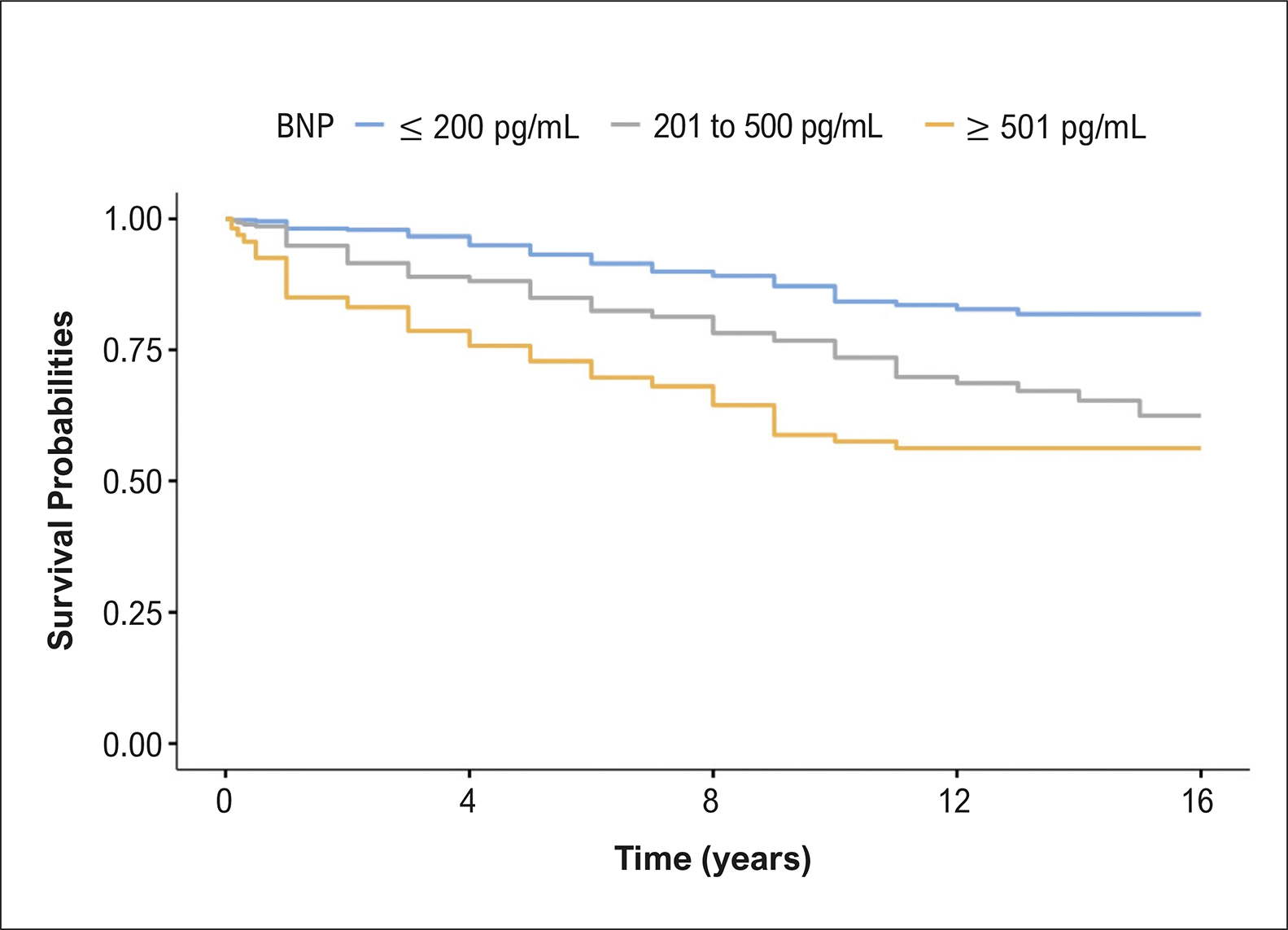
During follow-up, with a mean period of 7.7 years (minimum of 1 year and maximum of 16 years), the mortality rate was 18.6% (mean age of 62.0 ± 17.0 years, 53.9% female). The annual mortality rate was 1.3% among the patients studied. The survival curve revealed higher mortality for patients with BNP > 200 pg/mL (Figure 1), LA diameter ≥ 45 mm (Figure 2), and LVEF ≤ 50%, with log-rank test showing p < 0.001. Univariate analysis revealed that male sex was associated with risk of death (OR 1.58, 95% CI: 1.18 to 2.10, p < 0.001). We also performed multivariate analysis, which showed that the following variables were related to higher mortality: advanced age, increased LA diameter, reduced LVEF, and presence of atrial fibrillation or flutter (Table 4).

– Hypertrophic cardiomyopathy according to BNP values. Log-rank test, p < 0.001. BNP: B-type natriuretic peptide.

In the assessment based on different LVEF strata, the group with preserved LVEF (≥ 50%) included 1196 patients, and mortality occurred in 17.8%. The group with slightly reduced LVEF (40% to 49%) included 30 patients, and mortality was 30%. The group with reduced LVEF (< 40%) included 18 patients, with mortality of 55.5%. There was a significant difference in mortality (p = 0.001) according to LVEF stratification, as shown in Figure 3. The Central Illustration describes the main findings and their relationship with prognosis.

– Kaplan-Meier curves for mortality according to LVEF. Log-rank test, p = 0.001. LVEF: left ventricular ejection fraction.

: Clinical and Echocardiographic Particularities of Hypertrophic Cardiomyopathy in a Brazilian Population and its Prognostic Impact
Discussion
In our study, the majority of patients evaluated were men, with LVEF > 50%, asymmetric hypertrophy, and septal predominance. The overall annual mortality rate was 1.3%. Increased BNP, advanced age, LA enlargement, LVEF ≤ 50%, and the presence of atrial fibrillation or flutter were associated with a worse prognosis.
Patients with HCM present structural and functional alterations that can lead to heart failure, chest pain, arrhythmias, syncope, and even sudden death. In this context, TTE is a fundamental exam for the diagnosis, assessment, and follow-up of these patients. Moreover, it allows the identification of variables that may be related to increased mortality.2,8,19,20
The male sex (53.7%) showed a slightly higher percentage than the female sex, when compared to previous studies conducted in populations from North America and Europe, which showed that the male population could reach 62% to 71%. Probably, because we did not have many significant exclusion criteria, the frequency between the sexes was very similar, reflecting our reality in daily care.7,9-12
In HCM, LVEF is often normal or increased.7 Furthermore, septal size ≥ 28 mm, the presence of apical aneurysm, and LVEF ≤ 50% are characteristics that are related to the risk of sudden death.8 In our study population, 94% of patients had LVEF greater than 50%. These findings are in agreement with the literature, which indicates that less than 10% of patients have LVEF ≤ 50%.8 Our study revealed that 7.2% of patients had septal thickness ≥ 28 mm, and this group deserves special attention. With respect to left ventricular aneurysm, the literature describes it as a rare finding, between 2% and 5%, and it may be related to a higher risk of arrhythmias.8 In our study, ventricular aneurysm was very rare, found in only 1 patient. It was probably underestimated due to the limited assessment by non-contrast echocardiography, especially in cases of small aneurysms. Moreover, the use of magnetic resonance imaging has been shown to improve the diagnostic accuracy for this alteration.21-31
Data from the literature on global populations reveal that approximately 2/3 of patients with HCM have the obstructive form. In our study, approximately 1/3 of the cohort evaluated had the obstructive form. However, given that this was a database study and that LV outflow tract gradient is an important dynamic component, failure to perform the Valsalva maneuver or ineffective performance of the maneuver during TTE could explain these findings.2-8
High BNP increases the risk of cardiovascular events in patients with HCM.8,13,14 In this context, Geske et al., in a study of patients with HCM, demonstrated that BNP was an independent predictor of morbidity and mortality. This evidence is consistent with our results, which revealed that BNP > 200 pg/mL was related to mortality.
Atrial fibrillation or flutter may occur in patients with HCM and are associated with worsening of functional status, thromboembolic events, and increased mortality.16-22 The onset of this arrhythmia in HCM is multifactorial, but it is related to LA enlargement, which a common finding in patients with HCM. Our study revealed that 42.4% of all patients had LA enlargement, and this frequency was even higher in older patients, reaching 50% in patients over 70 years of age. A study from the United States published in 2014 found that the prevalence of atrial fibrillation in patients with HCM was 18%.23 Our study identified a smaller number (9.6%) of patients with atrial fibrillation or flutter. One of the explanations for this difference was the use of electrocardiogram in the first clinical assessment. Nonetheless, we are aware that many patients may develop atrial fibrillation during follow-up.
Previous studies have shown that women with HCM are more likely to have symptoms of heart failure, particularly dyspnea on exertion, fatigue, palpitations, chest pain, and New York Heart Association functional class III to IV, when compared to men. Moreover, they have a higher risk of disease-related events when compared to men.26,27 Our study did not evaluate difference in symptoms or clinical outcomes related to sex. We only evaluated echocardiogram criteria, which revealed that women had significantly smaller septum, LA diameter, posterior wall, and LV diameters.25-28Our findings may suggest a divergence with the literature. However, more in-depth investigation is needed in future studies in order to verify whether these echocardiographic changes are correlated with symptoms and outcomes.
Studies have shown show that 46% of patients may have a benign course, with normal life expectancy and no limitations. The adverse events that are found in a portion of patients include sudden death, chest pain, heart failure, and atrial fibrillation.8 Cardiovascular interventions, such as implantable cardioverter-defibrillator implantation, have reduced cardiovascular mortality rates to less than 1.0%/year.32 Our study showed an overall mortality rate of 1.3%/year. This difference can be explained by the fact that we used all-cause mortality for analysis, rather than only cardiovascular mortality. Moreover, as the study was conducted at a referral center, patients generally present greater complexity.8,9
In one study, Chen et al.33 evaluated 3605 Chinese patients over a mean follow-up period of 4.6 years, and they found an all-cause mortality rate of 6.3%. In another study from China, Kwak et al.34 demonstrated that, in the oldest group, with a mean age of 68 years, all-cause mortality at 5 years was 12%. Our mean follow-up time was 7.7 years, with mortality of 18.5%. In addition to the fact that they are distinct populations, one of the justifications for this difference is likely due to our longer follow-up period. A study conducted in China by Ma et al.,35 assessing 2268 patients, revealed that age, LVEF, and NT-proBNP were independent predictors of all-cause mortality. Our study also reported that these factors were significant for prognosis.
Patients with HCM have higher morbidity and mortality, especially when we find associated risk factors. Understanding the characteristics of the disease in our population allows for more accurate comprehension, based on national data. New national studies should be conducted to prospectively evaluate cardiovascular characteristics and progression.
Study limitations
This was a retrospective study based on medical records, which limits the collection of some clinical and echocardiographic data. Due to the fact that the investigation was conducted at a single center, even though it is a referral hospital, it is subject to participant selection bias, which most likely reflects patients on the most severe clinical spectrum of this disease. The cavity measurements assessed by echocardiography were not indexed. BNP levels were not available for analysis in all cases. Another limitation is that we did not have access to the cause of death for each patient and, therefore, assessed all-cause mortality.
Conclusions
This study revealed that the majority of patients had preserved LVEF, with asymmetric hypertrophy and septal predominance. Overall all-cause mortality was 1.3%/year. Increased BNP, advanced age, LA enlargement, LVEF ≤ 50%, and the presence of atrial fibrillation or flutter were associated with worse prognosis.
References
-
1 Ommen SR, Ho CY, Asif IM, Balaji S, Burke MA, Day SM, et al. 2024 AHA/ACC/AMSSM/HRS/PACES/SCMR Guideline for the Management of Hypertrophic Cardiomyopathy: A Report of the American Heart Association/American College of Cardiology Joint Committee on Clinical Practice Guidelines. Circulation. 2024;149(23):e1239-e1311. doi: 10.1161/CIR.0000000000001250
» https://doi.org/10.1161/CIR.0000000000001250 -
2 Arbelo E, Protonotarios A, Gimeno JR, Arbustini E, Barriales-Villa R, Basso C, et al. 2023 ESC Guidelines for the Management of Cardiomyopathies. Eur Heart J. 2023;44(37):3503-626. doi: 10.1093/eurheartj/ehad194.
» https://doi.org/10.1093/eurheartj/ehad194 -
3 Kwon S, Kim HK, Kim B, Lee HJ, Han KD, Hwang IC, et al. Comparison of Mortality and Cause of Death between Adults with and without Hypertrophic Cardiomyopathy. Sci Rep. 2022;12(1):6386. doi: 10.1038/s41598-022-10389-4.
» https://doi.org/10.1038/s41598-022-10389-4 -
4 Lorenzini M, Anastasiou Z, O'Mahony C, Guttman OP, Gimeno JR, Monserrat L, et al. Mortality Among Referral Patients with Hypertrophic Cardiomyopathy vs the General European Population. JAMA Cardiol. 2020;5(1):73-80. doi: 10.1001/jamacardio.2019.4534.
» https://doi.org/10.1001/jamacardio.2019.4534 -
5 Jacobsen MB, Petersen JK, Modin D, Butt JH, Thune JJ, Bundgaard H, et al. Long Term Mortality in Patients with Hypertrophic Cardiomyopathy - A Danish Nationwide Study. Am Heart J Plus. 2022;25:100244. doi: 10.1016/j.ahjo.2022.100244.
» https://doi.org/10.1016/j.ahjo.2022.100244 -
6 Wang Y, Gao W, Han X, Jiang J, Sandler B, Li X, et al. Cardiovascular Outcomes by Time-Varying New York Heart Association Class Among Patients with Obstructive Hypertrophic Cardiomyopathy: A Retrospective Cohort Study. J Med Econ. 2023;26(1):1495-506. doi: 10.1080/13696998.2023.2277076.
» https://doi.org/10.1080/13696998.2023.2277076 -
7 Lu DY, Pozios I, Haileselassie B, Ventoulis I, Liu H, Sorensen LL, et al. Clinical Outcomes in Patients with Nonobstructive, Labile, and Obstructive Hypertrophic Cardiomyopathy. J Am Heart Assoc. 2018;7(5):e006657. doi: 10.1161/JAHA.117.006657.
» https://doi.org/10.1161/JAHA.117.006657 -
8 Fernandes F, Simões MV, Correia EB, Marcondes-Braga FG, Coelho-Filho OR, Mesquita CT, et al. Guidelines on the Diagnosis and Treatment of Hypertrophic Cardiomyopathy - 2024. Arq Bras Cardiol. 2024;121(7):e202400415. doi: 10.36660/abc.20240415.
» https://doi.org/10.36660/abc.20240415 -
9 Maron BJ, Rowin EJ, Udelson JE, Maron MS. Clinical Spectrum and Management of Heart Failure in Hypertrophic Cardiomyopathy. JACC Heart Fail. 2018;6(5):353-63. doi: 10.1016/j.jchf.2017.09.011.
» https://doi.org/10.1016/j.jchf.2017.09.011 -
10 Habib M, Adler A, Fardfini K, Hoss S, Hanneman K, Rowin EJ, et al. Progression of Myocardial Fibrosis in Hypertrophic Cardiomyopathy: A Cardiac Magnetic Resonance Study. JACC Cardiovasc Imaging. 2021;14(5):947-58. doi: 10.1016/j.jcmg.2020.09.037.
» https://doi.org/10.1016/j.jcmg.2020.09.037 -
11 Melacini P, Basso C, Angelini A, Calore C, Bobbo F, Tokajuk B, et al. Clinicopathological Profiles of Progressive Heart Failure in Hypertrophic Cardiomyopathy. Eur Heart J. 2010;31(17):2111-23. doi: 10.1093/eurheartj/ehq136.
» https://doi.org/10.1093/eurheartj/ehq136 -
12 Neubauer S, Kolm P, Ho CY, Kwong RY, Desai MY, Dolman SF, et al. Distinct Subgroups in Hypertrophic Cardiomyopathy in the NHLBI HCM Registry. J Am Coll Cardiol. 2019;74(19):2333-45. doi: 10.1016/j.jacc.2019.08.1057.
» https://doi.org/10.1016/j.jacc.2019.08.1057 -
13 Matthia EL, Setteducato ML, Elzeneini M, Vernace N, Salerno M, Kramer CM, et al. Circulating Biomarkers in Hypertrophic Cardiomyopathy. J Am Heart Assoc. 2022;11(23):e027618. doi: 10.1161/JAHA.122.027618.
» https://doi.org/10.1161/JAHA.122.027618 -
14 Minami Y, Haruki S, Kanbayashi K, Maeda R, Itani R, Hagiwara N. B-Type Natriuretic Peptide and Risk of Sudden Death in Patients with Hypertrophic Cardiomyopathy. Heart Rhythm. 2018;15(10):1484-90. doi: 10.1016/j.hrthm.2018.04.030.
» https://doi.org/10.1016/j.hrthm.2018.04.030 -
15 Geske JB, McKie PM, Ommen SR, Sorajja P. B-Type Natriuretic Peptide and Survival in Hypertrophic Cardiomyopathy. J Am Coll Cardiol. 2013;61(24):2456-60. doi: 10.1016/j.jacc.2013.04.004.
» https://doi.org/10.1016/j.jacc.2013.04.004 -
16 Rowin EJ, Link MS, Maron MS, Maron BJ. Evolving Contemporary Management of Atrial Fibrillation in Hypertrophic Cardiomyopathy. Circulation. 2023;148(22):1797-811. doi: 10.1161/CIRCULATIONAHA.123.065037.
» https://doi.org/doi: 10.1161/CIRCULATIONAHA.123.065037 -
17 Mistrulli R, Ferrera A, Muthukkattil ML, Battistoni A, Gallo G, Barbato E, et al. Atrial Fibrillation in Patients with Hypertrophic Cardiomyopathy and Cardiac Amyloidosis: From Clinical Management to Catheter Ablation Indication. J Clin Med. 2024;13(2):501. doi: 10.3390/jcm13020501.
» https://doi.org/10.3390/jcm13020501 -
18 Guttmann OP, Rahman MS, O'Mahony C, Anastasakis A, Elliott PM. Atrial Fibrillation and Thromboembolism in Patients with Hypertrophic Cardiomyopathy: Systematic Review. Heart. 2014;100(6):465-72. doi: 10.1136/heartjnl-2013-304276.
» https://doi.org/10.1136/heartjnl-2013-304276 -
19 Garg L, Gupta M, Sabzwari SRA, Agrawal S, Agarwal M, Nazir T, et al. Atrial Fibrillation in Hypertrophic Cardiomyopathy: Prevalence, Clinical Impact, and Management. Heart Fail Rev. 2019;24(2):189-97. doi: 10.1007/s10741-018-9752-6.
» https://doi.org/10.1007/s10741-018-9752-6 -
20 Klopotowski M, Kwapiszewska A, Kukula K, Jamiolkowski J, Dabrowski M, Derejko P, et al. Clinical and Echocardiographic Parameters as Risk Factors for Atrial Fibrillation in Patients with Hypertrophic Cardiomyopathy. Clin Cardiol. 2018;41(10):1336-40. doi: 10.1002/clc.23050.
» https://doi.org/10.1002/clc.23050 -
21 Olivotto I, Cecchi F, Casey SA, Dolara A, Traverse JH, Maron BJ. Impact of Atrial Fibrillation on the Clinical Course of Hypertrophic Cardiomyopathy. Circulation. 2001;104(21):2517-24. doi: 10.1161/hc4601.097997.
» https://doi.org/10.1161/hc4601.097997 -
22 Kubo T, Baba Y, Ochi Y, Hirota T, Yamasaki N, Kawai K, et al. Clinical Significance of New-Onset Atrial Fibrillation in Patients with Hypertrophic Cardiomyopathy. ESC Heart Fail. 2021;8(6):5022-30. doi: 10.1002/ehf2.13563.
» https://doi.org/10.1002/ehf2.13563 -
23 Siontis KC, Geske JB, Ong K, Nishimura RA, Ommen SR, Gersh BJ. Atrial Fibrillation in Hypertrophic Cardiomyopathy: Prevalence, Clinical Correlations, and Mortality in a Large High-Risk Population. J Am Heart Assoc. 2014;3(3):e001002. doi: 10.1161/JAHA.114.001002.
» https://doi.org/10.1161/JAHA.114.001002 -
24 Chumakova OS, Baklanova TN, Milovanova NV, Zateyshchikov DA. Hypertrophic Cardiomyopathy in Underrepresented Populations: Clinical and Genetic Landscape Based on a Russian Single-Center Cohort Study. Genes. 2023;14(11):2042. doi: 10.3390/genes14112042.
» https://doi.org/10.3390/genes14112042 -
25 Butters A, Lakdawala NK, Ingles J. Sex Differences in Hypertrophic Cardiomyopathy: Interaction with Genetics and Environment. Curr Heart Fail Rep. 2021;18(5):264-73. doi: 10.1007/s11897-021-00526-x.
» https://doi.org/doi: 10.1007/s11897-021-00526-x -
26 Zhao H, Tan Z, Liu M, Yu P, Ma J, Li X, et al. Is There a Sex Difference in the Prognosis of Hypertrophic Cardiomyopathy? A Systematic Review and Meta-Analysis. J Am Heart Assoc. 2023;12(11):e026270. doi: 10.1161/JAHA.122.026270.
» https://doi.org/10.1161/JAHA.122.026270 -
27 Rowin EJ, Maron MS, Wells S, Patel PP, Koethe BC, Maron BJ. Impact of Sex on Clinical Course and Survival in the Contemporary Treatment Era for Hypertrophic Cardiomyopathy. J Am Heart Assoc. 2019;8(21):e012041. doi: 10.1161/JAHA.119.012041.
» https://doi.org/10.1161/JAHA.119.012041 -
28 Geske JB, Ong KC, Siontis KC, Hebl VB, Ackerman MJ, Hodge DO, et al. Women with Hypertrophic Cardiomyopathy Have Worse Survival. Eur Heart J. 2017;38(46):3434-40. doi: 10.1093/eurheartj/ehx527.
» https://doi.org/10.1093/eurheartj/ehx527 -
29 Maron MS, Rowin E, Spirito P, Maron BJ. Differing Strategies for Sudden Death Prevention in Hypertrophic Cardiomyopathy. Heart. 2023;109(8):589-94. doi: 10.1136/heartjnl-2020-316693.
» https://doi.org/10.1136/heartjnl-2020-316693 -
30 Arteaga E, Araújo AQ, Bernstein M, Ramires FJ, Ianni BM, Fernandes F, et al. Prognostic Value of the Collagen Volume Fraction in Hypertrophic Cardiomyopathy. Arq Bras Cardiol. 2009;92(3):210-4. doi: 10.1590/s0066-782x2009000300010.
» https://doi.org/10.1590/s0066-782x2009000300010 -
31 Lee DZJ, Montazeri M, Bataiosu R, Hoss S, Adler A, Nguyen ET, et al. Clinical Characteristics and Prognostic Importance of Left Ventricular Apical Aneurysms in Hypertrophic Cardiomyopathy. JACC Cardiovasc Imaging. 2022;15(10):1696-711. doi: 10.1016/j.jcmg.2022.03.029.
» https://doi.org/10.1016/j.jcmg.2022.03.029 -
32 Maron BJ, Desai MY, Nishimura RA, Spirito P, Rakowski H, Towbin JA, et al. Management of Hypertrophic Cardiomyopathy: JACC State-of-the-Art Review. J Am Coll Cardiol. 2022;79(4):390-414. doi: 10.1016/j.jacc.2021.11.021.
» https://doi.org/10.1016/j.jacc.2021.11.021 -
33 Chen QF, Zou J, Katsouras CS, You S, Zhou J, Ge HB, et al. Clinical Characteristics and Outcomes in Patients with Apical and Nonapical Hypertrophic Cardiomyopathy. J Am Heart Assoc. 2024;13(19):e036663. doi: 10.1161/JAHA.124.036663.
» https://doi.org/10.1161/JAHA.124.036663 -
34 Kwak S, Kim J, Park CS, Lee HJ, Park JB, Lee SP, et al. Distinct Phenotypic Groups and Related Clinical Outcomes in Patients with Hypertrophic Cardiomyopathy. J Am Heart Assoc. 2024;13(20):e036245. doi: 10.1161/JAHA.124.036245.
» https://doi.org/10.1161/JAHA.124.036245 -
35 Ma H, Zhou Y, He Y, Yu C, Liao Q, Xi H, et al. Prognosis for Patients with Apical Hypertrophic Cardiomyopathy: A Multicenter Cohort Study Based on Propensity Score Matching. Kardiol Pol. 2023;81(12):1247-56. doi: 10.33963/v.kp.98355.
» https://doi.org/10.33963/v.kp.98355
-
Study association
This study is not associated with any thesis or dissertation work.
-
Ethics approval and consent to participate
This study was approved by the Ethics Committee of the CAPPesq under the protocol number 5.123.838. All the procedures in this study were in accordance with the 1975 Helsinki Declaration, updated in 2013. Informed consent was obtained from all participants included in the study.
-
Sources of funding:
There were no external funding sources for this study.
Edited by
-
Editor responsible for the review:
Natália Olivetti



 BNP: B-type natriuretic peptide; LA: left atrial; LVEF: left ventricular ejection fraction.