
 Guideline for Perioperative Cardiovascular Evaluation of the Brazilian Society of Cardiology – 2024
B - Research funding under your direct/personal responsibility (directed to the department or institution) from the Brazilian or international pharmaceutical, orthosis, prosthesis, equipment and implants industry:–
Bruno Caramelli Nothing to be declared Caio de Assis Moura Tavares Financial declaration A - Economically relevant payments of any kind made to (i) you, (ii) your spouse/partner or any other person living with you, (iii) any legal person in which any of these is either a direct or indirect controlling owner, business partner, shareholder or participant; any payments received for lectures, lessons, training instruction, compensation, fees paid for participation in advisory boards, investigative boards or other committees, etc. From the brazilian or international pharmaceutical, orthosis, prosthesis, equipment and implants industry:–
Caio Julio Cesar dos Santos Fernandes Financial declaration A - Economically relevant payments of any kind made to (i) you, (ii) your spouse/partner or any other person living with you, (iii) any legal person in which any of these is either a direct or indirect controlling owner, business partner, shareholder or participant; any payments received for lectures, lessons, training instruction, compensation, fees paid for participation in advisory boards, investigative boards or other committees, etc. From the brazilian or international pharmaceutical, orthosis, prosthesis, equipment and implants industry:–
B - Research funding under your direct/personal responsibility (directed to the department or institution) from the Brazilian or international pharmaceutical, orthosis, prosthesis, equipment and implants industry:–
Other relationships Funding of continuing medical education activities, including travel, accommodation and registration in conferences and courses, from the brazilian or international pharmaceutical, orthosis, prosthesis, equipment and implants industry:–
Carisi Anne Polanczyk Nothing to be declared Carlos Jardim Nothing to be declared Carolina Leticia Zilli Vieira Nothing to be declared Claudio Pinho Nothing to be declared Daniela Calderaro Financial declaration A - Economically relevant payments of any kind made to (i) you, (ii) your spouse/partner or any other person living with you, (iii) any legal person in which any of these is either a direct or indirect controlling owner, business partner, shareholder or participant; any payments received for lectures, lessons, training instruction, compensation, fees paid for participation in advisory boards, investigative boards or other committees, etc. From the brazilian or international pharmaceutical, orthosis, prosthesis, equipment and implants industry: –
Other relationships Funding of continuing medical education activities, including travel, accommodation and registration in conferences and courses, from the brazilian or international pharmaceutical, orthosis, prosthesis, equipment and implants industry:–
Danielle Menosi Gualandro Financial declaration A - Economically relevant payments of any kind made to (i) you, (ii) your spouse/partner or any other person living with you, (iii) any legal person in which any of these is either a direct or indirect controlling owner, business partner, shareholder or participant; any payments received for lectures, lessons, training instruction, compensation, fees paid for participation in advisory boards, investigative boards or other committees, etc. From the brazilian or international pharmaceutical, orthosis, prosthesis, equipment and implants industry: –
Dirk Schreen Nothing to be declared Fabiana Goulart Marcondes-Braga Financial declaration A - Economically relevant payments of any kind made to (i) you, (ii) your spouse/partner or any other person living with you, (iii) any legal person in which any of these is either a direct or indirect controlling owner, business partner, shareholder or participant; any payments received for lectures, lessons, training instruction, compensation, fees paid for participation in advisory boards, investigative boards or other committees, etc. From the brazilian or international pharmaceutical, orthosis, prosthesis, equipment and implants industry:–
B - Research funding under your direct/personal responsibility (directed to the department or institution) from the Brazilian or international pharmaceutical, orthosis, prosthesis, equipment and implants industry: –
Fábio de Souza Financial declaration A - Economically relevant payments of any kind made to (i) you, (ii) your spouse/partner or any other person living with you, (iii) any legal person in which any of these is either a direct or indirect controlling owner, business partner, shareholder or participant; any payments received for lectures, lessons, training instruction, compensation, fees paid for participation in advisory boards, investigative boards or other committees, etc. From the brazilian or international pharmaceutical, orthosis, prosthesis, equipment and implants industry:–
Other relationships Funding of continuing medical education activities, including travel, accommodation and registration in conferences and courses, from the brazilian or international pharmaceutical, orthosis, prosthesis, equipment and implants industry: –
Flávio Tarasoutchi Other relationships Funding of continuing medical education activities, including travel, accommodation and registration in conferences and courses, from the brazilian or international pharmaceutical, orthosis, prosthesis, equipment and implants industry:–
Francisco Akira Malta Cardozo Financial declaration A - Economically relevant payments of any kind made to (i) you, (ii) your spouse/partner or any other person living with you, (iii) any legal person in which any of these is either a direct or indirect controlling owner, business partner, shareholder or participant; any payments received for lectures, lessons, training instruction, compensation, fees paid for participation in advisory boards, investigative boards or other committees, etc. From the brazilian or international pharmaceutical, orthosis, prosthesis, equipment and implants industry:–
Other relationships Funding of continuing medical education activities, including travel, accommodation and registration in conferences and courses, from the brazilian or international pharmaceutical, orthosis, prosthesis, equipment and implants industry: –
Gabriel Assis Lopes do Carmo Nothing to be declared Gabriel Kanhouche Nothing to be declared José Jayme Galvão de Lima Nothing to be declared Luciana Dornfeld Bichuette Nothing to be declared Luciana Sacilotto Nothing to be declared Luciana Savoy Fornari Nothing to be declared Luciano Ferreira Drager Nothing to be declared Luciano Janussi Vacanti Nothing to be declared Luis Henrique Wolff Gowdak Financial declaration A - Economically relevant payments of any kind made to (i) you, (ii) your spouse/partner or any other person living with you, (iii) any legal person in which any of these is either a direct or indirect controlling owner, business partner, shareholder or participant; any payments received for lectures, lessons, training instruction, compensation, fees paid for participation in advisory boards, investigative boards or other committees, etc. From the brazilian or international pharmaceutical, orthosis, prosthesis, equipment and implants industry:–
B - Research funding under your direct/personal responsibility (directed to the department or institution) from the Brazilian or international pharmaceutical, orthosis, prosthesis, equipment and implants industry:–
Other relationships Funding of continuing medical education activities, including travel, accommodation and registration in conferences and courses, from the brazilian or international pharmaceutical, orthosis, prosthesis, equipment and implants industry:–
Marcelo Luiz Campos Vieira Nothing to be declared Marcelo Luiz FlorianoMelo Martins Nothing to be declared Márcio Silva Miguel Lima Nothing to be declared Marcos Pita Lottenberg Nothing to be declared Márlon Juliano Romero Aliberti Nothing to be declared Mauricio Felippi de Sá Marchi Nothing to be declared Milena Ribeiro Paixão Nothing to be declared Mucio Tavares de Oliveira Junior Financial declaration B - Research funding under your direct/personal responsibility (directed to the department or institution) from the Brazilian or international pharmaceutical, orthosis, prosthesis, equipment and implants industry:–
Pai Ching Yu Nothing to be declared Patricia Ramos Cury Nothing to be declared Pedro Silvio Farsky Financial declaration A - Economically relevant payments of any kind made to (i) you, (ii) your spouse/partner or any other person living with you, (iii) any legal person in which any of these is either a direct or indirect controlling owner, business partner, shareholder or participant; any payments received for lectures, lessons, training instruction, compensation, fees paid for participation in advisory boards, investigative boards or other committees, etc. From the brazilian or international pharmaceutical, orthosis, prosthesis, equipment and implants industry:–
Other relationships Funding of continuing medical education activities, including travel, accommodation and registration in conferences and courses, from the brazilian or international pharmaceutical, orthosis, prosthesis, equipment and implants industry: –
Ranna Santos Pessoa Nothing to be declared Rinaldo Focaccia Siciliano Nothing to be declared Tarso Augusto Duenhas Accorsi Financial declaration A - Economically relevant payments of any kind made to (i) you, (ii) your spouse/partner or any other person living with you, (iii) any legal person in which any of these is either a direct or indirect controlling owner, business partner, shareholder or participant; any payments received for lectures, lessons, training instruction, compensation, fees paid for participation in advisory boards, investigative boards or other committees, etc. From the brazilian or international pharmaceutical, orthosis, prosthesis, equipment and implants industry: –
Other relationships Funding of continuing medical education activities, including travel, accommodation and registration in conferences and courses, from the brazilian or international pharmaceutical, orthosis, prosthesis, equipment and implants industry: –
Vinícius Machado Correia Financial declaration A - Economically relevant payments of any kind made to (i) you, (ii) your spouse/partner or any other person living with you, (iii) any legal person in which any of these is either a direct or indirect controlling owner, business partner, shareholder or participant; any payments received for lectures, lessons, training instruction, compensation, fees paid for participation in advisory boards, investigative boards or other committees, etc. From the brazilian or international pharmaceutical, orthosis, prosthesis, equipment and implants industry: –
Wilson Mathias Junior Nothing to be declared
Table of Contents
1. Definition of the Problem 8
1.1. Objective of the Guideline 8
1.2. Methodology and Levels of Evidence 8
1.3. Particularities of the Perioperative Period 8
1.4. Creation of the Perioperative Risk Team 8
2. General and Additional Preoperative Assessment 11
2.1. Preoperative Risk Stratification 11
2.1.1. Severe/unstable CV Conditions in the Perioperative Period 11
2.1.2. Estimation of Intrinsic Risk Related to Type of Surgery 11
2.1.3. Functional Capacity Assessment 11
2.1.4. Tools for Estimating Perioperative CV Risk 12
2.1.5. Frailty Assessment 13
2.1.5.1. Frailty Assessment Before Noncardiac Surgery 14
2.1.5.2. Impact of Frailty Assessment on Noncardiac Surgery 14
2.2. Electrocardiography 15
2.3. Echocardiography 16
2.4. Noninvasive Tests for Myocardial Ischemia 17
2.4.1. Exercise Electrocardiogram 17
2.4.2. Myocardial Perfusion Imaging 17
2.4.3. Dobutamine Stress Echocardiography 18
2.4.4. Summary of Recommendations for Noninvasive Functional
Imaging for Myocardial Ischaemia 18
2.5. Coronary Cineangiography 18
2.6. Coronary Computed Tomography Angiography 18
3. Specific Diseases and Procedures in the Perioperative
Period 19
3.1. Heart Failure 19
3.2. Arterial Hypertension 20
3.2.1. Preoperative BP Management 20
3.2.2. Intraoperative BP Management 21
3.2.3. Postoperative BP Management 21
3.2.4. Perioperative Management of Antihypertensives 21
3.2.4.1. Renin-Angiotensin-Aldosterone System Inhibitors 21
3.2.4.2. Calcium Channel Blockers 22
3.2.4.3. Diuretics 22
3.2.4.4. Central Sympatholytic Drugs 22
3.2.5. Final Considerations 22
3.3. Patient Management in Low-Risk Procedures 22
3.3.1. Dental Procedures 22
3.3.1.1. Antithrombotic agents 22
3.3.1.2. Cardiac Implantable Electronic Devices 23
3.3.1.3. Local Anesthetics 23
3.3.2. Dermatological Surgery 23
3.3.3. Endoscopic Procedures 24
3.3.3.1. Management of Antiplatelet Therapy in Endoscopic
Procedures 24
3.3.3.2. Management of Anticoagulants in Endoscopic Procedures 25
3.3.4. Ophthalmic Procedures 25
3.4. Valvular Heart Disease 26
3.4.1. Aortic Valve Stenosis 26
3.4.2. Mitral Valve Stenosis 27
3.4.3. Aortic and Mitral Regurgitation 27
3.4.4. Prosthetic Heart Valves 27
3.5. Solid Organ Transplantation 27
3.5.1 Liver 27
3.5.1.1. Cirrhotic Cardiomyopathy 28
3.5.1.2. Alcoholic Cardiomyopathy 28
3.5.1.3. Portopulmonary Hypertension 28
3.5.1.4. Hepatopulmonary Syndrome 29
3.5.1.5. Coronary Artery Disease 30
3.5.1.5.1. When to Investigate CAD? 30
3.5.1.5.2. Which Test to Request for CAD Investigation? 30
3.5.1.5.3. When and How to Intervene in CAD? 30
3.5.2. Kidney 30
3.5.2.1. CAD Risk Stratification 32
4. Measures for Reducing CV Surgical Risk 33
4.1. Perioperative Pharmacological Therapy 33
4.1.1. Beta-Blockers 33
4.1.2. Statins 33
4.1.3. Anti-Platelet Medications 34
4.1.3.1. Monotherapy with ASA 34
4.1.3.2. Monotherapy with Antiplatelet Medications Other than ASA 35
4.1.3.3. Dual Antiplatelet Therapy 35
4.2. Myocardial Revascularization 36
4.3. Perioperative Anticoagulation Management 36
4.3.1. Warfarin 37
4.3.1.1. Patients with Moderate Thromboembolic Risk 37
4.3.1.2. Urgent or Emergency Procedures 37
4.3.2. Direct Oral Anticoagulants 38
4.3.2.1. Dabigatran 39
4.3.2.2. Rivaroxaban 39
4.3.2.3. Apixaban 39
4.3.2.4. Edoxaban 40
4.3.2.5. Evaluation of the Anticoagulant Effect of DOACs 40
4.3.2.6. Interruption of DOACs Before Elective Surgery 40
4.4. Infective Endocarditis Prophylaxis 41
4.4.1. Dental Procedures 41
4.4.2. Respiratory Tract Procedures 43
4.4.3. Gastrointestinal and Genitourinary Procedures 43
4.4.4. Dermatological or Musculoskeletal Procedures 43
4.4.5. Body Piercing and Tattooing 43
5. Perioperative Biomarkers 44
5.1. Natriuretic Peptides 44
5.2. Cardiac Troponins and Surveillance of CV Complications 44
6. Diagnosis and Treatment of Perioperative CV
Complications 44
6.1. Perioperative Acute Myocardial Infarction/Injury (PMI) 44
6.2. Acute Atrial Fibrillation/Flutter 48
6.3. Acute Heart Failure 49
6.4. Venous Thromboembolism 50
References 51

1. Definition of the Problem
1.1. Objective of the Guideline
The rapid advancement of medical knowledge in recent years prompted the creation of guidelines to filter the best evidence and organize it for practical use in daily medical practice. The need for updates varies by field and reflects the quantity and quality of medical research on a given subject. Interest in perioperative care for noncardiac surgery is relatively recent, starting in 1996 after the publication of Mangano et al.’s study, which observed a reduction in cardiovascular (CV) complications in patients who received intravenous atenolol in the immediate preoperative period.1 In Brazil, publications began emerging in 2003, with the first Guideline for Perioperative Cardiovascular Evaluation being published in 2007 and updated in 2011 and 2017.2 This is the fourth update in 15 years, highlighting the topic’s importance in both research and daily professional practice.
Although the percentage of perioperative complications is small, their occurrence in Brazil is significantly higher than in other countries. Additionally, most procedures are low risk, but considering the annual volume of over 8 million surgical interventions in Brazil, there is a substantial number of patients at high risk of complications who require greater care.
The main objectives of the Guideline for Perioperative Cardiovascular Evaluation of the Brazilian Society of Cardiology – 2024 are:
To provide and standardize updated knowledge on CV risk stratification.
To establish adequate methods for risk stratification and diagnosis of complications, not only to improve efficiency but also to reduce costs associated with unnecessary tests.
To offer updated knowledge on the relationship between different comorbidities and CV diseases (CVDs) that may interact in the perioperative setting, such as the interaction and adequate discontinuation of medications or the safe timeframe for performing surgical procedures after CV interventions.
To offer recommendations on the optimal location for performing interventions in high-risk patients or on contraindications to procedures when the risk of complications outweighs the potential benefits.
1.2. Methodology and Levels of Evidence
The methodology and levels of evidence adopted by this Guideline are the same as those adopted by the SBC and referred to below:
Classes (grades) of recommendation:
-
✓
Class I – Conditions for which there is conclusive evidence or a consensus that the procedure is safe and useful/effective.
-
✓
Class II – Conditions for which there is conflicting evidence and/or a divergence of opinion about the safety and usefulness/efficacy of a procedure.
-
✓
Class IIA – Evidence or opinion in favor of the procedure. The majority agrees.
-
✓
Class IIB – Safety and usefulness are less well established, and there is no predominance of opinions in favor of the method.
-
✓
Class III – Conditions for which there is evidence and/or general agreement that a procedure is not useful/effective and, in some cases, may be harmful.
Levels of evidence:
-
✓
Level A – Data obtained from several large, randomized studies showing concurring results and/or a robust meta-analysis of randomized controlled trials.
-
✓
Level B – Data obtained from a less robust meta-analysis, a single randomized study, or from nonrandomized (observational) studies.
-
✓
Level C – Data obtained from consensual expert opinions.
All scientific evidence used in this Guideline comes from indexed journals with qualified editorial boards and peer reviewers.
1.3. Particularities of the Perioperative Period
In patients with heart disease, the perioperative period presents many peculiarities, such as the use of anticoagulation and antithrombotic therapy, which sometimes should not be interrupted and could increase the risk of intraoperative bleeding. Importantly, CV risk stratification tools, such as the ATP-III, Framingham, and CHADS-VASc scores, which are familiar to cardiologists, establish risk profiles over years and are not suitable for predicting short-term perioperative risk for noncardiac surgery (usually less than 30 days). For these reasons, the Guideline provides relevant information specifically for assessing the risk of CV events, prevention methods, and recommendations to make the perioperative period of noncardiac procedures safer for patients with heart disease. A summary of the new recommendations included in this Guideline is presented in Table 1.
1.4. Creation of the Perioperative Risk Team
This Guideline focuses mainly, but not exclusively, on cardiologists, given that the most serious complications involve the CV system, such as ischemic heart disease, pulmonary embolism (PE), and acute heart failure (HF). These complications are most associated with mortality, increased length of hospital stay, and higher costs, not only due to additional procedures but also due to the request for diagnostic tests. Patients entering operating rooms today are older and consequently have more comorbidities, requiring the involvement not only of the surgeon but also the anesthesiologist, cardiologist, and other specialists in their treatment. The follow-up of high-risk patients should be interdisciplinary, including adequate planning to determine the best time for the intervention and the risk/benefit ratio, as well as to optimize the treatment of underlying diseases and their complications.
Cardiologists are already familiar with the Heart Team, typically consisting of the cardiac surgeon, the interventional cardiologist, and the clinical cardiologist, who usually work in the preoperative period to decide the best approach for a given case. The perioperative care of patients undergoing noncardiac surgery, which includes both the entire intraoperative and postoperative periods, involves a greater number of specialists from different fields. The Guideline for Perioperative Cardiovascular Evaluation of the Brazilian Society of Cardiology – 2024 innovates by proposing the creation of a Perioperative Risk Team (PRT). The purpose of the PRT, indicated for more severe cases, is to gather all relevant patient information, such as previous diseases and treatments, prognosis, type of proposed surgery, and current cardiac status. With harmony and teamwork among various specialists, it will be possible to offer the best joint decision for the patient and their family. Since the most serious complications involve the CV system, the cardiologist should plan, when necessary, the formation of the PRT for severe and specific cases and establish its dynamics. Tools such as video platforms, for recording and storing interdisciplinary meetings, can be very useful to improve outcomes and provide guarantees for both health care professionals and the patient.
2. General and Additional Preoperative Assessment
2.1. Preoperative Risk Stratification
2.1.1. Severe/Unstable CV Conditions in the Perioperative Period
For elective procedures, the first step is to determine the patient’s baseline clinical condition. There are clinical circumstances in which the spontaneous risk of complications is very high, regardless of the surgical procedure to be performed. Identifying these circumstances is crucial because treating such conditions should take priority over elective procedures, which should be postponed whenever possible and reconsidered only after the patient has stabilized (Table 2).
2.1.2. Estimation of Intrinsic Risk Related to the Type of Surgery
The intrinsic risk of surgery is determined by the type and duration of the procedure, without considering the patient’s clinical characteristics. It is defined as the probability of CV events occurring perioperatively, regardless of the clinical variables of the patients. This risk is related to the duration of surgery, hemodynamic stress, and loss of blood and fluids. Patients with stable clinical conditions, who do not have high-risk CV conditions, may undergo low-intrinsic risk procedures without the need for additional evaluation. Despite the difficulty in determining the specific risk of a surgical procedure, as each procedure occurs under different circumstances, the European Society of Cardiology proposed a CV risk classification that considers the risk of CV death, acute myocardial infarction (AMI), and stroke within 30 days (Table 3).
Additionally, the urgency of the surgical intervention should always be considered. Emergency and urgent operations are associated with a higher incidence of CV complications. In situations where the prognosis of the underlying disease necessitating surgery demands an emergency intervention, the role of the cardiologist should be limited to suggesting surveillance measures (including the location for postoperative care) and interventions to reduce intra and postoperative risk. It is not recommended to order any additional tests that could delay the proposed surgery. For urgent procedures, there is sufficient time to optimize CV therapy or perform additional tests, such as transthoracic echocardiography (TTE), when indicated. However, functional tests for the assessment of myocardial ischemia should not be performed, as their results would not alter patient management since the proposed surgery cannot be postponed for coronary treatment. Furthermore, there are time-sensitive procedures, which are not urgencies, but whose delay could worsen the prognosis of the underlying disease. A common example is cancer surgery, whose delay can worsen the prognosis. In these cases, we recommend multidisciplinary discussion and the selection of the best individualized strategy.
2.1.3. Functional Capacity Assessment
Patients with reduced functional capacity (less than four metabolic equivalents [METs] or inability to climb two flights of stairs) are more likely to develop perioperative complications.4 Functional capacity can be objectively assessed via exercise electrocardiogram (ECG), which is not always feasible or desirable, or clinical history. A recent study demonstrated that patients who reported being able to climb two flights of stairs during the preoperative evaluation had a lower rate of postoperative CV events.5
Additionally, the estimate of functional capacity combined with the Revised Cardiac Risk Index (RCRI) showed greater accuracy in predicting postoperative events compared with the RCRI alone.5 In addition to the greater likelihood of poor perioperative outcomes, patients with reduced functional capacity may have their symptoms underestimated due to their limitations. Therefore, this may be considered when deciding whether to order additional tests such as functional tests for myocardial ischemia.
Chart 1 presents recommendations for preoperative assessment of functional capacity.
2.1.4. Tools for Estimating Perioperative CV Risk
Subjective assessments of perioperative CV risk are valuable, but objective risk estimation allows for the rational use of additional risk stratification tools and perioperative CV care. Furthermore, the calculation of CV risk better supports multidisciplinary discussions aimed at minimizing the patient’s overall risk.
This Guideline does not advocate for the adoption of a specific algorithm for perioperative CV risk stratification but recommends calculating the risk using one of the indices available in the literature after excluding severe/unstable cardiac conditions (Table 2).
Among the indices for estimating perioperative CV risk, the RCRI, or Lee score,6 and the more recently published American University of Beirut-HAS2 (AUB-HAS2) index, stand out for their practicality and accuracy in distinguishing different risk classifications.7 Both models integrate clinical characteristics and the type of proposed surgery, without different weightings among the variables for each index (Tables 4 and 5). Increasing rates of CV complications were observed according to the number of variables in the pivotal cohorts of the RCRI and AUB-HAS2.
In the Brazilian population, the superiority of one algorithm over the other has not yet been established. It is worth noting that although both indices estimate severe CV outcomes, the outcomes differ. The RCRI, already validated in a Brazilian population,8 estimates the risk of myocardial infarction (MI), acute pulmonary edema, third-degree atrioventricular block, and cardiorespiratory arrest within 30 days after surgery.6 The AUB-HAS2, on the other hand, has not yet been validated in a Brazilian population and estimates the risk of MI, stroke, and death, also within 30 days of surgery.7
In the older version of the RCRI used in the previous edition of this Guideline,2 the risk of postoperative CV events was 0.4% for Class I (no risk variables), 0.9% for Class II (1 variable), 7% for Class III (2 variables), and 11% for Class IV (3 or more variables). Other algorithms that estimate the risk of perioperative complications also included numerical percentage values that represent the rates observed in reference studies. More recent studies, however, suggest that these risk estimates are outdated and are currently higher to the increased severity of patients undergoing surgery. As published in the latest European guideline on perioperative assessment,3 there was a 4% risk for Class I, 6% for Class II, 10% for Class III, and 15% for Class IV.
Considering the significant variation in these figures by country and population, we opted for a semiquantitative classification, establishing low risk (Class I and II), intermediate risk (Class III), and high risk (Class IV) in preoperative risk assessment reports without specifying absolute values.
Among the limitations of these indices is the potential loss of accuracy in patients with reduced functional capacity. These patients have a worse perioperative prognosis9 and may be asymptomatic simply because they do not reach the threshold to trigger symptoms. Therefore, even if a patient with reduced functional capacity receives a low or intermediate CV risk estimate from the indices, this evaluation can be complemented by tests for coronary artery disease (CAD) and HF before high-risk operations, especially when the patient has CV risk factors (see item 2.4, functional tests). Another issue is the loss of index accuracy for specific operations, such as the RCRI, which loses the ability to discriminate between risk classifications and underestimates events in patients undergoing vascular operations, particularly abdominal aortic aneurysm repair.6,10-12 Specifically for vascular surgery, this Guideline recommends using the Vascular Study Group of New England Cardiac Risk Index (VSG-CRI) (Tables 6 and 7).11 Compared with the RCRI, the VSG-CRI showed better accuracy in predicting MI, clinically relevant arrhythmia, and HF in the postoperative period of vascular surgery.11 The AUB-HAS2 also maintains good accuracy in distinguishing between four classes of progressive risk in the perioperative period of vascular surgery, with higher absolute rates of perioperative complications than those observed for all operations combined.13,14
Risk factors and respective scores in the Vascular Study Group of New England Cardiac Risk Index (VSG-CRI)11
Risk classification according to the Vascular Study Group of New England Cardiac Risk Index (VSG-CRI)11
Other risk indices validated for perioperative CV assessment include the American College of Physicians Risk Index15,16 and the Multicenter Perioperative Evaluation Study (EMAPO) Index,8 the latter developed and validated in Brazil. For estimating overall risk not solely related to CV morbidity and mortality outcomes, the American College of Surgeons (ACS) NSQIP® Surgical Risk Calculator (www.riskcalculator.facs.org) can be used.17 This tool was developed from a database of over 1 million operations performed in the United States and considers, in addition to the specific type of surgery, 21 clinical variables, providing risk estimates for 8 different outcomes. The main limitation of this tool is that it is not quick or easily applicable, requiring a calculator and including some subjectively determined variables.
The choice of perioperative CV risk index should consider the evaluator’s experience and the vascular or nonvascular nature of the surgical procedure. It is worth noting that the risk of venous thromboembolism (VTE), which is highly prevalent and preventable in the perioperative period, is not covered by the indices discussed in this section and requires a dedicated approach.2
The flowchart for perioperative assessment is presented in Central Illustration.
2.1.5. Frailty Assessment
Frailty syndrome is an age-related, multidimensional clinical entity defined as a reduction in physiological reserve across several physiological systems, leading to a state of increased vulnerability to stressors such as acute illnesses and surgery. Frailty is associated with an increased risk of mortality and several other adverse outcomes, such as hospitalization, reduced functional capacity, and poor quality of life.18 Over the past decades, there has been growing global attention to the assessment of frailty in different clinical settings, reflecting the phenomenon of an aging population worldwide.19,20 In Brazil, the prevalence of frailty is 9% among people aged ≥ 50 years living in the community and 16% among those aged ≥ 65 years. These rates are similar to those in developed countries and higher in health care settings such as clinics and hospitals.21 Despite the lack of consensus on a gold standard method for defining frailty, the Physical Frailty Phenotype and the Frailty Index are considered the most valid measures for identifying this syndrome.22 Although associated with multimorbidity, aging, and limitations in basic and instrumental activities of daily living, frailty syndrome is considered a distinct entity separate from these other factors.22-24 The severity of frailty ranges from robust and prefrail to truly frail patients.
Although CVD can evidently cause frailty as a result of hospitalization for acute events, immobility, and physical limitation in individuals with established disease (eg, hospitalization for acute coronary syndrome, physical limitation due to HF symptoms),25 there is biological plausibility for common underlying pathophysiological processes in both conditions, indicating a bidirectional relationship.26 Frailty is associated with a series of physiological and functional changes, including chronic inflammation, endothelial dysfunction, autonomic dysfunction, activation of the renin-angiotensin-aldosterone system, and oxidative stress, which are risk factors for CVD.27 Indeed, in a cohort of patients without CVD,28 the presence of frailty was a risk factor for the incidence of CV events over a 6-year follow-up period. Other studies have also observed an independent association between frailty and adverse CV outcomes in several CV conditions.29
2.1.5.1. Frailty Assessment Before Noncardiac Surgery
Despite the lack of consensus on a single instrument for screening frailty in older adults, both at the population level and in specific situations such as the perioperative assessment for noncardiac surgery, it is recommended that frailty be objectively assessed using previously validated instruments. Table 8 presents some examples of instruments that can be used to assess frailty. It is noteworthy that there are other instruments available beyond those mentioned in Table 8. The recommendation to use instruments is based on the fact that the subjective assessment of physicians, especially those not specialized in geriatrics, can be influenced by confounding factors such as advanced age, the presence of multiple diseases, low weight, and the use of walking aids, which can lead to an erroneous assumption of frailty when it is not actually the case.30-32 Frailty assessment with validated instruments also provides more detailed information about the patient’s status rather than simply categorizing them as frail or nonfrail, which can be simplistic and superficial.
Frailty can also be assessed and classified based on different health domains: physical, cognitive, psychosocial, and nutritional. Depending on the tool used to define frailty, one or more domains are assessed, allowing for the characterization of patients into distinct clinical phenotypes.22
2.1.5.2. Impact of Frailty Assessment on Noncardiac Surgery
Frailty is a predictor of several adverse perioperative outcomes across multiple surgical specialties, including both elective and emergency procedures. Evidence indicates that frail patients are at higher risk for postoperative clinical complications such as infections, delirium, acute renal failure, cardiac arrhythmias, and MI, as well as issues related to wound healing, such as dehiscence and hernias. Consequently, frail patients tend to have prolonged lengths of hospital stay. These postoperative complications negatively impact the functional recovery and quality of life of frail patients undergoing surgery, often leading to a greater need for transfer to long-term care facilities and increased short- and long-term mortality compared with nonfrail patients.33-43 Despite this, the impact of incorporating frailty assessment into the prognostic performance of traditional risk scores (RCRI, VSG-CRI, AUB-HAS 2, etc.) remains uncertain due to the limited number of studies on this topic.44,45 The impact of this combination using other statistical methods still needs to be investigated to determine its significance and clinical utility.46
Despite existing limitations, perioperative frailty assessment is understood to (1) assist in the decision-making process by providing a more comprehensive assessment of the risks associated with the surgical procedure, thereby improving communication between the patient, family, and the several professionals involved in perioperative care (anesthesiologist, surgeon, clinician, cardiologist, geriatrician); and (2) identify a subgroup of patients at higher risk of complications, in whom early interventions can be implemented (eg, physical and nutritional rehabilitation; measures for prevention and early identification of delirium).
Chart 2 presents recommendations for perioperative frailty assessment in noncardiac surgery.
2.2. Electrocardiography
Electrocardiography (ECG) can detect arrhythmias, conduction disorders, myocardial ischemia or prior AMI, ventricular overload, and changes due to electrolyte disturbances or effects of medications. Additionally, a baseline ECG is important for comparative evaluation in the perioperative period for patients at high risk of CV events.
However, routine application of a test with limited specificity can lead to false-positive results in asymptomatic patients. ECG changes often cause concern among the surgical and anesthetic teams and can sometimes lead to unnecessary cancellation of the operation.47 It is estimated that approximately 50% of individuals over 40 years of age will present some ECG abnormality.48 The presence of abnormalities tends to increase with age and the presence of comorbidities, but has a low predictive power for complications.49-51
In a retrospective study of over 23,000 patients, those with abnormal ECG findings had a greater incidence of CV death within 30 days compared with those with normal ECG results.52 This finding was corroborated by two subsequent prospective studies which found similar results, with preoperative ECG abnormalities being predictors of perioperative CV events.53,54 In another retrospective study, a corrected QT interval between 480 ms and 519 ms was an independent predictor of mortality after noncardiac surgery.55 However, in the group of patients undergoing low-to-moderate risk surgery, the preoperative ECG provided limited prognostic information. Therefore, the prognostic interpretation of the ECG depends on the patient’s clinical history as well as their CV risk.
Thus, the main role of the preoperative ECG is to provide a baseline tracing for comparison in case of a suspected postoperative CV event. The indication for preoperative ECG should be judicious, considering the patient’s clinical history, type of surgery, and pre-existing conditions (Figure 1).
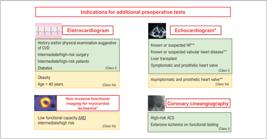
Indications for additional preoperative cardiac tests. *Only indicated for patients scheduled for intermediate- or high-risk surgery. **No evaluation in the past year (or 6 months in cases of valvular heart disease). ACS: acute coronary syndromes; CVD: cardiovascular disease; HF: heart failure.
Chart 3 presents recommendations for performing ECG.
2.3. Echocardiography
Echocardiography is a low-cost, noninvasive diagnostic method with extensive application in various fields of cardiology. It provides a comprehensive CV morphofunctional assessment, which is crucial for preoperative diagnostic and therapeutic decisions. In terms of morphology and anatomy, basic data assessed in routine echocardiography include cavity size, ventricular mass, structural assessment of valves and major vessels, and systolic and diastolic function. Hemodynamic evaluation is derived from mathematical calculations using data obtained from Doppler ultrasound, and color flow mapping accurately assesses intracardiac and transvalvular flow dynamics. Morphofunctional cardiac changes can be closely correlated with an increased perioperative risk.
In addition to conventional TTE, other modalities can be performed. These include transesophageal echocardiography, a semi-invasive technique that allows detailed anatomical assessment, intracavitary thrombus investigation, and aortic evaluation, among others; myocardial strain imaging for a more detailed evaluation of ventricular contractile function; the use of contrast agents (microbubbles) for myocardial perfusion assessment; and three-dimensional echocardiography, which allows for a more accurate volumetric evaluation and determination of left ventricular ejection fraction (LVEF).
However, it is important to note that preoperative resting echocardiography should not be performed routinely in the setting of noncardiac surgery. It should only be requested when, after an initial clinical evaluation, there is suspicion of heart disease. This initial evaluation should involve thorough history-taking and physical examination, and possibly common clinical tests such as laboratory tests, ECG, and chest radiography.56,57
TTE is the primary diagnostic method in patients with suspected or known HF, and its multiple modalities help estimate surgical risk.58-60 However, it should be used judiciously, as there is no evidence that TTE is associated with increased survival or shorter lengths of hospital stay. On the contrary, several studies suggest that more widespread use increases length of hospital stay without clinical benefit.61 Finally, in patients with known or suspected valvular heart disease (VHD), prosthetic heart valves, and intracardiac devices, TTE or transesophageal echocardiography should be used to help determine perioperative risk, as well as to guide prophylaxis for infective endocarditis (IE) (Figure 1).
Chart 4 presents recommendations for performing preoperative echocardiography.
2.4. Noninvasive Tests for Myocardial Ischemia
2.4.1. Exercise Electrocardiogram
Exercise ECG is a safe, useful, and effective tool in the investigation of myocardial ischemia,62 with its primary mechanism being the provocation of a mismatch in blood supply and demand. Thus, it is reasonable to think that the detection of abnormalities on exercise ECG could be reproducible during the perioperative period and its varying levels of stress. However, there is no robust evidence that using this strategy results in reduced perioperative mortality.63
Considering that the goal of risk stratification is to reduce perioperative risk, it would not be logical to perform exercise ECG on individuals already stratified as low risk by the recommended algorithms. In populations with a low prevalence of CAD, exercise ECG would not add value to perioperative clinical stratification, given that the lower the prevalence of CAD, the lower the positive predictive value of the test. In fact, exercise ECG could even delay the surgery due to the need for other more specific tests to differentiate true from false-positive results.64,65
Even in high-risk individuals, such as in the preoperative period of vascular surgery, the positive predictive value, sensitivity, and specificity of exercise ECG are low (10%, 74%, and 69%, respectively), although the negative predictive value is high (98%).66 Conversely, in a cohort study, preoperative exercise ECG for ischemia in high-risk patients with 3 or more clinical risk factors was associated with shorter lengths of hospital stay and lower hospital and 1-year mortality rates.64 Therefore, among asymptomatic individuals with a higher prevalence of the disease, exercise ECG could be requested only when the result would influence the prognosis and, consequently, the preoperative approach, either to guide more intensive clinical therapy or a myocardial revascularization procedure. In this setting, low exercise tolerance (below 4 METs) and the onset of ischemic response at a low workload are associated with a higher number of perioperative CV events.9
Chart 5 presents recommendations for preoperative exercise ECG.
2.4.2. Myocardial Perfusion Imaging (MPI)
Despite greater availability and lower cost, exercise ECG has limitations, particularly in patients with a high pretest probability of CAD, in those with baseline ECG abnormalities, and in those unable to perform physical activity on a treadmill.62
Thus, the use of non-invasive functional imaging for myocardial ischaemia presents higher accuracy in assessing the risk of perioperative CV events. This approach can be useful for further risk stratification in patients with reduced functional capacity, established CAD, or a high pretest probability of CAD without a prior diagnosis of chronic coronary syndrome.
In the perioperative period, there is a strong correlation between the degree of CAD and the severity of perfusion abnormalities, meaning that functional imaging tests functional testing has a greater predictive capacity in patients with significant CAD.67 A meta-analysis evaluating 1,179 patients undergoing vascular surgery revealed that large perfusion defects (> 20%) were associated with a higher rate of perioperative events, with an area under the curve of 0.78 (95%CI 0.65-0.89). However, it should be noted that even in a high-risk population (established vascular disease), only 23% of individuals presented with extensive ischemia.68
In patients with good functional capacity (≥ 4 METs), preoperative functional imaging tests in addition to the RCRI added only moderate value (AUC 0.77 vs AUC 0.70).69 Another consideration when requesting functional imaging tests is that recent studies (outside the perioperative context) demonstrated the benefit of clinical treatment over invasive strategies even in patients with moderate-to-severe ischemia.70
Finally, despite its usefulness in the diagnosis of CAD and CV risk stratification, preoperative functional imaging tests should be requested only when necessary. This is because it may lead to a propensity for coronary revascularization before noncardiac surgery, which has been shown in randomized clinical trials to not alter long-term outcomes, and may even delay noncardiac surgery for the minimum time required for dual antiplatelet therapy (DAPT).71 Even in patients with high ischemic burden on functional tests listed for kidney transplant, invasive approaches such as revascularization were not superior to optimized clinical treatment.72
2.4.3. Dobutamine Stress Echocardiography
Dobutamine stress echocardiography (DSE) is accurate and safe in the identification of patients with CAD and plays an important role as a predictor of CV events.73,74 Stress can be induced via physical exercise, on a treadmill or cycle ergometer (bicycle), or by using medications, specifically dobutamine and dipyridamole, combined with atropine if there are no contraindications. Dobutamine and exercise have similar diagnostic accuracy, which is superior to that of dipyridamole.75
Pharmacological stress echocardiography with dobutamine or dipyridamole is recognized for its significant diagnostic and prognostic value. Segmental contraction abnormalities involving a large extent of the left ventricle, appearing at early stages of drug infusion, not only confirm the diagnosis of CAD but also confer a high risk of short-term ischemic complications, significantly increasing perioperative CV risk. Conversely, the absence of segmental contraction abnormalities during stress is well correlated with the absence of severe coronary obstruction. Additionally, the appearance of such abnormalities in a small myocardial area at the late peak stress stage portends a better prognosis and lower risk of CV events. If DSE does not show residual ischemia in a patient with a prior infarction, the prognosis is good, and the likelihood of perioperative reinfarction, death, and acute pulmonary edema during noncardiac surgery is low.66 The use of DSE in the assessment of perioperative risk is well documented, with a positive predictive value ranging from 25% to 55% and a negative predictive value ranging from 93% to 100% for CV events in patients undergoing noncardiac surgery.66,76 The results are typically used to guide preoperative clinical management, especially the decision to perform coronary angiography with angioplasty or myocardial revascularization surgery before or after elective surgery. A meta-analysis of 15 studies comparing MPI and DSE in preoperative CV risk stratification demonstrated that the prognostic value of abnormalities in both imaging methods for perioperative ischemic events is similar.77
Finally, the use of microbubble contrast agents improves the visualization of left ventricular endocardial borders, increasing the sensitivity of DSE for detecting segmental contraction abnormalities. Furthermore, real-time assessment of myocardial perfusion may also be performed. Perfusion deficits detected in an ischemic wall are correlated with a worse prognosis, higher likelihood of short-term events and, consequently, increased perioperative CV risk.
2.4.4. Summary of Recommendations for Noninvasive Functional Imaging for Myocardial Ischaemia (Figure 1)
Chart 6 presents recommendations for preoperative myocardial perfusion imaging or stress echocardiography.
2.5. Coronary Cineangiography
Invasive coronary assessment with coronary cineangiography is not routinely recommended before noncardiac surgery and should not replace noninvasive tests for myocardial ischemia, when indicated. Performing coronary cineangiography unnecessarily may delay surgical planning and has not shown evidence of increased patient survival or reduced risk of perioperative MI, even in patients undergoing procedures with a high risk of CV complications.71,78
The indications for invasive coronary assessment are similar to those outside the preoperative setting, such as patients with acute coronary syndrome or extensive ischemia on functional testing (Figure 1).79 A small, randomized study evaluated the effect of routine preoperative coronary cineangiography on carotid endarterectomy. Despite fewer ischemic events in patients undergoing preoperative angioplasty, there was no difference in mortality. Moreover, surgery was performed on average 4 days after the procedure, which is neither recommended nor safe.80,81
Time-sensitive procedures, such as cancer surgery, should not be postponed for invasive assessment in asymptomatic patients. This recommendation is based on studies that failed to show any reduction in postoperative CV complications in asymptomatic patients undergoing preoperative coronary revascularization.82
Chart 7 presents recommendations for preoperative coronary cineangiography.
2.6. Coronary Computed Tomography Angiography
Coronary computed tomography angiography (CCTA) has high sensitivity for anatomical detection of coronary stenoses, including multivessel and left main CAD.83 However, the benefits of CCTA before noncardiac surgery remain uncertain.
The benefit of coronary anatomy assessment via CCTA was investigated by Li et al., who assessed 841 elderly patients undergoing high-risk noncardiac surgery without known or suspected CAD. Single-, two-, and three-vessel disease was found in 103 (12.2%), 45 (5.4%) and 16 (1.9%) patients, respectively. An Agatston score above 195 was independently associated with a higher risk of CAD. Significant CAD was found in 19.5% of patients. In multivariate analysis, the degree of stenosis was as an independent factor for the cancellation of scheduled surgery. The authors considered CCTA useful for ruling out or confirming significant CAD.84
The use of CCTA in addition to the RCRI was investigated by Ahn et al.,85 who evaluated retrospective studies including patients undergoing intermediate-risk procedures. The presence of lesions > 50% increased the incidence of major adverse cardiac events (MACE), reaching up to 29.7% in multi-vessel CAD vs 4.3% in its absence. The authors concluded that CCTA showed additive value to RCRI in risk reclassification. In a prospective study of 955 patients, Sheth et al. demonstrated that preoperative CCTA could improve estimation of risk for patients who will experience perioperative CV death or MI. However, risk reclassification using CCTA compared with the RCRI occurred in 22% of patients and could inappropriately overestimate the risk of complications by up to 5 times.86
The extent and severity of CAD on CCTA concerning MACE incidence was evaluated in a meta-analysis of 11 studies.87 It was observed that the severity and extent of CAD were associated with increased MACE risk (no CAD 2%, nonobstructive 4.1%, obstructive single-vessel 7.1%, obstructive multivessel 23.1%). Multi-vessel CAD presented the highest risk (odds ratio [OR] 8.9). Increased calcium scores were also associated with a higher perioperative MACE risk (calcium score ≥ 100, OR 5.1, ≥ 1,000 OR 10.4, both p < 0.001).
The PANDA trial directly compared the prognostic accuracy of CCTA vs DSE.88 The study included 215 patients with more than 1 risk factor for perioperative CV events who underwent both CCTA and DSE. DSE had an OR of 6.1 for CV events; the presence of significant CAD on CCTA had an OR of 18.8; and an elevated calcium score had an OR of 4.2. The authors concluded that CCTA might have higher prognostic value than DSE before noncardiac surgery, but the sample size of the study was too small for a definitive conclusion.
Due to the lack of more studies and evidence demonstrating event reduction with the use of CCTA, and primarily due to the concern of generating excessive "preventive" angioplasties, CCTA is not yet routinely recommended for risk stratification before noncardiac surgery.
3. Specific Diseases and Procedures in the Perioperative Period
3.1. Heart Failure
Heart failure (HF) affects approximately 1% to 2% of the general population in developed countries, approximately 5.7 million patients in the United States, and over 10% of the population older than 70 years.89,90 Circulatory system diseases are the leading cause of death in Brazil, accounting for approximately 29% of deaths in the country. Ischemic heart diseases and HF are responsible for approximately 39% of deaths due to circulatory system diseases.91
HF is a well-known risk factor for perioperative CV events. Data from a large registry of noncardiac operations, which included over 150,000 procedures, revealed that the presence of HF was associated with a 63% increase in the risk of perioperative mortality and a 51% increase in the risk of 30-day all-cause readmission compared with the group with CAD without HF.92
A recent analysis with data from a U.S. database including 21,560,996 noncardiac surgical procedures from 2012 to 2014 revealed the presence of perioperative HF in 4.9% of cases, which was associated with a higher in-hospital mortality rate (4.8% vs. 0.8%), conferring a 2.2 times increased risk of perioperative mortality. The risk was higher among patients with acute HF than those with chronic HF, and the risk was greater in patients with decompensated chronic HF compared with those with chronic HF alone (7.8% vs. 3.8%, p < 0.001).93
In another retrospective cohort study involving 609,735 patients undergoing noncardiac surgery, the 90-day mortality rate was higher among patients with symptomatic HF (10.1%), followed by patients with asymptomatic HF (4.9%). In patients without HF, the mortality rate was 1.2%. Thus, the presence of HF, whether symptomatic or not, increases the risk of early death.94
Reduced LVEF is considered a strong predictor of CV events in patients undergoing vascular surgery. However, most studies analyze LVEF by dichotomizing it as greater or less than 40%. A study involving 174 patients with HF revealed that only severely reduced LVEF (< 30%) was an independent predictor of mortality. The presence of moderately (30-40%) or mildly (40-50%) reduced LVEF, or HF with preserved LVEF (> 50%), were not independent predictors of 30-day mortality.95 Despite the predictive power of LVEF for CV events, routine echocardiography for all patients scheduled for noncardiac surgery is not indicated. A Canadian cohort study involving more than 250,000 patients (15% with preoperative echocardiography, n = 40,084) revealed that preoperative echocardiography is not associated with improved survival or reduced length of hospital stay after major noncardiac surgery.61
Preoperative elevation of natriuretic peptide levels is associated with a worse prognosis in the perioperative period, as it is associated with worsening ventricular function and a higher rate of CV events.96,97 Measuring these biomarkers can assist in the risk stratification of patients with HF.
Perioperative clinical management of patients with HF should involve special attention to volemia. Both hypovolemia, which can intensify hypotension, and hypervolemia, which can cause pulmonary edema and systemic venous congestion, should be avoided. The Austrian IMPROVE study aims to determine if the use of an inodilator, levosimendan, may benefit postoperative outcomes by evaluating the impact of the medication on natriuretic peptide levels. Its results may provide a new perioperative management approach for patients with HF undergoing noncardiac surgery.98
The flowchart for preoperative evaluation of patients with known or suspected HF in is shown in Figure 2.

Preoperative approach for patients with known or suspected heart failure. HF: heart failure; NYHA: New York Heart Association. #In patients with an echocardiogram from the past year and without new symptoms, it is not necessary to repeat the echocardiogram. *Optimized treatment of HF according to current guidelines (maximum tolerated dose of angiotensin-converting enzyme inhibitors/angiotensin receptor blockers/sacubitril/valsartan, beta-blockers, spironolactone, and sodium-glucose cotransporter type 2 [SGLT2] inhibitors for HF with reduced ejection fraction and adequate control of blood volume and blood pressure, spironolactone and SGLT2 inhibitors for patients with HF and preserved ejection fraction), evaluation and correction of myocardial ischemia, and correction of valvular heart disease in significant cases.
Chart 8 presents recommendations for the perioperative management of patients with HF.
3.2. Arterial Hypertension
Systemic hypertension is a highly prevalent clinical condition, both in the general population and in patients undergoing surgery, being responsible for approximately 50% of deaths from CAD and stroke.99 Although the importance of strict blood pressure (BP) control in the prevention of long-term CV events is well established, its perioperative management remains controversial.
3.2.1. Preoperative BP Management
A previous diagnosis of hypertension is an independent predictor of mortality in patients undergoing noncardiac surgery.100 However, there is no solid evidence linking BP measurement at the time of hospital admission with perioperative CV complications.101 This suggests that the perioperative risk attributed to hypertension is primarily due to its impact on target organ damage.102
Regarding hypotension, the literature provides more concrete data. A prospective cohort study involving 251,567 patients demonstrated that systolic BP levels < 119 mm Hg and diastolic levels < 63 mm Hg are associated with increased 30-day postoperative mortality.103 Therefore, BP management should also consider this probable J-curve phenomenon, in which low BP levels also represent a risk.
Regarding hypertension, it is recommended that high-risk elective operations be postponed only if systolic BP is ≥ 180 mm Hg and/or diastolic BP is ≥ 110 mm Hg. It is important to note that this threshold is based on older observational data104 and was defined before the current understanding of the risks associated with perioperative hypotension. Thus, the defined BP target should be maintained, but care must be taken to avoid hypotension.
Finally, patients with suspected secondary hypertension should be investigated before surgery. Although there is no robust evidence of increased perioperative risk in patients with secondary hypertension, those with undiagnosed pheochromocytoma are known to have very high surgical mortality rates.105
3.2.2. Intraoperative BP Management
When the introduction of antihypertensives is required intraoperatively, the medication should ideally be easily titratable, have a rapid onset of action, few side effects, and low cost. Several classes are available, including beta-blockers (esmolol and labetalol), calcium channel blockers (nicardipine), and nitrates (sodium nitroprusside and nitroglycerin).
Hypertensive patients are also more susceptible to hypotension, often due to intravascular volume depletion.106 Excessive concern about strict BP control in the preoperative period can lead to hypotension both during and after surgery, which is particularly associated with cardiac, renal, and cerebral injury, as well as increased mortality.107
In this setting, the INPRESS study (Effect of Individualized vs Standard Blood Pressure Management Strategies on Postoperative Organ Dysfunction Among High-Risk Patients Undergoing Major Surgery) compared an individualized strategy aimed at achieving a systolic BP within 10% of the reference value vs. a standard strategy where patients received vasoconstrictors only if systolic BP was < 80 mm Hg or 40% of the reference value. The individualized strategy reduced the incidence of systemic inflammatory response syndrome and organ dysfunction 7 days after surgery.108 A retrospective analysis involving patients undergoing elective noncardiac surgery underscored the deleterious effects of perioperative hypotension.109
3.2.3. Postoperative BP Management
A previous diagnosis of hypertension is the main risk factor for postoperative hypertension. However, other factors such as pain, hypercapnia, and agitation after anesthesia also contribute. Intravenous antihypertensive therapy should be considered in patients with sustained systolic BP ≥ 180 mm Hg and/or diastolic BP ≥ 110 mm Hg, as these levels increase the risk of bleeding, especially in cardiac, vascular, and endoscopic procedures, such as transurethral resection of the prostate. Hypotension should also be properly treated and prevented by maintaining an adequate volume status.
Chart 9 shows recommendations for perioperative BP management.
3.2.4. Perioperative Management of Antihypertensives
3.2.4.1. Renin-Angiotensin-Aldosterone System Inhibitors
The use of renin-angiotensin-aldosterone system (RAAS) inhibitors in the perioperative period is highly controversial due to conflicting study results. Large retrospective studies comparing the discontinuation vs. continuation of angiotensin-converting enzyme inhibitors (ACEIs) and angiotensin II receptor blockers (ARBs) perioperatively have not shown differences between groups in terms of hypotension incidence, need for fluid resuscitation, and 30-day mortality.110 Furthermore, discontinuing these medications on the day of surgery did not significantly increase the incidence of preoperative hypertension or result in higher surgery cancellation rates.111
However, Roshanov et al. observed a decrease in the intraoperative risk of hypotension and a reduction in the composite outcome of death, MI, and stroke with the discontinuation of these medications.112 Despite the potential for increased hypotension with the maintenance of ACEIs/ARBs, there was no observed increase in acute kidney injury.113 It is also noteworthy that if ARBs were discontinued and not reintroduced within 2 days after surgery, mortality rates increased.114
Thus, current evidence does not provide a definitive answer on whether to maintain or discontinue ACEIs/ARBs in the perioperative period, requiring individualized decision-making. The ongoing randomized trial STOP-or-NOT may provide additional information on this topic.115 For this, factors such as the type of surgery, patient BP variability, and bleeding risk should be considered. Importantly, if these medications are discontinued, they should ideally be reintroduced postoperatively as soon as clinically feasible.
3.2.4.2. Calcium Channel Blockers
In patients undergoing noncardiac surgery, no significant differences in BP levels were observed with the use of calcium channel blockers compared with other antihypertensive medications, and their use was not associated with increased occurrence of adverse events.116 Therefore, while the formal recommendation is to continue chronic therapy with calcium channel blockers perioperatively, this decision should be individualized, considering the risks of hypotension and the lack of deleterious effects from their discontinuation.
3.2.4.3. Diuretics
There is no robust evidence regarding the use of diuretics in the perioperative period. Their maintenance, including administration on the day of surgery, has been shown to be safe and not associated with increased risk of hypotension, need for fluid resuscitation, or use of vasopressor agents, nor with increased risk of postoperative CV events.117
3.2.4.4. Central Sympatholytic Drugs
Early studies on the use of clonidine in the perioperative period of noncardiac surgery showed a reduction in myocardial ischemia.118 However, larger, more recent studies have failed to confirm this association, instead linking its use to increased incidence of nonfatal cardiac arrest, particularly due to asystole or pulseless electrical activity. The use of clonidine is also associated with a significant increase in bradycardia and hypotension, with hypotension being an independent predictor of MI.119 Thus, clonidine may be used in cases of uncontrolled hypertension with insufficient time for effective management, but it should not be introduced to reduce CV events. Discontinuation in chronic users is not recommended due to the potential for rebound hypertension.
3.2.5. Final Considerations
The perioperative management of hypertension is highly challenging. While it is crucial to avoid extremely high BP levels (systolic BP > 180 mm Hg and diastolic BP > 110 mm Hg), it is equally important to prevent hypotension, which has been increasingly recognized in recent studies as a predictor of poor prognosis and increased risk of mortality. Therefore, aside from beta-blockers and central sympatholytic drugs, whose discontinuation may be harmful, the perioperative management of antihypertensives should be individualized to avoid significant BP increase, variability, and particularly hypotension. Perioperative clinical evaluation should include BP monitoring and vigilance for symptoms of hypotension such as dizziness, fainting, drowsiness, signs of low cardiac output, renal function deterioration, low urine output, and delirium.
Chart 10 shows recommendations for the perioperative management of antihypertensives.
3.3. Patient Management in Low-Risk Procedures
In this Guideline, "low-risk procedures" refers exclusively to the risk of bleeding and CV complications. We are aware that there are low risk surgical procedures in all medical specialties, but specific surgical considerations are beyond the scope of this document.
3.3.1. Dental Procedures
Maintaining adequate oral health is crucial for reducing the risk of systemic complications, especially in patients with CVD, leading to better glycemic control, reduced risk of bacteremia, and possibly better BP control.120-122 Oral diseases, such as periodontal diseases and endodontic infections, may pose a postoperative risk of bacteremia and sepsis in patients requiring intubation and in transplant recipients123 and should be treated before surgical procedures.124
Mouth rinsing with 15 mL of 0.2% chlorhexidine (most effective) or 5% povidone-iodine for 30 to 60 seconds is recommended before dental procedures and before intermediate- and high-risk procedures to reduce the risk of sepsis.125,126
3.3.1.1. Antithrombotic Agents
When patients on antithrombotic therapy, including anticoagulants and antiplatelet medications, undergo dental surgical treatment, a decision must be made regarding the continuation of anticoagulation therapy, balancing the risk of hemorrhagic complications against the risk of embolic complications. Decades of studies involving thousands of patients on anticoagulation therapy undergoing dental procedures have shown that hemorrhagic complications requiring more than local hemostatic measures are rare and never fatal. However, some embolic complications have been fatal or debilitating in patients in whom anticoagulation therapy was interrupted due to a dental procedure.127
Most dental procedures have a low risk of bleeding, with a bleeding rate of up to 3.63%.128 There is strong evidence for older medications (e.g., warfarin, antiplatelet medications) and limited evidence for newer oral anticoagulants that, for most patients, the occurrence of significant uncontrollable bleeding is very low. For these reasons, this Guideline does not recommend altering or interrupting anticoagulation or antiplatelet therapy before dental procedures.129,130
In general, dental procedures can be safely performed in patients on warfarin with an international normalized ratio (INR) < 3.5, provided local measures to reduce bleeding are employed. For patients with an INR above this range or those expected to have a higher risk of bleeding, the best strategy should be discussed between the dentist and the physician who prescribed anticoagulant therapy.131
Bleeding management in dental practice can include lysine analogs (tranexamic acid 250mg, 15 to 25 mg/kg, ie, 2 tablets of 250 mg, 2-3 times a day) and local application of hemostatic sponges and cyanoacrylates, among other methods.
3.3.1.2. Cardiac Implantable Electronic Devices
In addition to the risk of infection, the functioning of cardiac implantable electronic devices (CIEDs) may be impaired by the action of dental equipment during some procedures, particularly battery-operated composite curing light, ultrasonic scalers, and cleaning systems, when the distance between them and the CIED is less than 23 cm.132 In these cases, alternative dental equipment should be used.
Conversely, other dental electronic devices such as electric toothbrushes, electric scalpels, electric pulp testers, high- and low-speed handpieces, apex locators, and amalgamators have not shown interference.132,133
3.3.1.3. Local Anesthetics
Excessive use of local anesthetics combined with vasoconstrictors can increase heart rate (HR), BP, and subsequent myocardial oxygen demand. However, recent studies confirm that the use of 1-4 anesthetic cartridges with vasoconstrictor (lidocaine with epinephrine 1:800,00, 1:100,000, and 1:200,000) is relatively safe for patients with controlled CVD and hypertension.134,135
Chart 11 presents a summary of recommendations for dental procedures.
3.3.2. Dermatological Surgery
Dermatological surgical procedures have a low risk of both CV events and bleeding. Data from the literature suggest that approximately 50% of patients undergoing dermatological surgery are on antiplatelet or anticoagulant therapy.136,137 In these cases, the surgical team and anesthesiologist should be informed about current medications and necessary precautions, including more careful and prolonged hemostasis, as the risk associated with discontinuing antithrombotic therapy usually outweighs the risk of bleeding inherent to the procedure.
In patients on acetylsalicylic acid (ASA) for secondary prevention of CV events, discontinuation is not recommended before any dermatological surgical procedure.138,139
In patients on clopidogrel monotherapy, the risk of bleeding during surgery is increased, and some studies recommend discontinuing the medication at least 6 days before the procedure.140,141 However, no studies have demonstrated the safety of discontinuation in relation to CV events. Although there is a tendency to extrapolate evidence from ASA monotherapy to clopidogrel, there is insufficient evidence to support maintenance or discontinuation. Since bleeding in dermatological surgery is typically minimal and controllable, we recommend maintaining clopidogrel.
In patients on DAPT due to a coronary stent who are out of the critical thrombosis period, the recommendation is to discontinue the second antiplatelet medication,142,143 respecting the intervals described in this Guideline (see section on antiplatelet medications, item 4.1.3).144
In patients on warfarin, it is recommended not to discontinue the medication, adjusting the INR to ≤ 3.5 to minimize the risk of bleeding. However, some studies have not shown a correlation between INR level and increased risk of bleeding in patients on warfarin.142,145-148
In patients on direct oral anticoagulants (DOACs), despite limited evidence, it is recommended to maintain these medications during most dermatological procedures142,149,150 and schedule the surgery a few hours before the next dose to avoid peak serum concentration.
Chart 12 shows recommendations for patients scheduled for dermatological surgery.
3.3.3. Endoscopic Procedures
Endoscopic procedures are considered low risk for the occurrence of CV events.151 Cancelling endoscopic procedures for CV intervention is usually unnecessary, except for severe CV conditions, as outlined in the section on perioperative assessment algorithms of this Guideline (item 2.1.1). Additionally, most CV medications do not need to be discontinued and can be taken with a minimal amount of water. The most crucial decision is whether to maintain or discontinue antithrombotic medications in patients on antithrombotic therapy, as their maintenance during an endoscopic procedure may lead to bleeding, whereas their interruption poses a risk of thromboembolic events. The risk of bleeding in endoscopic procedures is variable, which should always be considered when determining the best strategy to be used. The risk varies according to the type of procedure and is particularly related to the presence of therapeutic interventions and biopsies. Table 9 describes the risk of bleeding associated with common endoscopic procedures in clinical practice.152 The risk of thromboembolic events from discontinuing antithrombotic therapy varies according to the therapy’s indication and the patient’s condition.
3.3.3.1. Management of Antiplatelet Therapy in Endoscopic Procedures
In endoscopic procedures with a low risk of bleeding, antiplatelet therapy can be maintained, whether as monotherapy (regardless of the medication) or DAPT.152-157 In procedures with a high risk of bleeding, several considerations should be made. Patients on DAPT due to recent stent placement or acute coronary syndrome (see itens 4.1 and 4.2) are at a higher risk of suffering CV events from discontinuation of antiplatelet therapy.144 Thus, elective endoscopic procedures with a high risk of bleeding should be postponed whenever possible until this high-risk period has passed. However, for procedures that must be performed during this period, the most accepted strategy is maintaining ASA and discontinuing the second antiplatelet medication,155,158 although evidence for this strategy is limited. ASA monotherapy for secondary prevention of CV events may be maintained in the perioperative period of endoscopic procedures, including high-risk ones, as most evidence shows a low risk of significant bleeding in these circumstances.156,157,159-169 Some studies have shown increased bleeding in procedures such as endoscopic submucosal dissection in patients with gastric neoplasia170 and mucosectomy for colonic tumors larger than 20 mm.171 Such cases should be assessed individually, considering the risk of thrombotic events from ASA discontinuation.155 The maintenance of clopidogrel monotherapy during percutaneous endoscopic gastrostomy may be considered as there is some evidence supporting its use in this setting.160 Conversely, evidence on the use of prasugrel and ticagrelor during endoscopic procedures with a high risk of bleeding is scarce. If antiplatelet therapy is to be discontinued, the interval between discontinuation and the endoscopic procedure should follow the recommendations in the section on antiplatelet management of this Guideline.144 Antiplatelet therapy can be resumed after the procedure once hemostasis has been achieved, and a loading dose may be considered in patients at high risk of CV events.158
3.3.3.2. Management of Anticoagulants in Endoscopic Procedures
Anticoagulation therapy with warfarin may be maintained during endoscopic procedures with a low risk of bleeding152,153,155-157,161,169 but should be discontinued in high-risk ones.156,162,168 There is no current evidence on the use of DOACs in this setting. It is suggested to maintain DOACs during procedures with a low risk of bleeding but discontinue them in high-risk ones.152,156,157,169 The intervals between discontinuation and resumption of DOACs and warfarin (including bridging therapy in high-thromboembolic-risk patients) should follow the recommendations in the section on perioperative anticoagulation management of this Guideline (item 4.3). Regarding the use of DOACs, if possible, the endoscopic procedure should be performed before the next dose to avoid peak concentration, which occurs in the first 2 hours after administration.157,169
Chart 13 presents recommendations for patients scheduled for endoscopic procedures.
3.3.4. Ophthalmic Procedures
Ophthalmic surgery is relatively common among the older population. CV comorbidities requiring antithrombotic therapy, and its perioperative management are topics of intense debate among ophthalmologists and cardiologists. In Brazil, the fear of hemorrhagic events, including periorbital hematomas, leads to the indiscriminate discontinuation of ASA and warfarin in 82.7% of patients undergoing glaucoma surgery.172 However, the available evidence, although limited, suggests that this fear is not justified. The rate of bleeding events reported in observational studies is low and without major consequences, particularly in cataract surgery using conventional anesthesia techniques.173-177 Certain types of ophthalmic surgery, however, have a higher risk of bleeding events, such as trabeculectomy for glaucoma178,179 and vitrectomy for retinal diseases.180,181 Nonetheless, the available evidence does not demonstrate an increased risk of significant hemorrhagic events in these procedures with the use of ASA.179,182-184 In these cases, management should be individualized, but it is generally recommended to maintain ASA perioperatively.184,185 Patients on DAPT due to recent stent placement or acute coronary syndrome (see itens 4.1 and 4.2) are at higher risk of CV events with the interruption of antiplatelet therapy. Thus, ophthalmic surgical procedures, whenever possible, should be postponed until this high-risk period has passed. For procedures that need to be performed during this period, the approach depends on the risk of hemorrhagic events associated with the intervention. For low-risk interventions (intravitreal injections, cataract surgery, and peribulbar anesthesia), ASA and P2Y12 receptor inhibitors should be maintained. However, for high-risk interventions, such as vitrectomy and trabeculectomy, the most accepted recommendation is to maintain ASA and suspend the second antiplatelet medication, following the intervals described in the specific section of this Guideline (item 4.2),144 although evidence for this strategy is limited. Similar to patients on ASA monotherapy, published evidence supports maintaining clopidogrel monotherapy in the perioperative period of cataract surgery.176,177 Evidence for glaucoma and retinal surgery is even scarcer, thus it is recommended to discontinue clopidogrel 5 days before the procedure.
In patients on warfarin, the available evidence supports its maintenance during procedures with a low risk of hemorrhagic events, such as cataract surgery, provided the INR is within the therapeutic range.173,174,184,186,187 A meta-analysis of observational studies with patients undergoing cataract surgery while on warfarin found a bleeding incidence of approximately 10%. The bleedings were mostly self-limiting and subconjunctival, and no patients experienced compromised visual acuity related to a bleeding event.175 For glaucoma and retinal surgery, warfarin should be discontinued, and perioperative management should follow the strategy described in the section on perioperative anticoagulation management of this Guideline (item 4.3), according to the individual thrombotic risk of each patient. To date, evidence on the risk of bleeding in patients on DOACs undergoing ophthalmic surgery has only been well established for cataract surgery. The risk of bleeding associated with warfarin in cataract surgery has been well studied, and most guidelines recommend maintaining warfarin if the INR is within the therapeutic range. DOACs present a lower risk of bleeding than warfarin.188 A systematic review and meta-analysis revealed that patients on DOACs have a 22% relative risk reduction in spontaneous intraocular bleeding compared with warfarin.189 Two small studies compared the discontinuation and maintenance of DOACs during cataract surgery and found no increase in complications when maintaining DOAC therapy.186,190 Therefore, most major centers tend to maintain the use of DOACs during cataract surgery, primarily due to the absence of evidence suggesting otherwise.184,187 In high-risk procedures, there is no evidence regarding the safety of DOAC use. They are typically interrupted 24 hours before surgery.
Recommendations for the management of patients with coronary stents and prosthetic heart valves should be individualized, considering the risk of both thrombotic and hemorrhagic events. For patients recommended to continue anticoagulant and/or antiplatelet therapy, the surgeon should be informed of the need to ensure adequate hemostasis. A suggestion that can be considered and discussed with the anesthesiologist—who makes the final decision—is to use a specific type of anesthesia less associated with hemorrhagic complications.177 As for antiplatelets, if they are interrupted, they should be resumed postoperatively as soon as possible. It is also recommended that procedures with a higher risk of CV events be performed in hospitals equipped for urgent hemodynamic intervention (coronary angioplasty) if needed.
Chart 14 presents recommendations for patients scheduled for ophthalmic surgery.
3.4. Valvular Heart Disease
Patients with valvular heart disease (VHD) who are candidates for noncardiac surgery have a higher risk of perioperative CV complications.191 This risk is related to the type and anatomical severity of the disease, the presence of symptoms, and the type of surgery.6 The main CV complications that may occur in this setting include pulmonary congestion, acute pulmonary edema, cardiogenic shock, MI, tachyarrhythmias, embolic events, bleeding, and IE.192
Upon suspicion of VHD following a detailed clinical history and physical examination, a TTE should be performed. The TTE aims to assess the anatomical severity of the disease, ventricular function, cardiac remodeling, and to estimate pulmonary artery systolic pressure (PASP). If uncertainty remains, other diagnostic methods may be used, such as transesophageal echocardiography, computed tomography, magnetic resonance imaging, and cardiac catheterization.
Symptomatic patients with significant VHD present high morbidity and mortality and should undergo valvular intervention,193,194 which should be performed before elective procedures. Conversely, emergency noncardiac procedures should be performed following specific recommendations for each VHD to minimize the chances of CV decompensation.
Heart valve stenosis carries a higher perioperative risk compared with valve regurgitation. Therefore, extra care should be taken with patients with aortic and/or mitral stenosis.3,195 If there is more than one valvular lesion and/or a combination of stenosis and regurgitation, the approach should be based on the most significant lesion.
3.4.1. Aortic Valve Stenosis
Aortic valve stenosis is a common type of VHD in elderly patients, affecting approximately 2% to 4% of adults over 75 years,196 and this number is expected to increase in the coming years. Significant aortic stenosis is a well-established risk factor for perioperative mortality, HF, and MI in noncardiac surgery.193
Asymptomatic patients with significant aortic stenosis can safely undergo low-risk noncardiac surgery as long as volume overload is avoided.191,193 In these cases, if there is doubt as to whether the patient is asymptomatic, a stress test should be performed. The Heart Team, including the surgeon and anesthesiologist, plays an important role in decision-making.
In symptomatic patients with significant aortic stenosis or those scheduled for moderate-to-high risk surgery, aortic valve intervention should be performed before noncardiac surgery.197-199 Aortic valve replacement is associated with lower in-hospital and 30-day mortality in individuals undergoing intermediate- or high-risk surgery.198,199 Additionally, a study by Mizuno et al.197 with 218 patients demonstrated that those with aortic stenosis undergoing high-risk noncardiac surgery experienced faster progression of aortic stenosis compared with those who did not undergo surgical intervention. The choice between surgery or transcatheter aortic valve implantation (TAVI) should be based on the latest guidelines on VHD. When noncardiac surgery needs to be performed promptly, TAVI should be considered due to the quicker recovery associated with this procedure.5 In cases where TAVI and heart valve surgery are unfeasible, balloon valvuloplasty is an alternative, although the risks of this procedure should be weighed and restenosis will occur later.193,197,200
If noncardiac surgery is urgent or time-sensitive, such as in the case of femur fracture, invasive hemodynamic monitoring during anesthesia is necessary, avoiding changes in volume status, especially due to the high risk of hypervolemia. In these cases, postoperative care in an intensive care unit (ICU) with continuous hemodynamic and ECG monitoring, as well as serial cardiac troponin (cTn) measurements, is recommended. In time-sensitive procedures, balloon valvuloplasty and TAVI may be considered depending on the anatomical severity of the disease and the availability of these resources.
3.4.2. Mitral Valve Stenosis
Patients with mild-to-moderate mitral valve stenosis can undergo noncardiac surgery safely, provided measures are taken to prevent tachycardia and volume overload before and after the procedure.
Patients with significant mitral stenosis, even in the absence of symptoms, have a higher risk of CV complications. In cases for which surgical or percutaneous correction is indicated, patients should undergo the procedure before any elective noncardiac surgery.201 In asymptomatic patients with significant mitral stenosis without an indication for interventional treatment, mitral valve intervention should be considered before any high-risk procedure.
In emergency noncardiac surgery, invasive hemodynamic monitoring and prevention of tachycardia and hypervolemia are recommended. Increased HR, especially if atrial fibrillation (AF) develops, can lead to congestion and pulmonary edema. Thus, the use of beta-blockers and/or diuretics can be implemented in the perioperative period.
3.4.3. Aortic and Mitral Regurgitation
Primary valve regurgitation is associated with increased CV risk during noncardiac surgery, but it is better tolerated than valve stenosis.202,203 Mild-to-moderate aortic and mitral regurgitation do not increase the risk of CV complications during noncardiac surgery. In the case of significant valvular dysfunction, provided there are no symptoms and ejection fraction is preserved, noncardiac operations can also be performed, with caution to avoid volume overload.
Conversely, patients with significant primary aortic or mitral regurgitation with an indication for heart valve replacement have an increased risk of CV complications and should undergo surgical correction before noncardiac surgery.191 In symptomatic patients with moderate-to-severe secondary mitral regurgitation, it is important to assess, in conjunction with the Heart Team, whether the patient is a candidate for surgery or transcatheter edge-to-edge repair before the elective noncardiac surgery.193
If the noncardiac procedure is urgent or an emergency, it should be performed after optimization of pharmacological treatment and hemodynamic stabilization, preferably using vasodilators and diuretics, with postoperative care in an ICU.
3.4.4. Prosthetic Heart Valves
Patients with normally functioning prosthetic heart valves and no left ventricular dysfunction can undergo noncardiac surgery without additional risk.
Chart 15 presents recommendations for patients with VHD.
3.5. Solid Organ Transplantation
3.5.1. Liver
Considered the treatment of choice for many end-stage liver diseases, liver transplantation has become a routine procedure in several centers. Advances in anesthetic approaches, increased surgical experience, and improved postoperative care have allowed practices that, just over a decade ago, would have been exceptions. Consequently, many groups have expanded transplant indications to increasingly older populations and more complex cases for which transplant was previously contraindicated.204 Additionally, indications for nonalcoholic steatohepatitis, typically associated with obesity, diabetes, hypertension, and CVDs, have become more frequent, being leading causes of liver transplantation in some centers.205
Studies show that up to 26% of patients undergoing liver transplantation have at least one critical coronary artery stenosis.206 Within 1 year after the transplant, 15.2% of patients have at least one CV event, and 2.8% die from it.207 Thus, CVD is the third most common cause of death following liver transplantation, after infections and graft rejection or dysfunction. The most common CV events are arrhythmias, pulmonary edema, ventricular dysfunction, sudden death and AMI.206 For these reasons, pretransplant CV evaluation is crucial to determine the correct management of these patients both preoperatively and postoperatively.
In addition to CAD-related events, other comorbidities commonly found in patients with liver disease can increase morbidity and mortality after liver transplantation. These include alcoholic cardiomyopathy, cirrhotic cardiomyopathy, pulmonary hypertension (PH), and hepatopulmonary syndrome (HPS).
Table 10208 summarizes the main cardiopulmonary comorbidities of liver transplant candidates, clinical findings, diagnosis, and expected outcomes after the transplant.
3.5.1.1. Cirrhotic Cardiomyopathy
Cirrhotic cardiomyopathy is characterized by the following triad in patients with cirrhosis209,210: systolic dysfunction (primarily due to a deficit in stress-induced contractile response, with a resting LVEF < 55%), diastolic dysfunction, and electrophysiological abnormalities (especially prolonged QT intervals, chronotropic incompetence, and bradycardia, as well as ventricular repolarization abnormalities). Left atrial enlargement, increased myocardial mass, and elevated type B natriuretic peptide (BNP), N-terminal pro BNP (NT-proBNP), and cTnI levels are also commonly found.
Despite these findings increasing morbidity and mortality in liver transplant candidates, no specific treatment for these abnormalities has shown benefit.
3.5.1.2. Alcoholic Cardiomyopathy
Alcoholic cardiomyopathy affects 21% to 32% of patients with dilated cardiomyopathy in some centers.211,212 Considering that alcoholic cirrhosis is among the leading causes of liver disease, the concomitant occurrence of cirrhosis with dilated cardiomyopathy is relatively common.
3.5.1.3. Portopulmonary Hypertension
The hyperdynamic state of patients with portal hypertension can lead to vasoconstriction and pulmonary vascular remodeling, resulting in PH. This condition affects 5% to 10% of transplant candidates and is not related to the etiology or severity of portal hypertension, even in the absence of liver cirrhosis.213,214 Portopulmonary hypertension (POPH) is a subset of group 1 PH due to its pathological and hemodynamic similarities with other causes of precapillary PH. POPH can be classified, based on mean pulmonary artery pressure (mPAP), as mild (> 25 and < 35 mm Hg), moderate (> 35 and < 45 mm Hg), or severe (> 45 mm Hg).213 Clinical status and medical tests present unique features.215,216
As the symptoms of POPH are nonspecific and its diagnosis directly impacts liver transplant eligibility, screening with TTE is recommended for all candidates. A PASP > 50 mm Hg has a high diagnostic accuracy for POPH in liver transplant candidates (97% sensitivity and 77% specificity for moderate-to-severe POPH).217 In addition, echocardiography is useful to assess right ventricular function and rule out left ventricular dysfunction and/or VHD that could contribute to postcapillary PH.216 The International Liver Transplantation Society guidelines suggest right heart catheterization for definitive POPH if estimated PASP on TTE is > 50 mm Hg. However, new evidence indicates increased diagnostic sensitivity with an estimated PASP cutoff > 40 mm Hg. Depending on service availability, a PASP between 40 and 45 mm Hg on echocardiography may indicate the need for cardiac catheterization, especially if there are other signs of PH (Figure 3).216,217
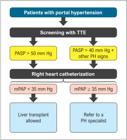
Evaluation of pulmonary artery pressure in liver transplant candidates. mPAP: mean pulmonary arterial pressure; PASP: pulmonary artery systolic pressure; PH: pulmonary hypertension; TTE: transthoracic echocardiogram. Adapted from Cartin-Ceba and Krowka.214
The diagnosis of POPH involves ruling out other causes of PH and the presence of the following hemodynamic criteria confirmed by right heart catheterization216:
-
• mPAP > 25 mm Hg.
-
• Pulmonary vascular resistance (PVR) > 3 Wood units (or 240 dyn.s.cm-5).
-
• Pulmonary capillary wedge pressure (PCWP) < 15 mm Hg. If PCWP > 15 mm Hg, likely due to hypervolemia (common in patients with cirrhosis), a transpulmonary pressure gradient (mPAP–PCWP) > 12 mm Hg suggests the presence of POPH.
Importantly, approximately 20% of patients with cirrhosis have a moderate increase in pulmonary pressure on echocardiography, but only a small fraction of these actually have POPH.218
Management of POPH depends on the presence of hemodynamic criteria assessed by right heart catheterization (Figure 3).214 In patients with a mPAP between 35 mm Hg and 50 mm Hg and increased PVR (> 240), liver transplantation cannot be performed unless there is a reduction in mPAP and PVR after pharmacological treatment of PH. The medications used in the treatment of PH have vasodilatory, antiplatelet, and antiproliferative effects, which can reduce pulmonary artery pressure.214 The main available medication classes are:
-
• Phosphodiesterase inhibitors (eg, sildenafil).
-
• Endothelin receptor antagonists (eg, bosentan, ambrisentan, macitentan).
-
• Soluble guanylate cyclase stimulators (eg, riociguat).
-
• Prostanoids (eg, epoprostenol, iloprost, treprostinil).
However, an equivalence in morbidity and mortality between patients with HP treated with these medications and those without a diagnosis of PH has not yet been demonstrated. Further studies are needed in this area.
Perioperative mortality in patients with severe POPH is close to 100%, making it a contraindication for isolated liver transplantation. In select centers, combined lung-liver transplantation should be considered.219
3.5.1.4. Hepatopulmonary Syndrome
Although sometimes mistaken for POPH, HPS presents different characteristics, such as hypoxia in the presence of liver disease that worsens in the upright position, with evidence of intrapulmonary vasodilation. Hypoxia occurs due to vasodilation in the pulmonary vascular bed, induced mainly by the accumulation of nitric oxide, leading to intrapulmonary arteriovenous shunting.220
HPS is diagnosed by the presence of the following triad214:
-
• Portal hypertension, chronic liver disease, or congenital portosystemic shunts.
-
• Hypoxemia (alveolar–arterial gradient > 15 mm Hg or > 20 mm Hg if age > 64 years).
-
• Intrapulmonary vasodilation, leading to shunting (visualization of bubbles in the left heart after 3 cardiac cycles on contrast echocardiography with microbubbles).
HPS affects 5% to 32% of transplant candidates and is not related to the etiology or severity of liver disease.221,222
The typical clinical presentation includes signs of chronic liver disease associated with cyanosis, nail clubbing, dyspnea when standing upright (platypnea), and hypoxia when also standing upright (orthodeoxia). Hypoxemia is a prominent finding in this syndrome.214
Screening is conducted with pulse oximetry. An oxygen saturation < 96% has a sensitivity of 100% and specificity of 88% for detecting PaO2 < 60 mm Hg.214 Therefore, arterial blood gas analysis is recommended in these patients. If arterial blood gas analysis shows an elevated alveolar–arterial gradient (≥ 15 mm Hg or > 20 mm Hg if age > 64 years) and a PaO2 < 80 mm Hg, a transthoracic echocardiogram with microbubbles should be performed.214 If there is an intracardiac shunt (eg, atrial septal defect, patent foramen ovale), the bubbles pass to the left heart 1 or 2 cardiac cycles after the visualization of bubbles in the right atrium. If these bubbles appear in the left atrium after 3 cardiac cycles, the presence of a pulmonary shunt is confirmed (the bubbles can pass through the dilated pulmonary vessels in HPS). It is important to note that echocardiography with microbubble contrast agents is sensitive but not specific for HPS, as many patients with cirrhosis have a positive test (intrapulmonary shunts) without meeting the criteria for HPS due to the absence of hypoxemia. A more specific test for HPS is Tc-99m macroaggregated albumin scintigraphy.214
The management of HPS consists of oxygen support, although it does not show sustained benefits in improving dyspnea and quality of life for these patients. Several pharmacological therapies have been tested, but none have shown proven benefits.214
Unlike PH, the treatment of choice for HPS is liver transplantation, although nondefinitive data correlate the degree of hypoxia with perioperative mortality.223
3.5.1.5. Coronary Artery Disease
Risk factors for CAD are as prevalent or even more prevalent in patients with cirrhosis than in the general population. In patients with advanced liver disease, the prevalence of diabetes and CAD is equal to or greater than in the general population. Particularly in patients with diabetes, the prevalence of CAD ranges from 2.5% to 27%.224,225 Conversely, the extent of the disease (degree of stenosis) does not appear to correlate with a worse prognosis.226 A comparison between liver and kidney transplant candidates revealed that the latter have a higher frequency of diabetes, hypertension, and CAD.227
3.5.1.5.1. When to Investigate CAD?
Indications for noninvasive tests to assess CV risk in liver transplant candidates vary among medical societies: some recommend it for all liver transplant candidates, while others suggest CV risk stratification before requesting a test.228 An interesting strategy for asymptomatic patients with CAD awaiting liver transplantation is to evaluate the presence of CV risk factors (male sex, hypertension, diabetes, dyslipidemia, past or current smoking, and a family history of early CAD).219 If the patient does not have any risk factor or has 1 risk factor, the negative predictive value for obstructive CAD is high, so there is no need to request a noninvasive test. However, if the patient has 2 or more risk factors, noninvasive testing is indicated.225
For symptomatic patients, usual CAD evaluation should be performed as recommended in specific guidelines, stratifying the patient according to symptoms (eg, angina and dyspnea) and risk factors (eg, sex, age).228,226 After stratification, the pretest probability of CAD is determined. If the probability is low/intermediate (< 15%), noninvasive tests (anatomic or functional) may be performed, or the investigation may be interrupted if the probability of CAD is very low. If the probability is high (> 15%), preference should be given to cardiac catheterization. In general, the preference for invasive approaches is given to patients with significant angina refractory to medical treatment, left ventricular dysfunction, or high-risk findings in noninvasive tests.228
3.5.1.5.2. Which Test to Request for CAD Investigation?
Once the patient’s risk has been determined, the best complementary method for CAD investigation can be chosen. Due to the characteristics of liver transplant candidates, this investigation lacks formal standardization. Many patients have mobility limitations, ascites, and neuropathy due to the chronic nature of the disease, preventing, for example, the performance of exercise ECG. Furthermore, approximately 50% of patients with cirrhosis have prolonged QT intervals, which can hinder test evaluation.216 Additionally, terminal liver disease is characterized by vasodilation and tachycardia, impairing the response induced by vasodilators (dipyridamole and adenosine) in myocardial scintigraphy. Beta-adrenergic receptors respond poorly to sympathetic stimuli, leading to inconclusive responses on DSE.215,217,218 Comparisons between the use of dobutamine vs. vasodilators as pharmacological stress agents in liver transplant candidates favor dobutamine, particularly DSE.218,219 The choice of which test to use should also respect the regional characteristics of each center.
CCTA has also been investigated in this setting and, when normal or showing nonobstructive coronary lesions, achieved a negative predictive value of 95% for MACE and 100% for clinical coronary events. A calcium score > 400 has high predictive power for early CV events in these patients.226 Care should be taken with CCTA and cardiac catheterization due to the use of contrast agents. Terminal liver disease is usually accompanied by renal dysfunction, which should be considered before indicating any of these tests.
3.5.1.5.3. When and How to Intervene in CAD?
The decision to treat CAD should be made when the lack of intervention could lead to excessive risk during and after noncardiac surgery. However, the best treatment option has not been established and should be individualized for each patient.
The indication for percutaneous procedures with stent placement (current consensus is that these should be drug-eluting stents) should consider the time-sensitive nature of liver transplant surgery, opting for the choice that has the least impact on waiting list time.
Surgical revascularization should, when possible, be postponed until after the transplant due to the high risk of hemorrhagic events or worsening liver condition associated with the surgery. Coronary artery bypass graft surgery (CABG) before transplantation should be reserved only for patients in whom the risk of death from CAD exceeds the risk of death from liver disease. CABG can be performed after liver transplantation with reasonable safety and low complication rates.229,230
Chart 16 presents recommendations for patients awaiting liver transplantation.
3.5.2. Kidney
Patients with stage 5 chronic kidney disease (CKD) (glomerular filtration rate < 15 mL/min/1.73 m2) undergoing dialysis or not constitute one of the highest CV risk groups, with CV mortality rates 5 to 100 times higher than those in the general population for the same age group. Although kidney transplant recipients have a lower rate of CV complications compared with patients maintained on dialysis, it is still higher than in the general population. In fact, CVD is the leading cause of post-transplant death, especially due to CAD.231 The post-transplant period presents a high risk of MI, particularly during the first year.232 In a cohort of 600 patients, Gill et al. reported a significant increase in the incidence of CV events in the first year after transplantation (39.6/100 patient-years, 95%CI 20.6-76.1) compared with the pre-transplant period.233 In the first 30 days after kidney transplantation, approximately half of deaths result from MI.234 Similarly, in the long-term follow-up, chronic ischemic heart disease accounts for over a third of deaths in patients with functioning grafts.234
Thus, the preoperative evaluation of kidney transplant candidates aims not only to reduce the short-term CV risk related to the procedure but also to reduce late CV events. During the evaluation of kidney transplant candidates, identifying the presence and extent of CAD is of fundamental importance as it allows the medical team to more accurately establish the risk/benefit of transplantation, the potential need for preoperative coronary intervention, the use of perioperative cardioprotective measures, and the control of risk factors postoperatively.235,236
The purpose of this section is to provide cardiologists with the most appropriate means of establishing CV risk in a very special population, almost always excluded from surgical risk stratification studies. The main goal is to identify, among kidney transplant candidates, those most likely of being diagnosed with CAD. Thus, the recommendations included in this section should only be applied to asymptomatic patients or those with atypical symptoms; for those individuals with clinical evidence and/or diagnostic findings suggestive of CAD, complementary investigation and treatment should follow the recommendations proposed for the general population contained in specific sections of this Guideline.
Identifying CAD is a significant challenge in kidney transplant candidates. Significant obstructive CAD, defined as stenosis ≥ 70% in major epicardial vessels (or ≥ 50% in the left main coronary artery), is described in kidney transplant candidates, being observed in up to 50% of individuals depending on the inclusion criteria for angiography.237,238 However, in the presence of advanced CAD, these patients are commonly asymptomatic or present with atypical symptoms. Noninvasive methods for detecting MI, such as exercise ECG, MPI, or pharmacological stress echocardiography—all routinely used in the general population—, show lower sensitivity and specificity in patients with CKD than in individuals with normal renal function, leading to a great number of false-negative results.239-241 More recently, the ISCHEMIA-CKD study showed in a population of patients with stage 4 and 5 CKD that myocardial revascularization based on severity of ischemia was not superior to optimized clinical treatment in the occurrence of composite CV outcomes. It is worth noting that the study did not specifically target patients on dialysis, as only 53.4% of included patients were on renal replacement therapy. Nonetheless, there was no significant interaction between the treatment effect (intervention vs. conservative treatment) for the primary outcome in the dialysis vs. nondialysis subgroups.242
The indiscriminate use of coronary cineangiography is not justified as it is an invasive method, not free of complications, costly, and results in more than 50% of normal results or without significant obstructive lesions. Conversely, observational studies suggest that patients with significant CAD have a high risk of events and CV death.243 Advances in CV imaging may lead to the replacement of invasive evaluation with noninvasive tests to document not only the presence and extent of coronary atherosclerosis but also its functional significance.244,245
To date, evidence does not support the indication of intervention based on the finding of obstructive CAD during pre-kidney transplant evaluation.72,246-248 In a retrospective study involving 406 patients undergoing invasive coronary angiography before kidney transplantation, the occurrence of CV events (CV death, acute coronary syndrome, or the need for post-transplant revascularization) was similar between patients with obstructive CAD and revascularization (23%) and those with CAD and no revascularization (26%) during a follow-up period of 5 years.249
Thus, the investigation of the presence and extent of obstructive CAD in kidney transplant candidates should be just one step in the overall risk assessment of this population, to identify individuals most likely to benefit from revascularization and who should therefore be referred for angiography. Such a strategy should reduce the number of unnecessary invasive tests, optimizing available economic resources,250 and lead to a more favorable relationship between the inherent risk of intervention (greater in these patients than in the general population) and long-term prognostic benefit.
3.5.2.1. CAD Risk Stratification
There is no consensus regarding the best strategy for investigating and treating CAD in kidney transplant candidates.251 This topic is controversial, with some suggesting the complete elimination of screening for occult CAD in asymptomatic individuals.252 This is primarily due to the lack of prospective studies in this population.253 However, it is known that the clinical parameters most strongly associated with ischemic heart disease after kidney transplantation are age > 50 years, diabetes, and previous evidence of CVD (clinical history and/or test findings). The prevalence of significant CAD (stenosis ≥ 70%) increases with the number of risk factors present. These three risk factors have served as the basis for formulating algorithms for investigating CAD in patients with CKD.254 Other factors considered predictors of CV events in kidney transplant candidates include systemic hypertension, ventricular dysfunction, left ventricular hypertrophy, smoking, dyslipidemia, and time on dialysis > 1 year. In general, traditional risk factors have less impact on renal patients, and risk scores such as the Framingham score underestimate the actual risk of events in these patients.255
Based on the results of mostly observational studies, we proposed a model for CV risk stratification in asymptomatic patients with CKD being screened for kidney transplantation, according to the presence or absence of the three cited risk factors (Figure 4).256 If there is any delay between the initial stratification and the transplant, we suggest a period of 3 years for restratification, provided the patient remains stable and without new symptoms or CV events.

Flowchart of perioperative assessment of kidney transplant candidates. CVD: cardiovascular disease; MPI: myocardial perfusion imaging; PO: postoperative.
High-sensitivity cTn (hs-cTn) levels can be used as an additional predictor of perioperative CV events in kidney transplantation. We recommend measuring hs-cTn levels in all patients before the procedure and serially at 24 and 48 hours after the transplant.257 An absolute increase in hs-cTn concentration on postoperative days 1 or 2 compared with preoperative values is indicative of perioperative infarction/injury. In the absence of baseline values, a significant increase in hs-cTn concentration on postoperative day 1 (eg, > 5 times the upper reference limit [URL]) or a significant change in these concentrations between days 1 and 2 (absolute rise or fall above the URL vs day 1) also indicates the occurrence of perioperativeinfarction/injury.
Finally, we recommend that decision-making regarding diagnostic investigation in asymptomatic kidney transplant candidates and in the face of potential obstructive CAD findings be made by a multidisciplinary team (Heart-Kidney Team).258
Chart 17 presents recommendations for the perioperative risk assessment of kidney transplant candidates.
4. Measures for Reducing CV Surgical Risk
4.1. Perioperative Pharmacological Therapy
4.1.1. Beta-Blockers
Although early studies suggested the benefits of beta-blockers in reducing perioperative CV events, this topic remains somewhat controversial, and the best approach seems to be individualized treatment.259 Indeed, the benefit of beta-blockers was shown to be directly associated with individual CV risk, with benefits observed in high-risk patients, whereas no such benefit was seen in low-risk ones.260 In this context, the POISE study (Effects of Extended-Release Metoprolol Succinate in Patients Undergoing Non-Cardiac Surgery) evaluated the use of beta-blockers in high-risk patients or those with established atherosclerotic disease and demonstrated a reduction in AMI, but with an increase in the incidence of stroke and overall mortality, which was likely associated with increased rates of bradycardia and hypotension.261 The major criticism of the study was that metoprolol was introduced 2 to 4 hours before the surgical procedure, potentially reaching a dose of 400 mg per day, which is rarely used in clinical practice and very close to the maximum recommended dose. This means there was insufficient time for more appropriate titration of the medication. However, when started at least a week in advance, beta-blockers demonstrated a reduction in long-term mortality in patients undergoing vascular surgery.262
Therefore, in general, we suggest that beta-blockers should be maintained in chronic users. Additionally, in patients who are not already on beta-blockers, they should not be introduced in the week (7 days) preceding the procedure.
Chart 18 shows recommendations for the perioperative management of beta-blockers.
4.1.2. Statins
Statins, in addition to reducing cholesterol levels, have pleiotropic effects, which reduce inflammation and stabilize atherosclerotic plaques. The use of statins for the prevention of CV events following vascular surgery is well-established, based on prospective, randomized, and placebo-controlled studies. In 2004, a randomized study involving 100 patients demonstrated that the use of 20 mg of atorvastatin was associated with a significant reduction in major CV events (death, AMI, stroke, unstable angina) in the perioperative period and at 6 months of follow-up, regardless of baseline cholesterol levels.263 In 2009, the use of fluvastatin slow-release (xl 80 mg) in 250 patients undergoing vascular surgery was shown to reduce the occurrence of postoperative MI and the combined outcome of AMI and cardiac arrest at 30 days compared with placebo (247 patients).264 These data were confirmed in a meta-analysis involving 23,536 patients, in which the perioperative use of statins in vascular surgery reduced overall mortality and the rates of AMI and stroke.265 Atorvastatin 20 mg in patients undergoing vascular surgery should preferably be introduced 2 weeks before the procedure and maintained for 30 days. After this period, the dose should be adjusted according to each patient’s LDL target.
Conversely, evidence on the use of statins for preventing CV complications in nonvascular surgery is more heterogeneous. Lindenauer et al.266 evaluated 780,591 patients undergoing noncardiac surgery (92% nonvascular) in a retrospective cohort study, of whom 77,082 (9.9%) received statins. Patients who received statins had lower in-hospital mortality. Another retrospective case-control study, including 989 patients who died within 30 days postoperatively and 1,879 controls, demonstrated that statin use was also associated with reduced mortality (OR 0.4, 95%CI 0.24-0.68).267 In a retrospective cohort study including 752 patients undergoing nonvascular surgery, a reduction in the composite outcome of nonfatal MI, AF, and 30-day mortality was seen in patients using statins.268 In an analysis of patients included in the VISION study, Berwanger et al. evaluated 2,842 patients who received statins and 4,492 controls and compared the occurrence of all-cause mortality, postoperative cardiac troponin elevation myocardial injury after noncardiac surgery (MINS) (ie, without evidence of a nonischemic etiology), or stroke at 30 days after surgery using propensity score matching. Statin use was associated with lower risk of the composite outcome (relative risk [RR] 0.83; IC95% 0.73-0.95; p = 0.007). Statins were also associated with a lower risk of all-cause mortality (RR 0.58, 95%CI 0.40-0.83, p = 0.003), CV mortality (RR 0.42, 95%CI 0.23-0.76, p = 0.004), and MINS (RR 0.86; 95%CI 0.73-0.98; p = 0.02). There was no reduction in non-CV mortality, MI, or stroke. It is noteworthy that, despite the propensity score matching analysis, patients in the statin group more frequently had CAD, diabetes, peripheral vascular disease, and were more likely to use ASA and ACE inhibitors/ARBs compared with patients not treated with statins. Therefore, despite having more risk factors, patients in the statin group had fewer CV events.269 In a recent retrospective study involving 180,000 patients, statin use on the day of surgery or postoperative day 1 was associated with lower 30-day mortality.270 However, the randomized LOAD study, in which 648 patients were randomized to a loading dose of atorvastatin 80 mg before surgery (or placebo) followed by a dose of 40 mg (or placebo) for 7 days, showed no difference in the combined outcome of all-cause mortality, nonfatal MI, and stroke at 30 days (hazard ratio 0.87, 95%CI 0.60-1.26, p = 0.46).271 It should be noted that the study lacked statistical power to draw definitive conclusions. Conversely, in a meta-analysis of randomized controlled trials, Putzu et al. demonstrated that perioperative statin use was associated with a lower occurrence of perioperative MI, without reducing mortality.272 Thus, due to conflicting results, routine statin prescription to reduce complications in patients undergoing nonvascular surgery is not recommended. However, patients with an indication for statin use due to comorbidities (CAD, diabetes, peripheral vascular disease) regardless of the perioperative context may benefit from statin initiation in the perioperative period of nonvascular surgery.
Statins are often discontinued postoperatively. The main reasons for discontinuing statins are postoperative ileus and the impossibility of administering oral medications, hemodynamic instability, concerns about side effects, and lack of awareness of the importance of maintaining statin use. The perioperative discontinuation of statins in chronic users is an independent predictor of CV events after vascular surgery.273,274 Perioperative statin use is safe. Although patients on statins have higher baseline creatine phosphokinase levels, elevations exceeding 5 times the reference value or rhabdomyolysis are rare.273 Therefore, in patients already using statins, the medication should be maintained throughout the perioperative period.
Chart 19 presents the recommendations for the perioperative use of statins.
4.1.3. Antiplatelet Medications
Surgical procedures performed while the patient is on antiplatelet therapy are associated with an increased risk of bleeding139,275; however, discontinuing antiplatelets can lead to a rebound effect276 and an increase in the occurrence of atherothrombotic events.139,277
4.1.3.1. Monotherapy with ASA
Regarding the use of ASA, several studies, such as the POISE-2,275 which evaluated 10,010 patients (70% in primary prevention), support the discontinuation of ASA for primary prevention 7 days before surgery and its nonreintroduction postoperatively.
Conversely, patients on ASA for secondary prevention should continue its use throughout the perioperative period to avoid rebound effects and increased risk of atherothrombotic events.139,278,279 The only exceptions include neurosurgery, in which any minimal bleeding can be catastrophic, and transurethral resection of the prostate,139 which lacks direct hemostasis is are associated with a higher bleeding risk. In these cases, ASA should be discontinued 7 days before surgery. However, for patients undergoing transurethral resection of the prostate with the use of greenlight lasers, ASA may be maintained.280,281 Additionally, there is no recommendation for the routine discontinuation of ASA for transrectal biopsy, a common urology procedure.280
There is no recommendation to introduce ASA before noncardiac surgery. If patients with established vascular disease were not previously on antiplatelet therapy, this Guideline recommends, by expert consensus, that this therapy should be planned at hospital discharge, because no studies currently support starting ASA before surgery.
4.1.3.2. Monotherapy with Antiplatelet Medications Other than ASA
Although ASA remains the most prescribed antiplatelet medication for monotherapy, many patients use a substitute due to allergy, gastrointestinal intolerance, a history of stroke, or according to new coronary syndrome guidelines.282 For patients on clopidogrel monotherapy, the risk of bleeding during surgery is increased.140 However, there are no studies demonstrating the safety of its discontinuation in relation to CV events. Therefore, the evidence on ASA maintenance or discontinuation cannot be extrapolated to clopidogrel.
The experts involved in the writing of this Guideline recommend maintaining clopidogrel during the perioperative period of noncardiac surgery with a low risk of bleeding, such as dermatological interventions. However, for procedures with a moderate-to-high-risk of bleeding, clopidogrel should be discontinued 5 days before the procedure.
For patients on antiplatelet monotherapy with ticagrelor, it should be stopped 5 days before the surgery. In the case of prasugrel, it should be discontinued 7 days before surgery. For patients on ticagrelor and prasugrel undergoing noncardiac surgery with a low risk of bleeding, such as dermatological interventions, there is insufficient scientific evidence. It is recommended that each case be individually assessed by a multidisciplinary team.
Chart 20 presents recommendations for the perioperative management of antiplatelet monotherapy.
4.1.3.3. Dual Antiplatelet Therapy
For updated recommendations on the management of DAPT, refer to the specific guideline "Focus on Managing Patients with Percutaneous Coronary Intervention – 2022." A summary of the recommendations is presented below.144
Chart 21 presents recommendations for the perioperative management of DAPT.
4.2. Myocardial Revascularization
Recent evidence has failed to demonstrate the beneficial role of prophylactic myocardial revascularization in patients with stable CAD in the preoperative period of noncardiac surgery.71,283 This is due to advancements in pharmacological therapy and, consequently, perioperative medication management, which make the potential benefit of prophylactic myocardial revascularization increasingly restricted. Therefore, the indications for myocardial revascularization before noncardiac surgery are the same as outside the perioperative period, aiming to improve the long-term prognosis and not just to reduce perioperative ischemic events.
For cases with unequivocal indications for myocardial revascularization in patients scheduled for noncardiac surgery, factors such as clinical stability, prognosis of the underlying condition that prompted the surgical indication, and potential risk of bleeding from the procedure should be considered in decision-making. Regarding the interval between myocardial revascularization and noncardiac surgery, no study has specifically investigated this topic. Therefore, the optimal interval refers to the necessary recovery time after cardiac surgery (approximately 30 days), with no minimum established period, requiring individual assessment considering the patient’s general clinical status and the urgency of the noncardiac surgery.
Conversely, the interval between myocardial revascularization and noncardiac surgery is crucial in cases of percutaneous coronary intervention (PCI).
The latest evidence on the safe interval between myocardial revascularization and noncardiac surgery is available in the specific guideline "Focus on Managing Patients with Percutaneous Coronary Intervention – 2022." Below is a summary of the recommendations. The classes of recommendation in this Guideline reflect available evidence, common sense, and clinical experience, particularly regarding the urgency of noncardiac surgery. For this reason, shorter intervals were attributed a weaker class of recommendation but are justifiable in individual cases of urgent noncardiac surgery.144
Chart 22 presents recommendations for the interval between myocardial revascularization and noncardiac surgery in patients undergoing elective percutaneous coronary interventions.
Recommendations for the interval between myocardial revascularization and noncardiac surgery in patients undergoing elective percutaneous coronary interventions
Chart 23 presents recommendations for the interval between myocardial revascularization and noncardiac surgery in patients undergoing percutaneous coronary interventions due to acute coronary syndromes.
Recommendations for the interval between myocardial revascularization and noncardiac surgery in patients undergoing percutaneous coronary interventions due to acute coronary syndromes
4.3. Perioperative Anticoagulation Management
Anticoagulation is increasingly common in clinical practice for patients with AF, VTE prevention or treatment, and in patients with mechanical heart valves. It is estimated that a quarter of these patients will require temporary interruption of anticoagulation for a scheduled surgical intervention within 2 years after starting therapy.284 The main challenge of perioperative anticoagulation management is that interruption of anticoagulation therapy temporarily increases thromboembolic risk, while its maintenance during invasive procedures can increase the risk of bleeding events, both of which increase the risk of mortality.285-288 When assessing perioperative thromboembolic risk, it is essential to recognize different risk situations. One involves patients receiving anticoagulation therapy for VTE prevention, while the other includes patients on anticoagulation therapy due to mechanical heart valves and/or AF to prevent arterial thromboembolism. Table 11 provides a proposed risk stratification for these patients, considering high-risk individuals as those with an annual thromboembolism risk of > 10%, moderate risk 5%-10%, and low risk < 5%.289-291Patients with mechanical heart valves are generally considered at high thromboembolic risk.
In addition to evaluating thromboembolic risk, the bleeding risk associated with certain surgical procedures while patients are on antithrombotic medications should also be considered. Table 12 describes the risk of bleeding associated with different surgical procedures.286 In general, procedures are divided into those with a high risk of major bleeding within 2 to 4 days (2%-4%) and those with a low risk (0%-2%). Major bleeding is typically defined as intracranial bleeding or bleeding leading to death, requiring reoperation, causing a hemoglobin drop ≥ 2 g/dL, or necessitating transfusion of ≥ 2 units of red blood cells.292
Furthermore, clinical conditions inherent to each patient can increase the risk of bleeding. Scores such as the HAS-BLED can quantify bleeding risk based on clinical characteristics of patients on anticoagulant therapy,293 with a HAS-BLED score ≥ 3 being associated with a higher risk.
Some procedures have a minimal risk of bleeding and can be performed without interruption of anticoagulant therapy, such as pacemaker/implantable cardioverter-defibrillator insertion, coronary angiography, minor dental procedures (extraction of up to 2 teeth, restorations, prostheses, endodontics, cleaning, and implants), cataract surgery, minor dermatologic procedures, arthrocentesis, and intra-articular injection.294
4.3.1. Warfarin289-291,295
Warfarin is a vitamin K antagonist whose anticoagulant effect takes days to wear off (half-life of 36 to 42 hours) and a similar time to be re-established after surgery. The flowchart for perioperative management of patients on warfarin is shown in Figure 5.

Perioperative management of patients anticoagulated with warfarin. *See item 3.3. **Severe thrombophilia: deficiency of protein C, S, antithrombin or presence of antiphospholipid antibodies. **Mild thrombophilia: heterozygous mutation of factor V Leiden or the prothrombin gene. AF: atrial fibrillation; INR: international normalized ratio; TIA: transient ischemic attack; VTE: venous thromboembolism.
High-risk thromboembolism patients may require bridging therapy with parenteral anticoagulants such as unfractionated heparin (UFH) or low-molecular-weight heparin (LMWH). These anticoagulants have a faster onset of action and a shorter half-life, allowing for the anticoagulant to be interrupted as close as possible to the surgery, minimizing thromboembolic risk. As warfarin metabolism can be influenced by factors such as patient age, renal function, and drug interactions, it is suggested to measure the INR the day before surgery to ensure it is < 1.5, and if not, reverse it with oral vitamin K (1-2 mg) and reassess the INR the next day.
The decision whether to perform bridging therapy in patients on warfarin depends on a joint analysis of thromboembolic risk (Table 11), bleeding risk (Table 12), and the patient himself/herself (Figure 5). The goal of heparin bridging is to minimize the time the patient is not on anticoagulation, thus reducing thromboembolic risk. However, observational studies and meta-analyses have shown that heparin bridging may increase bleeding.296,297 The BRIDGE study, which randomized 1,884 patients to receive bridging therapy with heparin or placebo, found that bridging did not significantly reduce thrombotic events but increased major bleeding. Notably, the mean CHADS2 score of the study population was 2.3, indicating a low thrombotic risk. This could mean that outcomes might differ for patients with a higher thrombotic risk (CHADS2 score > 4).298 Therefore, heparin bridging should not be indiscriminately administered to all anticoagulated patients.
4.3.1.1. Patients with Moderate Thromboembolic Risk
There is a lack of evidence on the best approach for patients with moderate thromboembolic risk regarding bridging therapy. Thus, the decision should be based on individual patient and surgery characteristics, with bridging therapy indicated only in specific cases at the discretion of the attending physician.
4.3.1.2. Urgent or Emergency Procedures
The therapeutic measures used to reverse oral anticoagulation with warfarin will depend on how quickly the prothrombin time (PT) measured by the INR needs to be corrected. In operations that can wait 18-24 hours, stopping warfarin and using intravenous vitamin K (2.5-5 mg) typically reduces the INR if it is within the therapeutic range.289-291
For rapid INR reduction, clotting factor deficiencies need to be treated with fresh frozen plasma (FFP) and prothrombin complex concentrate (PCC). Resolution No. 10 of January 23, 2004, of the Brazilian National Health Surveillance Agency (Agência Nacional de Vigilângia Sanitária, ANVISA) states that PCC is the treatment of choice for bleeding caused by coumarin anticoagulants and for rapid reversal of coumarin-associated complications. However, as PCC is not widely available in Brazilian hospitals, FFP is an acceptable alternative.299 The recommended dose of FFP is 15 mL/kg, avoiding volume overload,300 while there is no standardized dose for PCC. Table 13 outlines the doses used in some English services. However, regardless of the method used to replenish vitamin K-dependent coagulation factors, it is essential to administer vitamin K1 (2.5-5.0 mg orally or slowly intravenously) to maintain normal perioperative prothrombin levels.289-291
Chart 24 presents recommendations for patients anticoagulated with warfarin undergoing urgent/emergency surgery.
Recommendations for patients anticoagulated with warfarin undergoing urgent/emergency surgery
Chart 25 presents recommendations for patients anticoagulated with warfarin.
4.3.2. Direct Oral Anticoagulants
Since 2010, the development of anticoagulants that act directly on the coagulation cascade, known as DOACs, has provided a promising alternative to vitamin K antagonists, which were the only available anticoagulants until then.301 DOACs offer easier dosing regimens, pre-established doses, greater pharmacokinetic stability, and fewer drug and food interactions, without the need for serial PT/INR monitoring. These medications have been approved for use in the prevention of stroke in patients with nonvalvular AF, treatment of VTE (deep vein thrombosis/PE), prevention of recurrent VTE, and prevention of VTE in major orthopedic surgery.
The term "nonvalvular FA" has spawned confusion regarding which patients benefit from the use of DOACs. All pivotal trials of DOACs excluded patients with mechanical mitral valves or moderate-to-severe mitral stenosis, generating doubts regarding their use in other types of VHD. Currently, with new trials available, DOACs are authorized for anticoagulation in patients with the following valvular conditions: aortic regurgitation, mitral regurgitation, aortic stenosis, and mild mitral stenosis. Patients with bioprosthetic heart valves may also use DOACs instead of warfarin. In patients undergoing TAVI, DOACs may be used alone when anticoagulation is indicated. If anticoagulation is not indicated, it is not recommended to replace antiplatelets with DOACs.
In patients with moderate-to-severe mitral stenosis and those with a mechanical heart valve, current evidence contraindicates the use of DOACs. Regarding moderate-to-severe mitral stenosis, the INVICTUS study demonstrated that warfarin is superior to rivaroxaban in patients with rheumatic disease (reducing CV events and mortality), remaining the medication of choice in this population.302 Regarding the use of DOACs in patients with mechanical aortic valves, the PROACT-Xa trial was prematurely terminated due to an increase in thromboembolic events with the use of apixaban.
4.3.2.1. Dabigatran295,303-306
Dabigatran is an anticoagulant medication that acts as a reversible, direct thrombin inhibitor, preventing the conversion of fibrinogen to fibrin (factor IIa). It has a rapid onset of action, reaching peak activity between 30 and 120 minutes after administration, and a half-life of 12-17 hours. Excretion is predominantly renal (80%). Due to its rapid onset of action and shorter half-life, there is no need for bridging therapy with this medication. One concern associated with dabigatran use was the lack of specific antidotes until recently, when the available options were limited to PCC and hemodialysis, which had limited effect. The first antidote for reversal of direct thrombin inhibitors (dabigatran), idarucizumab, was approved by the U.S. Food and Drug Administration (FDA) in October 2015. Idarucizumab is a monoclonal antibody fragment that binds both free and thrombin-bound dabigatran with higher affinity than the binding affinity of dabigatran for factor II, completely reversing the anticoagulant effect of dabigatran. The recommended dose is 5 g administered intravenously as two bolus infusions of 2.5 g/50 mL 15 minutes apart.307
4.3.2.2. Rivaroxaban286,295,303,304,306
Rivaroxaban is a factor Xa inhibitor that blocks the conversion of prothrombin to thrombin. It also has a rapid onset of action, reaching peak activity between 2 and 4 hours after administration, and a short half-life (5-9 hours in young individuals and 11-13 hours in older patients). Rivaroxaban is eliminated by both hepatic metabolism and renal excretion (66%). Due to its rapid onset of action and short half-life, bridging therapy is also not necessary with this medication. Previously, only PCC was available for attempting to reverse the effects of rivaroxaban, as no specific antidotes were available. Currently, andexanet alfa is a specific antidote that binds factor Xa inhibitors in the active site, rapidly reversing the anticoagulant effect of apixaban and rivaroxaban within minutes after administration. Andexanet alfa can be administered as an 800 mg IV bolus followed by an 8 mg/min continuous infusion for 120 minutes (high dose) or as a 400 mg IV bolus followed by a 4 mg/min continuous infusion for 120 minutes (low dose).308
4.3.2.3. Apixaban286,295,304,306
Apixaban is another factor Xa inhibitor that prevents the conversion of prothrombin to thrombin. It has a rapid onset of action, reaching peak activity 3 hours after administration, and a short half-life (8-15 hours). Apixaban is eliminated by hepatic metabolism and renal (27%) and fecal excretion. Due to its rapid onset of action and short half-life, bridging therapy is also not necessary with this medication. Andexanet alfa is currently the specific antidote for factor Xa inhibitors, rapidly reversing the anticoagulant effect of apixaban and rivaroxaban within minutes after administration.308
4.3.2.4. Edoxaban309
Edoxaban is another factor Xa inhibitor. It has a rapid onset of action, reaching peak activity between 1 and 2 hours after administration, and a short half-life (10-14 hours). Edoxaban is eliminated by renal (50%) and biliary/intestinal (50%) excretion. Due to its rapid onset of action and short half-life, bridging therapy is also not necessary with this medication. Despite being the most recently approved DOAC, andexanet alfa is also available as an antidote for this edoxaban.310
4.3.2.5. Evaluation of the Anticoagulant Effect of DOACs
Although there is no universal test to accurately determine the anticoagulant effect of DOACs, conventional qualitative tests can be useful for this purpose and may be used in urgent situations. For dabigatran, thrombin time and activated partial thromboplastin time are used, and if elevated, it indicates the medication is still active. For factor Xa inhibitors, prothrombin activity and PT are used, but the INR should not be used.
4.3.2.6. Interruption of DOACs Before Elective Surgery
Previous guidelines have relied on the pharmacokinetics of DOACs to estimate the optimal time to interrupt them before elective surgery, evaluating data on half-life, renal clearance, and patient renal function. However, in 2019, the PAUSE study311 was published, which included 3,007 patients with AF using dabigatran, rivaroxaban, or apixaban. The study followed a standardized protocol for DOAC interruption and resumption based on DOAC pharmacokinetic properties, patient renal function, and procedure-associated bleeding risk, without using routine coagulation tests or heparin bridging. This strategy was associated with low rates of bleeding and thromboembolism, and these results can be extrapolated to clinical practice (Figure 6). Several guidelines have incorporated the PAUSE study protocol into their recommendations for DOAC interruption before surgical procedures.312,313 For patients undergoing surgical procedures with a low bleeding risk, DOAC can be suspended 1 day before surgery, and for those undergoing procedures with a high bleeding risk, DOAC should be discontinued 2 days before surgery. High-bleeding-risk procedures include any operation requiring neuraxial or epidural anesthesia, neurosurgery, major thoracic surgery (lobectomy, pneumonectomy, esophagectomy), major vascular surgery (aorta, lower limb revascularization, carotid endarterectomy), major abdominal and pelvic surgery (resection of hepatobiliary cancer, pancreatic cancer or pseudocyst, gastric and colorectal cancer; bower resection; resection of kidney, bladder, endometrial, or ovarian cancer; radical prostatectomy), major orthopedic surgery (hip arthroplasty or hip fracture, knee arthroplasty, metatarsal osteotomy), and other major cancer or reconstructive surgery (head and neck cancer).

CrCl: creatinine clearance; DOAC: direct oral anticoagulant. The black squares indicate the days on which the patient should not receive DOAC doses. High bleeding risk: any operation requiring neuraxial or epidural anesthesia, neurosurgery, major thoracic surgery (lobectomy, pneumonectomy, esophagectomy), major vascular surgery (aorta, lower limb revascularization, carotid endarterectomy), major abdominal and pelvic surgery (resection of hepatobiliary cancer, pancreatic cancer or pseudocyst, gastric and colorectal cancer; bower resection; resection of kidney, bladder, endometrial, or ovarian cancer; radical prostatectomy), major orthopedic surgery (hip arthroplasty or hip fracture, knee arthroplasty, metatarsal osteotomy), and other major cancer or reconstructive surgery (head and neck cancer).
Specifically for dabigatran, due to its high rate of renal excretion, if creatinine clearance is less than 50 mL/min, the medication should be interrupted 2 days before low-bleeding-risk operations and 4 days before high-bleeding-risk operations. In cases of regional anesthesia with an epidural catheter, wait at least 6 hours after catheter removal before the next DOAC dose.
Regarding edoxaban, as it is the most recently developed DOAC, studies evaluating its management in the perioperative period are scarce. The most accepted approach is to interrupt edoxaban 24 hours before low-bleeding-risk operations and 48 to 72 hours before high-bleeding-risk operations.
According to several guidelines,312 prophylactic doses of LMWH should be started 12 hours after surgery (if adequate hemostasis) in patients/procedures with a high risk of VTE. The prophylactic doses should be stopped concomitantly with the reintroduction of oral anticoagulation.t
Chart 26 presents recommendations for DOAC management.
4.4. Infective Endocarditis (IE) Prophylaxis
IE has high morbidity and mortality.314,315 Strategies to reduce its incidence are predominantly based on experimental models, observational studies, and expert opinions and show some divergences among the main guidelines.316-318
The primary determinant of IE is structural heart disease, particularly VHDs.315 In Brazil, the main cause of chronic VHD is rheumatic fever, whose prevalence and mortality have been increasing annually, predominantly affecting young adults.319 Endothelial injury caused by structural lesions promotes subendothelial exposure, leading to platelet activation followed by fibrin deposition. Circulating microorganisms adhere to damaged or inflamed endothelium and proliferate, culminating in IE. In the case of highly virulent microorganisms such as Staphylococcus aureus, intravenous drug use, and intravascular devices, IE can occur in individuals with structurally normal hearts.320
Because IE is most caused by bacterial infection, several studies have investigated the risk of bacteremia related to routine activities and invasive procedures. In low- and middle-income countries, such as Brazil, IE is mostly caused by Streptococcus spp., particularly the viridans group streptococci found in the oral cavity. In high-income countries, there is a greater incidence of health-care-associated IE caused by Staphylococcus aureus.315 We describe below the prophylaxis recommendations for each type of procedure based on the latest scientific evidence, focusing on the particularities of the Brazilian population.
4.4.1. Dental Procedures
Since the 1930s, several studies have correlated dental extraction and endodontic and/or periodontal manipulation with the presence of transient bacteremia.321-324 After experimental animal models confirmed the reduction of bacteremia after dental extraction with the use of prophylactic antibiotics,325,326 prophylaxis before dental procedures has been recommended for individuals with structural heart disease.
The positive impact of prophylaxis and the risks associated with antibiotic use have been evaluated in recent years. Observational studies have estimated the prevalence of IE related to dental procedures to be 2.7% to 13%.327-329 Routine activities such as chewing, tooth brushing, and flossing have also been correlated with transient bacteremia.325,330-333 Additionally, the use of antibiotics carries potential risks of adverse events, such as anaphylaxis, and frequent use increases the likelihood of antimicrobial resistance.333,334
Given the lack of strong scientific evidence and the potential risks of prescribing antibiotics vs. the high morbidity and mortality of IE, recommendations for prophylaxis before dental procedures have been revised. Currently, prophylaxis is recommended for individuals at high risk of IE, with some differences between guidelines.
In 2008, the UK National Institute for Health and Clinical Excellence (NICE) started not recommending prophylaxis against IE for individuals undergoing dental procedures.318 An observational study from England showed an increase in the incidence of IE after the restriction of prophylaxis from 2008 onwards, although it is not possible to establish a direct correlation with NICE recommendations.335,336 In 2016, the recommendation was updated to "prophylaxis is not routinely recommended," allowing the use of antibiotics on an individualized basis and specifically mentioning patients at high risk of IE.337 In 2020, another observational study found an increase in the incidence of IE in England, but did not detect any change in trends directly following the update of prophylaxis recommendations in 2008, either overall or in cases associated with oral streptococci.338
The American Heart Association (AHA) and the European Society of Cardiology (ESC) have recommended prophylaxis for individuals at high risk of IE since 2007339 and 2009, respectively.317 The population considered at high risk of developing IE-related complications is described in Table 14.317,339 A study showed an increase in the incidence of streptococcus IE in the United States compared with other etiologies following the update of prophylaxis recommendations in 2007, although there was no increase in the rates of hospitalization and valve surgery for IE.340 Other observational studies did not find an increase in the total number of IE cases or IE caused by microorganisms related to the oral cavity in the USA after the 2007 update of prophylaxis recommendations.335,341,342
Given the low access to dental care in Brazil, potentially leading to high rates of poor dental hygiene and thus a higher risk of bacteremia related to invasive dental procedures,343 coupled with the rarity of severe adverse events with antibiotic use and the lack of prophylaxis studies in our country, this Guideline recommends prophylaxis for patients with native VHD.
Therefore, IE prophylaxis is recommended for all patients with significant VHD (Table 14) undergoing dental procedures involving gingival and periodontal manipulation or mucosal incision (Table 15). The antibiotic should be administered in a single dose 30 to 60 minutes before the procedure (Table 16). If the antibiotic is not used before the procedure, it can be administered up to 2 hours after the procedure.
IE more commonly results from bacteremia due to daily routine activities than dental procedures. There is no doubt that maintaining good oral health is the best strategy for preventing IE. In individuals with periodontal and endodontic disease, the incidence and magnitude of bacteremia due to daily activities and surgical procedures are higher compared with individuals with healthy teeth.343 Therefore, we recommend daily dental care and biannual evaluation by a dentist.
Chart 27 presents recommendations for prophylaxis before dental procedures.
4.4.2. Respiratory Tract Procedures
Patients undergoing procedures that involve incision or biopsy of the respiratory mucosa, such as otorhinolaryngological surgery, should receive an antibiotic regimen similar to that used before dental procedures with a high risk of bacteremia. Antibiotic prophylaxis is not recommended for bronchoscopy, laryngoscopy, and orotracheal intubation. Patients undergoing treatment for an established infection, such as drainage of an abscess, should receive an antibiotic regimen that contains an antistaphylococcal medication.317
4.4.3. Gastrointestinal and Genitourinary Procedures
Despite limited evidence, we recommend prophylaxis before gastrointestinal and genitourinary procedures involving mucosal incision or biopsy in patients at high risk of IE. These include cesarean section and vaginal delivery, which have the potential for bacteremia, although there are no specific studies on the efficacy of this conduct.344
For upper gastrointestinal endoscopy and colonoscopy, we recommend prescribing prophylaxis whenever there is a high possibility of mucosal manipulation (eg, biopsies, resections). If unexpected mucosal manipulation occurs without prior prophylaxis administration, antibiotics can be administered up to 2 hours after the procedure. Prophylaxis is not indicated for other endoscopic exams, such as transesophageal echocardiography and endoscopic retrograde cholangiopancreatography.
The recommended antibiotic prophylactic regimen for gastrointestinal and genitourinary procedures is described in Table 17.
Prophylactic regimens to be administered 30 minutes before gastrointestinal and genitourinary procedures
Chart 28 presents recommendations for prophylaxis before gastrointestinal and genitourinary procedures.
4.4.4. Dermatological or Musculoskeletal Procedures
Patients undergoing treatment for an established infection, such as drainage of an abscess, should receive an antibiotic regimen that contains an agent active against staphylococci and streptococci.317
4.4.5. Body Piercing and Tattooing
There are case reports of IE after piercing and tattooing, particularly tongue piercing.345,346 Therefore, patients should be warned about the risks associated with piercing and tattooing, and these procedures should be discouraged.317
5. Perioperative Biomarkers
5.1. Natriuretic Peptides
Natriuretic peptides are released into the bloodstream by the myocardium in response to several physiological stimuli, such as myocardial stress and ischemia, among others. Several studies have demonstrated that elevated preoperative BNP levels are strong predictors of perioperative CV complications.347,348
In 2012, Biccard et al. conducted a prospective observational study involving 788 patients undergoing noncardiac surgery to investigate the clinical utility of preoperative BNP measurement compared with other biomarkers, such as cardiac troponins (cTn). Elevated preoperative levels of both cTn and BNP were independent predictors of CV events.349
A meta-analysis of 15 prospective observational studies including 4,856 patients found that elevated preoperative levels of BNP or NTproBNP were associated with a significantly increased risk (20 times higher) of MACE, CV mortality, and all-cause mortality (10 times higher) in the perioperative period (< 43 days post-surgery).350 However, the data from these studies did not establish a specific cutoff point for BNP levels nor did they determine whether these biomarkers add prognostic value to existing risk indices.351
A multicenter prospective observational study including 979 patients assessed the incremental value of hs-cTnT for risk prediction prior to noncardiac surgery in comparison with the RCRI score. The study also investigated the role of natriuretic peptides as risk predictors in noncardiac surgery. Both hs-cTnT and NTproBNP levels were higher among individuals who died compared to those who survived. The authors suggested that NTproBNP levels greater than 300 pg/mL confer a higher risk of mortality (4.8% vs 1.4%, p = 0.002).352
In 2014, a meta-analysis of 18 prospective observational studies assessed individual data from 2,179 patients undergoing noncardiac surgery and revealed that pre and postoperative BNP and NTproBNP levels enhance risk stratification for death or nonfatal MI within 30 days after the procedure. Preoperative BNP levels > 92 pg/mL or NTproBNP > 300 pg/mL increased the risk of death or nonfatal MI by 3.4 times (OR 3.4, 95%CI 2.6-4.5), while postoperative BNP levels > 400 pg/mL and NTproBNP > 900 pg/mL increased the risk by 2.7 and 1.8 times, respectively. Both pre and postoperative natriuretic peptide levels were independent predictors of death and nonfatal MI within 30 days. The meta-analysis also indicated that BNP levels < 30 pg/mL have a negative predictive value for perioperative CV events.
Additional postoperative natriuretic peptide measurement can improve risk stratification for CV events at 30 and 180 days after noncardiac surgery compared with preoperative measurement alone.353 More recently, a subanalysis of the VISION (Vascular Events in Noncardiac Surgery Patients Cohort Evaluation) study involving 10,402 patients over 45 years of age undergoing noncardiac surgery (vascular and nonvascular) confirmed previous findings that natriuretic peptides are good predictors of vascular death or MI in the postoperative period of noncardiac surgery.354 NTproBNP values between 100-200 pg/mL increased the risk of primary events by 2.3 times, values between 200-1500 pg/mL by 3.6 times, and values greater than 1,500 pg/mL by 5.5 times. The authors also showed that preoperative NTproBNP improves CV risk prediction in addition to the RCRI.355
Given the evidence presented, a slight modification in recommendations is proposed as described below.
Chart 29 presents recommendations for preoperative BNP/NTproBNP measurement for risk prediction of perioperative CV events.
Recommendations for preoperative BNP/NTproBNP measurement for risk prediction of perioperative cardiovascular events
5.2. Cardiac Troponins and Surveillance CV Complications
Despite the several algorithms and tools available for adequate risk stratification in patients undergoing noncardiac surgery, the accuracy in predicting adverse events remains suboptimal.356 Among the different tools that can assist in the prediction of CV events and postoperative mortality, the use of biomarkers has gained prominence due to the scientific evidence accumulated over recent decades, particularly the use of natriuretic peptides (BNP/NT-proBNP) and hs-cTnT/I.
Several studies have consolidated the role of hs-cTn in the optimization of preoperative risk stratification in noncardiac surgery. In a study with 979 patients undergoing noncardiac surgery, a preoperative hs-cTnT measurement above the 99th percentile was related to an increased risk for the combined outcome of mortality, MI, recovered cardiac arrest, and acute HF (RR 2.6, 95%CI 1.3-5.3). Hs-cTnT was also superior to the RCRI in predicting in-hospital mortality (AUC 0.809 vs 0.658, p = 0.006).352 A meta-analysis of 7 studies with over 4,000 patients revealed an increase in combined CV events (RR 2.9, 95%CI 1.9-4.4), short-term mortality (RR 5.4, 95%CI 3.21-9.06), and long-term mortality (RR 2.9, 95%CI 1.8-4.6) in patients with elevated hs-cTnT before noncardiac surgery.357
Hs-cTnI assays have also been evaluated in the perioperative setting. In the BASEL-PMI and Tropovasc studies, hs-cTnT and hs-cTnI levels were measured in 1,022 patients undergoing noncardiac surgery. Both hs-cTnI and hs-cTnT showed good accuracy in predicting combined CV events within 30 days after noncardiac and nonvascular surgery (AUC hs-cTnI 0.77, 95%CI 0.71-0.83; and AUC hs-cTnT 0.79, 95%CI 0.73-0.85). In patients undergoing vascular surgery, hs-cTnI performed better than hs-cTnT (AUC hs-cTnI 0.67, 95%CI 0.59-0.75; and AUC hs-cTnT 0.59, 95%CI 0.51-0.67).358
Finally, in hospitalized patients scheduled for surgery, several comorbidities and acute and chronic conditions can affect cTn levels (eg, anemia, sepsis, renal failure, HF).359,360 Different cohorts have observed that hs-cTn levels, particularly hs-cTnT, may be above the 99th percentile of the URL in up to 50% of patients undergoing noncardiac surgery.352,358,361-365
Therefore, although elevated preoperative cTn levels are a risk factor for postoperative CV events, there is no specific evidence or recommendations for additional investigations or specific measures to reduce risk in this population. Elevated values should be interpreted as chronic myocardial injury (defined by the Fourth Universal Definition of MI as at least one cTn value above the 99th percentile of the URL without dynamic changes).360 Abnormally high values in relation to known comorbidities should be investigated before elective operations. In other cases, the value should be considered the patient’s baseline and used for the correct interpretation of subsequent measurements, facilitating the correct diagnosis of acute perioperative myocardial infarction/injury (PMI) and differentiating acute from chronic biomarker elevations.361
As PMI is often asymptomatic, measuring of cTn levels on postoperative days 1 and 2 should be performed to diagnose PMI (see item 6.1). There is still no definitive consensus on which patients benefit the most cost-effectively from cTn screening. Indiscriminate cTn screening ensures no PMI is missed but results in many normal results. Conversely, cTn screening only on very high-risk patients increases the identification of PMI but misses the diagnosis in many patients.
In the VISION study, cTn levels were measured in all patients over 45 years of age undergoing surgery with regional or general anesthesia, who were hospitalized for at least 1 day.365 In the BASEL-PMI study, patients over 65 years of age or over 45 with known atherosclerotic disease (coronary, cerebrovascular, or peripheral) were screened.361 Recent data show that the relative percentage of individuals with myocardial injury after non-cardiac surgery (MINS) was higher among those classified as high risk by the RCRI (7.9%, 14.2%, 25.3%, and 38% in classes I, II, III, and IV, respectively). However, in absolute numbers, 30% of all MINS cases were in class I patients (ie, with a RCRI score of 0).366 Conversely, there is no evidence to recommend routine cTn measurements in patients without risk factors or RCRI class I undergoing low-complexity procedures.367 However, perioperative cTn screening in patients over 65 years of age or with a history of atherosclerotic disease has been shown to be cost-effective and has received support from some authors.361,368,369
The 2022 ESC Guidelines recommend cTn screening for patients with CV risk factors (age ≥ 65 years, dyslipidemia, hypertension, diabetes, smoking, family history), known CVD, or CVD symptoms.3 Considering the Brazilian context and the available evidence, this Guideline recommends systematic surveillance with ECG and cTn measurement before surgery and daily for 48 hours after surgery in patients with an intermediate- or high-risk estimate according to preoperative evaluation algorithms. Of note, approximately 80% to 90% of cTn changes, alone or not, occurred up to postoperative day 2,361,365 making it reasonable to measure cTn levels for 48 hours after surgery.
It is worth noting that with the availability of hs-cTn and new evidence, systematic ECG monitoring and automated ST-segment monitoring stopped being used due to significant variability in sensitivity (between 55% and 100%) and specificity (between 37% and 85%) for the detection of intra and postoperative ischemia, as its effectiveness depends on the technique used and the baseline characteristics of the study population (pretest probability of CAD).370-372
Chart 30 presents recommendations for perioperative surveillance.
6. Diagnosis and Treatment of Perioperative CV Complications
6.1. Perioperative Acute myocardial Infarction/Injury (PMI)
AMI is the most feared perioperative CV complication, occurring in 0.3% to 3% of low-risk patients with no history of CAD and in almost 33% of high-risk patients with a history of CAD.373 In recent observational studies, AMI incidence ranged from 0.4% to 0.9%, although it may be underdiagnosed without adequate monitoring.374,375 Although mortality rates have decreased in recent decades, hospital readmission or death within 30 days due to AMI still occurs in 1 in 3 patients,375-377 which is likely to be related to the presence of comorbidities, diagnostic difficulties, and limitations in using antithrombotic and antiplatelet therapies in patients with perioperative AMI.
Most patients with perioperative AMI are asymptomatic because some of the events occur during the intraoperative period and the administration of potent analgesia postoperatively.356,361,365,378,379 Additionally, when present, chest pain is often attributed to more obvious causes such as incisional or postural pain. Other perioperative manifestations, such as dyspnea and nausea, may be explained by alternative causes (atelectasis and medication effects), meaning that the hypothesis of perioperative AMI is often not considered by the medical team. Furthermore, only 25% of patients present ischemic changes on the ECG,361,380 and these changes need to be differentiated from other causes of ECG alterations, such as electrolyte disturbances, hypothermia, medication effects, or incorrect lead placement. Therefore, cTn screening is crucial for the diagnosis of AMI.
In recent years, postoperative cTn elevation has been associated with increased 30-day and 1-year mortality, regardless of the presence of other criteria from the universal definition of AMI (Figure 7). Considering all the particularities of perioperative AMI, we have chosen to use the term "perioperative acute myocardial infarction/injury (PMI)" for events occurring within the first 2 days after surgery during the phase of cTn screening.361

PMI is defined as the occurrence of an acute increase in cTn, defined as an absolute delta above or equal to the 99th percentile of the URL of the cTn assay between the preoperative value and the value on postoperative day 1 or 2, or between two postoperative concentrations if the preoperative value is missing (Figure 8).361,378 In the BASEL-PMI study, in which cTn screening was performed in patients over 65 years or over 45 years with atherosclerotic disease (coronary, cerebral, or peripheral) undergoing noncardiac surgery, the incidence of PMI using hs-cTnT (delta ≥ 14 ng/L) was 16%. In alignment with findings from previous studies, only 18% of patients were symptomatic. Patients with PMI had significantly higher mortality than patients without PMI at 30 days (9% vs 1%, p < 0.001, adjusted hazard ratio 2.7; 95%CI 1.5-4.8) and 1 year (23% vs 9%, p < 0.001, adjusted hazard ratio 1.6; 95%CI 1.2-2.2).361 These findings were later validated for hs-cTnI as well.378

PMI diagnosis. URL: 99th percentile upper reference limit value of the troponin assay – URL hs-cTnT (Roche) 14 ng/L, URL hs-cTnI (Abbott) 26 ng/L, URL hs-cTnI (Siemens) 40 ng/L. ECG: electrocardiogram; hs-cTn: high-sensitivity cardiac troponin; PMI: perioperative myocardial infarction/injury.
Hs-cTnI is produced by several manufacturers, and each has its own 99th percentile URL. The definition of PMI has been validated for Abbott (delta ≥ 26 ng/L) and Siemens (delta ≥ 40ng/L) hs-cTnI assays, but may be extrapolated to other assays.378 Although there was no difference in mortality between patients with PMI meeting one of the criteria from the universal definition of AMI or not, patients with PMI meeting any additional criteria from the AMI definition (Figure 7) had a higher incidence of MACE (including AMI, acute HF, arrhythmias, and CV death) than patients with PMI and increased cTn alone.378
Another condition specific to the perioperative period is MINS (myocardial injury after non-cardiac surgery), which is defined as a postoperative cTnT value ≥ 65 ng/L or a cTnT value between 20 and 65 ng/L AND an absolute delta ≥ 5 ng/L between pre and postoperative values. MINS also refers to cTn elevation caused by ischemia, excluding other causes such as sepsis, PE, tachyarrhythmias, and HF.365 The VISION study included patients over 45 years undergoing noncardiac surgery and found an incidence of MINS of 18%, with 93% of patients with MINS being asymptomatic. MINS occurrence was associated with a significant increase in mortality (adjusted hazard ratio 3.20; 95%CI 2.4-4.3). Furthermore, higher absolute postoperative cTn values, as well as greater acute deltas, were associated with increased mortality.365 It should be noted that the definition of MINS considers only the absolute postoperative cTn value and does not differentiate acute from chronic myocardial injury.
Traditionally, the etiology of perioperative AMI was considered a combination of type 1 AMI (plaque rupture due to increased platelet aggregation, intraplaque inflammation, decreased fibrinolysis, or increased catecholamine) and type 2 AMI (oxygen supply/demand mismatch due to anemia, tachycardia, hypoxemia, or hypotension).356,373 Pathologic and in vivo studies have shown that approximately 50% of perioperative AMIs are type 1.379,381-383 However, with the development of hs-cTn tests, increases in cTn can be detected in several other conditions in addition to AMI, such as sepsis, HF, renal failure, and even after strenuous exercise.359,384 Therefore, the etiology of PMI is much broader, as shown in Figure 9,385 and the prognosis of PMI also directly depends on its etiology. Patients with PMI due to extracardiac causes (eg, sepsis), HF, and tachyarrhythmias have the highest mortality.386

PMI and MINS etiology. PMI: perioperative acute myocardial infarction/injury, AMI: acute myocardial infarction, MINS: myocardial injury after noncardiac surgery; hs-cTnT: high-sensitivity troponin T.
ECG is mandatory in patients with PMI, and an echocardiogram should be considered to assess biventricular systolic function, VHD, and wall motion abnormalities.
Treatment of PMI fundamentally depends on its etiology. The adequate approach for a patient with PMI is shown in Figure 10. It is worth noting that in the presence of type 1 AMI, the choice of antiplatelet medication to be added to ASA depends on the bleeding risk, which should be discussed with the surgeon. In high-bleeding-risk patients, clopidogrel should be preferred. In cases of non-ST-elevation MI, the second antiplatelet medication should be administered during or after coronary cineangiography. Additionally, secondary causes of ischemia (tachycardia, hypotension, hypertension, anemia, pain) should always be treated.373 Although evidence for PMI treatment is scarce, retrospective studies suggest that optimized clinical management of CV risk factors (dyslipidemia, hypertension, diabetes, smoking cessation) is associated with improved prognosis and should therefore be performed in all patients.387

Flowchart for the management of patients with perioperative acute myocardial infarction/injury. *Clopidogrel or ticagrelor depending on bleeding risk; #Timing depending on bleeding risk after discussion by the multidisciplinary team; &If not performed preoperatively, perform on an outpatient basis or before discharge, as applicable; $On an outpatient basis or before discharge, as applicable. AMI: acute myocardial infarction; ASA: acetylsalicylic acid; CAD: coronary artery disease; CCA: coronary cineangiography; CT: computed tomography; DAPT: dual antiplatelet therapy; ECG: electrocardiogram; Hb: hemoglobin; LMWH: low-molecular-weight heparin; PMI: perioperative acute myocardial infarction/injury; UFH: unfractionated heparin.
A recent study showed that risk factors for MACE in patients with PMI likely due cardiac coronary causes include the presence of angina or dyspnea, an absolute increase in cTn 2 to 4 times the 99th percentile of the URL, emergency surgery, and high-risk surgery. Surprisingly, the presence of perioperative bleeding was a protective factor, probably because no other events occurred after the cause of ischemia was corrected.386
There is currently no evidence to determine the best time for performing noninvasive functional imaging for myocardial ischaemia tests in patients with PMI. However, considering that the risk of CV events and mortality after PMI is highest in the first 120 days after surgery, it is reasonable to perform the functional stress test before this period.
Regarding MINS treatment, the randomized MANAGE study investigated the use of dabigatran 110 mg twice daily in patients with MINS and demonstrated a reduction in the combined outcome of vascular mortality, nonfatal AMI, ischemic stroke, arterial thrombosis, amputation, and VTE.388
Although less frequent, perioperative AMI can occur after postoperative day 2, following the cTn screening period. In these cases, diagnosis should be made using the universal definition of MI criteria (Figure 7).360 Treatment is the same as for AMI outside the perioperative setting, respecting the particularities previously described (consider bleeding risk and multidisciplinary discussion with the surgeon).389
Chart 31 presents recommendations for the diagnosis and treatment of perioperative AMI.
6.2. Acute Atrial Fibrillation/Flutter
The diagnosis of AF/flutter requires assessment of heart rhythm, preferably with a standard 12-lead ECG to avoid artifacts and misdiagnoses. When using a single lead (including hospital monitors), AF is defined as an arrhythmia lasting ≥ 30 seconds.390 In noncardiac surgery, a meta-analysis showed a 10%391 incidence of postoperative AF (POAF), with a higher incidence in thoracic surgery.392 Although often self-limiting and asymptomatic, POAF is associated with a higher risk of late recurrence and cardioembolic events.393
POAF is defined as new-onset AF during or after the first hours of surgery, with a peak incidence between postoperative days 2 and 4.394 The pathogenesis of POAF results from the interaction between inflammation, pre-existing triggers, structural disease, and perioperative insults.395 Its occurrence after noncardiac surgery is associated with a 3-fold increase in the risk of stroke, a 5-fold increase in the risk of MI, and a 3-fold increase in mortality.396 Preventive measures against AF include adequate pre and postoperative regulation of hydroelectrolytic balance (normovolemia, monitoring, and replenishment of magnesium and potassium) and maintenance of current medications when appropriate in the hemodynamic setting.397
Several medications have been studied to reduce the incidence of AF and its deleterious effects. Preventive antiarrhythmic therapy with amiodarone or intravenous magnesium can be individually discussed in patients scheduled for thoracic surgery. The use of oral magnesium (total of 3.2 g over 3 days and 1.6 g on the day of surgery) in patients undergoing myocardial revascularization (POMAF-CS single-center, randomized controlled study) reduced the incidence of POAF compared with placebo.398 In a retrospective study by Tisdale et al., the use of intravenous amiodarone during anesthetic induction (43.75 mg/hour for 96 hours) in patients undergoing esophagectomy reduced the rate of perioperative AF but had no impact on length of hospital stay. Additionally, amiodarone resulted in hypotension, bradycardia, and prolonged corrected QT intervals, raising questions about whether it should be used routinely.399 Riber et al., in a randomized, double-blind, placebo-controlled study of patients undergoing pneumonectomy, demonstrated that administration of intravenous amiodarone 300 mg postoperatively, followed by 1200 mg orally per day for 5 days in hemodynamically stable patients, reduced the rate of perioperative AF (9% vs 32% in the control group).392 Khalil et al. compared three groups in the immediate postoperative period: group 1 received amiodarone (5 mg/kg bolus, followed by 15 mg/kg per day for 48 hours); group 2 received intravenous magnesium (80 mg/kg bolus, followed by 8 mg/kg/h for 48 hours); and the control group was derived from a retrospective analysis of patients undergoing lung resection. They found an AF rate of 10% in group 1, 12.5% in group 2, and 20.5% in the control group.400 Other nonantiarrhythmic medications have also been evaluated. Statins showed a potential role in preventing POAF in cardiac surgery, particularly myocardial revascularization.401,402 However, a later meta-analysis focusing on the postoperative period of noncardiac surgery did not observe any effect of statin use on POAF prevention.272 Colchicine is currently under investigation (Colchicine For The Prevention Of Perioperative Atrial Fibrillation In Patients Undergoing Thoracic Surgery [COP-AF], NCT03310125).403
The management of POAF in patients with conditions that decrease the likelihood of obtaining rhythm control involves initially correction of volemia and electrolyte imbalance, reduction of pain and postoperative inflammation, and HR control (< 100 to 110 bpm).404 In patients with preserved ejection fraction, calcium channel blockers and beta-blockers are preferred, while in patients with reduced ejection fraction, digoxin and specific beta-blockers are preferred. Digoxin may be less effective in hyperadrenergic states during surgery. When initiating treatment with a new medication, the dose should be slowly titrated to avoid hypotension, which is known to be deleterious in the postoperative period. In patients with rapid ventricular response and difficult chronotropic control, rhythm control (that is, conversion of AF) is the preferred choice, either by electrical or pharmacological cardioversion with monitoring of arrhythmia onset and the potential need for transesophageal echocardiography, as per the Brazilian AF guidelines.405
Regardless of the rhythm or HR control strategy, patients should be evaluated for anticoagulation initiation. The use of long-term anticoagulation for POAF was previously discussed individually for each patient. However, a meta-analysis of cardiac and noncardiac surgery observed a 37% higher risk of stroke, which was higher among patients undergoing noncardiac surgery. Therefore, long-term anticoagulation in patients with POAF and risk factors (CHA2DS2VASc) is recommended to prevent thromboembolic events, considering the net clinical benefit, bleeding risk based on the surgery performed, and patient preference (Figure 11).

Flowchart for the treatment of postoperative atrial fibrillation. *Dosages according to AF Guideline and clinical judgment. AF: atrial fibrillation; POAF: postoperative atrial fibrillation; HR: heart rate; ECV: electrical cardioversion; PE: pulmonary embolism.
Chart 32 presents recommendations for patients with POAF.
6.3. Acute Heart Failure
With the aging of the world’s population, the prevalence of HF is increasing.89 Older patients with multiple comorbidities are increasingly undergoing noncardiac surgery, which means that the occurrence of postoperative acute HF as a complication of noncardiac surgery is also likely to increase.93 Few studies have specifically evaluated the occurrence of postoperative acute HF. Its incidence in retrospective studies or studies evaluating postoperative acute HF as part of a combined endpoint of MACE ranges from 1% to 3.8%.406-412
A retrospective study with data from a U.S. database found that 4.9% of patients undergoing noncardiac surgery experienced HF during hospitalization (not specified if pre or postoperative), with an in-hospital mortality rate higher than that of patients without HF (5% vs 0.8%, p < 0.001). Most cases occurred in orthopedic and vascular operations.93
A recent prospective cohort study involving 11,162 noncardiac operations found an incidence of postoperative acute HF of 2.5%. Independent risk factors for postoperative acute HF include age, chronic HF, diabetes, AF, anemia, chronic obstructive pulmonary disease, CAD, peripheral artery disease, chronic myocardial injury, and emergency surgery. Approximately 50% of patients with postoperative acute HF had no known preoperative history of HF. The 1-year mortality rate for these patients was 44%, compared with 11% for those without postoperative acute HF (p < 0.001). Additionally, patients with postoperative acute HF had a rate of rehospitalization for HF of 15%.412
Therefore, postoperative acute HF is a perioperative complication associated with high mortality rates that is often overlooked.
Diagnosing postoperative acute HF is challenging because patients are bedridden, older, and often asymptomatic. Thus, in older patients with risk factors undergoing orthopedic or vascular operations, attention should be given to signs of postoperative acute HF. Diagnosis is clinical and supported by natriuretic peptide levels. An echocardiogram is recommended to assess biventricular systolic function and exclude significant VHD.
Treatment of postoperative acute HF should follow current guidelines for HF management outside the perioperative period.413 Medical therapy should be optimized before hospital discharge to prevent rehospitalizations for HF, and early outpatient follow-up should be scheduled for clinical reassessment and medication adjustment.
Chart 33 presents recommendations for patients with postoperative acute HF.
6.4. Venous Thromboembolism
The treatment of patients with perioperative venous thromboembolism (VTE) follows the principles applied to general patients. Special attention should be given to the bleeding risk associated with surgical procedures. In this setting, the surgical team should always be consulted before initiating antithrombotic therapy.
In the acute phase of VTE, LMWH or fondaparinux is preferred over UFH. If these medications are contraindicated or there is a high bleeding risk, UFH is the treatment of choice.414-417 For hemodynamically unstable patients, systemic thrombolysis is the treatment of choice, although often contraindicated postoperatively.418-421 In such cases, pulmonary embolectomy or catheter-directed therapy should be considered.422-425
For patients with an indication for oral anticoagulation, DOACs are preferred over vitamin K antagonists426-431 due to lower bleeding rates and greater convenience for patients and health care professionals (fixed doses, fewer drug and food interactions, and no need for serial blood tests to ensure a specific therapeutic range). If there is an absolute contraindication to anticoagulation, a vena cava filter may be considered.432-434
Anticoagulant therapy is recommended for a minimum duration of 3 months.435,436 Prolonging treatment generally reduces the recurrence of thromboembolic events but increases the risk of bleeding.437 Several criteria should be evaluated before deciding to prolong treatment, which is beyond the scope of this Guideline.438
Chart 34 presents recommendations for patients with postoperative VTE.
-
Development: Perioperative Assessment Group (Grupo de Estudos de Avaliação Perioperatória – GAPO) and Clinical Cardiology Department (Departamento de Cardiologia Clínica – DCC) of the Brazilian Society of Cardiology (Sociedade Brasileira de Cardiologia – SBC)
-
Writing committee: Danielle M. Gualandro, Luciana S. Fornari, Bruno Caramelli
-
SBC Clinical Practice Guidelines Committee: Carisi Anne Polanczyk (Coordinator), Humberto Graner Moreira, Mário de Seixas Rocha, Jose Airton de Arruda, Pedro Gabriel Melo de Barros e Silva – Period 2022-2023
-
Note: These guidelines are for information purposes and should not replace the clinical judgment of aphysician, who must ultimately determine the appropriate treatment for each patient.
References
-
1 Mangano DT, Layug EL, Wallace A, Tateo I. Effect of Atenolol on Mortality and Cardiovascular Morbidity after Noncardiac Surgery. Multicenter Study of Perioperative Ischemia Research Group. N Engl J Med. 1996;335(23):1713-20. doi: 10.1056/NEJM199612053352301.
» https://doi.org/10.1056/NEJM199612053352301 -
2 Gualandro DM, Yu PC, Caramelli B, Marques AC, Calderaro D, Fornari LS, et al. 3rd Guideline for Perioperative Cardiovascular Evaluation of the Brazilian Society of Cardiology. Arq Bras Cardiol. 2017;109(3 Suppl 1):1-104. doi: 10.5935/abc.20170140.
» https://doi.org/10.5935/abc.20170140 -
3 Halvorsen S, Mehilli J, Cassese S, Hall TS, Abdelhamid M, Barbato E, et al. 2022 ESC Guidelines on Cardiovascular Assessment and Management of Patients Undergoing Non-cardiac Surgery. Eur Heart J. 2022;43(39):3826-924. doi: 10.1093/eurheartj/ehac270.
» https://doi.org/10.1093/eurheartj/ehac270 -
4 Reilly DF, McNeely MJ, Doerner D, Greenberg DL, Staiger TO, Geist MJ, et al. Self-reported Exercise Tolerance and the Risk of Serious Perioperative Complications. Arch Intern Med. 1999;159(18):2185-92. doi: 10.1001/archinte.159.18.2185.
» https://doi.org/10.1001/archinte.159.18.2185 -
5 Buse GALL, Puelacher C, Gualandro DM, Genini AS, Hidvegi R, Bolliger D, et al. Association between Self-reported Functional Capacity and Major Adverse Cardiac Events in Patients at Elevated Risk Undergoing Noncardiac Surgery: A Prospective Diagnostic Cohort Study. Br J Anaesth. 2021;126(1):102-10. doi: 10.1016/j.bja.2020.08.041.
» https://doi.org/10.1016/j.bja.2020.08.041 -
6 Lee TH, Marcantonio ER, Mangione CM, Thomas EJ, Polanczyk CA, Cook EF, et al. Derivation and Prospective Validation of a Simple Index for Prediction of Cardiac Risk of Major Noncardiac Surgery. Circulation. 1999;100(10):1043-9. doi: 10.1161/01.cir.100.10.1043.
» https://doi.org/10.1161/01.cir.100.10.1043 -
7 Dakik HA, Chehab O, Eldirani M, Sbeity E, Karam C, Hassan OA, et al. A New Index for Pre-operative Cardiovascular Evaluation. J Am Coll Cardiol. 2019;73(24):3067-78. doi: 10.1016/j.jacc.2019.04.023.
» https://doi.org/10.1016/j.jacc.2019.04.023 -
8 Pinho C, Grandini PC, Gualandro DM, Calderaro D, Monachini M, Caramelli B. Multicenter Study of Perioperative Evaluation for Noncardiac Surgeries in Brazil (EMAPO). Clinics. 2007;62(1):17-22. doi: 10.1590/s1807-59322007000100004.
» https://doi.org/10.1590/s1807-59322007000100004 -
9 Wijeysundera DN, Pearse RM, Shulman MA, Abbott TEF, Torres E, Ambosta A, et al. Assessment of Functional Capacity Before Major Non-cardiac Surgery: An International, Prospective Cohort Study. Lancet. 2018;391(10140):2631-40. doi: 10.1016/S0140-6736(18)31131-0.
» https://doi.org/10.1016/S0140-6736(18)31131-0 -
10 Ford MK, Beattie WS, Wijeysundera DN. Systematic Review: Prediction of Perioperative Cardiac Complications and Mortality by the Revised Cardiac Risk Index. Ann Intern Med. 2010;152(1):26-35. doi: 10.7326/0003-4819-152-1-201001050-00007.
» https://doi.org/10.7326/0003-4819-152-1-201001050-00007 -
11 Bertges DJ, Goodney PP, Zhao Y, Schanzer A, Nolan BW, Likosky DS, et al. The Vascular Study Group of New England Cardiac Risk Index (VSG-CRI) Predicts Cardiac Complications More Accurately than the Revised Cardiac Risk Index in Vascular Surgery Patients. J Vasc Surg. 2010;52(3):674-83. doi: 10.1016/j.jvs.2010.03.031.
» https://doi.org/10.1016/j.jvs.2010.03.031 -
12 Gualandro DM, Puelacher C, Buse GL, Llobet GB, Yu PC, Cardozo FA, et al. Prediction of Major Cardiac Events after Vascular Surgery. J Vasc Surg. 2017;66(6):1826-35.e1. doi: 10.1016/j.jvs.2017.05.100.
» https://doi.org/10.1016/j.jvs.2017.05.100 -
13 Dakik HA, Sbaity E, Msheik A, Kaspar C, Eldirani M, Chehab O, et al. AUB-HAS2 Cardiovascular Risk Index: Performance in Surgical Subpopulations and Comparison to the Revised Cardiac Risk Index. J Am Heart Assoc. 2020;9(10):e016228. doi: 10.1161/JAHA.119.016228.
» https://doi.org/10.1161/JAHA.119.016228 -
14 Msheik A, Kaspar C, Mailhac A, Hoballah JJ, Tamim H, Dakik HA. Performance of the AUB-HAS2 Cardiovascular Risk Index in Vascular Surgery Patients. Vasc Med. 2021;26(5):535-41. doi: 10.1177/1358863X21996806.
» https://doi.org/10.1177/1358863X21996806 -
15 Guidelines for Assessing and Managing the Perioperative Risk from Coronary Artery Disease Associated with Major Noncardiac Surgery. American College of Physicians. Ann Intern Med. 1997;127(4):309-12. doi: 10.7326/0003-4819-127-4-199708150-00011.
» https://doi.org/10.7326/0003-4819-127-4-199708150-00011 -
16 Palda VA, Detsky AS. Perioperative Assessment and Management of Risk from Coronary Artery Disease. Ann Intern Med. 1997;127(4):313-28. doi: 10.7326/0003-4819-127-4-199708150-00012.
» https://doi.org/10.7326/0003-4819-127-4-199708150-00012 -
17 Bilimoria KY, Liu Y, Paruch JL, Zhou L, Kmiecik TE, Ko CY, et al. Development and Evaluation of the Universal ACS NSQIP Surgical Risk Calculator: A Decision Aid and Informed Consent Tool for Patients and Surgeons. J Am Coll Surg. 2013;217(5):833-42. doi: 10.1016/j.jamcollsurg.2013.07.385.
» https://doi.org/10.1016/j.jamcollsurg.2013.07.385 -
18 Morley JE, Vellas B, van Kan GA, Anker SD, Bauer JM, Bernabei R, et al. Frailty Consensus: A Call to Action. J Am Med Dir Assoc. 2013;14(6):392-7. doi: 10.1016/j.jamda.2013.03.022.
» https://doi.org/10.1016/j.jamda.2013.03.022 -
19 Hoogendijk EO, Afilalo J, Ensrud KE, Kowal P, Onder G, Fried LP. Frailty: Implications for Clinical Practice and Public Health. Lancet. 2019;394(10206):1365-75. doi: 10.1016/S0140-6736(19)31786-6.
» https://doi.org/10.1016/S0140-6736(19)31786-6 -
20 Clegg A, Young J, Iliffe S, Rikkert MO, Rockwood K. Frailty in Elderly People. Lancet. 2013;381(9868):752-62. doi: 10.1016/S0140-6736(12)62167-9.
» https://doi.org/10.1016/S0140-6736(12)62167-9 -
21 Andrade JM, Duarte YAO, Alves LC, Andrade FCD, Souza PRB Jr, Lima-Costa MF, et al. Frailty Profile in Brazilian Older Adults: ELSI-Brazil. Rev Saude Publica. 2018;52 (Suppl 2):17. doi: 10.11606/S1518-8787.2018052000616.
» https://doi.org/10.11606/S1518-8787.2018052000616 -
22 Dent E, Kowal P, Hoogendijk EO. Frailty Measurement in Research and Clinical Practice: A Review. Eur J Intern Med. 2016;31:3-10. doi: 10.1016/j.ejim.2016.03.007.
» https://doi.org/10.1016/j.ejim.2016.03.007 -
23 Gobbens RJ, van Assen MA, Luijkx KG, Wijnen-Sponselee MT, Schols JM. Determinants of Frailty. J Am Med Dir Assoc. 2010;11(5):356-64. doi: 10.1016/j.jamda.2009.11.008.
» https://doi.org/10.1016/j.jamda.2009.11.008 -
24 Kulminski A, Yashin A, Arbeev K, Akushevich I, Ukraintseva S, Land K, et al. Cumulative Index of Health Disorders as an Indicator of Aging-associated Processes in the Elderly: Results from Analyses of the National Long Term Care Survey. Mech Ageing Dev. 2007;128(3):250-8. doi: 10.1016/j.mad.2006.12.004.
» https://doi.org/10.1016/j.mad.2006.12.004 -
25 Pandey A, Kitzman D, Reeves G. Frailty Is Intertwined with Heart Failure: Mechanisms, Prevalence, Prognosis, Assessment, and Management. JACC Heart Fail. 2019;7(12):1001-11. doi: 10.1016/j.jchf.2019.10.005.
» https://doi.org/10.1016/j.jchf.2019.10.005 -
26 Stewart R. Cardiovascular Disease and Frailty: What are the Mechanistic Links? Clin Chem. 2019;65(1):80-6. doi: 10.1373/clinchem.2018.287318.
» https://doi.org/10.1373/clinchem.2018.287318 -
27 Abadir PM. The Frail Renin-angiotensin System. Clin Geriatr Med. 2011;27(1):53-65. doi: 10.1016/j.cger.2010.08.004.
» https://doi.org/10.1016/j.cger.2010.08.004 -
28 Damluji AA, Chung SE, Xue QL, Hasan RK, Moscucci M, Forman DE, et al. Frailty and Cardiovascular Outcomes in the National Health and Aging Trends Study. Eur Heart J. 2021;42(37):3856-65. doi: 10.1093/eurheartj/ehab468.
» https://doi.org/10.1093/eurheartj/ehab468 -
29 Ijaz N, Buta B, Xue QL, Mohess DT, Bushan A, Tran H, et al. Interventions for Frailty among Older Adults with Cardiovascular Disease: JACC State-of-the-Art Review. J Am Coll Cardiol. 2022;79(5):482-503. doi: 10.1016/j.jacc.2021.11.029.
» https://doi.org/10.1016/j.jacc.2021.11.029 -
30 Afilalo J. The Clinical Frailty Scale: Upgrade Your Eyeball Test. Circulation. 2017;135(21):2025-7. doi: 10.1161/CIRCULATIONAHA.116.025958.
» https://doi.org/10.1161/CIRCULATIONAHA.116.025958 -
31 Ahmed A, Sorajja P, Pai A, Plimpton N, Bae R, Pedersen WA, et al. Prospective Evaluation of the Eyeball Test for Assessing Frailty in Patients with Valvular Heart Disease. J Am Coll Cardiol. 2016;68(25):2911-2. doi: 10.1016/j.jacc.2016.10.016.
» https://doi.org/10.1016/j.jacc.2016.10.016 -
32 Eisen HJ. Frailty and Its Impact on Posttransplant Outcomes: Quantification of the "Eyeball Test". Transplantation. 2016;100(2):266-7. doi: 10.1097/TP.0000000000000990.
» https://doi.org/10.1097/TP.0000000000000990 -
33 Arya S, Kim SI, Duwayri Y, Brewster LP, Veeraswamy R, Salam A, et al. Frailty Increases the Risk of 30-day Mortality, Morbidity, and Failure to Rescue after Elective Abdominal Aortic Aneurysm Repair Independent of Age and Comorbidities. J Vasc Surg. 2015;61(2):324-31. doi: 10.1016/j.jvs.2014.08.115.
» https://doi.org/10.1016/j.jvs.2014.08.115 -
34 George EL, Chen R, Trickey AW, Brooke BS, Kraiss L, Mell MW, et al. Variation in Center-level Frailty Burden and the Impact of Frailty on Long-term Survival in Patients Undergoing Elective Repair for Abdominal Aortic Aneurysms. J Vasc Surg. 2020;71(1):46-55. doi: 10.1016/j.jvs.2019.01.074.
» https://doi.org/10.1016/j.jvs.2019.01.074 -
35 McIsaac DI, Bryson GL, van Walraven C. Association of Frailty and 1-Year Postoperative Mortality Following Major Elective Noncardiac Surgery: A Population-based Cohort Study. JAMA Surg. 2016;151(6):538-45. doi: 10.1001/jamasurg.2015.5085.
» https://doi.org/10.1001/jamasurg.2015.5085 -
36 Farhat JS, Velanovich V, Falvo AJ, Horst HM, Swartz A, Patton JH Jr, et al. Are the Frail Destined to Fail? Frailty Index as Predictor of Surgical Morbidity and Mortality in the Elderly. J Trauma Acute Care Surg. 2012;72(6):1526-30. doi: 10.1097/TA.0b013e3182542fab.
» https://doi.org/10.1097/TA.0b013e3182542fab -
37 George EM, Burke WM, Hou JY, Tergas AI, Chen L, Neugut AI, et al. Measurement and Validation of Frailty as a Predictor of Outcomes in Women Undergoing Major Gynaecological Surgery. BJOG. 2016;123(3):455-61. doi: 10.1111/1471-0528.13598.
» https://doi.org/10.1111/1471-0528.13598 -
38 George EL, Hall DE, Youk A, Chen R, Kashikar A, Trickey AW, et al. Association between Patient Frailty and Postoperative Mortality Across Multiple Noncardiac Surgical Specialties. JAMA Surg. 2021;156(1):e205152. doi: 10.1001/jamasurg.2020.5152.
» https://doi.org/10.1001/jamasurg.2020.5152 -
39 Adams P, Ghanem T, Stachler R, Hall F, Velanovich V, Rubinfeld I. Frailty as a Predictor of Morbidity and Mortality in Inpatient Head and Neck Surgery. JAMA Otolaryngol Head Neck Surg. 2013;139(8):783-9. doi: 10.1001/jamaoto.2013.3969.
» https://doi.org/10.1001/jamaoto.2013.3969 -
40 Shinall MC Jr, Arya S, Youk A, Varley P, Shah R, Massarweh NN, et al. Association of Preoperative Patient Frailty and Operative Stress with Postoperative Mortality. JAMA Surg. 2020;155(1):e194620. doi: 10.1001/jamasurg.2019.4620.
» https://doi.org/10.1001/jamasurg.2019.4620 -
41 Tavares CAM, Gualandro DM, Cardozo FAM, Oranges Filho M, Ando SM, Calderaro D, et al. Psoas Muscle Area and One-year Mortality in a Cohort of Patients Undergoing Vascular Surgery. J Am Geriatr Soc. 2022;70(7):2150-3. doi: 10.1111/jgs.17743.
» https://doi.org/10.1111/jgs.17743 -
42 Tjeertes EKM, van Fessem JMK, Mattace-Raso FUS, Hoofwijk AGM, Stolker RJ, Hoeks SE. Influence of Frailty on Outcome in Older Patients Undergoing Non-cardiac Surgery - A Systematic Review and Meta-Analysis. Aging Dis. 2020;11(5):1276-90. doi: 10.14336/AD.2019.1024.
» https://doi.org/10.14336/AD.2019.1024 -
43 Zhao H, Wei P, Feng Y. Association between Frailty and Clinical Outcomes and Quality of Life in Older Adults Following Hip Fracture Surgery: A Retrospective Cohort Study. Anesth Analg. 2022;134(5):1035-42. doi: 10.1213/ANE.0000000000005841.
» https://doi.org/10.1213/ANE.0000000000005841 -
44 Grudzinski AL, Aucoin S, Talarico R, Moloo H, Lalu MM, McIsaac DI. Comparing the Predictive Accuracy of Frailty Instruments Applied to Preoperative Electronic Health Data for Adults Undergoing Noncardiac Surgery. Br J Anaesth. 2022;129(4):506-14. doi: 10.1016/j.bja.2022.07.019.
» https://doi.org/10.1016/j.bja.2022.07.019 -
45 Hornor MA, Ma M, Zhou L, Cohen ME, Rosenthal RA, Russell MM, et al. Enhancing the American College of Surgeons NSQIP Surgical Risk Calculator to Predict Geriatric Outcomes. J Am Coll Surg. 2020;230(1):88-100. doi: 10.1016/j.jamcollsurg.2019.09.017.
» https://doi.org/10.1016/j.jamcollsurg.2019.09.017 -
46 Steyerberg EW, Vickers AJ, Cook NR, Gerds T, Gonen M, Obuchowski N, et al. Assessing the Performance of Prediction Models: A Framework for Traditional and Novel Measures. Epidemiology. 2010;21(1):128-38. doi: 10.1097/EDE.0b013e3181c30fb2.
» https://doi.org/10.1097/EDE.0b013e3181c30fb2 -
47 Goldberger AL, O’Konski M. Utility of the Routine Electrocardiogram Before Surgery and on General Hospital Admission. Critical Review and New Guidelines. Ann Intern Med. 1986;105(4):552-7. doi: 10.7326/0003-4819-105-4-552.
» https://doi.org/10.7326/0003-4819-105-4-552 -
48 García PA, Martín JJA, Hernández RMJ, Abad CG, Calle PT, Antolín JS, et al. Abnormal Electrocardiographic Findings in the Population Older Than 40 Years. Prevalence and Clinical Significance. Results of the OFRECE Study. Rev Esp Cardiol. 2019;72(10):820-6. doi: 10.1016/j.rec.2019.01.001.
» https://doi.org/10.1016/j.rec.2019.01.001 -
49 Liu LL, Dzankic S, Leung JM. Preoperative Electrocardiogram Abnormalities do Not Predict Postoperative Cardiac Complications in Geriatric Surgical Patients. J Am Geriatr Soc. 2002;50(7):1186-91. doi: 10.1046/j.1532-5415.2002.t01-1-50303.x.
» https://doi.org/10.1046/j.1532-5415.2002.t01-1-50303.x -
50 van Klei WA, Bryson GL, Yang H, Kalkman CJ, Wells GA, Beattie WS. The Value of Routine Preoperative Electrocardiography in Predicting Myocardial Infarction After Noncardiac Surgery. Ann Surg. 2007;246(2):165-70. doi: 10.1097/01.sla.0000261737.62514.63.
» https://doi.org/10.1097/01.sla.0000261737.62514.63 -
51 Martínez DG, Jiménez-Méndez C, Hernández RM, Hernández-Aceituno A, Roca AP, Torres RJA. Incidence of Electrocardiographic Alterations in the Preoperative Period of Non-cardiac Surgery. Rev Esp Anestesiol Reanim (Engl Ed). 2021;68(5):252-7. doi: 10.1016/j.redare.2020.11.003.
» https://doi.org/10.1016/j.redare.2020.11.003 -
52 Noordzij PG, Boersma E, Bax JJ, Feringa HH, Schreiner F, Schouten O, et al. Prognostic Value of Routine Preoperative Electrocardiography in Patients Undergoing Noncardiac Surgery. Am J Cardiol. 2006;97(7):1103-6. doi: 10.1016/j.amjcard.2005.10.058.
» https://doi.org/10.1016/j.amjcard.2005.10.058 -
53 Payne CJ, Payne AR, Gibson SC, Jardine AG, Berry C, Kingsmore DB. Is There Still a Role for Preoperative 12-lead Electrocardiography? World J Surg. 2011;35(12):2611-6. doi: 10.1007/s00268-011-1289-y.
» https://doi.org/10.1007/s00268-011-1289-y -
54 Biteker M, Duman D, Tekkeşin AI. Predictive Value of Preoperative Electrocardiography for Perioperative Cardiovascular Outcomes in Patients Undergoing Noncardiac, Nonvascular Surgery. Clin Cardiol. 2012;35(8):494-9. doi: 10.1002/clc.21003.
» https://doi.org/10.1002/clc.21003 -
55 Johannsdottir HX, Gudmundsdottir IJ, Karason S, Sigurdsson MI. Association between Pre-operative Prolonged Corrected QT Interval and All-cause Mortality After Non-cardiac Surgery. Acta Anaesthesiol Scand. 2023;67(3):284-92. doi: 10.1111/aas.14178.
» https://doi.org/10.1111/aas.14178 -
56 Montalescot G, Sechtem U, Achenbach S, Andreotti F, Arden C, Budaj A, et al. 2013 ESC Guidelines on the Management of Stable Coronary Artery Disease: The Task Force on the Management of Stable Coronary Artery Disease of the European Society of Cardiology. Eur Heart J. 2013;34(38):2949-3003. doi: 10.1093/eurheartj/eht296.
» https://doi.org/10.1093/eurheartj/eht296 -
57 Fleisher LA, Fleischmann KE, Auerbach AD, Barnason SA, Beckman JA, Bozkurt B, et al. 2014 ACC/AHA Guideline on Perioperative Cardiovascular Evaluation and Management of Patients Undergoing Noncardiac Surgery: Executive Summary: A Report of the American College of Cardiology/American Heart Association Task Force on Practice Guidelines. Circulation. 2014;130(24):2215-45. doi: 10.1161/CIR.0000000000000105.
» https://doi.org/10.1161/CIR.0000000000000105 -
58 Lang RM, Bierig M, Devereux RB, Flachskampf FA, Foster E, Pellikka PA, et al. Recommendations for Chamber Quantification. Eur J Echocardiogr. 2006;7(2):79-108. doi: 10.1016/j.euje.2005.12.014.
» https://doi.org/10.1016/j.euje.2005.12.014 -
59 Mor-Avi V, Lang RM, Badano LP, Belohlavek M, Cardim NM, Derumeaux G, et al. Current and Evolving Echocardiographic Techniques for the Quantitative Evaluation of Cardiac Mechanics: ASE/EAE Consensus Statement on Methodology and Indications Endorsed by the Japanese Society of Echocardiography. Eur J Echocardiogr. 2011;12(3):167-205. doi: 10.1093/ejechocard/jer021.
» https://doi.org/10.1093/ejechocard/jer021 -
60 Cowie B. Focused Transthoracic Echocardiography Predicts Perioperative Cardiovascular Morbidity. J Cardiothorac Vasc Anesth. 2012;26(6):989-93. doi: 10.1053/j.jvca.2012.06.031.
» https://doi.org/10.1053/j.jvca.2012.06.031 -
61 Wijeysundera DN, Beattie WS, Karkouti K, Neuman MD, Austin PC, Laupacis A. Association of Echocardiography Before Major Elective Non-cardiac Surgery with Postoperative Survival and Length of Hospital Stay: Population Based Cohort Study. BMJ. 2011;342:d3695. doi: 10.1136/bmj.d3695.
» https://doi.org/10.1136/bmj.d3695 -
62 Vacanti LJ, Sposito AC, Séspedes L, Sarpi M, Ramires JA, Bortnick AE. In Comparison to the Myocardial Perfusion Scintigraphy, A Treadmill Stress Test is a Viable, Efficient and Cost Effective Option to Predict Cardiovascular Events in Elderly Patients. Arq Bras Cardiol. 2007;88(5):531-6. doi: 10.1590/s0066-782x2007000500006.
» https://doi.org/10.1590/s0066-782x2007000500006 -
63 Kalesan B, Nicewarner H, Intwala S, Leung C, Balady GJ. Pre-operative Stress Testing in the Evaluation of Patients Undergoing Non-cardiac Surgery: A Systematic Review and Meta-analysis. PLoS One. 2019;14(7):e0219145. doi: 10.1371/journal.pone.0219145.
» https://doi.org/10.1371/journal.pone.0219145 -
64 Wijeysundera DN, Beattie WS, Austin PC, Hux JE, Laupacis A. Non-invasive Cardiac Stress Testing Before Elective Major Non-cardiac Surgery: Population Based Cohort Study. BMJ. 2010;340:b5526. doi: 10.1136/bmj.b5526.
» https://doi.org/10.1136/bmj.b5526 -
65 Grayburn PA, Hillis LD. Cardiac Events in Patients Undergoing Noncardiac Surgery: Shifting the Paradigm From Noninvasive Risk Stratification to Therapy. Ann Intern Med. 2003;138(6):506-11. doi: 10.7326/0003-4819-138-6-200303180-00017.
» https://doi.org/10.7326/0003-4819-138-6-200303180-00017 -
66 Kertai MD, Boersma E, Bax JJ, Heijenbrok-Kal MH, Hunink MG, L’talien GJ, et al. A Meta-analysis Comparing the Prognostic Accuracy of Six Diagnostic Tests for Predicting Perioperative Cardiac Risk in Patients Undergoing Major Vascular Surgery. Heart. 2003;89(11):1327-34. doi: 10.1136/heart.89.11.1327.
» https://doi.org/10.1136/heart.89.11.1327 -
67 Dowsley TF, Sheth T, Chow BJW. Complementary Pre-operative Risk Assessment Using Coronary Computed Tomography Angiography and Nuclear Myocardial Perfusion Imaging in Non-cardiac Surgery: A VISION-CTA Sub-study. J Nucl Cardiol. 2020;27(4):1331-7. doi: 10.1007/s12350-019-01779-9.
» https://doi.org/10.1007/s12350-019-01779-9 -
68 Etchells E, Meade M, Tomlinson G, Cook D. Semiquantitative Dipyridamole Myocardial Stress Perfusion Imaging for Cardiac Risk Assessment Before Noncardiac Vascular Surgery: A Meta-analysis. J Vasc Surg. 2002;36(3):534-40. doi: 10.1067/mva.2002.126563.
» https://doi.org/10.1067/mva.2002.126563 -
69 Kaw R, Nagarajan V, Jaikumar L, Halkar M, Mohananey D, Hernandez AV, et al. Predictive Value of Stress Testing, Revised Cardiac Risk Index, and Functional Status in Patients Undergoing Noncardiac Surgery. J Cardiothorac Vasc Anesth. 2019;33(4):927-32. doi: 10.1053/j.jvca.2018.07.020.
» https://doi.org/10.1053/j.jvca.2018.07.020 -
70 Maron DJ, Hochman JS, Reynolds HR, Bangalore S, O’Brien SM, Boden WE, et al. Initial Invasive or Conservative Strategy for Stable Coronary Disease. N Engl J Med. 2020;382(15):1395-407. doi: 10.1056/NEJMoa1915922.
» https://doi.org/10.1056/NEJMoa1915922 -
71 McFalls EO, Ward HB, Moritz TE, Goldman S, Krupski WC, Littooy F, et al. Coronary-artery Revascularization Before Elective Major Vascular Surgery. N Engl J Med. 2004;351(27):2795-804. doi: 10.1056/NEJMoa041905.
» https://doi.org/10.1056/NEJMoa041905 -
72 Herzog CA, Simegn MA, Xu Y, Costa SP, Mathew RO, El-Hajjar MC, et al. Kidney Transplant List Status and Outcomes in the ISCHEMIA-CKD Trial. J Am Coll Cardiol. 2021;78(4):348-61. doi: 10.1016/j.jacc.2021.05.001.
» https://doi.org/10.1016/j.jacc.2021.05.001 -
73 Mathias W Jr, Arruda A, Santos FC, Arruda AL, Mattos E, Osório A, et al. Safety of Dobutamine-atropine Stress Echocardiography: A Prospective Experience of 4,033 Consecutive Studies. J Am Soc Echocardiogr. 1999;12(10):785-91. doi: 10.1016/s0894-7317(99)70182-3.
» https://doi.org/10.1016/s0894-7317(99)70182-3 -
74 Chuah SC, Pellikka PA, Roger VL, McCully RB, Seward JB. Role of Dobutamine Stress Echocardiography in Predicting Outcome in 860 Patients with Known or Suspected Coronary Artery Disease. Circulation. 1998;97(15):1474-80. doi: 10.1161/01.cir.97.15.1474.
» https://doi.org/10.1161/01.cir.97.15.1474 -
75 Dagianti A, Penco M, Agati L, Sciomer S, Dagianti A, Rosanio S, et al. Stress Echocardiography: Comparison of Exercise, Dipyridamole and Dobutamine in Detecting and Predicting the Extent of Coronary Artery Disease. J Am Coll Cardiol. 1995;26(1):18-25. doi: 10.1016/0735-1097(95)00121-f.
» https://doi.org/10.1016/0735-1097(95)00121-f -
76 Das MK, Pellikka PA, Mahoney DW, Roger VL, Oh JK, McCully RB, et al. Assessment of Cardiac Risk Before Nonvascular Surgery: Dobutamine Stress Echocardiography in 530 Patients. J Am Coll Cardiol. 2000;35(6):1647-53. doi: 10.1016/s0735-1097(00)00586-6.
» https://doi.org/10.1016/s0735-1097(00)00586-6 -
77 Shaw LJ, Eagle KA, Gersh BJ, Miller DD. Meta-analysis of Intravenous Dipyridamole-thallium-201 Imaging (1985 to 1994) and Dobutamine Echocardiography (1991 to 1994) for Risk Stratification Before Vascular Surgery. J Am Coll Cardiol. 1996;27(4):787-98. doi: 10.1016/0735-1097(95)00549-8.
» https://doi.org/10.1016/0735-1097(95)00549-8 -
78 Poldermans D, Schouten O, Vidakovic R, Bax JJ, Thomson IR, Hoeks SE, et al. A Clinical Randomized Trial to Evaluate the Safety of a Noninvasive Approach in High-Risk Patients Undergoing Major Vascular Surgery: The DECREASE-V Pilot Study. J Am Coll Cardiol. 2007;49(17):1763-9. doi: 10.1016/j.jacc.2006.11.052.
» https://doi.org/10.1016/j.jacc.2006.11.052 -
79 Garcia S, Rider JE, Moritz TE, Pierpont G, Goldman S, Larsen GC, et al. Preoperative Coronary Artery Revascularization and Long-term Outcomes Following Abdominal Aortic Vascular Surgery in Patients with Abnormal Myocardial Perfusion Scans: A Subgroup Analysis of the Coronary Artery Revascularization Prophylaxis Trial. Catheter Cardiovasc Interv. 2011;77(1):134-41. doi: 10.1002/ccd.22699.
» https://doi.org/10.1002/ccd.22699 -
80 Illuminati G, Ricco JB, Greco C, Mangieri E, Calio F, Ceccanei G, et al. Systematic Preoperative Coronary Angiography and Stenting Improves Postoperative Results of Carotid Endarterectomy in Patients with Asymptomatic Coronary Artery Disease: A Randomised Controlled Trial. Eur J Vasc Endovasc Surg. 2010;39(2):139-45. doi: 10.1016/j.ejvs.2009.11.015.
» https://doi.org/10.1016/j.ejvs.2009.11.015 -
81 Berger PB, Kleiman NS, Pencina MJ, Hsieh WH, Steinhubl SR, Jeremias A, et al. Frequency of Major Noncardiac Surgery and Subsequent Adverse Events in the Year after Drug-eluting Stent Placement Results from the EVENT (Evaluation of Drug-Eluting Stents and Ischemic Events) Registry. JACC Cardiovasc Interv. 2010;3(9):920-7. doi: 10.1016/j.jcin.2010.03.021.
» https://doi.org/10.1016/j.jcin.2010.03.021 -
82 Raghunathan D, Palaskas NL, Yusuf SW, Eagle KA. Rise and Fall of Preoperative Coronary Revascularization. Expert Rev Cardiovasc Ther. 2020;18(5):249-59. doi: 10.1080/14779072.2020.1757432.
» https://doi.org/10.1080/14779072.2020.1757432 -
83 Raff GL, Gallagher MJ, O’Neill WW, Goldstein JA. Diagnostic Accuracy of Noninvasive Coronary Angiography Using 64-slice Spiral Computed Tomography. J Am Coll Cardiol. 2005;46(3):552-7. doi: 10.1016/j.jacc.2005.05.056.
» https://doi.org/10.1016/j.jacc.2005.05.056 -
84 Li XM, Xu ZZ, Wen ZP, Pei J, Dai W, Wang HM, et al. Usefulness of Preoperative Coronary Computed Tomography Angiography in High Risk Non-cardiovascular Surgery Old Patients with Unknown or Suspected Coronary Artery Disease. BMC Cardiovasc Disord. 2020;20(1):450. doi: 10.1186/s12872-020-01731-7.
» https://doi.org/10.1186/s12872-020-01731-7 -
85 Ahn JH, Park JR, Min JH, Sohn JT, Hwang SJ, Park Y, et al. Risk Stratification Using Computed Tomography Coronary Angiography in Patients Undergoing Intermediate-risk Noncardiac Surgery. J Am Coll Cardiol. 2013;61(6):661-8. doi: 10.1016/j.jacc.2012.09.060.
» https://doi.org/10.1016/j.jacc.2012.09.060 -
86 Sheth T, Chan M, Butler C, Chow B, Tandon V, Nagele P, et al. Prognostic Capabilities of Coronary Computed Tomographic Angiography Before Non-cardiac Surgery: Prospective Cohort Study. BMJ. 2015;350:h1907. doi: 10.1136/bmj.h1907.
» https://doi.org/10.1136/bmj.h1907 -
87 Koshy AN, Ha FJ, Gow PJ, Han HC, Amirul-Islam FM, Lim HS, et al. Computed Tomographic Coronary Angiography in Risk Stratification Prior to Non-cardiac Surgery: A Systematic Review and Meta-analysis. Heart. 2019;105(17):1335-42. doi: 10.1136/heartjnl-2018-314649.
» https://doi.org/10.1136/heartjnl-2018-314649 -
88 Ahn JH, Jeong YH, Park Y, Kwak CH, Jang JY, Hwang JY, et al. Head-to-head Comparison of Prognostic Accuracy in Patients Undergoing Noncardiac Surgery of Dobutamine Stress Echocardiography versus Computed Tomography Coronary Angiography (PANDA trial): A Prospective Observational Study. J Cardiovasc Comput Tomogr. 2020;14(6):471-7. doi: 10.1016/j.jcct.2020.02.001.
» https://doi.org/10.1016/j.jcct.2020.02.001 -
89 Savarese G, Becher PM, Lund LH, Seferovic P, Rosano GMC, Coats AJS. Global Burden of Heart Failure: A Comprehensive and Updated Review of Epidemiology. Cardiovasc Res. 2023;118(17):3272-87. doi: 10.1093/cvr/cvac013.
» https://doi.org/10.1093/cvr/cvac013 -
90 McDonagh TA, Metra M, Adamo M, Gardner RS, Baumbach A, Böhm M, et al. 2021 ESC Guidelines for the Diagnosis and Treatment of Acute and Chronic Heart Failure. Eur Heart J. 2021;42(36):3599-726. doi: 10.1093/eurheartj/ehab368.
» https://doi.org/10.1093/eurheartj/ehab368 -
91 Gaui EN, Oliveira GM, Klein CH. Mortality by Heart Failure and Ischemic Heart Disease in Brazil from 1996 to 2011. Arq Bras Cardiol. 2014;102(6):557-65. doi: 10.5935/abc.20140072.
» https://doi.org/10.5935/abc.20140072 -
92 Hammill BG, Curtis LH, Bennett-Guerrero E, O’Connor CM, Jollis JG, Schulman KA, et al. Impact of Heart Failure on Patients Undergoing Major Noncardiac Surgery. Anesthesiology. 2008;108(4):559-67. doi: 10.1097/ALN.0b013e31816725ef.
» https://doi.org/10.1097/ALN.0b013e31816725ef -
93 Smilowitz NR, Banco D, Katz SD, Beckman JA, Berger JS. Association between Heart Failure and Perioperative Outcomes in Patients Undergoing Non-cardiac Surgery. Eur Heart J Qual Care Clin Outcomes. 2021;7(1):68-75. doi: 10.1093/ehjqcco/qcz066.
» https://doi.org/10.1093/ehjqcco/qcz066 -
94 Lerman BJ, Popat RA, Assimes TL, Heidenreich PA, Wren SM. Association of Left Ventricular Ejection Fraction and Symptoms with Mortality after Elective Noncardiac Surgery among Patients with Heart Failure. JAMA. 2019;321(6):572-9. doi: 10.1001/jama.2019.0156.
» https://doi.org/10.1001/jama.2019.0156 -
95 Healy KO, Waksmonski CA, Altman RK, Stetson PD, Reyentovich A, Maurer MS. Perioperative Outcome and Long-term Mortality for Heart Failure Patients Undergoing Intermediate- and High-risk Noncardiac Surgery: Impact of Left Ventricular Ejection Fraction. Congest Heart Fail. 2010;16(2):45-9. doi: 10.1111/j.1751-7133.2009.00130.x.
» https://doi.org/10.1111/j.1751-7133.2009.00130.x -
96 Rajagopalan S, Croal BL, Reeve J, Bachoo P, Brittenden J. N-terminal Pro-B-type Natriuretic Peptide is an Independent Predictor of All-cause Mortality and MACE After Major Vascular Surgery in Medium-term Follow-up. Eur J Vasc Endovasc Surg. 2011;41(5):657-62. doi: 10.1016/j.ejvs.2010.12.017.
» https://doi.org/10.1016/j.ejvs.2010.12.017 -
97 Biccard BM, Buse GAL, Burkhart C, Cuthbertson BH, Filipovic M, Gibson SC, et al. The Influence of Clinical Risk Factors on Pre-operative B-type Natriuretic Peptide Risk Stratification of Vascular Surgical Patients. Anaesthesia. 2012;67(1):55-9. doi: 10.1111/j.1365-2044.2011.06958.x.
» https://doi.org/10.1111/j.1365-2044.2011.06958.x -
98 Reiterer C, Kabon B, Taschner A, Adamowitsch N, Graf A, Fraunschiel M, et al. Effect of Perioperative Levosimendan Administration on Postoperative N-terminal pro-B-type Natriuretic Peptide Concentration in Patients with Increased Cardiovascular Risk Factors Undergoing Non-cardiac Surgery: Protocol for the Double-blind, Randomised, Placebo-controlled IMPROVE Trial. BMJ Open. 2022;12(1):e058216. doi: 10.1136/bmjopen-2021-058216.
» https://doi.org/10.1136/bmjopen-2021-058216 -
99 Ford ES. Trends in Mortality From All Causes and Cardiovascular Disease among Hypertensive and Nonhypertensive Adults in the United States. Circulation. 2011;123(16):1737-44. doi: 10.1161/CIRCULATIONAHA.110.005645.
» https://doi.org/10.1161/CIRCULATIONAHA.110.005645 - 100 Browner WS, Li J, Mangano DT. In-hospital and Long-term Mortality in Male Veterans Following Noncardiac Surgery. The Study of Perioperative Ischemia Research Group. JAMA. 1992;268(2):228-32.
-
101 Crowther M, van der Spuy K, Roodt F, Nejthardt MB, Davids JG, Roos J, et al. The Relationship between Pre-operative Hypertension and Intra-operative Haemodynamic Changes Known to be Associated with Postoperative Morbidity. Anaesthesia. 2018;73(7):812-8. doi: 10.1111/anae.14239.
» https://doi.org/10.1111/anae.14239 -
102 Casadei B, Abuzeid H. Is There a Strong Rationale for Deferring Elective Surgery in Patients with Poorly Controlled Hypertension? J Hypertens. 2005;23(1):19-22. doi: 10.1097/00004872-200501000-00005.
» https://doi.org/10.1097/00004872-200501000-00005 -
103 Venkatesan S, Myles PR, Manning HJ, Mozid AM, Andersson C, Jϕrgensen ME, et al. Cohort Study of Preoperative Blood Pressure and Risk of 30-day Mortality After Elective Non-cardiac Surgery. Br J Anaesth. 2017;119(1):65-77. doi: 10.1093/bja/aex056.
» https://doi.org/10.1093/bja/aex056 -
104 Howell SJ, Sear JW, Foëx P. Hypertension, Hypertensive Heart Disease and Perioperative Cardiac Risk. Br J Anaesth. 2004;92(4):570-83. doi: 10.1093/bja/aeh091.
» https://doi.org/10.1093/bja/aeh091 -
105 Sellevold OF, Raeder J, Stenseth R. Undiagnosed Phaeochromocytoma in the Perioperative Period. Case Reports. Acta Anaesthesiol Scand. 1985;29(5):474-9. doi: 10.1111/j.1399-6576.1985.tb02236.x.
» https://doi.org/10.1111/j.1399-6576.1985.tb02236.x -
106 Koutsaki M, Patoulias D, Tsinivizov P, Doumas M, Kallistratos M, Thomopoulos C, et al. Evaluation, Risk Stratification and Management of Hypertensive Patients in the Perioperative Period. Eur J Intern Med. 2019;69:1-7. doi: 10.1016/j.ejim.2019.09.012.
» https://doi.org/10.1016/j.ejim.2019.09.012 -
107 Walsh M, Devereaux PJ, Garg AX, Kurz A, Turan A, Rodseth RN, et al. Relationship between Intraoperative Mean Arterial Pressure and Clinical Outcomes After Noncardiac Surgery: Toward an Empirical Definition of Hypotension. Anesthesiology. 2013;119(3):507-15. doi: 10.1097/ALN.0b013e3182a10e26.
» https://doi.org/10.1097/ALN.0b013e3182a10e26 -
108 Futier E, Lefrant JY, Guinot PG, Godet T, Lorne E, Cuvillon P, et al. Effect of Individualized vs Standard Blood Pressure Management Strategies on Postoperative Organ Dysfunction among High-Risk Patients Undergoing Major Surgery: A Randomized Clinical Trial. JAMA. 2017;318(14):1346-57. doi: 10.1001/jama.2017.14172.
» https://doi.org/10.1001/jama.2017.14172 -
109 Roshanov PS, Sheth T, Duceppe E, Tandon V, Bessissow A, Chan MTV, et al. Relationship between Perioperative Hypotension and Perioperative Cardiovascular Events in Patients with Coronary Artery Disease Undergoing Major Noncardiac Surgery. Anesthesiology. 2019;130(5):756-66. doi: 10.1097/ALN.0000000000002654.
» https://doi.org/10.1097/ALN.0000000000002654 -
110 Turan A, You J, Shiba A, Kurz A, Saager L, Sessler DI. Angiotensin Converting Enzyme Inhibitors are not Associated with Respiratory Complications or Mortality After Noncardiac Surgery. Anesth Analg. 2012;114(3):552-60. doi: 10.1213/ANE.0b013e318241f6af.
» https://doi.org/10.1213/ANE.0b013e318241f6af -
111 Twersky RS, Goel V, Narayan P, Weedon J. The Risk of Hypertension After Preoperative Discontinuation of Angiotensin-converting Enzyme Inhibitors or Angiotensin Receptor Antagonists in Ambulatory and Same-day Admission Patients. Anesth Analg. 2014;118(5):938-44. doi: 10.1213/ANE.0000000000000076.
» https://doi.org/10.1213/ANE.0000000000000076 -
112 Roshanov PS, Rochwerg B, Patel A, Salehian O, Duceppe E, Belley-Côté EP, et al. Withholding versus Continuing Angiotensin-converting Enzyme Inhibitors or Angiotensin II Receptor Blockers before Noncardiac Surgery: An Analysis of the Vascular events In noncardiac Surgery patIents cOhort evaluatioN Prospective Cohort. Anesthesiology. 2017;126(1):16-27. doi: 10.1097/ALN.0000000000001404.
» https://doi.org/10.1097/ALN.0000000000001404 -
113 STARSurg Collaborative. Association between Peri-operative Angiotensin-converting Enzyme Inhibitors and Angiotensin-2 Receptor Blockers and Acute Kidney Injury in Major Elective Non-cardiac Surgery: A Multicentre, Prospective Cohort Study. Anaesthesia. 2018;73(10):1214-22. doi: 10.1111/anae.14349.
» https://doi.org/10.1111/anae.14349 -
114 Lee SM, Takemoto S, Wallace AW. Association between Withholding Angiotensin Receptor Blockers in the Early Postoperative Period and 30-day Mortality: A Cohort Study of the Veterans Affairs Healthcare System. Anesthesiology. 2015;123(2):288-306. doi: 10.1097/ALN.0000000000000739.
» https://doi.org/10.1097/ALN.0000000000000739 -
115 Legrand M, Futier E, Leone M, Deniau B, Mebazaa A, Plaud B, et al. Impact of Renin-angiotensin System Inhibitors Continuation versus Discontinuation on Outcome After Major Surgery: Protocol of a Multicenter Randomized, Controlled Trial (STOP-or-NOT Trial). Trials. 2019;20(1):160. doi: 10.1186/s13063-019-3247-1.
» https://doi.org/10.1186/s13063-019-3247-1 -
116 Lin Y, Ma L. Blood Pressure Lowering Effect of Calcium Channel Blockers on Perioperative Hypertension: A Systematic Review and Meta-analysis. Medicine. 2018;97(48):e13152. doi: 10.1097/MD.0000000000013152.
» https://doi.org/10.1097/MD.0000000000013152 -
117 Khan NA, Campbell NR, Frost SD, Gilbert K, Michota FA, Usmani A, et al. Risk of Intraoperative Hypotension with Loop Diuretics: A Randomized Controlled Trial. Am J Med. 2010;123(11):1059. doi: 10.1016/j.amjmed.2010.07.019.
» https://doi.org/10.1016/j.amjmed.2010.07.019 -
118 Wijeysundera DN, Naik JS, Beattie WS. Alpha-2 Adrenergic Agonists to Prevent Perioperative Cardiovascular Complications: A Meta-analysis. Am J Med. 2003;114(9):742-52. doi: 10.1016/s0002-9343(03)00165-7.
» https://doi.org/10.1016/s0002-9343(03)00165-7 -
119 Devereaux PJ, Sessler DI, Leslie K, Kurz A, Mrkobrada M, Alonso-Coello P, et al. Clonidine in Patients Undergoing Noncardiac Surgery. N Engl J Med. 2014;370(16):1504-13. doi: 10.1056/NEJMoa1401106.
» https://doi.org/10.1056/NEJMoa1401106 -
120 Sandberg GE, Sundberg HE, Fjellstrom CA, Wikblad KF. Type 2 Diabetes and Oral Health: A Comparison between Diabetic and Non-diabetic Subjects. Diabetes Res Clin Pract. 2000;50(1):27-34. doi: 10.1016/s0168-8227(00)00159-5.
» https://doi.org/10.1016/s0168-8227(00)00159-5 -
121 Simpson TC, Clarkson JE, Worthington HV, MacDonald L, Weldon JC, Needleman I, et al. Treatment of Periodontitis for Glycaemic Control in People with Diabetes Mellitus. Cochrane Database Syst Rev. 2022;4(4):CD004714. doi: 10.1002/14651858.CD004714.pub4.
» https://doi.org/10.1002/14651858.CD004714.pub4 -
122 Del Pinto R, Landi L, Grassi G, Sforza NM, Cairo F, Citterio F, et al. Hypertension and Periodontitis: A Joint Report by the Italian Society of Hypertension (SIIA) and the Italian Society of Periodontology and Implantology (SIdP). High Blood Press Cardiovasc Prev. 2021;28(5):427-38. doi: 10.1007/s40292-021-00466-6.
» https://doi.org/10.1007/s40292-021-00466-6 -
123 Bertossi D, Barone A, Iurlaro A, Marconcini S, De Santis D, Finotti M, et al. Odontogenic Orofacial Infections. J Craniofac Surg. 2017;28(1):197-202. doi: 10.1097/SCS.0000000000003250.
» https://doi.org/10.1097/SCS.0000000000003250 -
124 Azoulay E, Timsit JF, Tafflet M, Lassence A, Darmon M, Zahar JR, et al. Candida Colonization of the Respiratory Tract and Subsequent Pseudomonas Ventilator-associated Pneumonia. Chest. 2006;129(1):110-7. doi: 10.1378/chest.129.1.110.
» https://doi.org/10.1378/chest.129.1.110 -
125 Mohd-Said S, Mohd-Dom TN, Suhaimi N, Rani H, McGrath C. Effectiveness of Pre-procedural Mouth Rinses in Reducing Aerosol Contamination During Periodontal Prophylaxis: A Systematic Review. Front. 2021;8:600769. doi: 10.3389/fmed.2021.600769.
» https://doi.org/10.3389/fmed.2021.600769 -
126 Cherry M, Daly CG, Mitchell D, Highfield J. Effect of Rinsing with Povidone-iodine on Bacteraemia Due to Scaling: A Randomized-Controlled Trial. J Clin Periodontol. 2007;34(2):148-55. doi: 10.1111/j.1600-051X.2006.01025.x.
» https://doi.org/10.1111/j.1600-051X.2006.01025.x -
127 Wahl MJ. The Mythology of Anticoagulation Therapy Interruption for Dental Surgery. J Am Dent Assoc. 2018;149(1):1-10. doi: 10.1016/j.adaj.2017.09.054.
» https://doi.org/10.1016/j.adaj.2017.09.054 -
128 Hiroshi I, Natsuko SY, Yutaka I, Masayori S, Hiroyuki N, Hirohisa I. Frequency of Hemorrhage after Tooth Extraction in Patients Treated with a Direct Oral Anticoagulant: A Multicenter Cross-sectional Study. PLoS One. 2022;17(4):e0266011. doi: 10.1371/journal.pone.0266011.
» https://doi.org/10.1371/journal.pone.0266011 -
129 Al-Mubarak S, Rass MA, Alsuwyed A, Alabdulaaly A, Ciancio S. Thromboembolic Risk and Bleeding in Patients Maintaining or Stopping Oral Anticoagulant Therapy During Dental Extraction. J Thromb Haemost. 2006;4(3):689-91. doi: 10.1111/j.1538-7836.2006.01825.x.
» https://doi.org/10.1111/j.1538-7836.2006.01825.x -
130 Blinder D, Manor Y, Martinowitz U, Taicher S. Dental Extractions in Patients Maintained on Oral Anticoagulant Therapy: Comparison of INR Value with Occurrence of Postoperative Bleeding. Int J Oral Maxillofac Surg. 2001;30(6):518-21. doi: 10.1054/ijom.2001.0172.
» https://doi.org/10.1054/ijom.2001.0172 -
131 Valgimigli M, Bueno H, Byrne RA, Collet JP, Costa F, Jeppsson A, et al. 2017 ESC Focused Update on Dual Antiplatelet Therapy in Coronary Artery Disease Developed in Collaboration with EACTS: The Task Force for Dual Antiplatelet Therapy in Coronary Artery Disease of the European Society of Cardiology (ESC) and of the European Association for Cardio-Thoracic Surgery (EACTS). Eur Heart J. 2018;39(3):213-60. doi: 10.1093/eurheartj/ehx419.
» https://doi.org/10.1093/eurheartj/ehx419 -
132 Roedig JJ, Shah J, Elayi CS, Miller CS. Interference of Cardiac Pacemaker and Implantable Cardioverter-defibrillator Activity During Electronic Dental Device Use. J Am Dent Assoc. 2010;141(5):521-6. doi: 10.14219/jada.archive.2010.0224.
» https://doi.org/10.14219/jada.archive.2010.0224 -
133 Al Rahabi MK, Ghabbani HM. Influence and Safety of Electronic Apex Locators in Patients with Cardiovascular Implantable Electronic Devices: A Systematic Review. Libyan J Med. 2019;14(1):1547071. doi: 10.1080/19932820.2018.1547071.
» https://doi.org/10.1080/19932820.2018.1547071 -
134 Godzieba A, Smektała T, Jędrzejewski M, Sporniak-Tutak K. Clinical Assessment of the Safe Use Local Anaesthesia with Vasoconstrictor Agents in Cardiovascular Compromised Patients: A Systematic Review. Med Sci Monit. 2014;20:393-8. doi: 10.12659/MSM.889984.
» https://doi.org/10.12659/MSM.889984 -
135 Seminario-Amez M, González-Navarro B, Ayuso-Montero R, Jané-Salas E, López-López J. Use of Local Anesthetics with a Vasoconstrictor Agent During Dental Treatment in Hypertensive and Coronary Disease Patients. A Systematic Review. J Evid Based Dent Pract. 2021;21(2):101569. doi: 10.1016/j.jebdp.2021.101569.
» https://doi.org/10.1016/j.jebdp.2021.101569 -
136 O’Neill JL, Taheri A, Solomon JA, Pearce DJ. Postoperative Hemorrhage Risk after Outpatient Dermatologic Surgery Procedures. Dermatol Surg. 2014;40(1):74-6. doi: 10.1111/dsu.12357.
» https://doi.org/10.1111/dsu.12357 -
137 Bordeaux JS, Martires KJ, Goldberg D, Pattee SF, Fu P, Maloney ME. Prospective Evaluation of Dermatologic Surgery Complications Including Patients on Multiple Antiplatelet and Anticoagulant Medications. J Am Acad Dermatol. 2011;65(3):576-83. doi: 10.1016/j.jaad.2011.02.012.
» https://doi.org/10.1016/j.jaad.2011.02.012 -
138 Dixon AJ, Dixon MP, Dixon JB. Bleeding Complications in Skin Cancer Surgery are Associated with Warfarin but not Aspirin Therapy. Br J Surg. 2007;94(11):1356-60. doi: 10.1002/bjs.5864.
» https://doi.org/10.1002/bjs.5864 -
139 Burger W, Chemnitius JM, Kneissl GD, Rücker G. Low-dose Aspirin for Secondary Cardiovascular Prevention - Cardiovascular Risks After its Perioperative Withdrawal versus Bleeding Risks with its Continuation - Review and Meta-analysis. J Intern Med. 2005;257(5):399-414. doi: 10.1111/j.1365-2796.2005.01477.x.
» https://doi.org/10.1111/j.1365-2796.2005.01477.x -
140 Mukerji G, Munasinghe I, Raza A. A Survey of the Peri-operative Management of Urological Patients on Clopidogrel. Ann R Coll Surg Engl. 2009;91(4):313-20. doi: 10.1308/003588409X391820.
» https://doi.org/10.1308/003588409X391820 -
141 Kapoor S, Singh G, Arya RC, Singh V, Garg A, Ralhan S, et al. Effect of Anti-platelet Therapy on Peri-operative Blood Loss in Patients Undergoing Off-pump Coronary Artery Bypass Grafting. Ann Card Anaesth. 2022;25(2):182-7. doi: 10.4103/aca.aca_12_22.
» https://doi.org/10.4103/aca.aca_12_22 -
142 Palamaras I, Semkova K. Perioperative Management of and Recommendations for Antithrombotic Medications in Dermatological Surgery. Br J Dermatol. 2015;172(3):597-605. doi: 10.1111/bjd.13362.
» https://doi.org/10.1111/bjd.13362 -
143 Cook-Norris RH, Michaels JD, Weaver AL, Phillips PK, Brewer JD, Roenigk RK, et al. Complications of Cutaneous Surgery in Patients Taking Clopidogrel-containing Anticoagulation. J Am Acad Dermatol. 2011;65(3):584-91. doi: 10.1016/j.jaad.2011.02.013.
» https://doi.org/10.1016/j.jaad.2011.02.013 -
144 Calderaro D, Bichuette LD, Maciel PC, Cardozo FAM, Ribeiro HB, Gualandro DM, et al. Update of the Brazilian Society of Cardiology’s Perioperative Cardiovascular Assessment Guideline: Focus on Managing Patients with Percutaneous Coronary Intervention - 2022. Arq Bras Cardiol. 2022;118(2):536-47. doi: 10.36660/abc.20220039.
» https://doi.org/10.36660/abc.20220039 -
145 Blasdale C, Lawrence CM. Perioperative International Normalized Ratio Level is a Poor Predictor of Postoperative Bleeding Complications in Dermatological Surgery Patients Taking Warfarin. Br J Dermatol. 2008;158(3):522-6. doi: 10.1111/j.1365-2133.2007.08419.x.
» https://doi.org/10.1111/j.1365-2133.2007.08419.x -
146 Isted A, Cooper L, Colville RJ. Bleeding on the Cutting Edge: A Systematic Review of Anticoagulant and Antiplatelet Continuation in Minor Cutaneous Surgery. J Plast Reconstr Aesthet Surg. 2018;71(4):455-67. doi: 10.1016/j.bjps.2017.11.024.
» https://doi.org/10.1016/j.bjps.2017.11.024 -
147 Scherer FD, Dressler C, Valles GA, Nast A. Risk of Complications Due to Antithrombotic Agents in Cutaneous Surgery: A Systematic Review and Meta-analysis. J Dtsch Dermatol Ges. 2021;19(10):1421-32. doi: 10.1111/ddg.14579.
» https://doi.org/10.1111/ddg.14579 -
148 Koenen W, Kunte C, Hartmann D, Breuninger H, Moehrle M, Bechara FG, et al. Prospective Multicentre Cohort Study on 9154 Surgical Procedures to Assess the Risk of Postoperative Bleeding - A DESSI Study. J Eur Acad Dermatol Venereol. 2017;31(4):724-31. doi: 10.1111/jdv.14080.
» https://doi.org/10.1111/jdv.14080 -
149 Löser C, Nast A, Zeymer U. Dermatosurgery in the Age of Novel Oral Anticoagulants/Direct Oral Anticoagulants. Hautarzt. 2019;70(11):850-3. doi: 10.1007/s00105-019-04483-4.
» https://doi.org/10.1007/s00105-019-04483-4 -
150 Steffel J, Verhamme P, Potpara TS, Albaladejo P, Antz M, Desteghe L, et al. The 2018 European Heart Rhythm Association Practical Guide on the Use of Non-vitamin K Antagonist Oral Anticoagulants in Patients with Atrial Fibrillation. Eur Heart J. 2018;39(16):1330-93. doi: 10.1093/eurheartj/ehy136.
» https://doi.org/10.1093/eurheartj/ehy136 -
151 Rogers BH, Silvis SE, Nebel OT, Sugawa C, Mandelstam P. Complications of Flexible Fiberoptic Colonoscopy and Polypectomy. Gastrointest Endosc. 1975;22(2):73-7. doi: 10.1016/s0016-5107(75)73705-7.
» https://doi.org/10.1016/s0016-5107(75)73705-7 -
152 Acosta RD, Abraham NS, Chandrasekhara V, Chathadi KV, Early DS, Eloubeidi MA, et al. The Management of Antithrombotic Agents for Patients Undergoing GI Endoscopy. Gastrointest Endosc. 2016;83(1):3-16. doi: 10.1016/j.gie.2015.09.035.
» https://doi.org/10.1016/j.gie.2015.09.035 -
153 Ono S, Fujishiro M, Kodashima S, Takahashi Y, Minatsuki C, Mikami-Matsuda R, et al. Evaluation of Safety of Endoscopic Biopsy without Cessation of Antithrombotic Agents in Japan. J Gastroenterol. 2012;47(7):770-4. doi: 10.1007/s00535-012-0538-7.
» https://doi.org/10.1007/s00535-012-0538-7 -
154 Whitson MJ, Dikman AE, von Althann C, Sanyal S, Desai JC, Bamji ND, et al. Is Gastroduodenal Biopsy Safe in Patients Receiving Aspirin and Clopidogrel?: A Prospective, Randomized Study Involving 630 Biopsies. J Clin Gastroenterol. 2011;45(3):228-33. doi: 10.1097/MCG.0b013e3181eb5efd.
» https://doi.org/10.1097/MCG.0b013e3181eb5efd -
155 Veitch AM, Vanbiervliet G, Gershlick AH, Boustiere C, Baglin TP, Smith LA, et al. Endoscopy in Patients on Antiplatelet or Anticoagulant Therapy, Including Direct Oral Anticoagulants: British Society of Gastroenterology (BSG) and European Society of Gastrointestinal Endoscopy (ESGE) Guidelines. Endoscopy. 2016;48(4):385-402. doi: 10.1055/s-0042-102652.
» https://doi.org/10.1055/s-0042-102652 -
156 Lim H, Gong EJ, Min BH, Kang SJ, Shin CM, Byeon JS, et al. Clinical Practice Guideline for the Management of Antithrombotic Agents in Patients Undergoing Gastrointestinal Endoscopy. Clin Endosc. 2020;53(6):663-77. doi: 10.5946/ce.2020.192.
» https://doi.org/10.5946/ce.2020.192 -
157 Veitch AM, Radaelli F, Alikhan R, Dumonceau JM, Eaton D, Jerrome J, et al. Endoscopy in Patients on Antiplatelet or Anticoagulant Therapy: British Society of Gastroenterology (BSG) and European Society of Gastrointestinal Endoscopy (ESGE) Guideline Update. Endoscopy. 2021;53(9):947-69. doi: 10.1055/a-1547-2282.
» https://doi.org/10.1055/a-1547-2282 -
158 Becker RC, Scheiman J, Dauerman HL, Spencer F, Rao S, Sabatine M, et al. Management of Platelet-directed Pharmacotherapy in Patients with Atherosclerotic Coronary Artery Disease Undergoing Elective Endoscopic Gastrointestinal Procedures. Am J Gastroenterol. 2009;104(12):2903-17. doi: 10.1038/ajg.2009.667.
» https://doi.org/10.1038/ajg.2009.667 -
159 Kien-Fong Vu C, Chang F, Doig L, Meenan J. A Prospective Control Study of the Safety and Cellular Yield of EUS-guided FNA or Trucut biopsy in Patients Taking Aspirin, Nonsteroidal Anti-inflammatory Drugs, or Prophylactic Low Molecular Weight Heparin. Gastrointest Endosc. 2006;63(6):808-13. doi: 10.1016/j.gie.2005.09.033.
» https://doi.org/10.1016/j.gie.2005.09.033 -
160 Richter JA, Patrie JT, Richter RP, Henry ZH, Pop GH, Regan KA, et al. Bleeding after Percutaneous Endoscopic Gastrostomy is linked to Serotonin Reuptake Inhibitors, Not Aspirin or Clopidogrel. Gastrointest Endosc. 2011;74(1):22-34. doi: 10.1016/j.gie.2011.03.1258.
» https://doi.org/10.1016/j.gie.2011.03.1258 -
161 Shiffman ML, Farrel MT, Yee YS. Risk of Bleeding after Endoscopic Biopsy or Polypectomy in Patients Taking Aspirin or Other NSAIDS. Gastrointest Endosc. 1994;40(4):458-62. doi: 10.1016/s0016-5107(94)70210-1.
» https://doi.org/10.1016/s0016-5107(94)70210-1 -
162 Hui AJ, Wong RM, Ching JY, Hung LC, Chung SC, Sung JJ. Risk of Colonoscopic Polypectomy Bleeding with Anticoagulants and Antiplatelet Agents: Analysis of 1657 Cases. Gastrointest Endosc. 2004;59(1):44-8. doi: 10.1016/s0016-5107(03)02307-1.
» https://doi.org/10.1016/s0016-5107(03)02307-1 -
163 Cotton PB, Garrow DA, Gallagher J, Romagnuolo J. Risk Factors for Complications after ERCP: A Multivariate Analysis of 11,497 Procedures Over 12 Years. Gastrointest Endosc. 2009;70(1):80-8. doi: 10.1016/j.gie.2008.10.039.
» https://doi.org/10.1016/j.gie.2008.10.039 -
164 Onal IK, Parlak E, Akdogan M, Yesil Y, Kuran SO, Kurt M, et al. Do Aspirin and Non-steroidal Anti-inflammatory Drugs Increase the Risk of Post-sphincterotomy Hemorrhage--A Case-control Study. Clin Res Hepatol Gastroenterol. 2013;37(2):171-6. doi: 10.1016/j.clinre.2012.04.010.
» https://doi.org/10.1016/j.clinre.2012.04.010 -
165 Freeman ML, Nelson DB, Sherman S, Haber GB, Herman ME, Dorsher PJ, et al. Complications of Endoscopic Biliary Sphincterotomy. N Engl J Med. 1996;335(13):909-18. doi: 10.1056/NEJM199609263351301.
» https://doi.org/10.1056/NEJM199609263351301 -
166 Yousfi M, Gostout CJ, Baron TH, Hernandez JL, Keate R, Fleischer DE, et al. Postpolypectomy Lower Gastrointestinal Bleeding: Potential Role of Aspirin. Am J Gastroenterol. 2004;99(9):1785-9. doi: 10.1111/j.1572-0241.2004.30368.x.
» https://doi.org/10.1111/j.1572-0241.2004.30368.x -
167 Nelson DB, Freeman ML. Major Hemorrhage from Endoscopic Sphincterotomy: Risk Factor Analysis. J Clin Gastroenterol. 1994;19(4):283-7. doi: 10.1097/00004836-199412000-00004.
» https://doi.org/10.1097/00004836-199412000-00004 -
168 Shalman D, Gerson LB. Systematic Review with Meta-analysis: The Risk of Gastrointestinal Haemorrhage Post-polypectomy in Patients Receiving Anti-platelet, Anti-coagulant and/or Thienopyridine Medications. Aliment Pharmacol Ther. 2015;42(8):949-56. doi: 10.1111/apt.13367.
» https://doi.org/10.1111/apt.13367 -
169 Kato M, Uedo N, Hokimoto S, Ieko M, Higuchi K, Murakami K, et al. Guidelines for Gastroenterological Endoscopy in Patients Undergoing Antithrombotic Treatment: 2017 Appendix on Anticoagulants Including Direct Oral Anticoagulants. Dig Endosc. 2018;30(4):433-40. doi: 10.1111/den.13184.
» https://doi.org/10.1111/den.13184 -
170 Cho SJ, Choi IJ, Kim CG, Lee JY, Nam BH, Kwak MH, et al. Aspirin Use and Bleeding Risk after Endoscopic Submucosal Dissection in Patients with Gastric Neoplasms. Endoscopy. 2012;44(2):114-21. doi: 10.1055/s-0031-1291459.
» https://doi.org/10.1055/s-0031-1291459 -
171 Metz AJ, Bourke MJ, Moss A, Williams SJ, Swan MP, Byth K. Factors that Predict Bleeding Following Endoscopic Mucosal Resection of Large Colonic Lesions. Endoscopy. 2011;43(6):506-11. doi: 10.1055/s-0030-1256346.
» https://doi.org/10.1055/s-0030-1256346 -
172 Balbino M, Boin P, Prata TS. Perioperative Management of Anticoagulant Users Scheduled for Glaucoma Surgery: A Survey among the Brazilian Glaucoma Society Members. Arq Bras Oftalmol. 2013;76(6):363-5. doi: 10.1590/s0004-27492013000600009.
» https://doi.org/10.1590/s0004-27492013000600009 -
173 Katz J, Feldman MA, Bass EB, Lubomski LH, Tielsch JM, Petty BG, et al. Risks and Benefits of Anticoagulant and Antiplatelet Medication Use Before Cataract Surgery. Ophthalmology. 2003;110(9):1784-8. doi: 10.1016/S0161-6420(03)00785-1.
» https://doi.org/10.1016/S0161-6420(03)00785-1 -
174 Kallio H, Paloheimo M, Maunuksela EL. Haemorrhage and Risk Factors Associated with Retrobulbar/Peribulbar Block: A Prospective Study in 1383 Patients. Br J Anaesth. 2000;85(5):708-11. doi: 10.1093/bja/85.5.708.
» https://doi.org/10.1093/bja/85.5.708 -
175 Jamula E, Anderson J, Douketis JD. Safety of Continuing Warfarin Therapy During Cataract Surgery: A Systematic Review and Meta-analysis. Thromb Res. 2009;124(3):292-9. doi: 10.1016/j.thromres.2009.01.007.
» https://doi.org/10.1016/j.thromres.2009.01.007 -
176 Calenda E, Lamothe L, Genevois O, Cardon A, Muraine M. Peribulbar Block in Patients Scheduled for Eye Procedures and Treated with Clopidogrel. J Anesth. 2012;26(5):779-82. doi: 10.1007/s00540-012-1406-6.
» https://doi.org/10.1007/s00540-012-1406-6 -
177 Kumar N, Jivan S, Thomas P, McLure H. Sub-Tenon’s Anesthesia with Aspirin, Warfarin, and Clopidogrel. J Cataract Refract Surg. 2006;32(6):1022-5. doi: 10.1016/j.jcrs.2006.02.035.
» https://doi.org/10.1016/j.jcrs.2006.02.035 -
178 Law SK, Song BJ, Yu F, Kurbanyan K, Yang TA, Caprioli J. Hemorrhagic Complications from Glaucoma Surgery in Patients on Anticoagulation Therapy or Antiplatelet Therapy. Am J Ophthalmol. 2008;145(4):736-46. doi: 10.1016/j.ajo.2007.12.007.
» https://doi.org/10.1016/j.ajo.2007.12.007 -
179 Cobb CJ, Chakrabarti S, Chadha V, Sanders R. The Effect of Aspirin and Warfarin Therapy in Trabeculectomy. Eye. 2007;21(5):598-603. doi: 10.1038/sj.eye.6702277.
» https://doi.org/10.1038/sj.eye.6702277 - 180 Chauvaud D. Anticoagulation and Vitreoretinal Surgery. Bull Acad Natl Med. 2007;191(4-5):879-84.
-
181 Fu AD, McDonald HR, Williams DF, Cantrill HL, Ryan EH Jr, Johnson RN, et al. Anticoagulation with Warfarin in Vitreoretinal Surgery. Retina. 2007;27(3):290-5. doi: 10.1097/01.iae.0000243033.39301.10.
» https://doi.org/10.1097/01.iae.0000243033.39301.10 -
182 Ryan A, Saad T, Kirwan C, Keegan DJ, Acheson RW. Maintenance of Perioperative Antiplatelet and Anticoagulant Therapy for Vitreoretinal Surgery. Clin Exp Ophthalmol. 2013;41(4):387-95. doi: 10.1111/ceo.12017.
» https://doi.org/10.1111/ceo.12017 -
183 Narendran N, Williamson TH. The Effects of Aspirin and Warfarin Therapy on Haemorrhage in Vitreoretinal Surgery. Acta Ophthalmol Scand. 2003;81(1):38-40. doi: 10.1034/j.1600-0420.2003.00020.x.
» https://doi.org/10.1034/j.1600-0420.2003.00020.x -
184 Herranz-Heras JC, Alcalde-Blanco L, Cañas-Zamarra I, Mencía-Gutiérrez E, Ferro-Osuna M, Pérez-Trigo S. Update on the Perioperative Management of Antiplatelets and Anticoagulants in Ophthalmic Surgery. Arch Soc Esp Oftalmol (Engl Ed). 2021;96(8):422-9. doi: 10.1016/j.oftale.2020.11.001.
» https://doi.org/10.1016/j.oftale.2020.11.001 -
185 Kong KL, Khan J. Ophthalmic Patients on Antithrombotic Drugs: A Review and Guide to Perioperative Management. Br J Ophthalmol. 2015;99(8):1025-30. doi: 10.1136/bjophthalmol-2014-306036.
» https://doi.org/10.1136/bjophthalmol-2014-306036 -
186 Barequet IS, Zehavi-Dorin T, Bourla N, Tamarin I, Moisseiev J, Salomon O. Safety of Cataract Surgery in Patients Treated with the New Oral Anticoagulants (NOACs). Graefes Arch Clin Exp Ophthalmol. 2019;257(12):2671-76. doi: 10.1007/s00417-019-04488-8.
» https://doi.org/10.1007/s00417-019-04488-8 -
187 Patel R, De Klerk T. Novel Oral Anticoagulants in Cataract Surgery-moving Towards a National Consensus Regarding Perioperative Continuation. Eye (Lond). 2019;33(11):1820. doi: 10.1038/s41433-019-0527-7.
» https://doi.org/10.1038/s41433-019-0527-7 -
188 Eikelboom J, Merli G. Bleeding with Direct Oral Anticoagulants vs Warfarin: Clinical Experience. Am J Med. 2016;129(11):33-40. doi: 10.1016/j.amjmed.2016.06.003.
» https://doi.org/10.1016/j.amjmed.2016.06.003 -
189 Sun MT, Wood MK, Chan W, Selva D, Sanders P, Casson RJ, et al. Risk of Intraocular Bleeding with Novel Oral Anticoagulants Compared with Warfarin: A Systematic Review and Meta-analysis. JAMA Ophthalmol. 2017;135(8):864-70. doi: 10.1001/jamaophthalmol.2017.2199.
» https://doi.org/10.1001/jamaophthalmol.2017.2199 -
190 Cheung JJC, Liu S, Li KKW. Phacoemulsification Cataract Surgery in Patients Receiving Novel Oral Anticoagulant Medications. Int Ophthalmol. 2019;39(3):623-30. doi: 10.1007/s10792-018-0862-x.
» https://doi.org/10.1007/s10792-018-0862-x -
191 Vahanian A, Beyersdorf F, Praz F, Milojevic M, Baldus S, Bauersachs J, et al. 2021 ESC/EACTS Guidelines for the Management of Valvular Heart Disease. Eur J Cardiothorac Surg. 2021;60(4):727-800. doi: 10.1093/ejcts/ezab389.
» https://doi.org/10.1093/ejcts/ezab389 -
192 Polanczyk CA, Goldman L, Marcantonio ER, Orav EJ, Lee TH. Supraventricular Arrhythmia in Patients Having Noncardiac Surgery: Clinical Correlates and Effect on Length of Stay. Ann Intern Med. 1998;129(4):279-85. doi: 10.7326/0003-4819-129-4-199808150-00003.
» https://doi.org/10.7326/0003-4819-129-4-199808150-00003 -
193 Tarasoutchi F, Montera MW, Ramos AIO, Sampaio RO, Rosa VEE, Accorsi TAD, et al. Update of the Brazilian Guidelines for Valvular Heart Disease - 2020. Arq Bras Cardiol. 2020;115(4):720-75. doi: 10.36660/abc.20201047.
» https://doi.org/10.36660/abc.20201047 -
194 Otto CM, Nishimura RA, Bonow RO, Carabello BA, Erwin JP 3rd, Gentile F, et al. 2020 ACC/AHA Guideline for the Management of Patients With Valvular Heart Disease: Executive Summary: A Report of the American College of Cardiology/American Heart Association Joint Committee on Clinical Practice Guidelines. Circulation. 2021;143(5):35-71. doi: 10.1161/CIR.0000000000000932.
» https://doi.org/10.1161/CIR.0000000000000932 -
195 Mehilli J, Winhard M. ESC Guidelines 2022 on Cardiovascular Assessment and Management of Patients Undergoing Non-cardiac Surgery: What is New? Herz. 2023;48(1):31-8. doi: 10.1007/s00059-022-05150-6.
» https://doi.org/10.1007/s00059-022-05150-6 -
196 Osnabrugge RL, Mylotte D, Head SJ, Van Mieghem NM, Nkomo VT, LeReun CM, et al. Aortic Stenosis in the Elderly: Disease Prevalence and Number of Candidates for Transcatheter Aortic Valve Replacement: A Meta-analysis and Modeling Study. J Am Coll Cardiol. 2013;62(11):1002-12. doi: 10.1016/j.jacc.2013.05.015.
» https://doi.org/10.1016/j.jacc.2013.05.015 -
197 Mizuno R, Yamagami ST, Higashi T, Nakada Y, Takeda Y, Okayama S, et al. Major Non-cardiac Surgery is a Risk Factor for Rapid Hemodynamic Progression of Non-rheumatic Aortic Stenosis. Circ J. 2015;79(4):867-72. doi: 10.1253/circj.CJ-14-1111.
» https://doi.org/10.1253/circj.CJ-14-1111 -
198 Taniguchi T, Morimoto T, Shiomi H, Ando K, Shirai S, Kanamori N, et al. Elective Non-Cardiac Surgery in Patients with Severe Aortic Stenosis - Observations From the CURRENT AS Registry. Circ J. 2020;84(7):1173-82. doi: 10.1253/circj.CJ-20-0026.
» https://doi.org/10.1253/circj.CJ-20-0026 -
199 Luis SA, Dohaei A, Chandrashekar P, Scott CG, Padang R, Lokineni S, et al. Impact of Aortic Valve Replacement for Severe Aortic Stenosis on Perioperative Outcomes Following Major Noncardiac Surgery. Mayo Clin Proc. 2020;95(4):727-37. doi: 10.1016/j.mayocp.2019.10.038.
» https://doi.org/10.1016/j.mayocp.2019.10.038 -
200 Tashiro T, Pislaru SV, Blustin JM, Nkomo VT, Abel MD, Scott CG, et al. Perioperative Risk of Major Non-cardiac Surgery in Patients with Severe Aortic Stenosis: A Reappraisal in Contemporary Practice. Eur Heart J. 2014;35(35):2372-81. doi: 10.1093/eurheartj/ehu044.
» https://doi.org/10.1093/eurheartj/ehu044 -
201 Reyes VP, Raju BS, Wynne J, Stephenson LW, Raju R, Fromm BS, et al. Percutaneous Balloon Valvuloplasty Compared with Open Surgical Commissurotomy for Mitral Stenosis. N Engl J Med. 1994;331(15):961-7. doi: 10.1056/NEJM199410133311501.
» https://doi.org/10.1056/NEJM199410133311501 -
202 Lai HC, Lai HC, Lee WL, Wang KY, Ting CT, Hung CJ, et al. Impact of Chronic Advanced Aortic Regurgitation on the Perioperative Outcome of Noncardiac Surgery. Acta Anaesthesiol Scand. 2010;54(5):580-8. doi: 10.1111/j.1399-6576.2009.02171.x.
» https://doi.org/10.1111/j.1399-6576.2009.02171.x -
203 Bajaj NS, Agarwal S, Rajamanickam A, Parashar A, Poddar KL, Griffin BP, et al. Impact of Severe Mitral Regurgitation on Postoperative Outcomes after Noncardiac Surgery. Am J Med. 2013;126(6):529-35. doi: 10.1016/j.amjmed.2012.12.005.
» https://doi.org/10.1016/j.amjmed.2012.12.005 -
204 optn.transplant.hrsa.gov [Internet]. Rockville: Health Resources and Services Administration; 2024 [cited 2024 May 27]. Available from: https://optn.transplant.hrsa.gov/
» https://optn.transplant.hrsa.gov/ -
205 Wong RJ, Aguilar M, Cheung R, Perumpail RB, Harrison SA, Younossi ZM, et al. Nonalcoholic Steatohepatitis is the Second Leading Etiology of Liver Disease among Adults Awaiting Liver Transplantation in the United States. Gastroenterology. 2015;148(3):547-55. doi: 10.1053/j.gastro.2014.11.039.
» https://doi.org/10.1053/j.gastro.2014.11.039 -
206 Mandell MS, Lindenfeld J, Tsou MY, Zimmerman M. Cardiac Evaluation of Liver Transplant Candidates. World J Gastroenterol. 2008;14(22):3445-51. doi: 10.3748/wjg.14.3445.
» https://doi.org/10.3748/wjg.14.3445 -
207 Nicolau-Raducu R, Gitman M, Ganier D, Loss GE, Cohen AJ, Patel H, et al. Adverse Cardiac Events after Orthotopic Liver Transplantation: A Cross-Sectional Study in 389 Consecutive Patients. Liver Transpl. 2015;21(1):13-21. doi: 10.1002/lt.23997.
» https://doi.org/10.1002/lt.23997 -
208 Martinez-Palli G, Cárdenas A. Pre Operative Cardio Pulmonary Assessment of the Liver Transplant Candidate. Ann Hepatol. 2011;10(4):421-33. doi: 10.1016/S1665-2681(19)31508-X.
» https://doi.org/10.1016/S1665-2681(19)31508-X -
209 Zaky A, Bendjelid K. Appraising Cardiac Dysfunction in Liver Transplantation: An Ongoing Challenge. Liver Int. 2015;35(1):12-29. doi: 10.1111/liv.12582.
» https://doi.org/10.1111/liv.12582 - 210 Rugină M, Predescu L, Sălăgean M, Gheorghe L, Gheorghe C, Tulbure D, et al. Pre-liver Transplantation, Cardiac Assessment. Chirurgia. 2012;107(3):283-90.
-
211 Capasso JM, Li P, Guideri G, Malhotra A, Cortese R, Anversa P. Myocardial Mechanical, Biochemical, and Structural Alterations Induced by Chronic Ethanol Ingestion in Rats. Circ Res. 1992;71(2):346-56. doi: 10.1161/01.res.71.2.346.
» https://doi.org/10.1161/01.res.71.2.346 -
212 la Vecchia LL, Bedogni F, Bozzola L, Bevilacqua P, Ometto R, Vincenzi M. Prediction of Recovery after Abstinence in Alcoholic Cardiomyopathy: Role of Hemodynamic and Morphometric Parameters. Clin Cardiol. 1996;19(1):45-50. doi: 10.1002/clc.4960190109.
» https://doi.org/10.1002/clc.4960190109 -
213 Swanson KL, Krowka MJ. Screen for Portopulmonary Hypertension, Especially in Liver Transplant Candidates. Cleve Clin J Med. 2008;75(2):121-2. doi: 10.3949/ccjm.75.2.121.
» https://doi.org/10.3949/ccjm.75.2.121 -
214 Cartin-Ceba R, Krowka MJ. Pulmonary Complications of Portal Hypertension. Clin Liver Dis. 2019;23(4):683-711. doi: 10.1016/j.cld.2019.06.003.
» https://doi.org/10.1016/j.cld.2019.06.003 -
215 Krowka MJ. Portopulmonary Hypertension. Semin Respir Crit Care Med. 2012;33(1):17-25. doi: 10.1055/s-0032-1301731.
» https://doi.org/10.1055/s-0032-1301731 -
216 Stauber RE, Olschewski H. Portopulmonary Hypertension: Short Review. Eur J Gastroenterol Hepatol. 2010;22(4):385-90. doi: 10.1097/MEG.0b013e3283337130.
» https://doi.org/10.1097/MEG.0b013e3283337130 -
217 Kim WR, Krowka MJ, Plevak DJ, Lee J, Rettke SR, Frantz RP, et al. Accuracy of Doppler Echocardiography in the Assessment of Pulmonary Hypertension in Liver Transplant Candidates. Liver Transpl. 2000;6(4):453-8. doi: 10.1053/jlts.2000.7573.
» https://doi.org/10.1053/jlts.2000.7573 -
218 Castro M, Krowka MJ. Hepatopulmonary Syndrome. A Pulmonary Vascular Complication of Liver Disease. Clin Chest Med. 1996;17(1):35-48. doi: 10.1016/s0272-5231(05)70297-5.
» https://doi.org/10.1016/s0272-5231(05)70297-5 -
219 Porres-Aguilar M, Zuckerman MJ, Figueroa-Casas JB, Krowka MJ. Portopulmonary Hypertension: State of the Art. Ann Hepatol. 2008;7(4):321-30. doi: 10.1016/S1665-2681(19)31832-0.
» https://doi.org/10.1016/S1665-2681(19)31832-0 -
220 Grace JA, Angus PW. Hepatopulmonary Syndrome: Update on Recent Advances in Pathophysiology, Investigation, and Treatment. J Gastroenterol Hepatol. 2013;28(2):213-9. doi: 10.1111/jgh.12061.
» https://doi.org/10.1111/jgh.12061 -
221 Rodríguez-Roisin R, Krowka MJ, Hervé P, Fallon MB; ERS Task Force Pulmonary-Hepatic Vascular Disorders (PHD) Scientific Committee. Pulmonary-Hepatic vascular Disorders (PHD). Eur Respir J. 2004;24(5):861-80. doi: 10.1183/09031936.04.00010904.
» https://doi.org/10.1183/09031936.04.00010904 -
222 Fallon MB, Krowka MJ, Brown RS, Trotter JF, Zacks S, Roberts KE, et al. Impact of Hepatopulmonary Syndrome on Quality of Life and Survival in Liver Transplant Candidates. Gastroenterology. 2008;135(4):1168-75. doi: 10.1053/j.gastro.2008.06.038.
» https://doi.org/10.1053/j.gastro.2008.06.038 -
223 Gupta S, Castel H, Rao RV, Picard M, Lilly L, Faughnan ME, et al. Improved Survival after Liver Transplantation in Patients with Hepatopulmonary Syndrome. Am J Transplant. 2010;10(2):354-63. doi: 10.1111/j.1600-6143.2009.02822.x.
» https://doi.org/10.1111/j.1600-6143.2009.02822.x - 224 Carey WD, Dumot JA, Pimentel RR, Barnes DS, Hobbs RE, Henderson JM, et al. The Prevalence of Coronary Artery Disease in Liver Transplant Candidates Over Age 50. Transplantation. 1995;59(6):859-64.
-
225 Tiukinhoy-Laing SD, Rossi JS, Bayram M, De Luca L, Gafoor S, Blei A, et al. Cardiac Hemodynamic and Coronary Angiographic Characteristics of Patients Being Evaluated for Liver Transplantation. Am J Cardiol. 2006;98(2):178-81. doi: 10.1016/j.amjcard.2006.01.089.
» https://doi.org/10.1016/j.amjcard.2006.01.089 -
226 Kong YG, Kang JW, Kim YK, Seo H, Lim TH, Hwang S, et al. Preoperative Coronary Calcium Score is Predictive of Early Postoperative Cardiovascular Complications in Liver Transplant Recipients. Br J Anaesth. 2015;114(3):437-43. doi: 10.1093/bja/aeu384. Epub 2014 Nov 21.
» https://doi.org/10.1093/bja/aeu384 -
227 Lentine KL, Costa SP, Weir MR, Robb JF, Fleisher LA, Kasiske BL, et al. Cardiac Disease Evaluation and Management among Kidney and Liver Transplantation Candidates: A Scientific Statement from the American Heart Association and the American College of Cardiology Foundation: Endorsed by the American Society of Transplant Surgeons, American Society of Transplantation, and National Kidney Foundation. Circulation. 2012;126(5):617-63. doi: 10.1161/CIR.0b013e31823eb07a.
» https://doi.org/10.1161/CIR.0b013e31823eb07a -
228 Knuuti J, Wijns W, Saraste A, Capodanno D, Barbato E, Funck-Brentano C, et al. 2019 ESC Guidelines for the Diagnosis and Management of Chronic Coronary Syndromes. Eur Heart J. 2020;41(3):407-77. doi: 10.1093/eurheartj/ehz425.
» https://doi.org/10.1093/eurheartj/ehz425 -
229 Bizouarn P, Ausseur A, Desseigne P, Le Teurnier Y, Nougarede B, Train M, et al. Early and Late Outcome after Elective Cardiac Surgery in Patients with Cirrhosis. Ann Thorac Surg. 1999;67(5):1334-8. doi: 10.1016/s0003-4975(99)00226-x.
» https://doi.org/10.1016/s0003-4975(99)00226-x -
230 Prabhakar G, Testa G, Abbasoglu O, Jeyarajah DR, Goldstein RM, Levy MF, et al. The Safety of Cardiac Operations in the Liver Transplant Recipient. Ann Thorac Surg. 1998;65(4):1060-4. doi: 10.1016/s0003-4975(98)00094-0.
» https://doi.org/10.1016/s0003-4975(98)00094-0 -
231 Hart A, Weir MR, Kasiske BL. Cardiovascular Risk Assessment in Kidney Transplantation. Kidney Int. 2015;87(3):527-34. doi: 10.1038/ki.2014.335.
» https://doi.org/10.1038/ki.2014.335 -
232 Kasiske BL, Maclean JR, Snyder JJ. Acute Myocardial Infarction and Kidney Transplantation. J Am Soc Nephrol. 2006;17(3):900-7. doi: 10.1681/ASN.2005090984.
» https://doi.org/10.1681/ASN.2005090984 -
233 Gill JS, Ma I, Landsberg D, Johnson N, Levin A. Cardiovascular Events and Investigation in Patients who are Awaiting Cadaveric Kidney Transplantation. J Am Soc Nephrol. 2005;16(3):808-16. doi: 10.1681/ASN.2004090810.
» https://doi.org/10.1681/ASN.2004090810 -
234 Ying T, Shi B, Kelly PJ, Pilmore H, Clayton PA, Chadban SJ. Death after Kidney Transplantation: An Analysis by Era and Time Post-Transplant. J Am Soc Nephrol. 2020;31(12):2887-99. doi: 10.1681/ASN.2020050566.
» https://doi.org/10.1681/ASN.2020050566 -
235 Kotta PA, Elango M, Papalois V. Preoperative Cardiovascular Assessment of the Renal Transplant Recipient: A Narrative Review. J Clin Med. 2021;10(11):2525. doi: 10.3390/jcm10112525.
» https://doi.org/10.3390/jcm10112525 -
236 Chadban SJ, Ahn C, Axelrod DA, Foster BJ, Kasiske BL, Kher V, et al. KDIGO Clinical Practice Guideline on the Evaluation and Management of Candidates for Kidney Transplantation. Transplantation. 2020;104(4 Suppl 1):11-103. doi: 10.1097/TP.0000000000003136.
» https://doi.org/10.1097/TP.0000000000003136 -
237 Lima JJG, Gowdak LHW, Reusing JO, David-Neto E, Bortolotto LA. Invasive Coronary Artery Disease Assessment and Myocardial Infarction in Patients on Renal Replacement Therapy. Int Urol Nephrol. 2022;54(8):2083-2092. doi: 10.1007/s11255-022-03115-6.
» https://doi.org/10.1007/s11255-022-03115-6 -
238 Lima JJG, Gowdak LHW, Paula FJ, Muela HCS, David-Neto E, Bortolotto LA. Evaluation of a Protocol for Coronary Artery Disease Investigation in Asymptomatic Elderly Hemodialysis Patients. Int J Nephrol Renovasc Dis. 2018;11:303-311. doi: 10.2147/IJNRD.S174018.
» https://doi.org/10.2147/IJNRD.S174018 -
239 Kanigicherla DAK, Bhogal T, Stocking K, Chinnadurai R, Gray S, Javed S, et al. Non-Invasive Cardiac Stress Studies May Not Offer Significant Benefit in Pre-Kidney Transplant Evaluation: A Retrospective Cohort Study. PLoS One. 2020;15(10):e0240912. doi: 10.1371/journal.pone.0240912.
» https://doi.org/10.1371/journal.pone.0240912 -
240 Winther S, Svensson M, Jϕrgensen HS, Rasmussen LD, Holm NR, Gormsen LC, et al. Prognostic Value of Risk Factors, Calcium Score, Coronary CTA, Myocardial Perfusion Imaging, and Invasive Coronary Angiography in Kidney Transplantation Candidates. JACC Cardiovasc Imaging. 2018;11(6):842-54. doi: 10.1016/j.jcmg.2017.07.012.
» https://doi.org/10.1016/j.jcmg.2017.07.012 -
241 Steinmetz T, Perl L, Zvi BR, Atamna M, Kornowski R, Shiyovich A, et al. The Prognostic Value of Pre-Operative Coronary Evaluation in Kidney Transplanted Patients. Front Cardiovasc Med. 2022;9:974158. doi: 10.3389/fcvm.2022.974158.
» https://doi.org/10.3389/fcvm.2022.974158 -
242 Bangalore S, Maron DJ, O’Brien SM, Fleg JL, Kretov EI, Briguori C, et al. Management of Coronary Disease in Patients with Advanced Kidney Disease. N Engl J Med. 2020;382(17):1608-18. doi: 10.1056/NEJMoa1915925.
» https://doi.org/10.1056/NEJMoa1915925 -
243 Lima JJ, Sabbaga E, Vieira ML, Paula FJ, Ianhez LE, Krieger EM, et al. Coronary Angiography is the Best Predictor of Events in Renal Transplant Candidates Compared with Noninvasive Testing. Hypertension. 2003;42(3):263-8. doi: 10.1161/01.HYP.0000087889.60760.87.
» https://doi.org/10.1161/01.HYP.0000087889.60760.87 -
244 Kassab K, Doukky R. Cardiac Imaging for the Assessment of Patients Being Evaluated for Kidney Transplantation. J Nucl Cardiol. 2022;29(2):543-57. doi: 10.1007/s12350-021-02561-6.
» https://doi.org/10.1007/s12350-021-02561-6 -
245 Dahl JN, Nielsen MB, Birn H, Rasmussen LD, Ivarsen P, Svensson M, et al. Prognostic Value of Computed Tomography Derived Fractional Flow Reserve for Predicting Cardiac Events and Mortality in Kidney Transplant Candidates. J Cardiovasc Comput Tomogr. 2022;16(5):442-51. doi: 10.1016/j.jcct.2022.03.003.
» https://doi.org/10.1016/j.jcct.2022.03.003 -
246 Siddiqui MU, Junarta J, Marhefka GD. Coronary Revascularization versus Optimal Medical Therapy in Renal Transplant Candidates with Coronary Artery Disease: A Systematic Review and Meta-Analysis. J Am Heart Assoc. 2022;11(4):e023548. doi: 10.1161/JAHA.121.023548.
» https://doi.org/10.1161/JAHA.121.023548 -
247 Ewing EC, Edwards AR. Cardiovascular Disease Assessment Prior to Kidney Transplantation. Methodist Debakey Cardiovasc J. 2022;18(4):50-61. doi: 10.14797/mdcvj.1117.
» https://doi.org/10.14797/mdcvj.1117 -
248 Chawla S, Lak HM, Sammour Y, Ahmed T, Gunasekaran D, Mutti J, et al. Outcomes of Interventional Management of Coronary Artery Disease in Kidney Transplant Recipients. Transplant Proc. 2022;54(3):663-70. doi: 10.1016/j.transproceed.2022.02.006.
» https://doi.org/10.1016/j.transproceed.2022.02.006 -
249 Felix R, Saparia T, Hirose R, Almers L, Chau Q, Jonelis T, et al. Cardiac Events after Kidney Transplantation According to Pretransplantation Coronary Artery Disease and Coronary Revascularization Status. Transplant Proc. 2016;48(1):65-73. doi: 10.1016/j.transproceed.2015.12.028.
» https://doi.org/10.1016/j.transproceed.2015.12.028 -
250 Ying T, Tran A, Webster AC, Klarenbach SW, Gill J, Chadban S, et al. Screening for Asymptomatic Coronary Artery Disease in Waitlisted Kidney Transplant Candidates: A Cost-Utility Analysis. Am J Kidney Dis. 2020;75(5):693-704. doi: 10.1053/j.ajkd.2019.10.001.
» https://doi.org/10.1053/j.ajkd.2019.10.001 -
251 Ferro CJ, Berry M, Moody WE, George S, Sharif A, Townend JN. Screening for Occult Coronary Artery Disease in Potential Kidney Transplant Recipients: Time for Reappraisal? Clin Kidney J. 2021;14(12):2472-82. doi: 10.1093/ckj/sfab103.
» https://doi.org/10.1093/ckj/sfab103 -
252 Sharif A. The Argument for Abolishing Cardiac Screening of Asymptomatic Kidney Transplant Candidates. Am J Kidney Dis. 2020;75(6):946-54. doi: 10.1053/j.ajkd.2019.05.033.
» https://doi.org/10.1053/j.ajkd.2019.05.033 -
253 Mohamed TI, Baqal OJ, Binzaid AA, Belhaj K, Ahmad JTH, AlHennawi HT, et al. Outcomes of Routine Coronary Angiography in Asymptomatic Patients with End-Stage Renal Disease Prior to Kidney Transplantation. Angiology. 2020;71(8):721-5. doi: 10.1177/0003319720927239.
» https://doi.org/10.1177/0003319720927239 -
254 Gowdak LH, Paula FJ, César LA, Bortolotto LA, Lima JJ. A New Risk Score Model to Predict the Presence of Significant Coronary Artery Disease in Renal Transplant Candidates. Transplant Res. 2013;2(1):18. doi: 10.1186/2047-1440-2-18.
» https://doi.org/10.1186/2047-1440-2-18 -
255 Huang JC, Chen SC, Su HM, Chang JM, Hwang SJ, Chen HC. Performance of the Framingham Risk Score in Patients Receiving Hemodialysis. Nephrology. 2013;18(7):510-5. doi: 10.1111/nep.12094.
» https://doi.org/10.1111/nep.12094 -
256 Lima JJ, Gowdak LH, Paula FJ, Muela HC, David-Neto E, Bortolotto LA. Coronary Artery Disease Assessment and Intervention in Renal Transplant Patients: Analysis from the KiHeart Cohort. Transplantation. 2016;100(7):1580-7. doi: 10.1097/TP.0000000000001157.
» https://doi.org/10.1097/TP.0000000000001157 -
257 Park BE, Bae MH, Park YJ, Kim HN, Kim N, Jang SY, et al. Preoperative Cardiac Troponin I as a Predictor of Postoperative Cardiac Events in Patients with End Stage Renal Disease Undergoing Non-Cardiac Surgery. Heart Vessels. 2023;38(2):265-73. doi: 10.1007/s00380-022-02159-z.
» https://doi.org/10.1007/s00380-022-02159-z -
258 Shroff GR, Carlson MD, Mathew RO. Coronary Artery Disease in Chronic Kidney Disease: Need for a Heart-Kidney Team-Based Approach. Eur Cardiol. 2021;16:e48. doi: 10.15420/ecr.2021.30.
» https://doi.org/10.15420/ecr.2021.30 -
259 Devereaux PJ, Beattie WS, Choi PT, Badner NH, Guyatt GH, Villar JC, et al. How Strong is the Evidence for the Use of Perioperative Beta Blockers in Non-Cardiac Surgery? Systematic Review and Meta-Analysis of Randomised Controlled Trials. BMJ. 2005;331(7512):313-21. doi: 10.1136/bmj.38503.623646.8F.
» https://doi.org/10.1136/bmj.38503.623646.8F -
260 Lindenauer PK, Pekow P, Wang K, Mamidi DK, Gutierrez B, Benjamin EM. Perioperative Beta-Blocker Therapy and Mortality after Major Noncardiac Surgery. N Engl J Med. 2005;353(4):349-61. doi: 10.1056/NEJMoa041895.
» https://doi.org/10.1056/NEJMoa041895 -
261 POISE Study Group; Devereaux PJ, Yang H, Yusuf S, Guyatt G, Leslie K, et al. Effects of Extended-Release Metoprolol Succinate in Patients Undergoing Non-Cardiac Surgery (POISE Trial): A Randomised Controlled Trial. Lancet. 2008;371(9627):1839-47. doi: 10.1016/S0140-6736(08)60601-7.
» https://doi.org/10.1016/S0140-6736(08)60601-7 -
262 Flu WJ, van Kuijk JP, Chonchol M, Winkel TA, Verhagen HJ, Bax JJ, et al. Timing of Pre-operative Beta-blocker Treatment in Vascular Surgery Patients: Influence on Post-operative Outcome. J Am Coll Cardiol. 2010;56(23):1922-9. doi: 10.1016/j.jacc.2010.05.056.
» https://doi.org/10.1016/j.jacc.2010.05.056 -
263 Durazzo AE, Machado FS, Ikeoka DT, De Bernoche C, Monachini MC, Puech-Leão P, et al. Reduction in Cardiovascular Events after Vascular Surgery with Atorvastatin: A Randomized Trial. J Vasc Surg. 2004;39(5):967-75. doi: 10.1016/j.jvs.2004.01.004.
» https://doi.org/10.1016/j.jvs.2004.01.004 -
264 Schouten O, Poldermans D, Visser L, Kertai MD, Klein J, van Urk H, et al. Fluvastatin and Bisoprolol for the Reduction of Perioperative Cardiac Mortality and Morbidity in High-risk Patients Undergoing Non-cardiac Surgery: Rationale and Design of the DECREASE-IV Study. Am Heart J. 2004;148(6):1047-52. doi: 10.1016/j.ahj.2004.05.046.
» https://doi.org/10.1016/j.ahj.2004.05.046 -
265 Antoniou GA, Hajibandeh S, Hajibandeh S, Vallabhaneni SR, Brennan JA, Torella F. Meta-analysis of the Effects of Statins on Perioperative Outcomes in Vascular and Endovascular Surgery. J Vasc Surg. 2015;61(2):519-532.e1. doi: 10.1016/j.jvs.2014.10.021.
» https://doi.org/10.1016/j.jvs.2014.10.021 -
266 Lindenauer PK, Pekow P, Wang K, Gutierrez B, Benjamin EM. Lipid-lowering Therapy and In-hospital Mortality Following Major Noncardiac Surgery. JAMA. 2004;291(17):2092-9. doi: 10.1001/jama.291.17.2092.
» https://doi.org/10.1001/jama.291.17.2092 -
267 Noordzij PG, Poldermans D, Schouten O, Schreiner F, Feringa HH, Dunkelgrun M, et al. Beta-blockers and Statins are Individually Associated with Reduced Mortality in Patients Undergoing Noncardiac, Nonvascular Surgery. Coron Artery Dis. 2007;18(1):67-72. doi: 10.1097/MCA.0b013e328010a461.
» https://doi.org/10.1097/MCA.0b013e328010a461 -
268 Raju MG, Pachika A, Punnam SR, Gardiner JC, Shishehbor MH, Kapadia SR, et al. Statin Therapy in the Reduction of Cardiovascular Events in Patients Undergoing Intermediate-Risk Noncardiac, Nonvascular Surgery. Clin Cardiol. 2013;36(8):456-61. doi: 10.1002/clc.22135.
» https://doi.org/10.1002/clc.22135 -
269 Berwanger O, Le Manach Y, Suzumura EA, Biccard B, Srinathan SK, Szczeklik W, et al. Association between Pre-operative Statin Use and Major Cardiovascular Complications among Patients Undergoing Non-cardiac Surgery: The Vision Study. Eur Heart J. 2016;37(2):177-85. doi: 10.1093/eurheartj/ehv456.
» https://doi.org/10.1093/eurheartj/ehv456 - 270 London MJ, Schwartz GG, Hur K, Henderson WG. Association of Perioperative Statin Use with Mortality and Morbidity after Major Noncardiac Surgery. JAMA Intern Med. 2017;177(2):231-42.
-
271 Berwanger O, Silva PGB, Barbosa RR, Precoma DB, Figueiredo EL, Hajjar LA, et al. Atorvastatin for High-risk Statin-naïve Patients Undergoing Noncardiac Surgery: The Lowering the Risk of Operative Complications Using Atorvastatin Loading Dose (LOAD) Randomized Trial. Am Heart J. 2017;184:88-96. doi: 10.1016/j.ahj.2016.11.001.
» https://doi.org/10.1016/j.ahj.2016.11.001 -
272 Putzu A, Silva CMPDC, Almeida JP, Belletti A, Cassina T, Landoni G, et al. Perioperative Statin Therapy in Cardiac and Non-cardiac Surgery: A Systematic Review and Meta-analysis of Randomized Controlled Trials. Ann Intensive Care. 2018;8(1):95. doi: 10.1186/s13613-018-0441-3.
» https://doi.org/10.1186/s13613-018-0441-3 -
273 Schouten O, Hoeks SE, Welten GM, Davignon J, Kastelein JJ, Vidakovic R, et al. Effect of Statin Withdrawal on Frequency of Cardiac Events after Vascular Surgery. Am J Cardiol. 2007;100(2):316-20. doi: 10.1016/j.amjcard.2007.02.093.
» https://doi.org/10.1016/j.amjcard.2007.02.093 -
274 Le Manach Y, Godet G, Coriat P, Martinon C, Bertrand M, Fléron MH, et al. The Impact of Postoperative Discontinuation or Continuation of Chronic Statin Therapy on Cardiac Outcome after Major Vascular Surgery. Anesth Analg. 2007;104(6):1326-33. doi: 10.1213/01.ane.0000263029.72643.10.
» https://doi.org/10.1213/01.ane.0000263029.72643.10 -
275 Devereaux PJ, Mrkobrada M, Sessler DI, Leslie K, Alonso-Coello P, Kurz A, et al. Aspirin in Patients Undergoing Noncardiac Surgery. N Engl J Med. 2014;370(16):1494-503. doi: 10.1056/NEJMoa1401105.
» https://doi.org/10.1056/NEJMoa1401105 -
276 Beving H, Zhao C, Albåge A, Ivert T. Abnormally High Platelet Activity after Discontinuation of Acetylsalicylic Acid Treatment. Blood Coagul Fibrinolysis. 1996;7(1):80-4. doi: 10.1097/00001721-199601000-00010.
» https://doi.org/10.1097/00001721-199601000-00010 -
277 Biondi-Zoccai GG, Lotrionte M, Agostoni P, Abbate A, Fusaro M, Burzotta F, et al. A Systematic Review and Meta-analysis on the Hazards of Discontinuing or Not Adhering to Aspirin among 50,279 Patients at risk for Coronary Artery Disease. Eur Heart J. 2006;27(22):2667-74. doi: 10.1093/eurheartj/ehl334.
» https://doi.org/10.1093/eurheartj/ehl334 -
278 Oscarsson A, Gupta A, Fredrikson M, Järhult J, Nyström M, Pettersson E, et al. To Continue or Discontinue Aspirin in the Perioperative Period: A Randomized, Controlled Clinical Trial. Br J Anaesth. 2010;104(3):305-12. doi: 10.1093/bja/aeq003.
» https://doi.org/10.1093/bja/aeq003 -
279 Mantz J, Samama CM, Tubach F, Devereaux PJ, Collet JP, Albaladejo P, et al. Impact of Preoperative Maintenance or Interruption of Aspirin on Thrombotic and Bleeding Events after Elective Non-Cardiac Surgery: The Multicentre, Randomized, Blinded, Placebo-controlled, STRATAGEM Trial. Br J Anaesth. 2011;107(6):899-910. doi: 10.1093/bja/aer274.
» https://doi.org/10.1093/bja/aer274 -
280 Culkin DJ, Exaire EJ, Green D, Soloway MS, Gross AJ, Desai MR, et al. Anticoagulation and Antiplatelet Therapy in Urological Practice: ICUD/AUA Review Paper. J Urol. 2014;192(4):1026-34. doi: 10.1016/j.juro.2014.04.103.
» https://doi.org/10.1016/j.juro.2014.04.103 -
281 Lee DJ, Rieken M, Halpern J, Zhao F, Pueschel H, Chughtai B, et al. Laser Vaporization of the Prostate with the 180-W XPS-Greenlight Laser in Patients with Ongoing Platelet Aggregation Inhibition and Oral Anticoagulation. Urology. 2016;91:167-73. doi: 10.1016/j.urology.2016.01.021.
» https://doi.org/10.1016/j.urology.2016.01.021 -
282 Byrne RA, Rossello X, Coughlan JJ, Barbato E, Berry C, Chieffo A, et al. 2023 ESC Guidelines for the Management of Acute Coronary Syndromes. Eur Heart J. 2023;44(38):3720-826. doi: 10.1093/eurheartj/ehad191.
» https://doi.org/10.1093/eurheartj/ehad191 -
283 Wong EY, Lawrence HP, Wong DT. The Effects of Prophylactic Coronary Revascularization or Medical Management on Patient Outcomes after Noncardiac Surgery--A Meta-analysis. Can J Anaesth. 2007;54(9):705-17. doi: 10.1007/BF03026867.
» https://doi.org/10.1007/BF03026867 -
284 Steffel J, Collins R, Antz M, Cornu P, Desteghe L, Haeusler K, et al. 2021 European Heart Rhythm Association Practical Guide on the Use of Non-Vitamin K Antagonist Oral Anticoagulants in Patients with Atrial Fibrillation. Europace. 2021 9;23(10):1612-76. doi: 10.1093/europace/euab065.
» https://doi.org/10.1093/europace/euab065 -
285 Douketis JD. Perioperative Management of Patients who are Receiving Warfarin Therapy: An Evidence-based and Practical Approach. Blood. 2011;117(19):5044-9. doi: 10.1182/blood-2011-02-329979.
» https://doi.org/10.1182/blood-2011-02-329979 -
286 Spyropoulos AC, Douketis JD. How I Treat Anticoagulated Patients Undergoing an Elective Procedure or Surgery. Blood. 2012;120(15):2954-62. doi: 10.1182/blood-2012-06-415943.
» https://doi.org/10.1182/blood-2012-06-415943 -
287 Jaffer AK. Perioperative Management of Warfarin and Antiplatelet Therapy. Cleve Clin J Med. 2009;76(Suppl 4):S37-44. doi: 10.3949/ccjm.76.s4.07.
» https://doi.org/10.3949/ccjm.76.s4.07 -
288 Gallego P, Apostolakis S, Lip GY. Bridging Evidence-based Practice and Practice-based Evidence in Periprocedural Anticoagulation. Circulation. 2012;126(13):1573-6. doi: 10.1161/CIRCULATIONAHA.112.135681.
» https://doi.org/10.1161/CIRCULATIONAHA.112.135681 -
289 Kearon C, Akl EA, Comerota AJ, Prandoni P, Bounameaux H, Goldhaber SZ, et al. Antithrombotic Therapy for VTE Disease: Antithrombotic Therapy and Prevention of Thrombosis, 9th ed: American College of Chest Physicians Evidence-Based Clinical Practice Guidelines. Chest. 2012;141(2 Suppl):e419S-e496S. doi: 10.1378/chest.11-2301.
» https://doi.org/10.1378/chest.11-2301 -
290 Kearon C, Akl EA, Ornelas J, Blaivas A, Jimenez D, Bounameaux H, et al. Antithrombotic Therapy for VTE Disease: CHEST Guideline and Expert Panel Report. Chest. 2016;149(2):315-52. doi: 10.1016/j.chest.2015.11.026.
» https://doi.org/10.1016/j.chest.2015.11.026 -
291 Stevens SM, Woller SC, Kreuziger LB, Bounameaux H, Doerschug K, Geersing GJ, et al. Antithrombotic Therapy for VTE Disease: Second Update of the CHEST Guideline and Expert Panel Report. Chest. 2021;160(6):e545-e608. doi: 10.1016/j.chest.2021.07.055.
» https://doi.org/10.1016/j.chest.2021.07.055 -
292 Mehran R, Rao SV, Bhatt DL, Gibson CM, Caixeta A, Eikelboom J, et al. Standardized Bleeding Definitions for Cardiovascular Clinical Trials: A Consensus Report from the Bleeding Academic Research Consortium. Circulation. 2011;123(23):2736-47. doi: 10.1161/CIRCULATIONAHA.110.009449.
» https://doi.org/10.1161/CIRCULATIONAHA.110.009449 -
293 Omran H, Bauersachs R, Rübenacker S, Goss F, Hammerstingl C. The HAS-BLED Score Predicts Bleedings During Bridging of Chronic Oral Anticoagulation. Results from the National Multicentre BNK Online bRiDging REgistRy (BORDER). Thromb Haemost. 2012;108(1):65-73. doi: 10.1160/TH11-12-0827.
» https://doi.org/10.1160/TH11-12-0827 -
294 Shaw JR, Kaplovitch E, Douketis J. Periprocedural Management of Oral Anticoagulation. Med Clin North Am. 2020;104(4):709-26. doi: 10.1016/j.mcna.2020.02.005.
» https://doi.org/10.1016/j.mcna.2020.02.005 -
295 Lorga Filho AM, Azmus AD, Soeiro AM, Quadros AS, Avezum A, Marques AC, et al. Brazilian Guidelines on Platelet Antiaggregants and Anticoagulants in Cardiology. Arq Bras Cardiol. 2013;101(3 Suppl 3):1-95. doi: 10.5935/abc.2013S009.
» https://doi.org/10.5935/abc.2013S009 -
296 Siegal D, Yudin J, Kaatz S, Douketis JD, Lim W, Spyropoulos AC. Periprocedural Heparin Bridging in Patients Receiving Vitamin K Antagonists: Systematic Review and Meta-analysis of Bleeding and Thromboembolic Rates. Circulation. 2012;126(13):1630-9. doi: 10.1161/CIRCULATIONAHA.112.105221.
» https://doi.org/10.1161/CIRCULATIONAHA.112.105221 -
297 Tafur AJ, McBane R 2nd, Wysokinski WE, Litin S, Daniels P, Slusser J, et al. Predictors of Major Bleeding in Peri-procedural Anticoagulation Management. J Thromb Haemost. 2012;10(2):261-7. doi: 10.1111/j.1538-7836.2011.04572.x.
» https://doi.org/10.1111/j.1538-7836.2011.04572.x -
298 Douketis JD, Spyropoulos AC, Kaatz S, Becker RC, Caprini JA, Dunn AS, et al. Perioperative Bridging Anticoagulation in Patients with Atrial Fibrillation. N Engl J Med. 2015;373(9):823-33. doi: 10.1056/NEJMoa1501035.
» https://doi.org/10.1056/NEJMoa1501035 - 299 Brasil. Ministério da Saúde. Agência Nacional de Vigilância Sanitária (ANVISA). RDC nº10, de 23 de janeiro de 2004. Diretrizes para o uso de plasma fresco congelado PFC e plasma virus inativo. Diário Oficial da União, Brasília, 26 jan. 2004.
-
300 O’Shaughnessy DF, Atterbury C, Maggs PB, Murphy M, Thomas D, Yates S, et al. Guidelines for the Use of Fresh-frozen Plasma, Cryoprecipitate and Cryosupernatant. Br J Haematol. 2004;126(1):11-28. doi: 10.1111/j.1365-2141.2004.04972.x.
» https://doi.org/10.1111/j.1365-2141.2004.04972.x -
301 Fanaroff AC, Vora AN, Lopes RD. Non-vitamin K Antagonist Oral Anticoagulants in Patients with Valvular Heart Disease. Eur Heart J Suppl. 2022;24(Suppl A):A19-A31. doi: 10.1093/eurheartj/suab151.
» https://doi.org/10.1093/eurheartj/suab151 -
302 Connolly SJ, Karthikeyan G, Ntsekhe M, Haileamlak A, El Sayed A, El Ghamrawy A, et al. Rivaroxaban in Rheumatic Heart Disease-associated Atrial Fibrillation. N Engl J Med. 2022;387(11):978-88. doi: 10.1056/NEJMoa2209051.
» https://doi.org/10.1056/NEJMoa2209051 -
303 Schulman S, Crowther MA. How I Treat with Anticoagulants in 2012: New and Old Anticoagulants, and When and How to Switch. Blood. 2012;119(13):3016-23. doi: 10.1182/blood-2011-10-378950.
» https://doi.org/10.1182/blood-2011-10-378950 -
304 Lai A, Davidson N, Galloway SW, Thachil J. Perioperative Management of Patients on New Oral Anticoagulants. Br J Surg. 2014;101(7):742-9. doi: 10.1002/bjs.9485.
» https://doi.org/10.1002/bjs.9485 -
305 Stangier J, Rathgen K, Stähle H, Gansser D, Roth W. The Pharmacokinetics, Pharmacodynamics and Tolerability of Dabigatran Etexilate, a New Oral Direct Thrombin Inhibitor, in Healthy Male Subjects. Br J Clin Pharmacol. 2007;64(3):292-303. doi: 10.1111/j.1365-2125.2007.02899.x.
» https://doi.org/10.1111/j.1365-2125.2007.02899.x -
306 Fonseca NM, Alves RR, Pontes JP; Sociedade Brasileira de Anestesiologia. SBA Recommendations for Regional Anesthesia Safety in Patients Taking Anticoagulants. Braz J Anesthesiol. 2014;64(1):1-15. doi: 10.1016/j.bjane.2013.04.010.
» https://doi.org/10.1016/j.bjane.2013.04.010 -
307 Pollack CV Jr, Reilly PA, Eikelboom J, Glund S, Verhamme P, Bernstein RA, et al. Idarucizumab for Dabigatran Reversal. N Engl J Med. 2015;373(6):511-20. doi: 10.1056/NEJMoa1502000.
» https://doi.org/10.1056/NEJMoa1502000 -
308 Josef AP, Garcia NM. Systemic Anticoagulation and Reversal. Surg Clin North Am. 2022;102(1):53-63. doi: 10.1016/j.suc.2021.09.011.
» https://doi.org/10.1016/j.suc.2021.09.011 -
309 Zalpour A, Oo TH. Update on Edoxaban for the Prevention and Treatment of Thromboembolism: Clinical Applications Based on Current Evidence. Adv Hematol. 2015;2015:920361. doi: 10.1155/2015/920361.
» https://doi.org/10.1155/2015/920361 -
310 Carpenter E, Singh D, Dietrich E, Gums J. Andexanet Alfa for Reversal of Factor Xa Inhibitor-associated Anticoagulation. Ther Adv Drug Saf. 2019;10:2042098619888133. doi: 10.1177/2042098619888133.
» https://doi.org/10.1177/2042098619888133 -
311 Douketis JD, Spyropoulos AC, Duncan J, Carrier M, Le Gal G, Tafur AJ, et al. Perioperative Management of Patients with Atrial Fibrillation Receiving a Direct Oral Anticoagulant. JAMA Intern Med. 2019;179(11):1469-78. doi: 10.1001/jamainternmed.2019.2431.
» https://doi.org/10.1001/jamainternmed.2019.2431 -
312 Squizzato A, Poli D, Barcellona D, Ciampa A, Grandone E, Manotti C, et al. Management of DOAC in Patients Undergoing Planned Surgery or Invasive Procedure: Italian Federation of Centers for the Diagnosis of Thrombotic Disorders and the Surveillance of the Antithrombotic Therapies (FCSA) Position Paper. Thromb Haemost. 2022;122(3):329-35. doi: 10.1055/a-1715-5960.
» https://doi.org/10.1055/a-1715-5960 -
313 Saja K; Haemostasis and Thrombosis Taskforce of the British Society for Haematology. Addendum to the Guideline on the Peri-operative Management of Anti-coagulation and Anti-platelet Therapy. Br J Haematol. 2022;197(2):188-89. doi: 10.1111/bjh.18114.
» https://doi.org/10.1111/bjh.18114 -
314 Abdulhak AAB, Baddour LM, Erwin PJ, Hoen B, Chu VH, Mensah GA, et al. Global and Regional Burden of Infective Endocarditis, 1990-2010: A Systematic Review of the Literature. Glob Heart. 2014;9(1):131-43. doi: 10.1016/j.gheart.2014.01.002.
» https://doi.org/10.1016/j.gheart.2014.01.002 -
315 Habib G, Erba PA, Iung B, Donal E, Cosyns B, Laroche C, et al. Clinical Presentation, Aetiology and Outcome of Infective Endocarditis. Results of the ESC-EORP EURO-ENDO (European Infective Endocarditis) Registry: A Prospective Cohort Study. Eur Heart J. 2019;40(39):3222-32. doi: 10.1093/eurheartj/ehz620.
» https://doi.org/10.1093/eurheartj/ehz620 -
316 Wilson WR, Gewitz M, Lockhart PB, Bolger AF, DeSimone DC, Kazi DS, et al. Prevention of Viridans Group Streptococcal Infective Endocarditis: A Scientific Statement from the American Heart Association. Circulation. 2021;143(20):e963-e978. doi: 10.1161/CIR.0000000000000969.
» https://doi.org/10.1161/CIR.0000000000000969 -
317 Habib G, Lancellotti P, Antunes MJ, Bongiorni MG, Casalta JP, Del Zotti F, et al. Endorsed by: European Association for Cardio-Thoracic Surgery (EACTS), the European Association of Nuclear Medicine (EANM). Eur Heart J. 2015;36(44):3075-128. doi: 10.1093/eurheartj/ehv319.
» https://doi.org/10.1093/eurheartj/ehv319 - 318 Centre for Clinical Practice at NICE (UK). Prophylaxis Against Infective Endocarditis: Antimicrobial Prophylaxis Against Infective Endocarditis in Adults and Children Undergoing Interventional Procedures. London: National Institute for Health and Clinical Excellence (UK); 2008.
-
319 Figueiredo ET, Azevedo L, Rezende ML, Alves CG. Rheumatic Fever: A Disease without Color. Arq Bras Cardiol. 2019;113(3):345-54. doi: 10.5935/abc.20190141.
» https://doi.org/10.5935/abc.20190141 -
320 Werdan K, Dietz S, Löffler B, Niemann S, Bushnaq H, Silber RE, et al. Mechanisms of Infective Endocarditis: Pathogen-host Interaction and Risk States. Nat Rev Cardiol. 2014;11(1):35-50. doi: 10.1038/nrcardio.2013.174.
» https://doi.org/10.1038/nrcardio.2013.174 -
321 Overholser CD, Moreillon P, Glauser MP. Experimental Bacterial Endocarditis after Dental Extractions in Rats with Periodontitis. J Infect Dis. 1987;155(1):107-12. doi: 10.1093/infdis/155.1.107.
» https://doi.org/10.1093/infdis/155.1.107 -
322 Okell CC, Elliott SD. Bacteraemia and Oral Sepsis with Special Reference to the Aetiology of Subacute Endocarditis. Lancet. 1935;226(5851):869-72. doi: 10.1016/S0140-6736(00)47788-3.
» https://doi.org/10.1016/S0140-6736(00)47788-3 -
323 Bender IB, Seltzer S, Yermish M. The Incidence of Bacteremia in Endodontic Manipulation: Preliminary Report. 1960. J Endod. 2003;29(11):697-700. doi: 10.1097/00004770-200311000-00003.
» https://doi.org/10.1097/00004770-200311000-00003 -
324 Forner L, Larsen T, Kilian M, Holmstrup P. Incidence of Bacteremia after Chewing, Tooth Brushing and Scaling in Individuals with Periodontal Inflammation. J Clin Periodontol. 2006;33(6):401-7. doi: 10.1111/j.1600-051X.2006.00924.x.
» https://doi.org/10.1111/j.1600-051X.2006.00924.x -
325 Glauser MP, Bernard JP, Moreillon P, Francioli P. Successful Single-dose Amoxicillin Prophylaxis Against Experimental Streptococcal Endocarditis: Evidence for Two Mechanisms of Protection. J Infect Dis. 1983;147(3):568-75. doi: 10.1093/infdis/147.3.568.
» https://doi.org/10.1093/infdis/147.3.568 -
326 Shanson DC, Akash S, Harris M, Tadayon M. Erythromycin Stearate, 1.5 g, for the Oral Prophylaxis of Streptococcal Bacteraemia in Patients Undergoing dental extraction: Efficacy and Tolerance. J Antimicrob Chemother. 1985;15(1):83-90. doi: 10.1093/jac/15.1.83.
» https://doi.org/10.1093/jac/15.1.83 -
327 van der Meer JT, van Wijk W, Thompson J, Vandenbroucke JP, Valkenburg HA, Michel MF. Efficacy of Antibiotic Prophylaxis for Prevention of Native-Valve Endocarditis. Lancet. 1992;339(8786):135-9. doi: 10.1016/0140-6736(92)90207-j.
» https://doi.org/10.1016/0140-6736(92)90207-j -
328 Strom BL, Abrutyn E, Berlin JA, Kinman JL, Feldman RS, Stolley PD, et al. Dental and Cardiac Risk Factors for Infective Endocarditis. A Population-based, Case-control Study. Ann Intern Med. 1998;129(10):761-9. doi: 10.7326/0003-4819-129-10-199811150-00002.
» https://doi.org/10.7326/0003-4819-129-10-199811150-00002 -
329 Duval X, Alla F, Hoen B, Danielou F, Larrieu S, Delahaye F, et al. Estimated Risk of Endocarditis in Adults with Predisposing Cardiac Conditions Undergoing Dental Procedures with or without Antibiotic Prophylaxis. Clin Infect Dis. 2006;42(12):e102-7. doi: 10.1086/504385.
» https://doi.org/10.1086/504385 -
330 Roberts GJ. Dentists are Innocent! "Everyday" Bacteremia is the Real Culprit: A Review and Assessment of the Evidence that Dental Surgical Procedures are a Principal Cause of Bacterial Endocarditis in Children. Pediatr Cardiol. 1999 20(5):317-25. doi: 10.1007/s002469900477.
» https://doi.org/10.1007/s002469900477 - 331 Lucas V, Roberts GJ. Odontogenic Bacteremia Following Tooth Cleaning Procedures in Children. Pediatr Dent. 2000;22(2):96-100.
-
332 Seymour RA, Lowry R, Whitworth JM, Martin MV. Infective Endocarditis, Dentistry and Antibiotic Prophylaxis; Time for a Rethink? Br Dent J. 2000;189(11):610-6. doi: 10.1038/sj.bdj.4800845.
» https://doi.org/10.1038/sj.bdj.4800845 -
333 Lockhart PB, Brennan MT, Sasser HC, Fox PC, Paster BJ, Bahrani-Mougeot FK. Bacteremia Associated with Toothbrushing and Dental Extraction. Circulation. 2008;117(24):3118-25. doi: 10.1161/CIRCULATIONAHA.107.758524.
» https://doi.org/10.1161/CIRCULATIONAHA.107.758524 -
334 Lee P, Shanson D. Results of a UK Survey of Fatal Anaphylaxis after Oral Amoxicillin. J Antimicrob Chemother. 2007;60(5):1172-3. doi: 10.1093/jac/dkm315.
» https://doi.org/10.1093/jac/dkm315 -
335 Duval X, Delahaye F, Alla F, Tattevin P, Obadia JF, Le Moing V, et al. Temporal Trends in Infective Endocarditis in the Context of Prophylaxis Guideline Modifications: Three Successive Population-Based Surveys. J Am Coll Cardiol. 2012;59(22):1968-76. doi: 10.1016/j.jacc.2012.02.029.
» https://doi.org/10.1016/j.jacc.2012.02.029 -
336 Dayer MJ, Jones S, Prendergast B, Baddour LM, Lockhart PB, Thornhill MH. Incidence of Infective Endocarditis in England, 2000-13: A Secular Trend, Interrupted Time-series Analysis. Lancet. 2015;385(9974):1219-28. doi: 10.1016/S0140-6736(14)62007-9.
» https://doi.org/10.1016/S0140-6736(14)62007-9 -
337 Thornhill MH, Dayer M, Lockhart PB, McGurk M, Shanson D, Prendergast B, et al. A Change in the NICE Guidelines on Antibiotic Prophylaxis. Br Dent J. 2016;221(3):112-4. doi: 10.1038/sj.bdj.2016.554.
» https://doi.org/10.1038/sj.bdj.2016.554 -
338 Quan TP, Muller-Pebody B, Fawcett N, Young BC, Minaji M, Sandoe J, et al. Investigation of the Impact of the NICE Guidelines Regarding Antibiotic Prophylaxis During Invasive Dental Procedures on the Incidence of Infective Endocarditis in England: An Electronic Health Records Study. BMC Med. 2020;18(1):84. doi: 10.1186/s12916-020-01531-y.
» https://doi.org/10.1186/s12916-020-01531-y -
339 Wilson W, Taubert KA, Gewitz M, Lockhart PB, Baddour LM, Levison M, et al. Prevention of Infective Endocarditis: Guidelines from the American Heart Association: A Guideline from the American Heart Association Rheumatic Fever, Endocarditis, and Kawasaki Disease Committee, Council on Cardiovascular Disease in the Young, and the Council on Clinical Cardiology, Council on Cardiovascular Surgery and Anesthesia, and the Quality of Care and Outcomes Research Interdisciplinary Working Group. Circulation. 2007;116(15):1736-54. doi: 10.1161/CIRCULATIONAHA.106.183095.
» https://doi.org/10.1161/CIRCULATIONAHA.106.183095 -
340 Pant S, Patel NJ, Deshmukh A, Golwala H, Patel N, Badheka A, et al. Trends in Infective Endocarditis Incidence, Microbiology, and Valve Replacement in the United States from 2000 to 2011. J Am Coll Cardiol. 2015;65(19):2070-6. doi: 10.1016/j.jacc.2015.03.518.
» https://doi.org/10.1016/j.jacc.2015.03.518 -
341 Desimone DC, Tleyjeh IM, Sá DDC, Anavekar NS, Lahr BD, Sohail MR, et al. Incidence of Infective Endocarditis Caused by Viridans Group Streptococci Before and after Publication of the 2007 American Heart Association’s Endocarditis Prevention Guidelines. Circulation. 2012;126(1):60-4. doi: 10.1161/CIRCULATIONAHA.112.095281.
» https://doi.org/10.1161/CIRCULATIONAHA.112.095281 -
342 Pasquali SK, He X, Mohamad Z, McCrindle BW, Newburger JW, Li JS, et al. Trends in Endocarditis Hospitalizations at US Children’s Hospitals: Impact of the 2007 American Heart Association Antibiotic Prophylaxis Guidelines. Am Heart J. 2012;163(5):894-9. doi: 10.1016/j.ahj.2012.03.002.
» https://doi.org/10.1016/j.ahj.2012.03.002 -
343 Strom BL, Abrutyn E, Berlin JA, Kinman JL, Feldman RS, Stolley PD, et al. Risk Factors for Infective Endocarditis: Oral Hygiene and Nondental Exposures. Circulation. 2000;102(23):2842-8. doi: 10.1161/01.cir.102.23.2842.
» https://doi.org/10.1161/01.cir.102.23.2842 - 344 Tiossi CL, Rodrigues FO, Santos AR, Franken RA, Mimica L, Tedesco JJ. Bacteremia Induced by Labor. Is Prophylaxis for Infective Endocarditis Necessary?. Arq Bras Cardiol. 1994;62(2):91-4.
-
345 Yu CH, Minnema BJ, Gold WL. Bacterial Infections Complicating Tongue Piercing. Can J Infect Dis Med Microbiol. 2010;21(1):e70-4. doi: 10.1155/2010/987059.
» https://doi.org/10.1155/2010/987059 -
346 Armstrong ML, DeBoer S, Cetta F. Infective Endocarditis after Body Art: A Review of the Literature and Concerns. J Adolesc Health. 2008;43(3):217-25. doi: 10.1016/j.jadohealth.2008.02.008.
» https://doi.org/10.1016/j.jadohealth.2008.02.008 -
347 Rodseth RN, Buse GAL, Bolliger D, Burkhart CS, Cuthbertson BH, Gibson SC, et al. The Predictive Ability of Pre-operative B-type Natriuretic Peptide in Vascular Patients for Major Adverse Cardiac Events: An Individual Patient Data Meta-analysis. J Am Coll Cardiol. 2011;58(5):522-9. doi: 10.1016/j.jacc.2011.04.018.
» https://doi.org/10.1016/j.jacc.2011.04.018 -
348 Karthikeyan G, Moncur RA, Levine O, Heels-Ansdell D, Chan MT, Alonso-Coello P, et al. Is a Pre-operative Brain Natriuretic Peptide or N-terminal pro-B-type Natriuretic Peptide Measurement an Independent Predictor of Adverse Cardiovascular Outcomes within 30 Days of Noncardiac Surgery? A Systematic Review and Meta-analysis of Observational Studies. J Am Coll Cardiol. 2009;54(17):1599-606. doi: 10.1016/j.jacc.2009.06.028.
» https://doi.org/10.1016/j.jacc.2009.06.028 -
349 Biccard BM, Naidoo P, Vasconcellos K. What is the Best Pre-operative Risk Stratification Tool for Major Adverse Cardiac Events Following Elective Vascular Surgery? A Prospective Observational Cohort Study Evaluating Pre-operative Myocardial Ischaemia Monitoring and Biomarker Analysis. Anaesthesia. 2012;67(4):389-95. doi: 10.1111/j.1365-2044.2011.07020.x.
» https://doi.org/10.1111/j.1365-2044.2011.07020.x -
350 Ryding AD, Kumar S, Worthington AM, Burgess D. Prognostic Value of Brain Natriuretic Peptide in Noncardiac Surgery: A Meta-analysis. Anesthesiology. 2009;111(2):311-9. doi: 10.1097/ALN.0b013e3181aaeb11.
» https://doi.org/10.1097/ALN.0b013e3181aaeb11 -
351 Beattie WS, Wijeysundera DN. Perioperative Cardiac Biomarkers: The Utility and Timing. Curr Opin Crit Care. 2013;19(4):334-41. doi: 10.1097/MCC.0b013e3283632f07.
» https://doi.org/10.1097/MCC.0b013e3283632f07 -
352 Weber M, Luchner A, Seeberger M, Mueller C, Liebetrau C, Schlitt A, et al. Incremental Value of High-sensitive Troponin T in Addition to the Revised Cardiac Index for Peri-operative Risk Stratification in Non-cardiac Surgery. Eur Heart J. 2013;34(11):853-62. doi: 10.1093/eurheartj/ehs445.
» https://doi.org/10.1093/eurheartj/ehs445 -
353 Rodseth RN, Biccard BM, Le Manach Y, Sessler DI, Buse GAL, Thabane L, et al. The Prognostic Value of Pre-operative and Post-operative B-type Natriuretic Peptides in Patients Undergoing Noncardiac Surgery: B-type Natriuretic Peptide and N-terminal Fragment of pro-B-type Natriuretic Peptide: A Systematic Review and Individual Patient Data Meta-analysis. J Am Coll Cardiol. 2014;63(2):170-80. doi: 10.1016/j.jacc.2013.08.1630.
» https://doi.org/10.1016/j.jacc.2013.08.1630 -
354 Botto F, Alonso-Coello P, Chan MT, Villar JC, Xavier D, Srinathan S, et al. Myocardial Injury after Noncardiac Surgery: A Large, International, Prospective Cohort Study Establishing Diagnostic Criteria, Characteristics, Predictors, and 30-day Outcomes. Anesthesiology. 2014;120(3):564-78. doi: 10.1097/ALN.0000000000000113.
» https://doi.org/10.1097/ALN.0000000000000113 -
355 Duceppe E, Patel A, Chan MTV, Berwanger O, Ackland G, Kavsak PA, et al. Preoperative N-Terminal Pro-B-Type Natriuretic Peptide and Cardiovascular Events after Noncardiac Surgery: A Cohort Study. Ann Intern Med. 2020;172(2):96-104. doi: 10.7326/M19-2501.
» https://doi.org/10.7326/M19-2501 -
356 Devereaux PJ, Sessler DI. Cardiac Complications in Patients Undergoing Major Noncardiac Surgery. N Engl J Med. 2015;373(23):2258-69. doi: 10.1056/NEJMra1502824.
» https://doi.org/10.1056/NEJMra1502824 -
357 Zhao BC, Liu WF, Deng QW, Zhuang PP, Liu J, Li C, et al. Meta-analysis of Preoperative High-sensitivity Cardiac Troponin Measurement in Non-cardiac Surgical Patients at Risk of Cardiovascular Complications. Br J Surg. 2020;107(2):e81-e90. doi: 10.1002/bjs.11305.
» https://doi.org/10.1002/bjs.11305 -
358 Gualandro DM, Puelacher C, LuratiBuse G, Lampart A, Strunz C, Cardozo FA, et al. Comparison of High-sensitivity Cardiac Troponin I and T for the Prediction of Cardiac Complications after Non-cardiac Surgery. Am Heart J. 2018;203:67-73. doi: 10.1016/j.ahj.2018.06.012.
» https://doi.org/10.1016/j.ahj.2018.06.012 -
359 Jarolim P. High Sensitivity Cardiac Troponin Assays in the Clinical Laboratories. Clin Chem Lab Med. 2015;53(5):635-52. doi: 10.1515/cclm-2014-0565.
» https://doi.org/10.1515/cclm-2014-0565 -
360 Thygesen K, Alpert JS, Jaffe AS, Chaitman BR, Bax JJ, Morrow DA, et al. Fourth Universal Definition of Myocardial Infarction (2018). Eur Heart J. 2019;40(3):237-269. doi: 10.1093/eurheartj/ehy462.
» https://doi.org/10.1093/eurheartj/ehy462 -
361 Puelacher C, Buse GL, Seeberger D, Sazgary L, Marbot S, Lampart A, et al. Perioperative Myocardial Injury after Noncardiac Surgery: Incidence, Mortality, and Characterization. Circulation. 2018;137(12):1221-1232. doi: 10.1161/CIRCULATIONAHA.117.030114.
» https://doi.org/10.1161/CIRCULATIONAHA.117.030114 -
362 Shen JT, Xu M, Wu Y, Wen SH, Li X, Zhao BC, et al. Association of Pre-operative Troponin Levels with Major Adverse Cardiac Events and Mortality after Noncardiac Surgery: A Systematic Review and Meta-analysis. Eur J Anaesthesiol. 2018;35(11):815-24. doi: 10.1097/EJA.0000000000000868.
» https://doi.org/10.1097/EJA.0000000000000868 -
363 Nagele P, Brown F, Gage BF, Gibson DW, Miller JP, Jaffe AS, et al. High-sensitivity Cardiac Troponin T in Prediction and Diagnosis of Myocardial Infarction and Long-term Mortality after Noncardiac Surgery. Am Heart J. 2013;166(2):325-332.e1. doi: 10.1016/j.ahj.2013.04.018.
» https://doi.org/10.1016/j.ahj.2013.04.018 -
364 Gillmann HJ, Meinders A, Grohennig A, Larmann J, Bünte C, Calmer S, et al. Perioperative Levels and Changes of High-sensitivity Troponin T are Associated with Cardiovascular Events in Vascular Surgery Patients. Crit Care Med. 2014;42(6):1498-506. doi: 10.1097/CCM.0000000000000249.
» https://doi.org/10.1097/CCM.0000000000000249 -
365 Writing Committee for the VISION Study Investigators; Devereaux PJ, Biccard BM, Sigamani A, Xavier D, Chan MTV, et al. Association of Postoperative High-Sensitivity Troponin Levels with Myocardial Injury and 30-Day Mortality among Patients Undergoing Noncardiac Surgery. JAMA. 2017;317(16):1642-51. doi: 10.1001/jama.2017.4360.
» https://doi.org/10.1001/jama.2017.4360 -
366 Roshanov PS, Sessler DI, Chow CK, Garg AX, Walsh MW, Lam NN, et al. Predicting Myocardial Injury and Other Cardiac Complications After Elective Noncardiac Surgery with the Revised Cardiac Risk Index: The VISION Study. Can J Cardiol. 2021;37(8):1215-224. doi: 10.1016/j.cjca.2021.03.015.
» https://doi.org/10.1016/j.cjca.2021.03.015 -
367 Costa MCDBG, Furtado MV, Borges FK, Ziegelmann PK, Suzumura ÉA, Berwanger O, et al. Perioperative Troponin Screening Identifies Patients at Higher Risk for Major Cardiovascular Events in Noncardiac Surgery. Curr Probl Cardiol. 2021;46(3):100429. doi: 10.1016/j.cpcardiol.2019.05.002.
» https://doi.org/10.1016/j.cpcardiol.2019.05.002 -
368 Buse GL, Manns B, Lamy A, Guyatt G, Polanczyk CA, Chan MTV, et al. Troponin T Monitoring to Detect Myocardial Injury after Noncardiac Surgery: A Cost-Consequence Analysis. Can J Surg. 2018;61(3):185-194. doi: 10.1503/cjs.010217.
» https://doi.org/10.1503/cjs.010217 -
369 Duceppe E, Parlow J, MacDonald P, Lyons K, McMullen M, Srinathan S, et al. Canadian Cardiovascular Society Guidelines on Perioperative Cardiac Risk Assessment and Management for Patients who Undergo Noncardiac Surgery. Can J Cardiol. 2017;33(1):17-32. doi: 10.1016/j.cjca.2016.09.008.
» https://doi.org/10.1016/j.cjca.2016.09.008 -
370 Landesberg G. Monitoring for Myocardial Ischemia. Best Pract Res Clin Anaesthesiol. 2005;19(1):77-95. doi: 10.1016/j.bpa.2004.07.006.
» https://doi.org/10.1016/j.bpa.2004.07.006 -
371 Landesberg G, Mosseri M, Wolf Y, Vesselov Y, Weissman C. Perioperative Myocardial Ischemia and Infarction: Identification by Continuous 12-lead Electrocardiogram with Online ST-segment Monitoring. Anesthesiology. 2002;96(2):264-70. doi: 10.1097/00000542-200202000-00007.
» https://doi.org/10.1097/00000542-200202000-00007 -
372 Raby KE, Barry J, Creager MA, Cook EF, Weisberg MC, Goldman L. Detection and Significance of Intraoperative and Postoperative Myocardial Ischemia in Peripheral Vascular Surgery. JAMA. 1992 8;268(2):222-7. doi: 10.1001/jama.1992.03490020070033.
» https://doi.org/10.1001/jama.1992.03490020070033 -
373 Gualandro DM, Calderaro D, Yu PC, Caramelli B. Acute Myocardial Infarction after Noncardiac Surgery. Arq Bras Cardiol. 2012;99(5):1060-7. doi: 10.1590/s0066-782x2012005000098.
» https://doi.org/10.1590/s0066-782x2012005000098 -
374 Hallqvist L, Granath F, Bell M. Myocardial Infarction after Noncardiac Surgery in Sweden: A National, Retrospective Observational Cohort Study. Br J Anaesth. 2020;125(1):47-54. doi: 10.1016/j.bja.2020.03.015.
» https://doi.org/10.1016/j.bja.2020.03.015 -
375 Smilowitz NR, Gupta N, Guo Y, Berger JS, Bangalore S. Perioperative Acute Myocardial Infarction Associated with Non-cardiac Surgery. Eur Heart J. 2017;38(31):2409-17. doi: 10.1093/eurheartj/ehx313.
» https://doi.org/10.1093/eurheartj/ehx313 -
376 Smilowitz NR, Beckman JA, Sherman SE, Berger JS. Hospital Readmission after Perioperative Acute Myocardial Infarction Associated with Noncardiac Surgery. Circulation. 2018;137(22):2332-39. doi: 10.1161/CIRCULATIONAHA.117.032086.
» https://doi.org/10.1161/CIRCULATIONAHA.117.032086 -
377 Parashar A, Agarwal S, Krishnaswamy A, Sud K, Poddar KL, Bassi M, et al. Percutaneous Intervention for Myocardial Infarction after Noncardiac Surgery: Patient Characteristics and Outcomes. J Am Coll Cardiol. 2016;68(4):329-38. doi: 10.1016/j.jacc.2016.03.602.
» https://doi.org/10.1016/j.jacc.2016.03.602 -
378 Gualandro DM, Puelacher C, Buse GL, Glarner N, Cardozo FA, Vogt R, et al. Incidence and Outcomes of Perioperative Myocardial Infarction/injury Diagnosed by High-Sensitivity Cardiac Troponin I. Clin Res Cardiol. 2021;110(9):1450-63. doi: 10.1007/s00392-021-01827-w.
» https://doi.org/10.1007/s00392-021-01827-w -
379 Gualandro DM, Campos CA, Calderaro D, Yu PC, Marques AC, Pastana AF, et al. Coronary Plaque Rupture in Patients with Myocardial Infarction after Noncardiac Surgery: Frequent and Dangerous. Atherosclerosis. 2012;222(1):191-5. doi: 10.1016/j.atherosclerosis.2012.02.021.
» https://doi.org/10.1016/j.atherosclerosis.2012.02.021 -
380 Devereaux PJ, Xavier D, Pogue J, Guyatt G, Sigamani A, Garutti I, et al. Characteristics and Short-term Prognosis of Perioperative Myocardial Infarction in Patients Undergoing Noncardiac Surgery: A Cohort Study. Ann Intern Med. 2011;154(8):523-8. doi: 10.7326/0003-4819-154-8-201104190-00003.
» https://doi.org/10.7326/0003-4819-154-8-201104190-00003 -
381 Dawood MM, Gutpa DK, Southern J, Walia A, Atkinson JB, Eagle KA. Pathology of Fatal Perioperative Myocardial Infarction: Implications Regarding Pathophysiology and Prevention. Int J Cardiol. 1996;57(1):37-44. doi: 10.1016/s0167-5273(96)02769-6.
» https://doi.org/10.1016/s0167-5273(96)02769-6 -
382 Cohen MC, Aretz TH. Histological Analysis of Coronary Artery Lesions in Fatal Postoperative Myocardial Infarction. Cardiovasc Pathol. 1999;8(3):133-9. doi: 10.1016/s1054-8807(98)00032-5.
» https://doi.org/10.1016/s1054-8807(98)00032-5 -
383 Sheth T, Natarajan MK, Hsieh V, Valettas N, Rokoss M, Mehta S, et al. Incidence of Thrombosis in Perioperative and Non-operative Myocardial Infarction. Br J Anaesth. 2018;120(4):725-33. doi: 10.1016/j.bja.2017.11.063.
» https://doi.org/10.1016/j.bja.2017.11.063 -
384 Gualandro DM, Puelacher C, Mueller C. High-sensitivity Cardiac Troponin in Acute Conditions. Curr Opin Crit Care. 2014;20(5):472-7. doi: 10.1097/MCC.0000000000000132.
» https://doi.org/10.1097/MCC.0000000000000132 -
385 Puelacher C, Gualandro DM, Buse GL, Bolliger D, Marbot S, Kindler C, et al. Etiology of Peri-Operative Myocardial Infarction/Injury after Noncardiac Surgery and Associated Outcome. J Am Coll Cardiol. 2020;76(16):1910-1912. doi: 10.1016/j.jacc.2020.08.043.
» https://doi.org/10.1016/j.jacc.2020.08.043 -
386 Puelacher C, Gualandro DM, Glarner N, Buse GL, Lampart A, Bolliger D, et al. Long-term Outcomes of Perioperative Myocardial Infarction/Injury after Non-cardiac Surgery. Eur Heart J. 2023;44(19):1690-1701. doi: 10.1093/eurheartj/ehac798.
» https://doi.org/10.1093/eurheartj/ehac798 -
387 Foucrier A, Rodseth R, Aissaoui M, Ibanes C, Goarin JP, Landais P, et al. The Long-term Impact of Early Cardiovascular Therapy Intensification for Postoperative Troponin Elevation after Major Vascular Surgery. Anesth Analg. 2014;119(5):1053-63. doi: 10.1213/ANE.0000000000000302.
» https://doi.org/10.1213/ANE.0000000000000302 -
388 Devereaux PJ, Duceppe E, Guyatt G, Tandon V, Rodseth R, Biccard BM, et al. Dabigatran in Patients with Myocardial Injury after Non-cardiac Surgery (MANAGE): An International, Randomised, Placebo-Controlled Trial. Lancet. 2018;391(10137):2325-34. doi: 10.1016/S0140-6736(18)30832-8.
» https://doi.org/10.1016/S0140-6736(18)30832-8 -
389 Nicolau JC, Feitosa GS Filho, Petriz JL, Furtado RHM, Précoma DB, Lemke W, et al. Brazilian Society of Cardiology Guidelines on Unstable Angina and Acute Myocardial Infarction without ST-Segment Elevation - 2021. Arq Bras Cardiol. 2021;117(1):181-264. doi: 10.36660/abc.20210180.
» https://doi.org/10.36660/abc.20210180 -
390 Steinberg JS, O’Connell H, Li S, Ziegler PD. Thirty-Second Gold Standard Definition of Atrial Fibrillation and Its Relationship with Subsequent Arrhythmia Patterns: Analysis of a Large Prospective Device Database. Circ Arrhythm Electrophysiol. 2018;11(7):e006274. doi: 10.1161/CIRCEP.118.006274.
» https://doi.org/10.1161/CIRCEP.118.006274 -
391 Chebbout R, Heywood EG, Drake TM, Wild JRL, Lee J, Wilson M, et al. A Systematic Review of the Incidence of and Risk Factors for Postoperative Atrial Fibrillation Following General Surgery. Anaesthesia. 2018;73(4):490-98. doi: 10.1111/anae.14118.
» https://doi.org/10.1111/anae.14118 -
392 Riber LP, Christensen TD, Jensen HK, Hoejsgaard A, Pilegaard HK. Amiodarone Significantly Decreases Atrial Fibrillation in Patients Undergoing Surgery for Lung Cancer. Ann Thorac Surg. 2012;94(2):339-46. doi: 10.1016/j.athoracsur.2011.12.096.
» https://doi.org/10.1016/j.athoracsur.2011.12.096 -
393 Lin MH, Kamel H, Singer DE, Wu YL, Lee M, Ovbiagele B. Perioperative/Postoperative Atrial Fibrillation and Risk of Subsequent Stroke and/or Mortality. Stroke. 2019;50(6):1364-71. doi: 10.1161/STROKEAHA.118.023921.
» https://doi.org/10.1161/STROKEAHA.118.023921 -
394 Echahidi N, Pibarot P, O’Hara G, Mathieu P. Mechanisms, Prevention, and Treatment of Atrial Fibrillation after Cardiac Surgery. J Am Coll Cardiol. 2008;51(8):793-801. doi: 10.1016/j.jacc.2007.10.043.
» https://doi.org/10.1016/j.jacc.2007.10.043 -
395 Seo C, Michie C, Hibbert B, Davis DR. Systematic Review of Pre-clinical Therapies for Post-operative Atrial Fibrillation. PLoS One. 2020;15(11):e0241643. doi: 10.1371/journal.pone.0241643.
» https://doi.org/10.1371/journal.pone.0241643 -
396 AlTurki A, Marafi M, Proietti R, Cardinale D, Blackwell R, Dorian P, et al. Major Adverse Cardiovascular Events Associated with Postoperative Atrial Fibrillation after Noncardiac Surgery: A Systematic Review and Meta-Analysis. Circ Arrhythm Electrophysiol. 2020;13(1):e007437. doi: 10.1161/CIRCEP.119.007437.
» https://doi.org/10.1161/CIRCEP.119.007437 -
397 Bessissow A, Khan J, Devereaux PJ, Alvarez-Garcia J, Alonso-Coello P. Postoperative Atrial Fibrillation in Non-cardiac and Cardiac Surgery: An Overview. J Thromb Haemost. 2015;13(Suppl 1):S304-12. doi: 10.1111/jth.12974.
» https://doi.org/10.1111/jth.12974 -
398 Tohme J, Sleilaty G, Jabbour K, Gergess A, Hayek G, Jebara V, et al. Preoperative Oral Magnesium Loading to Prevent Postoperative Atrial Fibrillation Following Coronary Surgery: A Prospective Randomized Controlled Trial. Eur J Cardiothorac Surg. 2022;62(5):ezac269. doi: 10.1093/ejcts/ezac269.
» https://doi.org/10.1093/ejcts/ezac269 -
399 Tisdale JE, Jaynes HA, Watson MR, Corya AL, Shen C, Kesler KA. Amiodarone for Prevention of Atrial Fibrillation Following Esophagectomy. J Thorac Cardiovasc Surg. 2019;158(1):301-310.e1. doi: 10.1016/j.jtcvs.2019.01.095.
» https://doi.org/10.1016/j.jtcvs.2019.01.095 -
400 Khalil MA, Al-Agaty AE, Ali WG, Azeem MSA. A Comparative Study between Amiodarone and Magnesium Sulfate as Antiarrhythmic Agents for Prophylaxis Against Atrial Fibrillation Following Lobectomy. J Anesth. 2013;27(1):56-61. doi: 10.1007/s00540-012-1478-3.
» https://doi.org/10.1007/s00540-012-1478-3 -
401 Yuan X, Du J, Liu Q, Zhang L. Defining the Role of Perioperative Statin Treatment in Patients after Cardiac Surgery: A Meta-analysis and Systematic Review of 20 Randomized Controlled Trials. Int J Cardiol. 2017;228:958-66. doi: 10.1016/j.ijcard.2016.11.116.
» https://doi.org/10.1016/j.ijcard.2016.11.116 -
402 Chopra V, Wesorick DH, Sussman JB, Greene T, Rogers M, Froehlich JB, et al. Effect of Perioperative Statins on Death, Myocardial Infarction, Atrial Fibrillation, and Length of Stay: A Systematic Review and Meta-analysis. Arch Surg. 2012;147(2):181-9. doi: 10.1001/archsurg.2011.897.
» https://doi.org/10.1001/archsurg.2011.897 -
403 Bessissow A, Agzarian J, Shargall Y, Srinathan S, Neary J, Tandon V, et al. Colchicine for Prevention of Perioperative Atrial Fibrillation in patients undergoing Lung Resection Surgery: A Pilot Randomized Controlled Study. Eur J Cardiothorac Surg. 2018;53(5):945-51. doi: 10.1093/ejcts/ezx422.
» https://doi.org/10.1093/ejcts/ezx422 -
404 Personett HA, Smoot DL, Stollings JL, Sawyer M, Oyen LJ. Intravenous Metoprolol versus Diltiazem for Rate Control in Noncardiac, Nonthoracic Postoperative Atrial Fibrillation. Ann Pharmacother. 2014;48(3):314-9. doi: 10.1177/1060028013512473.
» https://doi.org/10.1177/1060028013512473 -
405 Magalhães LP, Figueiredo MJO, Cintra FD, Saad EB, Kuniyoshi RR, Lorga M Filho A, et al. Executive Summary of the II Brazilian Guidelines for Atrial Fibrillation. Arq Bras Cardiol. 2016;107(6):501-8. doi: 10.5935/abc.20160190.
» https://doi.org/10.5935/abc.20160190 -
406 Shay D, Scheffenbichler FT, Kelly BJ, Lihn AL, Deng H, Nourmahnad A, et al. Effects of Anticholinesterase Reversal Under General Anesthesia on Postoperative Cardiovascular Complications: A Retrospective Cohort Study. Anesth Analg. 2020;130(3):685-95. doi: 10.1213/ANE.0000000000004099.
» https://doi.org/10.1213/ANE.0000000000004099 -
407 Sabaté S, Mases A, Guilera N, Canet J, Castillo J, Orrego C, et al. Incidence and Predictors of Major Perioperative Adverse Cardiac and Cerebrovascular Events in Non-cardiac Surgery. Br J Anaesth. 2011;107(6):879-90. doi: 10.1093/bja/aer268.
» https://doi.org/10.1093/bja/aer268 -
408 Widimský P, Moťovská Z, Havlůj L, Ondráková M, Bartoška R, Bittner L, et al. Perioperative Cardiovascular Complications versus Perioperative Bleeding in Consecutive Patients with Known Cardiac Disease Undergoing Non-cardiac Surgery. Focus on Antithrombotic Medication. The Prague-14 Registry. Neth Heart J. 2014;22(9):372-9. doi: 10.1007/s12471-014-0575-3.
» https://doi.org/10.1007/s12471-014-0575-3 -
409 Reis PV, Lopes AI, Leite D, Moreira J, Mendes L, Ferraz S, et al. Major Cardiac Events in Patients Admitted to Intensive Care after Vascular Noncardiac Surgery: A Retrospective Cohort. Semin Cardiothorac Vasc Anesth. 2019;23(3):293-99. doi: 10.1177/1089253218825442.
» https://doi.org/10.1177/1089253218825442 -
410 Liu Z, Xu G, Xu L, Zhang Y, Huang Y. Perioperative Cardiac Complications in Patients Over 80 Years of Age with Coronary Artery Disease Undergoing Noncardiac Surgery: The Incidence and Risk Factors. Clin Interv Aging. 2020;15:1181-91. doi: 10.2147/CIA.S252160.
» https://doi.org/10.2147/CIA.S252160 -
411 Faxén UL, Hallqvist L, Benson L, Schrage B, Lund LH, Bell M. Heart Failure in Patients Undergoing Elective and Emergency Noncardiac Surgery: Still a Poorly Addressed Risk Factor. J Card Fail. 2020;26(12):1034-42. doi: 10.1016/j.cardfail.2020.06.015.
» https://doi.org/10.1016/j.cardfail.2020.06.015 -
412 Gualandro DM, Puelacher C, Chew MS, Andersson H, Buse GL, Glarner N, et al. Acute Heart Failure after Non-cardiac Surgery: Incidence, Phenotypes, Determinants and Outcomes. Eur J Heart Fail. 2023;25(3):347-57. doi: 10.1002/ejhf.2773.
» https://doi.org/10.1002/ejhf.2773 -
413 Rohde LEP, Montera MW, Bocchi EA, Clausell NO, Albuquerque DC, Rassi S, et al. Diretriz Brasileira de Insuficiência Cardíaca Crônica e Aguda. Arq Bras Cardiol. 2018;111(3):436-539. doi: 10.5935/abc.20180190.
» https://doi.org/10.5935/abc.20180190 -
414 Cossette B, Pelletier ME, Carrier N, Turgeon M, Leclair C, Charron P, et al. Evaluation of Bleeding Risk in Patients Exposed to Therapeutic Unfractionated or Low-molecular-weight Heparin: A Cohort Study in the Context of a Quality Improvement Initiative. Ann Pharmacother. 2010;44(6):994-1002. doi: 10.1345/aph.1M615.
» https://doi.org/10.1345/aph.1M615 -
415 Büller HR, Davidson BL, Decousus H, Gallus A, Gent M, Piovella F, et al. Fondaparinux or Enoxaparin for the Initial Treatment of Symptomatic Deep Venous Thrombosis: A Randomized Trial. Ann Intern Med. 2004;140(11):867-73. doi: 10.7326/0003-4819-140-11-200406010-00007.
» https://doi.org/10.7326/0003-4819-140-11-200406010-00007 -
416 Büller HR, Davidson BL, Decousus H, Gallus A, Gent M, Piovella F, et al. Subcutaneous Fondaparinux versus Intravenous Unfractionated Heparin in the Initial Treatment of Pulmonary Embolism. N Engl J Med. 2003;349(18):1695-702. doi: 10.1056/NEJMoa035451.
» https://doi.org/10.1056/NEJMoa035451 -
417 Robertson L, Jones LE. Fixed Dose Subcutaneous Low Molecular Weight Heparins versus Adjusted Dose Unfractionated Heparin for the Initial Treatment of Venous Thromboembolism. Cochrane Database Syst Rev. 2017;2(2):CD001100. doi: 10.1002/14651858.CD001100.pub4.
» https://doi.org/10.1002/14651858.CD001100.pub4 -
418 Sharifi M, Bay C, Skrocki L, Rahimi F, Mehdipour M; "MOPETT" Investigators. Moderate Pulmonary Embolism Treated with Thrombolysis (from the "MOPETT" Trial). Am J Cardiol. 2013;111(2):273-7. doi: 10.1016/j.amjcard.2012.09.027.
» https://doi.org/10.1016/j.amjcard.2012.09.027 -
419 Kline JA, Nordenholz KE, Courtney DM, Kabrhel C, Jones AE, Rondina MT, et al. Treatment of Submassive Pulmonary Embolism with Tenecteplase or Placebo: Cardiopulmonary Outcomes at 3 Months: Multicenter Double-blind, Placebo-Controlled Randomized Trial. J Thromb Haemost. 2014;12(4):459-68. doi: 10.1111/jth.12521.
» https://doi.org/10.1111/jth.12521 -
420 Meyer G, Vicaut E, Danays T, Agnelli G, Becattini C, Beyer-Westendorf J, et al. Fibrinolysis for Patients with Intermediate-risk Pulmonary Embolism. N Engl J Med. 2014;370(15):1402-11. doi: 10.1056/NEJMoa1302097.
» https://doi.org/10.1056/NEJMoa1302097 -
421 Marti C, John G, Konstantinides S, Combescure C, Sanchez O, Lankeit M, et al. Systemic Thrombolytic Therapy for Acute Pulmonary Embolism: A Systematic Review and Meta-analysis. Eur Heart J. 2015;36(10):605-14. doi: 10.1093/eurheartj/ehu218.
» https://doi.org/10.1093/eurheartj/ehu218 -
422 Wu MY, Liu YC, Tseng YH, Chang YS, Lin PJ, Wu TI. Pulmonary Embolectomy in High-risk Acute Pulmonary Embolism: The Effectiveness of a Comprehensive Therapeutic Algorithm Including Extracorporeal Life Support. Resuscitation. 2013;84(10):1365-70. doi: 10.1016/j.resuscitation.2013.03.032.
» https://doi.org/10.1016/j.resuscitation.2013.03.032 -
423 Keeling WB, Sundt T, Leacche M, Okita Y, Binongo J, Lasajanak Y, et al. Outcomes after Surgical Pulmonary Embolectomy for Acute Pulmonary Embolus: A Multi-Institutional Study. Ann Thorac Surg. 2016;102(5):1498-1502. doi: 10.1016/j.athoracsur.2016.05.004.
» https://doi.org/10.1016/j.athoracsur.2016.05.004 -
424 Pasrija C, Kronfli A, Rouse M, Raithel M, Bittle GJ, Pousatis S, et al. Outcomes after Surgical Pulmonary Embolectomy for Acute Submassive and Massive Pulmonary Embolism: A Single-center Experience. J Thorac Cardiovasc Surg. 2018;155(3):1095-1106.e2. doi: 10.1016/j.jtcvs.2017.10.139.
» https://doi.org/10.1016/j.jtcvs.2017.10.139 -
425 Lee T, Itagaki S, Chiang YP, Egorova NN, Adams DH, Chikwe J. Survival and Recurrence after Acute Pulmonary Embolism Treated with Pulmonary Embolectomy or Thrombolysis in New York State, 1999 to 2013. J Thorac Cardiovasc Surg. 2018;155(3):1084-1090.e12. doi: 10.1016/j.jtcvs.2017.07.074.
» https://doi.org/10.1016/j.jtcvs.2017.07.074 -
426 Schulman S, Kearon C, Kakkar AK, Mismetti P, Schellong S, Eriksson H, et al. Dabigatran versus Warfarin in the Treatment of Acute Venous Thromboembolism. N Engl J Med. 2009;361(24):2342-52. doi: 10.1056/NEJMoa0906598.
» https://doi.org/10.1056/NEJMoa0906598 -
427 Bauersachs R, Berkowitz SD, Brenner B, Buller HR, Decousus H, Gallus AS, et al. Oral Rivaroxaban for Symptomatic Venous Thromboembolism. N Engl J Med. 2010;363(26):2499-510. doi: 10.1056/NEJMoa1007903.
» https://doi.org/10.1056/NEJMoa1007903 -
428 Agnelli G, Buller HR, Cohen A, Curto M, Gallus AS, Johnson M, et al. Oral apixaban for the Treatment of Acute Venous Thromboembolism. N Engl J Med. 2013;369(9):799-808. doi: 10.1056/NEJMoa1302507.
» https://doi.org/10.1056/NEJMoa1302507 -
429 Büller HR, Décousus H, Grosso MA, Mercuri M, Middeldorp S, Prins MH, et al. Edoxaban versus Warfarin for the Treatment of Symptomatic Venous Thromboembolism. N Engl J Med. 2013;369(15):1406-15. doi: 10.1056/NEJMoa1306638.
» https://doi.org/10.1056/NEJMoa1306638 -
430 Büller HR, Prins MH, Lensin AW, Decousus H, Jacobson BF, Minar E, et al. Oral Rivaroxaban for the Treatment of Symptomatic Pulmonary Embolism. N Engl J Med. 2012;366(14):1287-97. doi: 10.1056/NEJMoa1113572.
» https://doi.org/10.1056/NEJMoa1113572 -
431 Schulman S, Kakkar AK, Goldhaber SZ, Schellong S, Eriksson H, Mismetti P, et al. Treatment of Acute Venous Thromboembolism with Dabigatran or Warfarin and Pooled Analysis. Circulation. 2014;129(7):764-72. doi: 10.1161/CIRCULATIONAHA.113.004450.
» https://doi.org/10.1161/CIRCULATIONAHA.113.004450 -
432 Decousus H, Leizorovicz A, Parent F, Page Y, Tardy B, Girard P, et al. A Clinical Trial of Vena Caval Filters in the Prevention of Pulmonary Embolism in Patients with Proximal Deep-vein Thrombosis. Prévention du Risque d’Embolie Pulmonaire par Interruption Cave Study Group. N Engl J Med. 1998;338(7):409-15. doi: 10.1056/NEJM199802123380701.
» https://doi.org/10.1056/NEJM199802123380701 -
433 PREPIC Study Group. Eight-year Follow-up of Patients with Permanent Vena Cava Filters in the Prevention of Pulmonary Embolism: The PREPIC (Prevention du Risque d’Embolie Pulmonaire par Interruption Cave) Randomized Study. Circulation. 2005;112(3):416-22. doi: 10.1161/CIRCULATIONAHA.104.512834.
» https://doi.org/10.1161/CIRCULATIONAHA.104.512834 -
434 Mismetti P, Laporte S, Pellerin O, Ennezat PV, Couturaud F, Elias A, et al. Effect of a Retrievable Inferior Vena Cava Filter Plus Anticoagulation vs Anticoagulation Alone on Risk of Recurrent Pulmonary Embolism: A Randomized Clinical Trial. JAMA. 2015;313(16):1627-35. doi: 10.1001/jama.2015.3780.
» https://doi.org/10.1001/jama.2015.3780 -
435 Agnelli G, Prandoni P, Becattini C, Silingardi M, Taliani MR, Miccio M, et al. Extended Oral Anticoagulant Therapy after a First Episode of Pulmonary Embolism. Ann Intern Med. 2003;139(1):19-25. doi: 10.7326/0003-4819-139-1-200307010-00008.
» https://doi.org/10.7326/0003-4819-139-1-200307010-00008 -
436 Iorio A, Kearon C, Filippucci E, Marcucci M, Macura A, Pengo V, et al. Risk of Recurrence after a First Episode of Symptomatic Venous Thromboembolism Provoked by a Transient Risk Factor: A Systematic Review. Arch Intern Med. 2010;170(19):1710-6. doi: 10.1001/archinternmed.2010.367.
» https://doi.org/10.1001/archinternmed.2010.367 -
437 Weitz JI, Lensing AWA, Prins MH, Bauersachs R, Beyer-Westendorf J, Bounameaux H, et al. Rivaroxaban or Aspirin for Extended Treatment of Venous Thromboembolism. N Engl J Med. 2017;376(13):1211-22. doi: 10.1056/NEJMoa1700518.
» https://doi.org/10.1056/NEJMoa1700518 -
438 Konstantinides SV, Meyer G, Becattini C, Bueno H, Geersing GJ, Harjola VP, et al. 2019 ESC Guidelines for the Diagnosis and Management of Acute Pulmonary Embolism Developed in Collaboration with the European Respiratory Society (ERS). Eur Heart J. 2020;41(4):543-603. doi: 10.1093/eurheartj/ehz405.
» https://doi.org/10.1093/eurheartj/ehz405


