Intrarater Reliability and Agreement of Blood Pressure, Arterial Stiffness, and Heart Rate Variability Assessments in Patients With Parkinson’s Disease
Abstract
To assess the intrarater reliability and agreement of blood pressure (BP), arterial stiffness, and heart rate variability (HRV) assessments in patients with Parkinson`s disease (PD). Twenty patients with PD visited the laboratory three times, during which brachial and central BP (auscultatory and applanation tonometry, respectively), arterial stiffness (carotid-femoral pulse wave velocity and augmentation index), and HRV assessments were performed at rest. Brachial and central systolic BP presented greater values on visit 1 when compared to visits 2 and 3 (122±13 vs. 116±16 vs. 120±15, p=0.029). There were no significant differences (p>0.05) among the experimental visits for other parameters. Brachial and central BP showed an intraclass correlation coefficient (ICC) above 0.842 and a standard error of measurement (SEM) lower than 5.0%. Bland–Altman plots indicated low agreement between visits 1 and 2 and good agreement between visits 2 and 3. Arterial stiffness indices exhibited ICC values between 0.781 and 0.886, and SEM ranged from 7.3% to 25.2%. Bland–Altman plots indicated moderate to good agreement among visits for arterial stiffness parameters. HRV indices presented ICC values ranging from 0.558 to 0.854 and SEM values ranging from 5.1% to 76.0%. Bland–Altman plots indicated moderate agreement among visits for HRV parameters. In PD patients, brachial and central BP present low intrarater reliability and agreement between visits 1 and 2 and good intrarater reliability and agreement between visits 2 and 3. In general, arterial stiffness and HRV assessments present acceptable intrarater reliability and agreement among visits, except for cardiac sympathovagal balance.
Parkinson Disease; Blood Pressure; Vascular Stiffness; Heart Rate
Resumo
Avaliar a confiabilidade e a concordância intra-avaliador das avaliações da pressão arterial (PA), rigidez arterial e variabilidade da frequência cardíaca (VFC) em pacientes com doença de Parkinson (DP). Vinte pacientes com DP realizaram três visitas ao laboratório, durante as quais foram realizadas avaliações da PA braquial e central (tonometria de aplanação e auscultatório, respectivamente), rigidez arterial (velocidade da onda de pulso carotídeo-femoral e índice de aumento) e VFC em repouso. A PA sistólica braquial e central apresentou valores maiores na visita 1 quando comparada às visitas 2 e 3 (122±13 vs. 116±16 vs. 120±15, p=0,029). Não houve diferenças significativas (p>0,05) entre as visitas experimentais para outros parâmetros. A PA braquial e central apresentou um coeficiente de correlação intraclasse (CCI) acima de 0,842 e um erro padrão de medida (EPM) menor que 5,0%. Os gráficos de Bland–Altman indicaram baixa concordância entre as visitas 1 e 2 e boa concordância entre as visitas 2 e 3. Os índices de rigidez arterial exibiram valores de CCI entre 0,781 e 0,886, e o EPM variou de 7,3% a 25,2%. Os gráficos de Bland–Altman indicaram concordância moderada a boa entre as visitas para os parâmetros de rigidez arterial. Os índices de VFC apresentaram valores de CCI variando de 0,558 a 0,854 e valores de EPM que variam de 5,1% a 76,0%. Os gráficos de Bland–Altman indicaram concordância moderada entre as visitas para os parâmetros de VFC. Em pacientes com DP, a PA braquial e central apresenta baixa confiabilidade intra-avaliador e concordância entre as visitas 1 e 2 e boa confiabilidade intra-avaliador e concordância entre as visitas 2 e 3. Em geral, as avaliações de rigidez arterial e VFC apresentam confiabilidade intra-avaliador e concordância aceitáveis entre as visitas, exceto para o equilíbrio simpático-vagal cardíaco.
Doença de Parkinson; Pressão Arterial; Rigidez Vascular; Frequência cardíaca
Introduction
Parkinson’s Disease (PD) is a progressive neurodegenerative disorder, characterized by dysfunction of the nigrostriatal dopaminergic system, resulting in motor symptoms such as bradykinesia, resting tremor, rigidity, and postural instability,1 which reduce the quality of life of these patients.2 PD also involves degeneration of the peripheral autonomic nervous system, including decreases in noradrenergic fibers and norepinephrine availability in the myocardium, which contributes to cardiovascular dysregulation.1 Emerging evidence has shown that alterations in cardiovascular function and regulation might be associated with the debilitating symptoms of PD.
In PD patients, elevated resting blood pressure (BP) increases the risk of mild cognitive impairment fourfold.3 Increased arterial stiffness is associated with orthostatic hypotension and supine hypertension,4 while reduced cardiac autonomic modulation is linked to freezing of gait5 and cognitive impairment.6 Consequently, there is growing interest in evaluating cardiovascular outcomes in PD patients. Despite this, it is unclear whether assessments of cardiovascular variables present good intrarater reliability and agreement, which are essential factors for assessing true changes in response to interventions. Therefore, in the current study, we evaluated the intrarater reliability and agreement of brachial and central BP (auscultatory and applanation tonometry, respectively), arterial stiffness carotid-femoral pulse wave velocity and augmentation index, and heart rate variability (HRV) assessments in patients with PD.
Methods
Participants
The present study is a secondary analysis of data obtained from a previously published study.7 For that study, the power obtained was 0.99%, with an effect size of 0.70 for brachial systolic BP response for the three experimental sessions performed by 20 patients, considering an alpha error of 0.05 (G*Power v. 3.1.9.4, Universität Kiel, Germany). Therefore, the sample size of the present study was the same as the previous one. Non-probability sampling was used to recruit patients from the Brazil Parkinson Association in São Paulo, Brazil. The eligibility criteria included having a confirmed diagnosis of PD, being at least 50 years old, not being in PD stages 4-5 (according to the modified Hoehn and Yahr scale), not having any other neurological disorder apart from PD, not having any cardiac disease or electrocardiographic abnormalities at rest and during maximal exercise tests, and not using medications that directly impact cardiac autonomic regulation (e.g., beta-blockers), except for those prescribed for PD treatment. This study follows the Guidelines for Reporting Reliability and Agreement Studies (GRRAS) checklist.8 The study was approved by the local ethics committee (CAAE: 95350718.6.0000.5511), and written informed consent was obtained from all patients.
Procedures
Patients visited the laboratory on three separate occasions, at the same time of day, with an interval of at least 72 hours between visits, as previously described.7 The same experimental procedures were employed on all occasions. Prior to each visit, patients were instructed not to perform exercise for 48 hours before experimental sessions, to consume a light meal two hours before, to take their PD medication 30 minutes before, and to refrain from consuming caffeinated drinks on experimental session days.
At each visit, when arriving at the laboratory, patients were placed at rest in the supine position. After 10 minutes, the assessments were performed in the following order: brachial BP, central BP, arterial stiffness, and HRV. Data were collected and analyzed by the same experienced researcher, who was not blinded.
Outcomes
Blood Pressure
Brachial BP was measured by the auscultatory method using a mercury sphygmomanometer. Three consecutive measurements were performed until differences of less than 4 mmHg were reached among the measurements, with 1-minute intervals between them on the arm less affected by PD and with the appropriate cuff size for the arm circumference. The average of the three values was calculated for data analysis.
Central blood pressure was assessed by the pulse wave analysis technique, recorded in the radial artery of the arm less affected by PD, using applanation tonometry (SphygmoCor Atcor Medical, Sydney, Australia),9 and the difference between central systolic BP and central diastolic BP assessed the central pulse pressure.
Arterial Stiffness
Arterial stiffness parameters were obtained using the SphygmoCor device (Atcor Medical, Sidney, Australia).10 For arterial stiffness the pressure waveforms at the carotid and femoral artery sites obtained using applanation tonometry simultaneously with electrocardiogram recording were used to calculate pulse wave velocity. The pulse wave analysis obtained in the radial artery included the augmentation index and augmentation index 75 (normalized units corresponding to a heart rate of 75 bpm).
Heart rate variability
Cardiac autonomic modulation was evaluated through the analysis of HRV.11 R-R intervals were registered using a heart rate monitor (V800; Polar Electro, Kempele, Finland), and data analysis was performed with specific software (Kubios HRV, Kubios Oy, Kuopil, Finland). The following indices were obtained: i) standard deviation of all R-R intervals (SDNN; marker of total HRV); ii) root mean square of the squared differences between adjacent normal R-R intervals (RMSSD; marker of predominant vagal modulation); iii) low-frequency component of R-R interval variability (LFR-R; marker of predominant sympathetic modulation); iv) high-frequency component of R-R interval variability (HFR-R; marker of vagal modulation); and v) cardiac sympathovagal balance (LF/HF). LFR-R and HFR-R were expressed in both absolute and normalized units.
Statistical analyses
Values are expressed as mean ± standard deviation. The normality of all data was checked by the Shapiro-Wilk test, with non-normal data being transformed via natural logarithm (ln) prior to further analysis. The presence of systematic bias was evaluated, comparing the values between the three experimental sessions using one-way ANOVAs. A Newman-Keuls post-hoc test was employed when a main effect was identified. Reliability was examined via the intraclass coefficient correlation (ICC) two-way mixed model,12 with results ranging from 0.0 to 1.0 and higher values indicating better reliability. The agreement was evaluated by Bland-Altman plots, with 95% limits of agreement (LOA) and the standard error of measurement (SEM) expressed in the actual units of measurement. To improve comparability between the different variables studied, SEM was also expressed as a proportion of the measured values (SEM%), calculated using the following equation: SEM% = {SEM / [(session 1 mean value + session 2 mean value + session 3 mean value) / 3]}. Lower SEM% values indicate better agreement. The minimal detectable change was calculated using the following equation: 1.64×√2×SEM.
Results
The sample characteristics are described in Table 1. All patients were men, and the majority (n=15; 75% of patients) were in Hoehn & Yahr modified stages 2 and 2.5. All patients were taking levodopa, and 40% were using a dopamine agonist.
Blood pressure
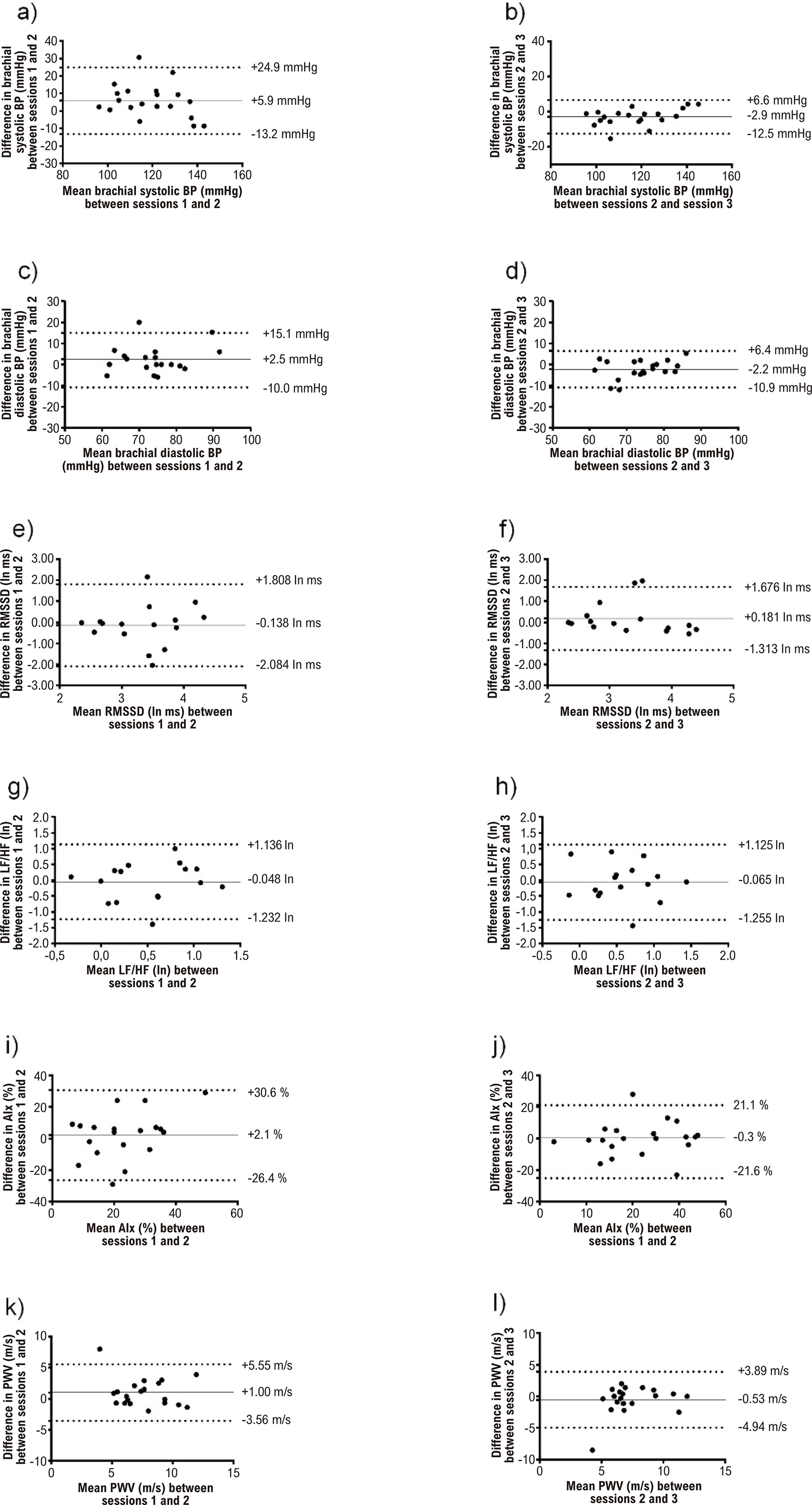
There was a significant main effect of the session (p<0.05) for brachial and central systolic BP. For both brachial and central systolic BP, post-hoc analysis revealed greater values in visit 1 when compared to visits 2 and 3. There were no significant differences for brachial and central diastolic BP (p>0.05) or central pulse pressure (p>0.05) among the experimental visits. ICCs ranged from 0.725 (central pulse pressure) to 0.921 (brachial systolic BP), and SEM% from 2.5% (brachial systolic BP) to 18.0% (central pulse pressure) (Table 2). Bland–Altman plots indicated low agreement between visits 1 and 2 and good agreement between visits 2 and 3 (Figure 1).

– Bland and Altman plots (systematic bias ± limits of agreement) for individual values of brachial systolic blood pressure, brachial diastolic blood pressure, root mean square of successive differences between normal heartbeats (RMSSD), cardiac sympathovagal balance (LF/HF), augmentation index (AIx) and pulse wave velocity (PWV). Ln: natural logarithm.
Arterial stiffness
There were no significant differences among experimental visits in the mean values of any arterial stiffness variable (p>0.05). ICCs ranged from 0.781 (augmentation index) to 0.886 (pulse wave velocity), and SEM% from 7.3% (pulse wave velocity) to 25.2% (augmentation index) (Table 2). Bland–Altman plots indicated moderate to good agreement among visits for the augmentation index and pulse wave velocity parameters, respectively (Figure 1).
Heart rate variability
There were no significant differences among experimental visits in the mean values of any variable (p>0.05). The ICCs ranged from 0.568 (normalized HFR-R) to 0.854 (ln HFR-R in absolute units) and SEM% from 5.1% (R-R interval) to 76.0% (LF/HFR-R) (Table 2). Bland–Altman plots indicated moderate agreement among visits for the majority of HRV parameters, except for LF/HFR-R (Figure 1).
Discussion
The main findings of the current study were that, in PD patients, BP presented low intrarater reliability and agreement between visits 1 and 2 and good reliability and agreement between visits 2 and 3. In general, the intrarater reliability and agreement of arterial stiffness and HRV indices were acceptable.
Brachial and central BP measurements presented low intrarater reliability and agreement between visits 1 and 2. Both central and brachial systolic BP were elevated in the first experimental visit, indicating the presence of systematic bias. Clinical guidelines13,14 recommend the measurement of BP in two or more visits to determine resting BP in order to minimize intervenient factors, such as the office environment, BP measurement procedures, and others. This is aligned with our data performed in PD patients, which demonstrated decreases in BP from the first to the second visit and stabilization in this variable between the second and third visits. This result indicates that trials with PD patients should employ one familiarization session before experimental sessions in order to obtain stable systolic BP data.
Good reliability and agreement were previously reported for brachial15 and central BPs in healthy individuals.16 Thus, the current results suggest that although PD is associated with impaired BP reactivity (e.g., orthostatic hypotension and supine hypertension),1,17 this does not translate into low reliability and agreement of resting BP assessments after the second visit.
Moderate to good reliability and agreement were found for arterial stiffness assessments, with pulse wave velocity being the most reliable measurement. These results are in line with previous data obtained in healthy older adults.18 This result is relevant since pulse wave velocity has been considered a marker of cardiovascular risk.19,20 Thus, the present data indicate that PD does not impair intrarater reliability and agreement for arterial stiffness. Indeed, it remains controversial whether PD directly affects arterial stiffness outcomes.21,22
The HRV indices presented acceptable intrarater reliability and agreement, except for ln LF/HFR-R. Indeed, the SEM magnitude was similar across log-transformed variables; however, when SEM was considered in relation to the respective mean values, ln LF/HFR-R exhibited a very large SEM%. Additionally, some caution is also required when employing normalized HFR-R for tracking HRV changes in PD patients. Nevertheless, the present results are similar to those obtained by previous studies23-27 in individuals without PD, which suggests that PD does not affect HRV reliability and agreement. Thus, although PD patients present abnormal values in some HRV indices,28 the current results suggest that the between-day variability of these parameters does not seem to be augmented in relation to non-PD individuals.
It is important to discuss our MDD results in light of the effect sizes found in previous clinical trials with PD patients. Mixed results have been reported regarding the responsiveness of BP to clinical interventions, with positive results29 or no effects.30-33 DiFrancisco-Donoghue et al.(2019)29 found an increase in systolic BP of 38 mmHg 60 minutes after a nicotine gum intervention in PD patients who suffer from low BP. Another study34 assessed the acute cardiovascular effects of levodopa oral administration in moderate PD patients and reported a decrease in systolic BP of 19 mmHg. This effect is greater than the MDD determined in the current study for systolic BP (6.8 mmHg), indicating that true individual changes can be detected in PD patients. Few clinical trials have also employed HRV indices as outcomes, with reports of both positive results17 or no effects.31,35 The only study that found statistically significant positive results evaluated the effects of progressive resistance training and found changes of 14 nu in LF R-R (nu).17 This effect is slightly lower than the MDD (16 nu) found in the current study, suggesting that true changes in LF R-R (nu) can be detected in a reasonable number of individuals, as many patients in the study by Kanegusuku et al. (2017)17 showed changes ≥ 16 nu. In contrast, there is a lack of studies investigating arterial stiffness responsiveness to clinical interventions. Based on the current MDD results, future studies with PD patients should analyze the individual responses, examining which patients showed true changes after interventions (observed changes ≥ MDD), mainly in BP and HRV parameters. Studies are still needed regarding arterial stiffness responsiveness to clinical interventions.
The current results have significant implications for both research and clinical settings. SEM data can be utilized for calculating the sample sizes required for clinical trials,36 while MDD represents the smallest amount of change required between repeated tests to indicate a real change when evaluating individual responses to interventions.37 Therefore, the SEM and MDD values determined in the current study should be considered when conducting research or clinical evaluations in PD patients. Furthermore, it is recommended that PD patients perform one familiarization session on the measurement of BP in order to obtain stable systolic BP data.
Lastly, it is important to mention the limitations of the current study. Some caution is required before extrapolating the current findings to PD patients with other characteristics, such as women, patients in more severe PD stages, and those with cardiovascular comorbidities. Another limitation of the current study includes the lack of a comparative control group composed of a paired sample of non-PD individuals. On the other hand, it is important to highlight that the current study employed a comprehensive reproducibility analysis involving systematic bias, reliability, and agreement evaluations, as well as three experimental sessions.
Conclusion
In patients with PD, central and brachial BP assessments showed low intrarater reliability and agreement between visits 1 and 2 and good reliability and agreement between visits 2 and 3. Arterial stiffness assessments presented acceptable intrarater reliability and agreement, with pulse wave velocity being the most reliable index. HRV indices showed acceptable intrarater reliability and agreement, except LF/HFR-R.
Acknowledgments
We gratefully acknowledge the volunteers and the Brazil Parkinson Association.
References
-
1 Sharabi Y, Vatine GD, Ashkenazi A. Parkinson's Disease Outside the Brain: Targeting the Autonomic Nervous System. Lancet Neurol. 2021;20(10):868-76. doi: 10.1016/S1474-4422(21)00219-2.
» https://doi.org/10.1016/S1474-4422(21)00219-2 -
2 Kanegusuku H, Ritti-Dias RM, Barbosa PYI, Guelfi ETN, Okamoto E, Miranda CS, et al. Influence of Motor Impairment on Exercise Capacity and Quality of Life in Patients with Parkinson Disease. J Exerc Rehabil. 2021;17(4):241-6. doi: 10.12965/jer.2142290.145.
» https://doi.org/10.12965/jer.2142290.145 -
3 Nicoletti A, Luca A, Baschi R, Cicero CE, Mostile G, Davì M, et al. Vascular Risk Factors, White Matter Lesions and Cognitive Impairment in Parkinson's Disease: The PACOS Longitudinal Study. J Neurol. 2021;268(2):549-58. doi: 10.1007/s00415-020-10189-8.
» https://doi.org/10.1007/s00415-020-10189-8 -
4 Kim JS, Lee SH, Oh YS, Park JW, An JY, Choi HS, et al. Arterial Stiffness and Cardiovascular Autonomic Dysfunction in Patients with Parkinson's Disease. Neurodegener Dis. 2017;17(2-3):89-96. doi: 10.1159/000450613.
» https://doi.org/10.1159/000450613 -
5 Heimler B, Koren O, Inzelberg R, Rosenblum U, Hassin-Baer S, Zeilig G, et al. Heart-rate Variability as a New Marker for Freezing Predisposition in Parkinson's Disease. Parkinsonism Relat Disord. 2023;113:105476. doi: 10.1016/j.parkreldis.2023.105476.
» https://doi.org/10.1016/j.parkreldis.2023.105476 -
6 Terroba-Chambi C, Abulafia C, Vigo DE, Merello M. Heart Rate Variability and Mild Cognitive Impairment in Parkinson's Disease. Mov Disord. 2020;35(12):2354-5. doi: 10.1002/mds.28234.
» https://doi.org/10.1002/mds.28234 -
7 Kanegusuku H, Cucato GG, Longano P, Okamoto E, Piemonte MEP, Correia MA, et al. Acute Cardiovascular Responses to Self-selected Intensity Exercise in Parkinson's Disease. Int J Sports Med. 2022;43(2):177-82. doi: 10.1055/a-1529-6480.
» https://doi.org/10.1055/a-1529-6480 -
8 Kottner J, Audigé L, Brorson S, Donner A, Gajewski BJ, Hróbjartsson A, et al. Guidelines for Reporting Reliability and Agreement Studies (GRRAS) were Proposed. J Clin Epidemiol. 2011;64(1):96-106. doi: 10.1016/j.jclinepi.2010.03.002.
» https://doi.org/10.1016/j.jclinepi.2010.03.002 -
9 Siebenhofer A, Kemp C, Sutton A, Williams B. The Reproducibility of Central Aortic Blood Pressure Measurements in Healthy Subjects Using Applanation Tonometry and Sphygmocardiography. J Hum Hypertens. 1999;13(9):625-9. doi: 10.1038/sj.jhh.1000887.
» https://doi.org/10.1038/sj.jhh.1000887 -
10 van Bortel LM, Duprez D, Starmans-Kool MJ, Safar ME, Giannattasio C, Cockcroft J, et al. Clinical Applications of Arterial Stiffness, Task Force III: Recommendations for User Procedures. Am J Hypertens. 2002;15(5):445-52. doi: 10.1016/s0895-7061(01)02326-3.
» https://doi.org/10.1016/s0895-7061(01)02326-3 - 11 Heart Rate Variability. Standards of Measurement, Physiological Interpretation, and Clinical Use. Task Force of the European Society of Cardiology and the North American Society of Pacing and Electrophysiology. Eur Heart J. 1996;17(3):354-81.
-
12 Vet HC, Terwee CB, Knol DL, Bouter LM. When to Use Agreement versus Reliability Measures. J Clin Epidemiol. 2006;59(10):1033-9. doi: 10.1016/j.jclinepi.2005.10.015.
» https://doi.org/10.1016/j.jclinepi.2005.10.015 -
13 Feitosa ADM, Barroso WKS, Mion D Jr, Nobre F, Mota-Gomes MA, Jardim PCBV, et al. Brazilian Guidelines for In-office and Out-of-office Blood Pressure Measurement - 2023. Arq Bras Cardiol. 2024;121(4):e20240113. doi: 10.36660/abc.20240113.
» https://doi.org/10.36660/abc.20240113 -
14 Williams B, Mancia G, Spiering W, Rosei EA, Azizi M, Burnier M, et al. 2018 Practice Guidelines for the Management of Arterial Hypertension of the European Society of Hypertension and the European Society of Cardiology: ESH/ESC Task Force for the Management of Arterial Hypertension. J Hypertens. 2018;36(12):2284-309. doi: 10.1097/HJH.0000000000001961.
» https://doi.org/10.1097/HJH.0000000000001961 -
15 Stanforth PR, Gagnon J, Rice T, Bouchard C, Leon AS, Rao DC, et al. Reproducibility of Resting Blood Pressure and Heart Rate Measurements. The HERITAGE Family Study. Ann Epidemiol. 2000;10(5):271-7. doi: 10.1016/s1047-2797(00)00047-8.
» https://doi.org/10.1016/s1047-2797(00)00047-8 -
16 Mitchelmore A, Stoner L, Lambrick D, Jobson S, Faulkner J. Reliability of Oscillometric Central Blood Pressure and Central Systolic Loading in Individuals Over 50 Years: Effects of Posture and Fasting. Atherosclerosis. 2018;269:79-85. doi: 10.1016/j.atherosclerosis.2017.12.030.
» https://doi.org/10.1016/j.atherosclerosis.2017.12.030 -
17 Kanegusuku H, Silva-Batista C, Peçanha T, Nieuwboer A, Silva ND Jr, Costa LA, et al. Effects of Progressive Resistance Training on Cardiovascular Autonomic Regulation in Patients with Parkinson Disease: A Randomized Controlled Trial. Arch Phys Med Rehabil. 2017;98(11):2134-41. doi: 10.1016/j.apmr.2017.06.009.
» https://doi.org/10.1016/j.apmr.2017.06.009 -
18 Perissiou M, Bailey TG, Windsor M, Leicht AS, Golledge J, Askew CD. Reliability of Arterial Stiffness Indices at Rest and Following a Single Bout of Moderate-intensity Exercise in Older Adults. Clin Physiol Funct Imaging. 2019;39(1):42-50. doi: 10.1111/cpf.12537.
» https://doi.org/10.1111/cpf.12537 -
19 Lee GP, Kim HL. Incremental Value of the Measures of Arterial Stiffness in Cardiovascular Risk Assessment. Rev Cardiovasc Med. 2022;23(1):6. doi: 10.31083/j.rcm2301006.
» https://doi.org/10.31083/j.rcm2301006 -
20 Helleputte S, van Bortel L, Verbeke F, Roodt JO, Calders P, Lapauw B, et al. Arterial Stiffness in Patients with Type 1 Diabetes and Its Comparison to Cardiovascular Risk Evaluation Tools. Cardiovasc Diabetol. 2022;21(1):97. doi: 10.1186/s12933-022-01537-1.
» https://doi.org/10.1186/s12933-022-01537-1 -
21 Günaydin ZY, Özer FF, Karagöz A, Bektas O, Karatas MB, Vural A, et al. Evaluation of Cardiovascular Risk in Patients with Parkinson Disease Under Levodopa Treatment. J Geriatr Cardiol. 2016;13(1):75-80. doi: 10.11909/j.issn.1671-5411.2016.01.003.
» https://doi.org/10.11909/j.issn.1671-5411.2016.01.003 -
22 Balal M, Demirkiran M, Paydas S. Central Aortic Pressure and Arterial Stiffness in Parkinson's Disease: A Comparative Study. Parkinsons Dis. 2022;2022:6723950. doi: 10.1155/2022/6723950.
» https://doi.org/10.1155/2022/6723950 -
23 Pinna GD, Maestri R, Torunski A, Danilowicz-Szymanowicz L, Szwoch M, La Rovere MT, et al. Heart Rate Variability Measures: A Fresh Look at Reliability. Clin Sci. 2007;113(3):131-40. doi: 10.1042/CS20070055.
» https://doi.org/10.1042/CS20070055 -
24 Zöllei E, Csillik A, Rabi S, Gingl Z, Rudas L. Respiratory Effects on the Reproducibility of Cardiovascular Autonomic Parameters. Clin Physiol Funct Imaging. 2007;27(4):205-10. doi: 10.1111/j.1475-097X.2007.00736.x.
» https://doi.org/10.1111/j.1475-097X.2007.00736.x -
25 Parati G, Omboni S, Villani A, Glavina F, Castiglioni P, Di Rienzo M, et al. Reproducibility of Beat-by-beat Blood Pressure and Heart Rate Variability. Blood Press Monit. 2001;6(4):217-20. doi: 10.1097/00126097-200108000-00011.
» https://doi.org/10.1097/00126097-200108000-00011 -
26 Fecchio RY, Brito LC, Peçanha T, Forjaz CLM. Consistency of Hemodynamic and Autonomic Mechanisms Underlying Post-exercise Hypotension. J Hum Hypertens. 2021;35(11):1003-11. doi: 10.1038/s41371-020-00452-w.
» https://doi.org/10.1038/s41371-020-00452-w -
27 Sandercock GRH, Bromley PD, Brodie DA. The Reliability of Short-term Measurements of Heart Rate Variability. Int J Cardiol. 2005;103(3):238-47. doi: 10.1016/j.ijcard.2004.09.013.
» https://doi.org/10.1016/j.ijcard.2004.09.013 -
28 Heimrich KG, Lehmann T, Schlattmann P, Prell T. Heart Rate Variability Analyses in Parkinson's Disease: A Systematic Review and Meta-analysis. Brain Sci. 2021;11(8):959. doi: 10.3390/brainsci11080959.
» https://doi.org/10.3390/brainsci11080959 -
29 Di Francisco-Donoghue J, Jung MK, Leder A. Nicotine Gum as a Therapeutic Approach for Low Blood Pressure in Parkinson's Disease: A Randomized Pilot Study. Nicotine Tob Res. 2019;21(2):253-6. doi: 10.1093/ntr/ntx263.
» https://doi.org/10.1093/ntr/ntx263 -
30 Ridgel AL, Walter BL, Tatsuoka C, Walter EM, Colón-Zimmermann K, Welter E, et al. Enhanced Exercise Therapy in Parkinson's disease: A comparative effectiveness trial. J Sci Med Sport. 2016;19(1):12-7. doi: 10.1016/j.jsams.2015.01.005.
» https://doi.org/10.1016/j.jsams.2015.01.005 -
31 Fernandes B, Barbieri FA, Arthuso FZ, Silva FA, Moretto GF, Imaizumi LFI, et al. High-Intensity Interval versus Moderate-intensity Continuous Training in Individuals with Parkinson's Disease: Hemodynamic and Functional Adaptation. J Phys Act Health. 2020;17(1):85-91. doi: 10.1123/jpah.2018-0588.
» https://doi.org/10.1123/jpah.2018-0588 - 32 Ganesan M, Pal PK, Gupta A, Sathyaprabha TN. Treadmill Gait Training Improves Baroreflex Sensitivity in Parkinson's Disease. Clin Auton Res. 2014;24(3):111-8. doi: 10.1007/s10286-014-0236-z.
-
33 Petramfar P, Hajari F, Yousefi G, Azadi S, Hamedi A. Efficacy of Oral Administration of Licorice as an Adjunct Therapy on Improving the Symptoms of Patients with Parkinson's Disease, A Randomized Double Blinded Clinical Trial. J Ethnopharmacol. 2020;247:112226. doi: 10.1016/j.jep.2019.112226.
» https://doi.org/10.1016/j.jep.2019.112226 -
34 Noack C, Schroeder C, Heusser K, Lipp A. Cardiovascular Effects of Levodopa in Parkinson's Disease. Parkinsonism Relat Disord. 2014;20(8):815-8. doi: 10.1016/j.parkreldis.2014.04.007.
» https://doi.org/10.1016/j.parkreldis.2014.04.007 -
35 Erola T, Haapaniemi T, Heikkinen E, Huikuri H, Myllyä V. Subthalamic Nucleus Deep Brain Stimulation does not Alter Long-term Heart Rate Variability in Parkinson's Disease. Clin Auton Res. 2006;16(4):286-8. doi: 10.1007/s10286-006-0354-3.
» https://doi.org/10.1007/s10286-006-0354-3 -
36 Hopkins WG. Measures of Reliability in Sports Medicine and Science. Sports Med. 2000;30(1):1-15. doi: 10.2165/00007256-200030010-00001.
» https://doi.org/10.2165/00007256-200030010-00001 - 37 Swinton PA, Hemingway BS, Saunders B, Gualano B, Dolan E. A Statistical Framework to Interpret Individual Response to Intervention: Paving the Way for Personalized Nutrition and Exercise Prescription. Front Nutr. 2018;5:41. doi: 10.3389/fnut.2018.00041.