Endothelial Function by Flow-Mediated Dilation (FMD) in the Brachial Artery in Hypertensive Patients
Abstract
Background High blood pressure (BP) values have traditionally been associated with the risk of ischemic heart disease, stroke, chronic kidney disease, and early mortality. The brachial artery FMD after cuff deflation has become the standard parameter for quantifying endothelial function, being a useful surrogate outcome due to its non-invasiveness, close correlation with coronary endothelial function, and association with the incidence of long-term coronary events.
Objectives To test hypotheses of correlation between the FMD and several blood parameters and to compare parameters between altered and non-altered FMD groups, and between hypertensive patients in the resistant hypertension groups (RHTN and non-RHTN).
Methods Seventy-two volunteers from a referral hypertension outpatient clinic participated in this prospective cross-sectional study, in which several patient variables were compared between the altered FMD (n = 38) and non-altered FMD (n = 34) groups, and also between the RHTN (n = 49) and non-RHTN (n = 23) groups. The variables that would explain the FMD were also investigated in this study. Statistical analyses were performed using parametric methods when the assumptions were met, and non-parametric methods otherwise. The significance level adopted in the statistical analysis was 5%.
Results The results showed a significant positive correlation between the FMD and LDL (p = 0.204, p = 0.042) and between FMD and triglycerides (p = 0.247, p = 0.037). Glycated hemoglobin was higher in the RHTN group (p = 0.020), potassium was higher in the non-RHTN group (p = 0.029), and C-reactive protein was higher in the RHTN group (p = 0.04). For the other comparisons, no statistically significant differences were found.
Conclusion LDL and triglycerides are FMD predictors, and the RHTN and non-RHTN groups differ in terms of the amount of potassium, protein C, and glycated hemoglobin. The altered and non-altered FMD groups differ only in terms of triglycerides.
Brachial Artery; Vascular Endothelium; Hypertension

Resumo
Fundamento Valores elevados de pressão arterial (PA) têm sido tradicionalmente associados ao risco de doença isquêmica do coração, acidente vascular cerebral, doença renal crônica e mortalidade precoce. A dilatação mediada por fluxo (FMD) da artéria braquial após a desinsuflação do manguito tornou-se o parâmetro padrão para quantificar a função endotelial, sendo um desfecho substituto útil em função de sua não invasividade, estreita correlação com a função endotelial coronariana e associação com a incidência de eventos coronarianos em longo prazo.
Objetivos Testar hipóteses de correlação entre a FMD e diversos parâmetros sanguíneos e comparar os parâmetros entre grupos com a FMD alterada e não alterada e entre hipertensos dos grupos hipertensão arterial resistente (HAR e não HAR).
Métodos Setenta e dois voluntários de um ambulatório de referência em Hipertensão Arterial foram incluídos neste estudo transversal prospectivo, no qual foram comparadas diversas variáveis mensuradas nos pacientes, entre os grupos FMD alterada (n = 38) e não alterada (n = 34), e também entre os grupos HAR (n = 49) e não HAR (n = 23). Investigamos também quais variáveis explicariam a FMD. As análises estatísticas foram conduzidas por meio de métodos paramétricos quando os pressupostos foram atendidos e não paramétricos quando estes não foram atendidos. O nível de significância adotado nas análises estatísticas foi de 5%.
Resultados Os resultados mostraram uma correlação positiva significativa entre FMD e LDL (p = 0,204, p = 0,042) e entre FMD e triglicerídeos (p = 0,247, p = 0,037). Hemoglobina glicada foi maior no grupo HAR (p = 0,020), potássio foi maior no grupo não HAR (p = 0,029) e proteína C reativa foi maior no grupo HAR (p = 0,04). Não houve diferenças estatísticas significativas para as demais comparações.
Conclusões O LDL e os triglicerídeos são preditores da FMD e os grupos HAR e não HAR diferem quanto à quantidade de potássio, proteína C e hemoglobina glicada. Os grupos de FMD alterada e não alterada diferem somente em relação aos triglicerídeos.
Artéria Braquial; Endotélio Vascular; Hipertensão
Introduction
High blood pressure (BP) values have traditionally been associated with the risk of ischemic heart disease, stroke, chronic kidney disease, and early mortality.1 Resistant hypertension (RHTN) is defined as office BP, remaining ≥ 140/90 mmHg despite treatment with three or more antihypertensive drugs that have synergistic actions at the maximum recommended or tolerated doses. At least one of these drugs should preferably be a thiazide diuretic. When BP control is achieved with four or more antihypertensive drugs, the patient is classified as resistant but controlled hypertensive (BP < 140/90 mmHg).1-4 In Brazil, the multicenter ReHOT (Resistant Hypertension Optimal Treatment) study found a prevalence of 11.7% for this comorbidity.5
The primary function of the arterial system is to “efficiently distribute blood to peripheral organs and maintain vascular homeostasis”.3,6 Endothelial dysfunction is the first functional alteration detectable in the atherosclerotic process.7 This is due to a decreased bioavailability of nitric oxide found not only in patients with clinically evident atherosclerotic disease, but also in patients with risk factors.8 It is an early manifestation of both atherosclerotic disease and type 2 diabetes mellitus (DM2), and its attenuation may occur soon after the initiation of therapies with antiatherosclerotic effects.2,4
In 1992, Celejamer et al.9 developed the flow-mediated dilation (FMD), a noninvasive method to assess early changes in vascular function in systemic arteries. The brachial artery FMD after cuff deflation has become the standard parameter for quantifying endothelial function, being a useful surrogate outcome due to its non-invasiveness, close correlation with coronary endothelial function, and association with the incidence of long-term coronary events.7 Numerous factors can potentially confound the measurement of FMD.10Besides, there is no consensus on aspects such as the location and time of cuff compression, which alters the quantitative result of the examination.11
The objective of this study was to test hypotheses of correlation between the FMD and several blood parameters and to compare parameters between groups with altered and non-altered FMD, and between hypertensive patients in the resistant hypertension groups (RHTN and non-RHTN).
Methods
Data collection
Seventy-two volunteers from an Arterial Hypertension referral outpatient clinic were included in this prospective cross-sectional study. The patients were informed about the preparation for the examination in advance.
The clinical and demographic characteristics of the patients were collected from electronic medical records. The local ethics committee approved the study (CAE # 81701717.6.0000.0049 and Opinion # 2.635.984), and written informed consent was obtained from all patients before the examinations.
Right brachial artery flow-mediated dilation (FMD) examination
During the examination, vasodilation in the brachial artery is assessed and occurs in response to the significantly increased blood flow, as induced by a period of circulatory occlusion. Reactive hyperemia is induced by the rapid release of a pneumatic pressure cuff placed around the forearm and inflated to suprasystolic pressure for five minutes. This procedure increases shear stress along the vessel in a parallel and laminar manner, activating mechanoreceptors in endothelial cells and promoting NO release.12,14
The pressure exerted on the arm causes vascular ischemia and subsequent dilation of the vessels.15 After five minutes, the deflation control valve is slowly opened. Once it reaches zero on the device, the posterior diameter (D2) is measured. Another 60 seconds are then allowed before applying the formula (D2 – D1)/D1 × 100, where values of 10% or less indicate alterations, as established by Regattieri et al.16 This calculation provides the values for determining FMD, which is the focus of this study.
All exams were conducted by the same volunteer physician, a specialist in ultrasound for 20 years. During the procedure, patients were also offered the opportunity to undergo carotid and vertebral Doppler (c/v), following the methodology outlined in the 2017 Brazilian Guideline for Dyslipidemia.17 These exams were always performed before measuring the FMD. The laboratory markers used for the study were: total cholesterol, HDL-cholesterol, LDL-cholesterol, glycated hemoglobin (HbAc1), uric acid, and C-reactive protein (CRP), as collected from electronic medical records.18
Statistical analysis
Data were compiled and analyzed using SPSS® software (version 25.0, Chicago, IL Statistical Package for the Social Sciences). In all tests, an alpha significance level of 5% was applied. Categorical variables were expressed as frequency and percentage, and continuous variables were expressed as mean and standard deviation. The chi-square test was used to test associations between categorical factors. When comparing the correlation pattern between quantitative and ordinal variables, the Spearman’s correlation analysis was used, as it better captures positive or negative correlation patterns without the need for perfect linearity. When comparing continuous variables between the RHTN (resistant and non-resistant) and FMD (altered and unaltered) groups, the t-test for independent samples was used. In all tests, normality was tested by the Shapiro Wilk test and homogeneity of variances was tested by the Levene test. Data did not deviate from normality and showed homogeneous variances in all comparisons. Linear regression analyses were also performed between LDL and FMD, and between triglycerides and FMD, with the assumptions of homoscedasticity and normality of the residues tested.
Results
For objective representation, the Central Illustration illustrates the data in a clear and practical manner.
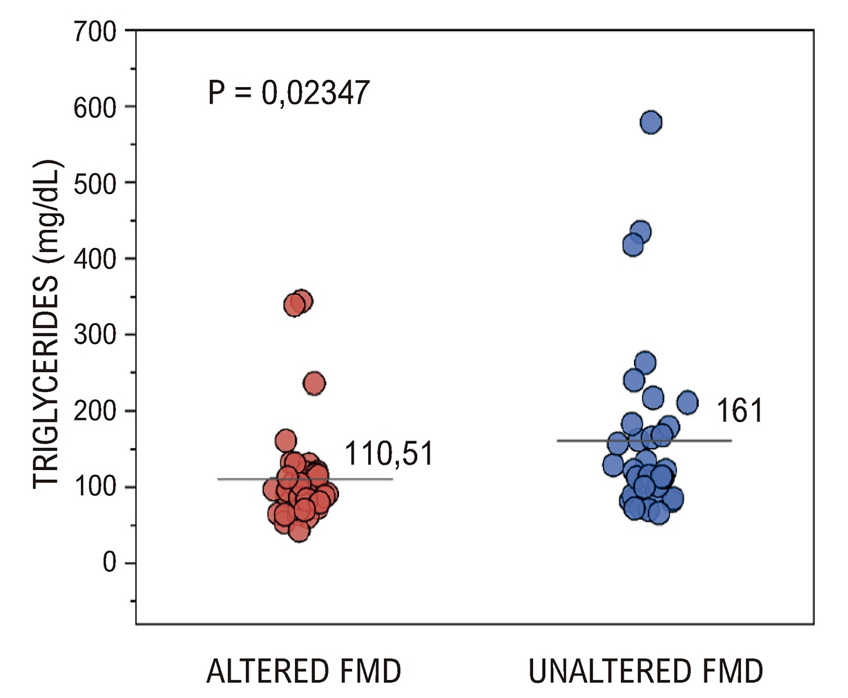
Initially, several variables between the altered and unaltered FMD groups were compared (Table 1). Significant differences can be noted for triglycerides (p = 0.023), which was higher in the unaltered FMD group (Figure 1).

– T-test comparing the amount of triglycerides between the altered and non-altered FMD groups.
When comparing the blood pressure groups, a higher median value was found for the non-RHTN group (43.9) compared to the RHTN group (33), p = 0.039. In addition, the correlations between FMD and all quantitative variables of the study were tested (Table 2).
When analyzing the descriptive statistics of our sample, the maximum value obtained in the altered FMD group was 11.32, with a mean of 5.25, standard deviation of 3.43 and the minimum value was zero. Unaltered FMD presents a much greater variation (mean of 18.2 and standard deviation of 7.43), with a minimum value of 10.14 and a maximum value of 44.72 (Table 3). When taking the maximum FMD group value as a cutoff point, this value would be 11.32. However, studies designed specifically for this issue of the cutoff point are recommended to further clarify this issue.
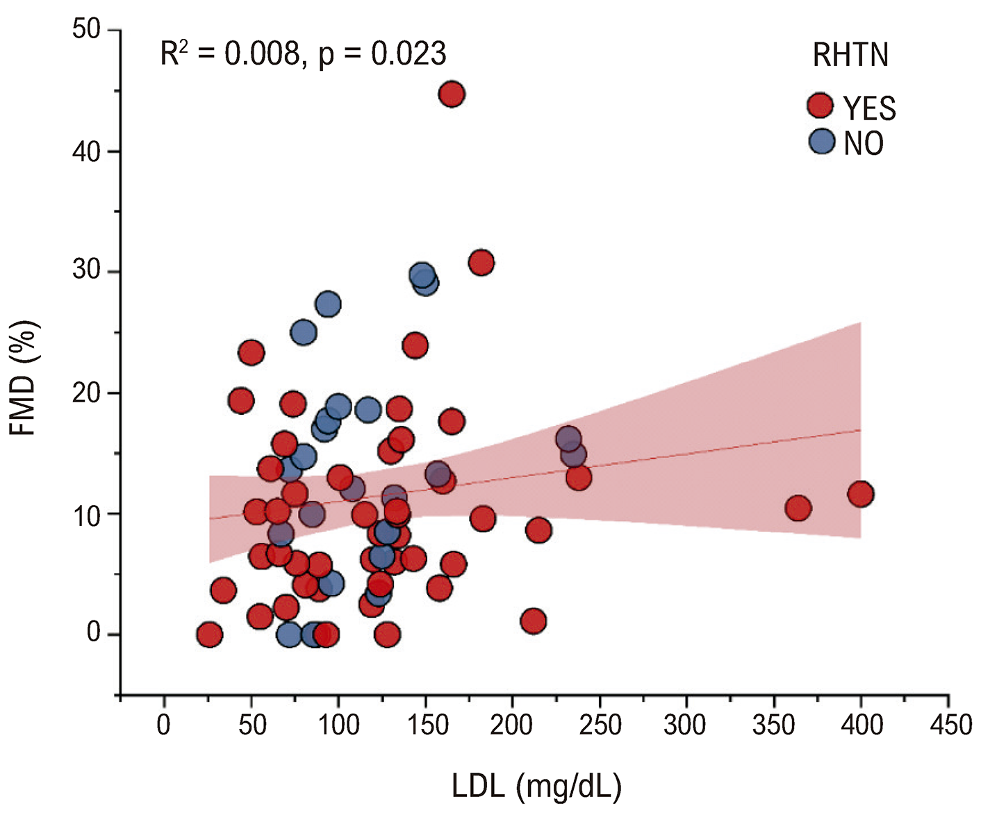
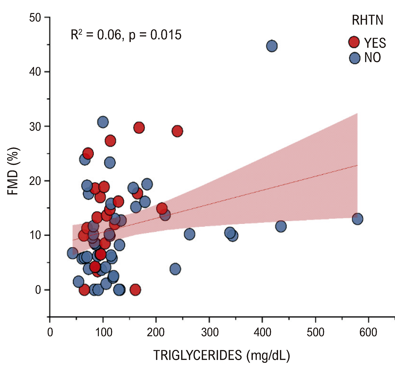
A significant positive correlation between FMD and LDL and between FMD and triglycerides was found. Regression analyses were conducted for the two significant relationships above (Figures 1 and 2).
The higher the LDL values, the higher the FMD values. LDL correctly predicts and explains 0.8% of the variation in FMD (Figure 2).

– Linear regression between FMD and LDL. FMD: flow-mediated dilation; RHTN: resistant hypertension.
Table 4 shows the association between the categorical factors and the RHTN and non-RHTN groups.
The use of vasodilators is more associated with RHTN patients (0.006). The comparison of the LDL means between the groups that use and do not use statins showed no significant differences (p = 0.336). Table 5 presents comparisons of several parameters between the RHTN and non-RHTN groups.
Significant differences were found for glycated hemoglobin (HbAc1) (Figure 4), potassium (Figure 5), and C-reactive protein (p = 0.02; 0.029 and 0.04, respectively) (Figure 6). Patients with RHTN have higher HbAc1 and Protein C values, while the non-RHTN group has higher potassium values.


– Comparison for C-reactive protein between RHTN and non-RHTN groups. Significant differences were found (p = 0.04), and C-reactive protein has a higher mean value in the RHTN group. RHTN: resistant hypertension; CRP: C-reactive protein. Source: Authors.
Significant differences were found (p = 0.0201), and the glycated hemoglobin mean value is higher in the RHTN group.
Significant differences were found (p = 0.029), and potassium has a higher mean value in the RHTN group.
Significant differences were found (p = 0.04), and C-reactive protein has a higher mean value in the RHTN group.
Discussion
Endothelial dysfunction is a functional alteration observed in the atherosclerotic process, resulting from reduced bioavailability of nitric oxide. Identifying the factors that contribute to endothelial dysfunction remains a challenge in health research. This condition is not only found in patients with clinically evident atherosclerotic disease but also in those with risk factors. High blood pressure affects individuals of various ages, lifestyles, and health conditions.
A key challenge in correlational studies between these two areas is identifying significant associations between continuous variables and categorical factors that help explain the risk of developing these conditions. Two major obstacles arise: first, the absence of a controlled experimental design in most cases, making it difficult to confirm associations; second, the typically low effect size of the association between risk factors and endothelial dysfunction, which complicates detection in small samples.
This study aimed to correlate FMD and hypertension (both resistant and non-resistant) with various categorical and continuous factors. Many expected associations were statistically non-significant (where one would expect them to be significant), while some unexpected correlations were significant, occasionally with an opposite sign. Each will be explained in detail in the following paragraphs.
The mean triglyceride levels were compared between altered and unaltered FMD groups, revealing a surprising pattern opposite to expectations. Mean triglycerides were higher in patients without altered FMD compared to those with altered FMD, representing a significance p of 0.015 (Figure 3). Kaplangoray et al.19 demonstrated that low dilation is associated with higher levels of lipids in the bloodstream, just as did the studies by Holewijn et al.20 and Fernandes.21

– Linear regression between FMD and triglycerides. FMD: flow-mediated dilation; RHTN: resistant hypertension.
When multiple statistical tests yield results that contradict well-established theoretical frameworks in the line of research, as is the case here, it is crucial to consider effect sizes and the number of statistical tests performed adopt correction criteria that minimize the risk of type 1 errors in interpreting p-values, and consider the sample size in relation to studies with opposite findings.22-24Considering these factors, despite the observed significant differences, the effect size—represented by the t statistic—was quite small (approximately 2.31), and the p-value was not even below 0.01. This weakens its reliability for rejecting the null hypothesis.
Regarding resistant and non-resistant RHTN and quantitative variables, two significant results were found in the comparisons between RHTN and HbAc1 (Figure 4) and between non-RHTN and potassium (Figure 5). As for the result of glycated hemoglobin, again the result contradicts what was theoretically expected. Glycated hemoglobin is higher in the RHTN group than in the non-RHTN group, with a very small difference between the mean values and the p-value of 0.021, based on data from 69 patients. This result contradicts the literature. Shimizu et al.25in a study performed in Japan, found a significant negative relationship between HbAc1 levels and blood pressure, and Ghost et al.26found that a lower amount of glycated hemoglobin leads to higher blood pressure levels. Therefore, Bonferroni’s rigorous criterion was used to qualify such opposite correlation found as non-significant (only p-values lower than 0.0031 would be significant).
For potassium, a significant result was found, in accordance with theoretical expectations, that is, the higher the potassium level, the lower the blood pressure, as found by Fonseca et al.27 According to Santos & Vasconcelos,28 a diet rich in potassium is highly recommended for hypertensive patients, since this nutrient is associated with a reduction in cardiovascular diseases and blood pressure levels. Applying the same level of scrutiny, the effect size remained small, with only a subtle difference between means (0.29). Given the number of statistical tests performed and the limited sample size of just 68 individuals for this comparison, these results should be interpreted with caution, even though they align with scientific literature predictions.
Regarding the association between medications and RHTN and non-RHTN, significant associations consistent with the literature were found. The first is the association between resistant hypertensive patients and the vasodilator variable (17 to 1), where, by definition, people with RHTN are more associated with the use of vasodilators, as they are very useful in the treatment of several medical conditions, including hypertension.29
One of such vasodilators, clonidine, was more frequently associated with altered hypertension, as expected. As a central agonist that reduces sympathetic tone, clonidine can be highly effective in managing hypertensive crises, including postoperative situations.30 Spironolactone was also more frequently associated with altered hypertension individuals, which was anticipated. Experimental evidence has demonstrated that spironolactone significantly reduces both systolic and diastolic blood pressure in hypertensive patients.31
One of the challenges in understanding which factors affect FMD is a definitive cutoff point, with theoretical scientific and statistical support, which defines a value from which it is possible to safely classify whether a given group of patients has altered FMD. This classification does not yet exist in the literature.
Few studies present this technology for determining brachial artery FMD. Therefore, the dilation represented by FMD, although seemingly easy to implement, has shown gaps in its real-world results. Such gaps include subtle postural changes, transducer tilt, measurements in systole or diastole, variations in the measurement site, quality of the image, and the diameter of the brachial artery. Arteries with less than 3.0 mm are more prone to errors, since a variation of 5% would be close to the detection limit. In addition, several external factors can influence the results of the exam, including exam duration, caffeine intake, inadequate fasting, environmental noise, patient discomfort, and the examiner’s expertise.
In this study, the FMD values of the right brachial artery presented values that were not very similar to the endovascular reality of the sample profile. A key consideration was the four-hour fasting period. Additionally, ensuring rigorous technique and employing technologies that accurately measure vessel diameter are essential for refining and standardizing the methodology. These improvements could enhance future studies and establish the technique as a practical tool for cardiovascular risk prevention.
Conclusion
Impaired endothelial function in hypertensive patients serves as a predictor for certain clinical variables and shows a weak correlation with LDL and triglycerides. The altered and non-altered FMD groups differ only in terms of triglycerides. The hypertensive groups (RHTN and non-RHTN) differ in terms of potassium, C-reactive protein, and glycated hemoglobin. The study has the limitation of not being an experimental study and also of lacking a large sample. Low-intensity effects are more reliably detected in large sample sizes. This study included fewer than 40 patients per group, yet, some effects were still observed. These findings suggest that future studies with larger samples may uncover stronger correlations, as theoretically predicted.
References
-
1 Lewington S, Clarke R, Qizilbash N, Peto R, Collins R; Prospective Studies Collaboration. Age-Specific Relevance of Usual Blood Pressure to Vascular Mortality: A Meta-Analysis of Individual Data for One Million Adults in 61 Prospective Studies. Lancet. 2002;360(9349):1903-13. doi: 10.1016/s0140-6736(02)11911-8.
» https://doi.org/10.1016/s0140-6736(02)11911-8 -
2 Krieger EM, Drager LF, Giorgi DMA, Pereira AC, Barreto-Filho JAS, Nogueira AR, et al. Spironolactone versus Clonidine as a Fourth-Drug Therapy for Resistant Hypertension: The ReHOT Randomized Study (Resistant Hypertension Optimal Treatment). Hypertension. 2018;71(4):681-90. doi: 10.1161/HYPERTENSIONAHA.117.10662.
» https://doi.org/10.1161/HYPERTENSIONAHA.117.10662 -
3 Barroso WKS, Rodrigues CIS, Bortolotto LA, Mota-Gomes MA, Brandão AA, Feitosa ADM, et al. Brazilian Guidelines of Hypertension - 2020. Arq Bras Cardiol. 2021;116(3):516-658. doi: 10.36660/abc.20201238.
» https://doi.org/10.36660/abc.20201238 -
4 Yugar-Toledo JC, Moreno H Jr, Gus M, Rosito GBA, Scala LCN, Muxfeldt ES, et al. Brazilian Position Statement on Resistant Hypertension - 2020. Arq Bras Cardiol. 2020;114(3):576-96. doi: 10.36660/abc.20200198.
» https://doi.org/10.36660/abc.20200198 -
5 Carey RM, Calhoun DA, Bakris GL, Brook RD, Daugherty SL, Dennison-Himmelfarb CR, et al. Resistant Hypertension: Detection, Evaluation, and Management: A Scientific Statement from the American Heart Association. Hypertension. 2018;72(5):e53-e90. doi: 10.1161/HYP.0000000000000084.
» https://doi.org/10.1161/HYP.0000000000000084 -
6 Tomiyama H, Yamashina A. Non-Invasive Vascular Function Tests: Their Pathophysiological Background and Clinical Application. Circ J. 2010;74(1):24-33. doi: 10.1253/circj.cj-09-0534.
» https://doi.org/10.1253/circj.cj-09-0534 -
7 Gottdiener JS, Reda DJ, Massie BM, Materson BJ, Williams DW, Anderson RJ. Effect of Single-Drug Therapy on Reduction of Left Ventricular Mass in Mild to Moderate Hypertension: Comparison of Six Antihypertensive Agents. The Department of Veterans Affairs Cooperative Study Group on Antihypertensive Agents. Circulation. 1997;95(8):2007-14. doi: 10.1161/01.cir.95.8.2007.
» https://doi.org/10.1161/01.cir.95.8.2007 -
8 Hansson L, Lindholm LH, Niskanen L, Lanke J, Hedner T, Niklason A, et al. Effect of Angiotensin-Converting-Enzyme Inhibition Compared with Conventional Therapy on Cardiovascular Morbidity and Mortality in Hypertension: The Captopril Prevention Project (CAPPP) Randomised Trial. Lancet. 1999;353(9153):611-6. doi: 10.1016/s0140-6736(98)05012-0.
» https://doi.org/10.1016/s0140-6736(98)05012-0 -
9 Celermajer DS, Sorensen KE, Gooch VM, Spiegelhalter DJ, Miller OI, Sullivan ID, et al. Non-Invasive Detection of Endothelial Dysfunction in Children and Adults at Risk of Atherosclerosis. Lancet. 1992;340(8828):1111-5. Doi: 10.1016/0140-6736(92)93147-f.
» https://doi.org/10.1016/0140-6736(92)93147-f -
10 Tomiyama H, Shiina K. State of the Art Review: Brachial-Ankle PWV. J Atheroscler Thromb. 2020;27(7):621-36. doi: 10.5551/jat.RV17041.
» https://doi.org/10.5551/jat.RV17041 -
11 Bots ML, Westerink J, Rabelink TJ, Koning EJ. Assessment of Flow-Mediated Vasodilatation (FMD) of the Brachial Artery: Effects of Technical Aspects of the FMD Measurement on the FMD Response. Eur Heart J. 2005;26(4):363-8. doi: 10.1093/eurheartj/ehi017.
» https://doi.org/10.1093/eurheartj/ehi017 -
12 Corretti MC, Anderson TJ, Benjamin EJ, Celermajer D, Charbonneau F, Creager MA, et al. Guidelines for the Ultrasound Assessment of Endothelial-Dependent Flow-Mediated Vasodilation of the Brachial Artery: A Report of the International Brachial Artery Reactivity Task Force. J Am Coll Cardiol. 2002;39(2):257-65. doi: 10.1016/s0735-1097(01)01746-6.
» https://doi.org/10.1016/s0735-1097(01)01746-6 -
13 Harris RA, Nishiyama SK, Wray DW, Richardson RS. Ultrasound Assessment of Flow-Mediated Dilation. Hypertension. 2010;55(5):1075-85. doi: 10.1161/HYPERTENSIONAHA.110.150821.
» https://doi.org/10.1161/HYPERTENSIONAHA.110.150821 -
14 Thijssen DHJ, Black M, Pyke KE, Padilla J, Atkinson G, Harris R, et al. Assessment of Flow-Mediated Dilation in Humans: A Methodological and Physiological Guideline. Am J Physiol Heart Circ Physiol. 2010;300(1):H2-H12. doi: 10.1152/ajpheart.00471.2010.
» https://doi.org/10.1152/ajpheart.00471.2010 -
15 Betik AC, Luckham VB, Hughson RL. Flow-Mediated Dilation in Human Brachial Artery After Different Circulatory Occlusion Conditions. Am J Physiol Heart Circ Physiol. 2004;286(1):H442-8. doi: 10.1152/ajpheart.00314.2003.
» https://doi.org/10.1152/ajpheart.00314.2003 - 16 Regattieri NAT, Leite SP, Koch HA, Montenegro CAB. Dilatação Fluxo-Mediada da Artéria Braquial: Desenvolvimento da Técnica, Estudo em Pacientes de Risco para Aterosclerose e em um Grupo Controle. Rev Bras Ultrason. 2006;9:9-13.
-
17 Faludi AA, Izar MCO, Saraiva JFK, Chacra APM, Bianco HT, Afiune A Neto, et al. Atualização da Diretriz Brasileira de Dislipidemias e Prevenção da Aterosclerose - 2017. Arq Bras Cardiol. 2017;109(2 Suppl 1):1-76. doi: 10.5935/abc.20170121.
» https://doi.org/10.5935/abc.20170121 -
18 Nascimento CRD, Ribeiro JLM, Mendes R, Barbosa RHA, Lopes JM, Tenório PP. The Relationship between Uric Acid/Albumin Ratio and Carotid Intima-Media Thickness in Patients with HypertensionReply. Arq Bras Cardiol. 2024;121(1):e20230691. doi: 10.36660/abc.20230691.
» https://doi.org/10.36660/abc.20230691 -
19 Kaplangoray M, Toprak K, Basanalan F, Palice A, Aydin C, Demirkiran A, et al. Investigation of the Relationship between Triglycerides-Glucose Index and Coronary Slow Flow: A Retrospective Case-Control Study. Arq Bras Cardiol. 2023;120(6):e20220679. doi: 10.36660/abc.20220679.
» https://doi.org/10.36660/abc.20220679 -
20 Holewijn S, Graaf J, den Heijer M, Stalenhoef AFH. Endothelial Dysfunction as Measured by Flow-Mediated Dilation (FMD) in Cardiovascular Risk Assessment. Artery Res. 2007;66(1). doi: 10.1016/j.artres.2007.07.115.
» https://doi.org/10.1016/j.artres.2007.07.115 - 21 Fernandes VO. Early Endothelial Dysfunction in Patients with Generalized Congenital Lipodystrophy (Berardinelli-Seip Syndrome) Assessed by Flow-Mediated Dilatation of the Brachial Artery and Peripheral Arterial Tonomy [dissertation]. Fortaleza: Universidade Federal do Ceará; 2015.
-
22 Nuzzo R. Scientific Method: Statistical Errors. Nature. 2014;506(7487):150-2. doi: 10.1038/506150a.
» https://doi.org/10.1038/506150a -
23 Cabin R, Mitchell, R. To Bonferroni or Not to Bonferroni: When and How are the Questions. Bull Ecol Soc Am. 2000;81(3):246-8. doi:10.2307/20168454.
» https://doi.org/10.2307/20168454 - 24 Leite AO, Sposito AC. Relação entre os Níveis de Lipoproteína de Alta Densidade (HDL) e a Função Endotelial Avaliada por Dilatação Mediada por Fluxo (FMD) em Diabéticos. Proceedings of the 29th Congresso de Iniciação Científica Unicamp; 2021. Campinas: Unicamp; 2021.
-
25 Shimizu Y, Nakazato M, Sekita T, Kadota K, Arima K, Yamasaki H, et al. Association between the Hemoglobin Levels and Hypertension in Relation to the BMI Status in a Rural Japanese Population: The Nagasaki Islands Study. Intern Med. 2014;53(5):435-40. doi: 10.2169/internalmedicine.53.1353.
» https://doi.org/10.2169/internalmedicine.53.1353 -
26 Ghosh T, Rehman T, Ahamed F. Relationship between Hemoglobin and Blood Pressure Levels in the Context of Chronic Morbidity Among Older Adults Residing in a Developing Country: A Community-Level Comparative Cross-Sectional Study. Cureus. 2021;13(11):e19540. doi: 10.7759/cureus.19540.
» https://doi.org/10.7759/cureus.19540 - 27 Fonseca HAR, Zamit TP, Machado VA. Relationship between Dietary Potassium and Blood Pressure. Rev Bras Hipertens. 2015;22(1):9-15.
- 28 Santos TMP, Vasconcelos SML. Ingestão de Na+ e K+ versus HAS: Bases para seu Manejo e Protocolo de Pesquisa. Rev Bras Hipertens. 2012;19(2):55-5.
- 29 Hariri L, Patel JB. Vasodilators. Treasure Island: StatPearls Publishing; 2023.
- 30 Stocche RM, Kamt JG, Garcia LV. Clonidina Intravenosa no Controle da Hipertensão Arterial Perioperatória em Cirurgias de Catarata. Retrospectiva do Estudo. Rev Bras Anestesiol. 2020;50(4):289-93.
-
Study association:
This article is part of the thesis of master submitted by Elaine Alves Santos Tessier, from Programa de Pós-graduação em medicina e saúde da Universidade Federal da Bahia (UFBA).
-
Ethics approval and consent to participate:
This study was approved by the Ethics Committee of the Hospital Universitário Prof. Edgard Santos - UFBA under the protocol number 2.635.984. All the procedures in this study were in accordance with the 1975 Helsinki Declaration, updated in 2013. Informed consent was obtained from all participants included in the study.
-
Sources of funding:
This study was funded by CNPq.
Edited by
-
Editor responsible for the review:
Paulo B. Veiga Jardim