Operative Experience and Clinical Outcomes in Cardiac Myxoma Patients: A Retrospective Single-Center Study
Abstract
Background Cardiac myxoma is the second most common primary cardiac tumor; however, contemporary single-center series integrating operative strategy with clinical outcomes remain limited.
Objectives To describe the operative approaches and clinical outcomes of patients undergoing surgical resection of cardiac myxomas at a single center.
Methods We conducted a retrospective review of 74 consecutive patients who underwent surgical resection of histologically confirmed cardiac myxomas between 2014 and 2024 at a tertiary referral center in Brazil. Clinical, imaging, surgical, and follow-up data were collected. The primary outcome was the incidence of thromboembolic events. A significance level of 5% was adopted for all statistical analyses.
Results The median age was 60 years (range 52-66), and 68.9% of patients were female. Median follow-up was 60 months (interquartile range 40-79). Thromboembolic events occurred in 14 patients (18.9%), including stroke (13.5%), embolic myocardial infarction (2.7%), and pulmonary embolism (2.7%). In multivariable Cox analysis, diabetes was independently associated with a lower hazard of thromboembolic events (HR 0.11; 95% CI 0.006-0.740; p < 0.05), whereas postoperative atrial fibrillation was associated with a higher thromboembolic risk (HR 6.74; 95% CI 1.490-30.510; p < 0.05). Tumor recurrence occurred in 2.7% of cases. Only one death was recorded during follow-up.
Conclusion Cardiac myxomas are associated with significant thromboembolic and hemodynamic risks. Early surgical resection provides excellent long-term outcomes and should be considered even in asymptomatic patients. In our cohort, tumor morphology showed a non-significant trend toward a higher risk of thromboembolic events.
Keywords:
Mixoma; Doenças Cardiovasculares; Surgery; Latin America
Highlights/Key points
•Cardiac myxomas are the second most common primary cardiac tumors, and surgical resection remains the treatment of choice.
•In this single-center study of 74 patients, operative outcomes were excellent, with a low recurrence rate (2.7%) and high long-term survival (98.6%).
•Thromboembolic events occurred in 18.9% of patients, with diabetes associated with a lower hazard of these events.
•These findings support the safety and long-term effectiveness of surgical resection for cardiac myxomas, even with conservative use of interatrial patch reconstruction.
Resumo
Fundamento O mixoma cardíaco é o segundo tumor cardíaco primário mais comum; no entanto, séries contemporâneas realizadas em centro único que integrem estratégia operatória e desfechos clínicos ainda são limitadas.
Objetivos Descrever as abordagens operatórias e os desfechos clínicos de pacientes submetidos à ressecção cirúrgica de mixomas cardíacos em um único centro.
Métodos Realizamos uma revisão retrospectiva de 74 pacientes consecutivos submetidos à ressecção cirúrgica de mixomas cardíacos com confirmação histológica entre 2014 e 2024 em um centro terciário de referência no Brasil. Foram coletados dados clínicos, de imagem, cirúrgicos e de seguimento. O desfecho primário foi a incidência de eventos tromboembólicos. Foi adotado um nível de significância de 5% para todas as análises estatísticas.
Resultados A idade mediana foi de 60 anos (intervalo 52-66), e 68,9% dos pacientes eram do sexo feminino. O seguimento mediano foi de 60 meses (intervalo interquartil 40-79). Eventos tromboembólicos ocorreram em 14 pacientes (18,9%), incluindo acidente vascular encefálico (13,5%), infarto do miocárdio embólico (2,7%) e embolia pulmonar (2,7%). Na análise multivariada de Cox, o diabetes foi independentemente associado a menor risco de eventos tromboembólicos (HR 0,11; intervalo de confiança de 95% [IC 95%] 0,006-0,740; p < 0,05), enquanto a fibrilação atrial pós-operatória foi associada a maior risco tromboembólico (HR 6,74; IC 95% 1,490-30,510; p < 0,05). Recorrência tumoral ocorreu em 2,7% dos casos. Apenas um óbito foi registrado durante o seguimento.
Conclusão Os mixomas cardíacos estão associados a riscos tromboembólicos e hemodinâmicos significativos. A ressecção cirúrgica precoce proporciona excelentes desfechos em longo prazo e deve ser considerada mesmo em pacientes assintomáticos. Em nossa coorte, a morfologia tumoral apresentou uma tendência não significativa para maior risco de eventos tromboembólicos.
Palavras-chave:
Mixoma; Cardiovascular Disease; Cirurgia; América Latina
Destaques/Pontos-chave
• Os mixomas cardíacos são os segundos tumores cardíacos primários mais comuns, e a ressecção cirúrgica permanece o tratamento de escolha.
• Neste estudo de centro único com 74 pacientes, os desfechos operatórios foram excelentes, com baixa taxa de recorrência (2,7%) e elevada sobrevida em longo prazo (98,6%).
• Eventos tromboembólicos ocorreram em 18,9% dos pacientes, com o diabetes associado a menor risco desses eventos.
• Esses achados sustentam a segurança e a eficácia em longo prazo da ressecção cirúrgica dos mixomas cardíacos, mesmo com uso conservador da reconstrução com patch interatrial.
Introduction
Cardiac myxomas are the second most common primary cardiac tumors, after papillary fibroelastomas; however, they remain the most frequent symptomatic and surgically treated cardiac neoplasms, accounting for the majority of resected benign cardiac tumors in clinical practice.1 Despite their benign histology, their intracardiac location can lead to substantial morbidity, including obstruction, embolic events, and systemic manifestations.2 With an estimated annual incidence of 0.5-1 case per million, these rare tumors occur predominantly in women between the third and sixth decades of life and are most commonly located in the left atrium (LA).2,3 Surgical resection is the treatment of choice and is associated with favorable outcomes, although recurrence remains a concern, particularly in familial cases.4,5
In Latin America, especially in Brazil, cardiac myxomas account for approximately 70%-75% of benign cardiac tumors reported in surgical series. However, their overall population incidence remains low, at approximately 0.5-1 case per million per year. Therefore, although rare in the general population, they represent the most common surgically treated cardiac neoplasm in the region.6
This study presents clinical and surgical data on cardiac myxomas from a Latin American perspective, where available evidence remains limited. By examining recurrence patterns and predictors of adverse outcomes, we aim to provide region-specific insights that may improve risk assessment and inform the long-term management of affected patients.
Methods
Study design and ethical approval
This retrospective study analyzed the medical records of patients diagnosed with cardiac myxomas and treated at a tertiary hospital in Brazil between 2014 and 2024. The study protocol was approved by the Institutional Review Board of the same institution (Protocol SDC-COP 26501, approved on January 12, 2025). All data were fully anonymized to ensure patient confidentiality in accordance with ethical standards.
Study population
Eligible participants were adult patients (≥ 18 years) with a confirmed diagnosis of intracardiac myxoma based on imaging studies, who underwent surgical resection followed by histopathological confirmation, and who were followed in the institution’s outpatient clinics for at least 1 month. Patients aged > 45 years or those with cardiovascular risk factors underwent standard preoperative institutional evaluation, including assessment of coronary artery patency to minimize the risk of perioperative myocardial infarction. Inclusion required complete medical documentation, including clinical notes and complementary test results, available in the institutional electronic medical record system.
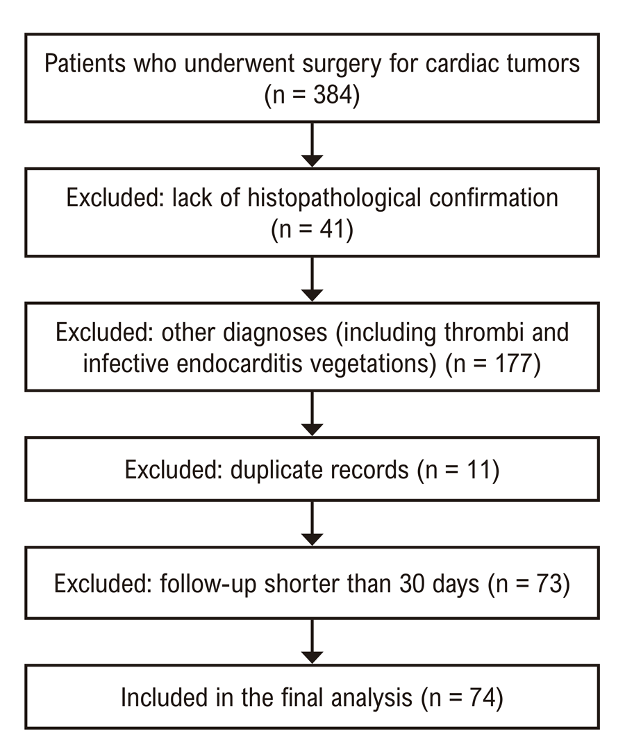
Exclusion criteria comprised duplicate records, incomplete medical documentation, surgery performed at another institution, follow-up shorter than 30 days, and unconfirmed diagnosis of intracardiac myxoma.
Data collection and variables
Follow-up data were obtained exclusively from the institutional electronic medical record, with no direct patient contact. The following variables were collected: i) demographic and clinical characteristics: age, sex, race, and comorbidities, such as diabetes mellitus, hypertension, smoking history, dyslipidemia, and peripheral arterial disease; ii) presentation and diagnostic data: initial symptoms and the imaging modality used for diagnosis; iii) surgical data: year of surgical intervention, surgical technique, and the need for additional cardiac procedures; iv) echocardiographic parameters (recorded preoperatively and postoperatively using standardized protocols performed by certified echocardiographers): left ventricular ejection fraction (LVEF), LA diameter, left and right ventricular (RV) dimensions, RV function, and pulmonary artery systolic pressure (PASP); v) tumor characteristics: size, morphology, surface features, mobility, degree of obstruction, and anatomical location; vi) complications and outcomes: thromboembolic events, tumor recurrence, and major adverse cardiovascular events.
Major cardiovascular events during follow-up were identified through a comprehensive review of clinical records, hospital discharge summaries, and, when applicable, official death certificates. To ensure data accuracy and consistency, all collected information was independently reviewed by two researchers. Discrepancies were resolved by consensus or adjudicated by a third researcher.
Thromboembolic events were defined as any clinically documented thromboembolic episode confirmed by imaging or surgical findings, including ischemic stroke, transient ischemic attack, systemic embolism, or peripheral arterial occlusion attributable to tumor fragments or thrombus. Events were further classified according to timing as preoperative, early postoperative, or late postoperative.
Statistical analysis
Continuous variables were expressed as median and interquartile range, as none followed a normal distribution according to the Kolmogorov–Smirnov test. Categorical variables were summarized as absolute frequencies and percentages.
Comparisons between groups were performed using the Mann–Whitney U test for continuous variables and the chi-square test or Fisher’s exact test for categorical variables, as appropriate. Paired comparisons of preoperative and postoperative echocardiographic parameters were evaluated using the Wilcoxon signed-rank test. Event-free survival for thromboembolic outcomes was assessed using the Kaplan–Meier method, with curves compared descriptively.
To identify predictors of thromboembolic events, univariate Cox proportional hazards regression analysis was initially performed. Variables with a p-value < 0.10 were entered into the multivariable model to determine independent associations.
All statistical analyses were conducted using IBM SPSS Statistics for Windows, version 27.0 (IBM Corp., Armonk, N.Y., USA). A two-tailed p-value < 0.05 was considered statistically significant.
Results
Study population and baseline characteristics
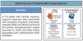
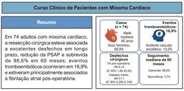
The study sample consisted of 74 patients diagnosed with cardiac myxoma according to the predefined inclusion and exclusion criteria (Diagram 1). The main findings are summarized in Central Illustration.

– Flowchart illustrating the selection process of patients included in the study. Most patients with follow-up shorter than 30 days were referred back to their originating hospitals after the first postoperative consultation.
Baseline demographic and clinical characteristics are presented in Table 1. The cohort was composed predominantly of middle-aged women, with frequent cardiovascular comorbidities, particularly hypertension and dyslipidemia. Among patients with concomitant neoplasms, thyroid and gastrointestinal tumors were the most common. Two patients had confirmed Carney complex, both with positive genetic testing identifying PRKAR1A gene mutations.
Clinical presentation
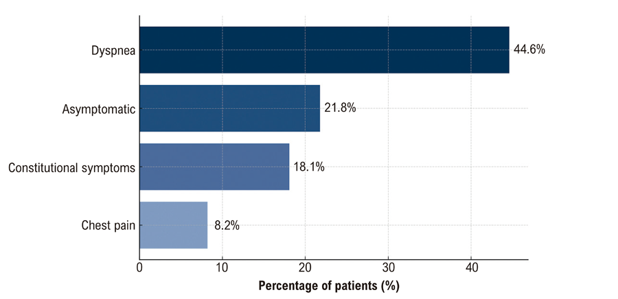
Dyspnea was the most common presenting symptom, reflecting the obstructive nature of cardiac myxomas, particularly those located in the LA. Notably, 21.8% of patients were asymptomatic (Figure 1), with tumors identified incidentally on imaging. Constitutional symptoms, including fever, weight loss, anemia, and asthenia, were documented in 18.1% of cases. Additional reported symptoms included chest pain and palpitations, possibly related to thromboembolic events or arrhythmias. Furthermore, 36.5% of tumors exhibited an obstructive pattern, contributing to exertional symptoms.
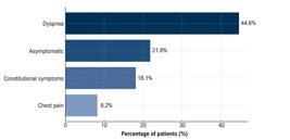
– Presenting symptoms in patients with cardiac myxoma. Distribution of the main clinical manifestations at initial presentation.
Surgical procedures and operative strategy
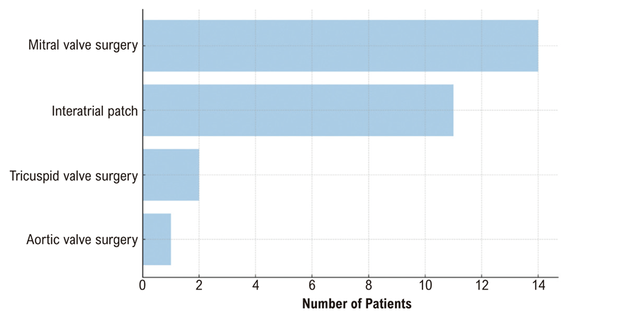
Among the 74 patients, mitral valve surgery was the most frequent associated procedure (Figure 2). In 12 cases, intervention was required due to direct myxoma involvement of the valve leaflets, while in two patients the procedure was performed opportunistically. Other associated procedures included interatrial patch repair, tricuspid valve surgery, and aortic valve surgery.

All patients underwent median sternotomy. LA myxomas were approached either through a right atrial (RA) transseptal route via the fossa ovalis when septal involvement or broad-based implantation was suspected, allowing en bloc excision with a cuff of interatrial septum followed by patch closure, or through direct left atriotomy for pedunculated or free-wall lesions. Concomitant valve procedures were performed when clinically indicated.
Echocardiographic findings
Preoperatively, most patients had preserved biventricular ejection fraction and normal left ventricular diastolic diameter. LA dilation was observed in 41.9% of cases, and elevated PASP was present in 20.3% of the cohort (Table 2).
Postoperatively, most patients maintained preserved biventricular ejection fraction, although the proportion with severe reductions in LVEF increased by 12.8%. Normal LA size increased by 10.8%, and normalization of PASP was observed in 6% of patients (Table 2).
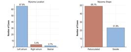
Myxomas were predominantly located in the LA (Figure 3), generally with a smooth surface, and had median dimensions of 45 × 35 mm (Table 2). A Wilcoxon signed-rank test (Figure 4) demonstrated statistically significant differences between preoperative and postoperative measurements for both LVEF and PASP. Specifically, both parameters showed significant reductions, with PASP decreasing primarily in patients with obstructive myxomas.
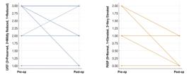
– Comparison of preoperative and postoperative echocardiographic parameters. Comparison of LVEF and PASP demonstrating a significant postoperative reduction in PASP and a transient decline in LVEF. LVEF: left ventricular ejection fraction. PASP: pulmonary artery systolic pressure.
Complications and clinical outcomes
Thromboembolic events were observed in 18.9% of patients, with ischemic stroke as the most frequent manifestation. All stroke cases occurred in patients with preoperative atrial fibrillation. Events occurred in 8.1% preoperatively and in 5.4% postoperatively (Table 3). Less frequent complications included atrial fibrillation, obstructive shock, and superior vena cava syndrome (Figure 5).

The mortality rate was low, with only one death recorded during a median follow-up period of 60 months. Event-free survival was further illustrated using a Kaplan–Meier curve, which demonstrated a progressive decline in freedom from thromboembolic events over time (Figure 6).

– Kaplan–Meier curve for thromboembolic event-free survival. Event-free survival up to 60 months, showing that most patients remained free from thromboembolic events during follow-up.
Tumor recurrence
Myxoma recurrence was uncommon. Both recurrent cases involved multiple episodes (three recurrences each) and were associated with Carney complex. One case involved an 18-year-old female with three left ventricular recurrences, and the other a 44-year-old male with sequential involvement of the LA, RA, and eventually a biatrial recurrence.
Comparative analysis according to thromboembolic events
Patients who experienced thromboembolic events (n = 14) were compared with those who did not (n = 60) using chi-square or Fisher’s exact tests for categorical variables and the Mann–Whitney U test for continuous variables due to non-normal distribution (Figures 7 and 8). Age was similar between groups (median: 57.5 vs. 60.5 years). Diabetes mellitus was significantly more prevalent in the event-free group (7.1% vs. 38.3%).

– Boxplots of clinical variables according to thromboembolic event status. Distribution of age and tumor size stratified by the occurrence of thromboembolic events.

– Comparison of clinical and tumor characteristics by thromboembolic event status. AF: atrial fibrillation.
Other factors, including tumor size, location, and echocardiographic parameters (LVEF, LA dilation, and PASP), showed no statistically significant differences between groups (Table 4). These findings are consistent with the Cox proportional hazards model results presented in Table 5 and Table 6, in which diabetes and atrial fibrillation emerged as independent predictors of embolic events.
Predictors of thromboembolic events
The multivariable Cox regression model (Table 6) showed that LA dilation was associated with a lower, although non-significant, risk of thromboembolic events. Diabetes and atrial fibrillation were significantly associated with the hazard of thromboembolic events (HR = 0.11; 95% CI: 0.006-0.740; p < 0.05). Mobile tumors showed a trend toward a higher risk of thromboembolism, although this increase was not statistically significant.
Univariate or multivariate analyses of myxoma recurrence and mortality were not performed due to the small number of events. A focused analysis was conducted to explore variables associated with an increased risk of thromboembolic events. Univariate analysis identified diabetes as a potential protective factor, contrasting with its established role in increasing thromboembolic risk in the general population. This finding suggests that the myxoma itself may be the primary driver of thromboembolic risk in this population, supporting the rationale for early surgical removal.
Discussion
This study reinforces the critical role of early surgical intervention in the management of cardiac myxomas and highlights its association with excellent long-term outcomes.7 In this cohort of 74 patients, predominantly with sporadic tumors, the median follow-up was 60 months, during which survival reached 98.6% and tumor recurrence remained low at 2.7%. These findings are consistent with previous reports demonstrating the effectiveness of prompt resection in reducing mortality and recurrence, particularly when complete excision of the tumor and its attachment is achieved.8,9
The lower frequency of constitutional symptoms observed in our cohort contrasts with the higher prevalence reported in other series. This difference may be explained by referral patterns, as many patients were evaluated at our center at more advanced stages of disease, when obstructive and embolic manifestations may predominate over systemic symptoms.10-12
All procedures in our cohort were performed via median sternotomy, which remains the standard approach at our institution to ensure adequate exposure and complete tumor excision. Minimally invasive and robotic techniques, such as those using the DaVinci Xi® system, were not available during the study period. However, these approaches have been associated with shorter recovery times and improved postoperative quality of life in selected centers.13,14
The interatrial septum, particularly the fossa ovalis, is recognized as the most common site of origin for cardiac myxomas. En bloc resection with removal of a portion of the interatrial septum followed by patch reconstruction is often recommended to minimize recurrence. In our cohort, only 14.9% of patients underwent interatrial patch reconstruction. This relatively low rate reflects a tailored, real-world surgical strategy at our tertiary center, where operative decisions were guided by tumor size, attachment morphology, and intraoperative findings.15-18
In many cases, small pedunculated tumors allowed for complete excision with a narrow margin and primary septal closure, particularly in older or high-risk patients.18-20
The incidence of thromboembolic events in our cohort (18.9%), primarily ischemic stroke (13.5%), is comparable to rates reported in prior studies. Stefanou et al. (2018) described a stroke rate of 11% among patients with cardiac myxomas, while Pinede et al. (2001) reported thromboembolic events in approximately 30% of cases. Importantly, our multivariable Cox regression analysis did not identify tumor location or age as significant predictors of thromboembolic risk. Tumor mobility showed a trend toward increased risk, although this association did not reach statistical significance, likely due to the limited number of events. This observation is consistent with the meta-analysis by Liu et al. (2020), which highlighted the lack of consistent morphological predictors across studies.3,21,22
Postoperative atrial fibrillation emerged as an independent predictor of thromboembolic events in our cohort, with more than a sixfold increase in hazard. This finding reinforces the well-established association between new-onset atrial fibrillation and heightened embolic risk following cardiac surgery and underscores the importance of close rhythm monitoring and timely anticoagulation in this population.23,24
In the multivariable model, diabetes mellitus was associated with a lower hazard of thromboembolic events. This observation has not been consistently reported in previous studies and should be interpreted cautiously. It is likely exploratory and potentially confounded, rather than indicative of a true protective biological effect.25-27 Possible explanations include closer clinical monitoring, more frequent use of antithrombotic or statin therapy, or unmeasured differences in cardiovascular risk management among diabetic patients. Because of the small number of events and the retrospective design, this finding should be considered hypothesis-generating and requires confirmation in larger prospective studies.
The cohort demonstrated a significant postoperative reduction in PASP (Wilcoxon Z = −2.070; p = 0.038), consistent with relief of LA inflow obstruction, particularly in patients with obstructive myxomas.
Conversely, LVEF declined in the early postoperative period (preserved LVEF 93.2% preoperatively vs 78.9% postoperatively; severe reduction 2.7% vs 15.5%; Wilcoxon Z = 3.052; p = 0.002). This pattern likely reflects transient perioperative ventricular dysfunction rather than a sustained deterioration in systolic function, especially considering that postoperative echocardiograms were performed within 30 days of surgery. These findings suggest that surgery promptly alleviates hemodynamic burden, while recovery of ventricular function may occur more gradually and warrants longitudinal echocardiographic follow-up.
Mikus et al. (2025) reported preserved LVEF in the postoperative period, indicating that the changes observed in our cohort may represent early, transient alterations rather than long-term impairment.14
Lastly, the survival rate observed in our study (98.6% over a median follow-up of 60 months) compares favorably with global surgical series, including the 93%-97% long-term survival reported by Jiang et al. (2019) and Perek et al. (2011). These results reinforce the long-term safety and effectiveness of surgical treatment for cardiac myxomas when performed in specialized centers.7,9,28
Study limitations
The retrospective, single-center design and the relatively small sample size may limit the generalizability of these findings. Additionally, due to the long data collection period inherent to retrospective studies, certain potentially relevant variables, such as tumor morphology and surface characteristics, which may be associated with mobility and thromboembolic risk, were not consistently documented.
Furthermore, the observed postoperative reduction in LVEF may partly reflect perioperative myocardial injury or infarction, which was not systematically assessed or recorded in our cohort, limiting the interpretation of this finding.
Conclusion
Our study reinforces that cardiac myxoma, when treated promptly, is associated with an excellent prognosis. At the same time, it highlights the significant thromboembolic risk related to this condition, which appears to be largely independent of tumor morphology. These findings support early surgical resection, even in asymptomatic patients and those with non-obstructive lesions. Continuous follow-up remains essential to monitor for late complications and potential recurrence.
References
-
1 Maleszewski JJ, Basso C, Bois MC, Glass C, Klarich KW, Leduc C, et al. The 2021 WHO Classification of Tumors of the Heart. J Thorac Oncol. 2022;17(4):510-8. doi: 10.1016/j.jtho.2021.10.021.
» https://doi.org/10.1016/j.jtho.2021.10.021 -
2 Nektaria M, Theologou S, Christos C, George S, Rokeia E, Dimitrios S, et al. Cardiac Myxomas: A Single-Center Case Series of 145 Patients Over a 32-Year Period Study. Ann Card Anaesth. 2023;26(1):17-22. doi: 10.4103/aca.aca_290_20.
» https://doi.org/10.4103/aca.aca_290_20 -
3 Stefanou MI, Rath D, Stadler V, Richter H, Hennersdorf F, Lausberg HF, et al. Cardiac Myxoma and Cerebrovascular Events: A Retrospective Cohort Study. Front Neurol. 2018;9:823. doi: 10.3389/fneur.2018.00823.
» https://doi.org/10.3389/fneur.2018.00823 -
4 Oktaviono YH, Saputra PBT, Arnindita JN, Afgriyuspita LS, Kurniawan RB, Pasahari D, et al. Clinical Characteristics and Surgical Outcomes of Cardiac Myxoma: A Meta-Analysis of Worldwide Experience. Eur J Surg Oncol. 2024;50(2):107940. doi: 10.1016/j.ejso.2023.107940.
» https://doi.org/10.1016/j.ejso.2023.107940 -
5 Bjessmo S, Ivert T. Cardiac Myxoma: 40 Years' Experience in 63 Patients. Ann Thorac Surg. 1997;63(3):697-700. doi: 10.1016/s0003-4975(96)01368-9.
» https://doi.org/10.1016/s0003-4975(96)01368-9 -
6 Dias RR, Fernandes F, Ramires FJA, Mady C, Albuquerque CP, Jatene FB. Mortality and Embolic Potential of Cardiac Tumors. Arq Bras Cardiol. 2014;103(1):13-8. doi: 10.5935/abc.20140096.
» https://doi.org/10.5935/abc.20140096 - 7 Perek B, Misterski M, Stefaniak S, Ligowski M, Puslecki M, Jemielity M. Early and Long-Term Outcome of Surgery for Cardiac Myxoma: Experience of a Single Cardiac Surgical centre. Kardiol Pol. 2011;69(6):558-64.
-
8 Islam AKMM. Cardiac Myxomas: A Narrative Review. World J Cardiol. 2022;14(4):206-19. doi: 10.4330/wjc.v14.i4.206.
» https://doi.org/10.4330/wjc.v14.i4.206 -
9 Jiang CX, Wang JG, Qi RD, Wang W, Gao LJ, Zhao JH, et al. Long-Term Outcome of Patients with Atrial Myxoma after Surgical Intervention: Analysis of 403 Cases. J Geriatr Cardiol. 2019;16(4):338-43. doi: 10.11909/j.issn.1671-5411.2019.04.003.
» https://doi.org/10.11909/j.issn.1671-5411.2019.04.003 -
10 Okongwu CC, Olaofe OO. Cardiac Myxoma: A Comprehensive Review. J Cardiothorac Surg. 2025;20(1):151. doi: 10.1186/s13019-024-03333-2.
» https://doi.org/10.1186/s13019-024-03333-2 -
11 Duman U, Furat C, Keskin G, Kahraman D, Hafiz E. Outcomes of Patients with Cardiac Myxoma: A Retrospective Multicentre Study. Open Cardiovasc Med J. 2019;13:24-31. doi:10.2174/1874192401913010024.
» https://doi.org/10.2174/1874192401913010024 -
12 Nagre SW, Bhosle KN. Cardiac Myxomas - Symptomology, Investigations and Surgical Treatment - Single Centre Experience of Fifteen Years at Grant Medical College, Mumbai, Maharashtra. J Cardiovasc Med Cardiol. 2018;5(2):1-6. doi:10.17352/2455-2976.000062.
» https://doi.org/10.17352/2455-2976.000062 -
13 Santos K, Albuquerque LG, Consoli L, Gamarra-Valverde N, Abdelmajeed A, Plonek T. Robotic-Assisted Versus Conventional Median Sternotomy for the Surgical Excision of Cardiac Myxomas: A Systematic Review and Meta-Analysis. J Cardiothorac Surg. 2025;33:24. doi:10.1186/s43057-025-00169-x.
» https://doi.org/10.1186/s43057-025-00169-x -
14 Mikus E, Fiorentino M, Sangiorgi D, Costantino A, Calvi S, Tenti E, et al. Comparative Analysis of Surgical Outcomes in Cardiac Myxoma Resection: Sternotomy Versus Right Mini-Thoracotomy. Cardiovasc Innov Appl. 2025;(1):1-11. doi:10.15212/CVIA.2025.0015.
» https://doi.org/10.15212/CVIA.2025.0015 -
15 Rahouma M, Arisha MJ, Elmously A, Ahmed MME, Spadaccio C, Mehta K, et al. Cardiac Tumors Prevalence and Mortality: A Systematic Review and Meta-Analysis. Int J Surg. 2020;76:178-89. doi: 10.1016/j.ijsu.2020.02.039.
» https://doi.org/10.1016/j.ijsu.2020.02.039 - 16 Nguyen T, Vaidya Y. Atrial Myxoma. Treasure Island: StatPearls Publishing; 2025.
-
17 Berdajs DA, Ferrari E. Surgical Treatment for Heart Myxomas. Multimed Man Cardiothorac Surg. 2012;2012:mms016. doi: 10.1093/mmcts/mms016.
» https://doi.org/10.1093/mmcts/mms016 -
18 Alhasso AA, Ahmed OF, Mohammed-Saeed DH, Kakamad FH, Almodhaffer SS, Zaid ZA, et al. Operative Management and Outcomes in Patients with Myxomas: A Single-Center Experience. Front Surg. 2023;10:1084447. doi: 10.3389/fsurg.2023.1084447.
» https://doi.org/10.3389/fsurg.2023.1084447 -
19 MacGillivray TE, Reardon MJ. Surgical Treatment of Benign Cardiac Tumors. Oper Tech Thorac Cardiovasc Surg. 2021;26(4):212-8. doi:10.1053/j.optechstcvs.2020.12.006.
» https://doi.org/10.1053/j.optechstcvs.2020.12.006 -
20 Ashinze P, Banerjee S, Egbunu E, Salawu W, Idris-Agbabiaka A, Obafemi E, et al. Cardiac Myxomas: A Review of Current Treatment Approaches and Emerging Molecular Therapies. J Cardiothorac Surg. 2024;32:22. doi:10.1186/s43057-024-00141-1.
» https://doi.org/10.1186/s43057-024-00141-1 -
21 Pinede L, Duhaut P, Loire R. Clinical Presentation of Left Atrial Cardiac Myxoma. A series of 112 Consecutive Cases. Medicine. 2001;80(3):159-72. doi: 10.1097/00005792-200105000-00002.
» https://doi.org/10.1097/00005792-200105000-00002 -
22 He DK, Zhang YF, Liang Y, Ye SX, Wang C, Kang B, et al. Risk Factors for Embolism in Cardiac Myxoma: A Retrospective Analysis. Med Sci Monit. 2015;21:1146-54. doi: 10.12659/MSM.893855.
» https://doi.org/10.12659/MSM.893855 -
23 Suero OR, Ali AK, Barron LR, Segar MW, Moon MR, Chatterjee S. Postoperative Atrial Fibrillation (POAF) after Cardiac Surgery: Clinical Practice Review. J Thorac Dis. 2024;16(2):1503-20. doi: 10.21037/jtd-23-1626.
» https://doi.org/10.21037/jtd-23-1626 -
24 Woldendorp K, Farag J, Khadra S, Black D, Robinson B, Bannon P. Postoperative Atrial Fibrillation after Cardiac Surgery: A Meta-Analysis. Ann Thorac Surg. 2021;112(6):2084-93. doi: 10.1016/j.athoracsur.2020.10.055.
» https://doi.org/10.1016/j.athoracsur.2020.10.055 -
25 Qureshi MA, Bakht D, Ahmed O, Haseeb S, Gupta K, Baqal O, et al. Evaluating Risk Factors of Embolism in Patients with Cardiac Myxoma: A Systematic Review and Meta-Analysis. Am Heart J Plus. 2025;56:100559. doi: 10.1016/j.ahjo.2025.100559.
» https://doi.org/10.1016/j.ahjo.2025.100559 -
26 He DK, Zhang YF, Liang Y, Ye SX, Wang C, Kang B, et al. Risk Factors for Embolism in Cardiac Myxoma: A Retrospective Analysis. Med Sci Monit. 2015;21:1146-54. doi: 10.12659/MSM.893855.
» https://doi.org/10.12659/MSM.893855 -
27 Liu Y, Wang J, Guo L, Ping L. Risk Factors of Embolism for the Cardiac Myxoma Patients: A Systematic Review and Metanalysis. BMC Cardiovasc Disord. 2020;20(1):348. doi: 10.1186/s12872-020-01631-w.
» https://doi.org/10.1186/s12872-020-01631-w -
28 Qiao ML, Ma L, Wang CB, Fang LB, Fan ZX, Niu TT, et al. Clinical Features, Risk Factors and Survival in Cardiac Myxoma-Related Ischemic Stroke: A Multicenter Case-Control Study. J Neurol Sci. 2023;444:120517. doi: 10.1016/j.jns.2022.120517.
» https://doi.org/10.1016/j.jns.2022.120517
-
Ethics statement:
This study was conducted in accordance with the ethical standards of the Institutional Review Board (Protocol SDC-COP 26501). All data were anonymized to ensure patient confidentiality, and the requirement for informed consent was waived due to the retrospective design.
-
Study association:
This study is not associated with any thesis or dissertation work.
-
Ethics approval and consent to participate:
This study was approved by the Ethics Committee of the Hospital das Clínicas da Faculdade de Medicina da Universidade de São Paulo (HCFM/USP) under the protocol number CAAE 86890725.3.0000.0068. PARECER NR 7.497.978. All the procedures in this study were in accordance with the 1975 Helsinki Declaration, updated in 2013.
-
Use of Artificial Intelligence:
The authors did not use any artificial intelligence tools in the development of this work.
-
Data Availability Statement:
All datasets supporting the results of this study are available upon request from the corresponding author.
-
Sources of funding:
There were no external funding sources for this study.
Edited by
-
Editor responsible for the review:
Henrique Ribeiro
All datasets supporting the results of this study are available upon request from the corresponding author.

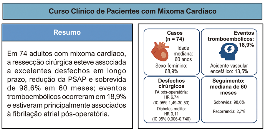 DM: diabetes melito; IC: intervalo de confiança; FA: fibrilação atrial; HR: razão de risco; PSAP: pressão sistólica da artéria pulmonar.
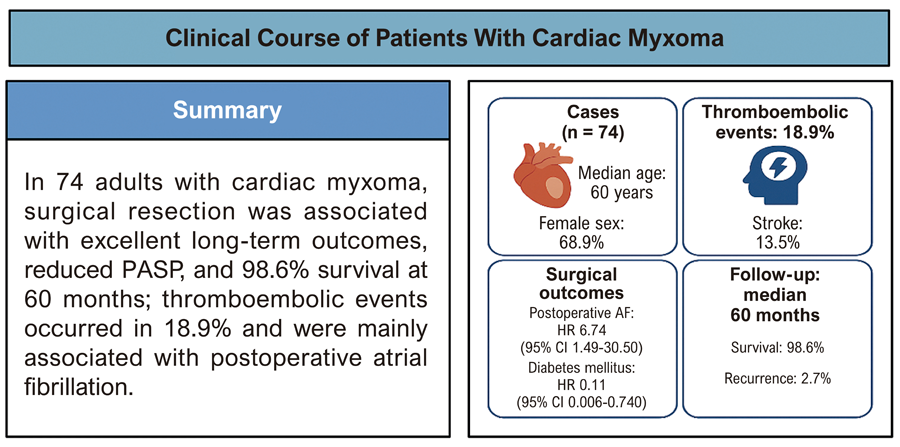 AF: atrial fibrillation; DM: diabetes mellitus; HR: hazard ratio; PASP: pulmonary artery systolic pressure.