PRKAG2 Cardiomyopathy: A Case-Control Study on the Diagnostic Yield Of Histopathology and Ultrastructural Analysis from Endomyocardial Biopsy
Abstract
Background The histopathological features of PRKAG2 cardiomyopathy have been reported in a fragmentary manner.
Objective We aimed to systematically evaluate the cardiac pathological features of PRKAG2 cardiomyopathy in a large patient cohort and assess their diagnostic potential compared to genetic sequencing.
Methods We conducted an observational, cross-sectional, case-control study including 18 patients with PRKAG2 cardiomyopathy and 11 heart transplant recipients as controls. All patients underwent percutaneous right ventricular endomyocardial biopsy. Tissue samples were analyzed using H&E staining, Periodic Acid-Schiff staining for glycogen, Masson’s trichrome for fibrosis, and ultrastructural assessment by transmission electron microscopy. Statistical significance was set at p < 0.05 for all analyses.
Results PRKAG2 cardiomyopathy hearts exhibited significant cardiomyocyte enlargement, normal-appearing mitochondria, extensive vacuolization of most myofibers, minimal interstitial fibrosis (only two patients had mild fibrosis), and no inflammatory cell infiltration. Transmission electron microscopy revealed abundant cytosolic glycogen, primarily in the perinuclear region, with additional deposits in intermyofibrillar and subsarcolemmal areas. This pronounced glycogen accumulation, consistently observed in all PRKAG2 patients, was absent in controls.
Conclusion Histological and ultrastructural examination of right ventricular endomyocardial biopsy samples reveals a distinct set of features that strongly suggest PRKAG2 cardiomyopathy.
Keywords
AMP-Activated Protein Kinases; Cardiomegaly; Microscopy, Electron, Transmission

Resumo
Fundamento As características histopatológicas da cardiomiopatia PRKAG2 foram relatadas de forma fragmentada.
Objetivo Nosso objetivo foi avaliar sistematicamente as características patológicas cardíacas da cardiomiopatia PRKAG2 em uma grande coorte de pacientes e avaliar seu potencial diagnóstico em comparação com o sequenciamento genético.
Métodos Realizamos um estudo observacional, transversal, caso-controle, incluindo 18 pacientes com cardiomiopatia associada ao gene PRKAG2 e 11 receptores de transplante cardíaco como grupo controle. Todos os pacientes foram submetidos a biópsia endomiocárdica percutânea do ventrículo direito. As amostras de tecido foram analisadas por meio de coloração com hematoxilina-eosina (H&E), coloração com ácido periódico-Schiff para glicogênio, tricrômico de Masson para fibrose e avaliação ultraestrutural por microscopia eletrônica de transmissão. A significância estatística foi definida em p < 0,05 para todas as análises.
Resultados Os corações com cardiomiopatia associada ao gene PRKAG2 apresentaram aumento significativo dos cardiomiócitos, mitocôndrias com aparência normal, extensa vacuolização da maioria das miofibras, fibrose intersticial mínima (apenas dois pacientes apresentaram fibrose leve) e ausência de infiltração de células inflamatórias. A microscopia eletrônica de transmissão revelou abundante glicogênio citosólico, principalmente na região perinuclear, com depósitos adicionais nas áreas intermiofibrilares e subsarcolemáticas. Esse acúmulo pronunciado de glicogênio, observado consistentemente em todos os pacientes com PRKAG2, estava ausente nos controles.
Conclusão O exame histológico e ultraestrutural de amostras de biópsia endomiocárdica do ventrículo direito revela um conjunto distinto de características que sugerem fortemente a cardiomiopatia associada ao gene PRKAG2.
Palavras-chave
Proteínas Quinases Ativadas por AMP; Cardiomegalia; Microscopia Eletrônica de Transmissão

Introduction
Mutations in the PRKAG2 gene, which encodes the regulatory γ2 subunit of AMP-activated protein kinase, lead to a highly penetrant phenotype of ventricular hypertrophy, pre-excitation, atrial tachyarrhythmias, progressive sinus bradycardia, and cardiac conduction abnormalities.1 The hallmark pathological feature – abnormal cardiomyocyte glycogen accumulation – initially led to its classification as a glycogen storage cardiomyopathy and a phenocopy of sarcomeric hypertrophic cardiomyopathy.2
Since its first descriptions, the morphological characteristics of PRKAG2 cardiomyopathy have been primarily reported using right ventricular septal endomyocardial biopsy (EMB) samples,2-15 a limited number of autopsies from patients who died suddenly,2,14,16,17,18 and explanted hearts from transplant recipients.7,19 Pathological findings appear to depend on the timing of the biopsy within the disease’s natural history, with more pronounced abnormalities observed in patients with severe clinical manifestations.1-19For instance, explanted hearts from transplant recipients with end-stage heart failure often exhibit distinct features compared to those of young patients who succumbed to sudden cardiac death.
Since the identification of the PRKAG2 gene,20,21 genetic sequencing has become the gold standard for diagnosis. However, the potential role of cardiac pathology assessment has never been systematically evaluated. Right ventricular (RV) EMB is a safe diagnostic tool that was widely used in the evaluation of suspected glycogen storage cardiomyopathies before genetic testing became widely available.22,23 In Brazil and other lower-income countries, access to genetic testing remains limited, whereas EMB is covered by the public healthcare system. It is unclear, however, whether EMB provides diagnostic accuracy comparable to genetic sequencing, which has a reported yield of 100%.
Accordingly, we conducted a cross-sectional case-control study to systematically assess the histological and ultrastructural features of PRKAG2 cardiomyopathy using: (1) a cohort of 18 patients from five families carrying the R302Q (the most frequently reported pathogenic variant) and H401Q variants of the PRKAG2 gene; (2) a control group of 11 structurally normal transplanted hearts. Histological and ultrastructural analysis of RV-EMB revealed a set of features suggestive of PRKAG2 cardiomyopathy, including cardiomyocyte enlargement, pronounced vacuolization, absence of interstitial fibrosis, substantial glycogen accumulation, and preserved mitochondrial volumetric density (Central Illustration).
Methods
Study design
This prospective observational case-control study was approved by the Scientific Ethics Committee of Universidade Federal de Minas Gerais (SISNEP CAAE: 0511.0.203.418-11). Written informed consent for EMB was obtained from all participants. The study was designed to assess the diagnostic performance of EMB in patients with genetically confirmed PRKAG2 cardiomyopathy, using transplant recipients as controls.
Study population
The case group comprised 18 patients with pathogenic PRKAG2 variants (16 with the R302Q mutation and two with the H401Q mutation, recently reported by our group12). The control group consisted of 11 heart transplant recipients with no clinical evidence of graft rejection who underwent routine EMB within 10 days post-transplant for rejection surveillance. Transplanted hearts were structurally and electrocardiographically normal based on echocardiography and electrocardiography.
Genetic analysis
Genomic DNA from PRKAG2 cardiomyopathy patients, isolated from whole blood leukocytes, underwent sequencing using a 52-gene cardiomyopathy panel. A genomic library was prepared with an AmpliSeq custom panel and sequenced using a PGM Ion Torrent system. Sequences were aligned to the hg19 reference genome, and nucleotide variants were filtered following the American College of Medical Genetics and Genomics and Association for Molecular Pathology guidelines.24,25 Family members underwent mutation-specific screening by Sanger sequencing using an ABI 3500XL genetic analyzer (Thermo Fisher Scientific, Waltham, MA, USA).
Endomyocardial biopsy
Under local anesthesia, an experienced operator performed EMB using a Cook® myocardial biopsy forceps (Bloomington, IN, USA), targeting the right interventricular septum to obtain five tissue samples. Three fragments were fixed in 10% formalin for 12 hours at room temperature for light microscopy. The remaining two were fixed in Karnovsky’s solution (2.5% glutaraldehyde and 2% paraformaldehyde in 0.1M cacodylate buffer, pH 7.4) overnight at 4°C for electron microscopy. No patient experienced complications.
Light microscopy
Formalin-fixed EMB fragments were embedded in paraffin, sectioned (2 µm), and stained with hematoxylin-eosin (H&E), periodic acid-Schiff (PAS), and Masson’s trichrome. Cardiomyocyte diameter (≥12 myocytes per patient) was measured using ImageJ software v1.49 (National Institutes of Health, USA). Photomicrographs were taken at ×400 magnification using an Axio Imager Z2 - Apotome 2 microscope (Zeiss, USA).
Transmission electron microscopy
Samples were post-fixed in 1% OsO4, stained overnight with 2% uranyl acetate, dehydrated in ethanol, and embedded in Epon 812 (EMS, USA). Ultrathin sections (50 nm) were cut using an ultramicrotome with a diamond knife, mounted on copper grids, and stained with Reynolds lead citrate. Transmission electron microscopy (TEM) was performed at 80 kV using a FEI Tecnai G2-12 Spirit, equipped with a SIS-MegaView 3 CCD camera. Micrographs were taken at ×9,900 magnification. Images were randomly selected from central cardiomyocyte regions and analyzed using ImageJ software. Volume densities (Vv) of glycogen, mitochondria, and myofibrils were determined using the classic point counting method with a 165-point grid (500 × 500 nm) projected onto each image, following Lopes Cantuária et al.26 At least 10 cardiomyocytes per patient underwent morphometric analysis.
Statistical analysis
As PRKAG2 cardiomyopathy is an extremely rare disease, the study sample was defined by convenience, including all eligible patients available during the study period. Microscopy images were analyzed in a blinded manner by two investigators. Any discrepancies were resolved by consensus with a third investigator. Data were analyzed using GraphPad Prism v6.1 (GraphPad Software, USA) and presented as mean ± standard deviation (SD). Categorical variables were presented as absolute and relative frequencies. Morphometric data were normally distributed (Shapiro-Wilk test). Pearson’s correlation assessed the relationship between cardiomyocyte diameter and Vv from TEM analysis. Group comparisons were performed using two-tailed Fisher’s exact test and Student’s unpaired t-test. Statistical significance was set at p < 0.05.
Results
Patients
The study included 18 patients with PRKAG2 cardiomyopathy from five unrelated families (Table 1): 10 males (55.5%), mean age 38.5 ± 11.8 years. Ventricular pre-excitation was highly prevalent (15/18; 83.3%), consistently associated with a fasciculo-ventricular pathway, and no atrioventricular accessory pathway was identified. Atrial arrhythmias occurred in 11 patients (61.1%), comprising atrial tachycardia in six (33.3%), atrial flutter in three (16.7%), and atrial fibrillation in two (11.1%). The control group comprised 11 heart transplant recipients, 8 males (72.7%), with a mean age of 54.9 ± 9.6 years (donor mean age: 27 ± 8.7 years, significantly younger than the PRKAG2 group, p = 0.043). Four donors had systemic hypertension, were on antihypertensive therapy, and showed mild hypertrophy on echocardiography. Together, these findings highlight ventricular pre-excitation and atrial arrhythmias as defining clinical features of PRKAG2 cardiomyopathy.
Endomyocardial biopsy assessment
Histological analysis
Patients with PRKAG2 cardiomyopathy exhibited marked cardiomyocyte enlargement, pronounced vacuolization, and absence of interstitial fibrosis, except for two cases (patients 8 and 14) with mild fibrosis (Tables 1 and 2). No inflammatory cell infiltration was observed (Figure 1). In contrast, control samples showed mild inflammatory infiltrate in 2 of 11 cases (18.1%), normal cardiomyocyte architecture, mild interstitial fibrosis in 3 cases (27.2%), and cytosolic vacuoles in a minority of myocytes in 2 cases (18.1%) (Figure 2). Transplant recipients whose donors had systemic hypertension exhibited larger cardiomyocytes than those whose donors were normotensive (15.2 ± 1.9 µm vs. 19.7 ± 1.7 µm; p = 0.0037).
– Histopathological findings in a patient with PRKAG2 cardiomyopathy. Histological sections stained with Masson’s trichrome (A, C) reveal normal myocardial architecture, normal density of mitochondria, absence of fibrosis or inflammation, and pronounced vacuolization of myocytes. In selected spots, vacuolization was not seen (insert B), in contrast with most areas with intense vacuolization of cardiomyocytes (insert C). Transmission electron micrographs (panels D, G) exhibited large amounts of perinuclear (D) and/or intermyofibrillar glycogen (E, F). Panels F and G show β-particles of glycogen as black dots with 30 nm; *: vacuolization; Gly: glycogen; Lp: lipofuscin; Mf: myofibril; Mit: mitochondria.

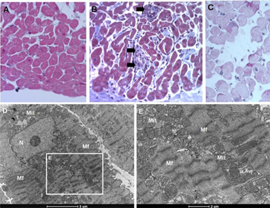
– Histopathological findings from endomyocardial biopsy of a heart transplant recipient. Sections stained with Masson’s trichrome (A, D) revealed normal myocardial architecture without fibrosis, vacuolization, or inflammation, and normal density of mitochondria. A few patients exhibited inflammatory infiltrates (arrows) (B) and some small areas with vacuolization (C, D). Despite the presence of the latter, transmission electron micrographs do not reveal any glycogen deposits (E, F). *: vacuolization; Gly: glycogen; Mf: myofibril; Mit: mitochondria.
Transmission electron microscopy
PRKAG2 cardiomyocytes displayed extensive glycogen accumulation, predominantly in the perinuclear region, with additional deposits in intermyofibrillar and subsarcolemmal areas. Mitochondria and sarcomeres appeared preserved in most myocytes (Figure 1D-G). Some cardiomyocytes contained β-glycogen particles interspersed among mitochondria. Lipofuscin granules, corresponding to lysosomal residues, were observed in both PRKAG2 and control samples. Patients with the H401Q mutation exhibited identical histological and ultrastructural findings as those with the R302Q variant. Notably, none of the control samples showed glycogen accumulation, even in cases with mild vacuolization (Figure 2).
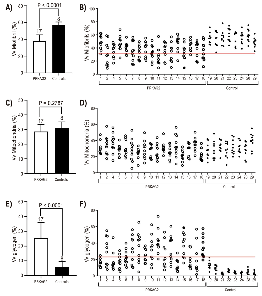
Quantitative TEM analysis revealed a 6-fold increase in glycogen density (p < 0.0001) in PRKAG2 patients, ~35% decrease in myofibrillar density (p < 0.0001), and no difference in mitochondrial density between groups (Figure 3). Individual volumetric density (Vv) values showed a clear threshold separating PRKAG2 from controls. PRKAG2 cases had at least one TEM image with Vv myofibrils < 30% and Vv glycogen > 22%. Control cases did not meet these thresholds. A positive correlation was observed between cardiomyocyte diameter and glycogen accumulation, while myofibril content showed a negative correlation. Mitochondrial volume density showed no correlation with cardiomyocyte diameter (Table 3).

– Volumetric densities (Vv) of myofibrils (A, B), mitochondria (C, D), and glycogen (E, F) from PRKAG2 (white bars/circles) and controls (black bars/dots) patients. Data presented as means ± standard deviation (A, C, E) (Number of patients described above bar, Student’s unpaired t-test) or individual values of each patient (B, D, F) (10 fields per patient); Red line indicates a threshold in the parameters between PRKAG2 and control patients.
– Pearson correlation between cardiomyocyte diameter and volumetric densities obtained by TEM
Missing data
Technical issues during TEM processing prevented a reliable assessment in 1 of 18 PRKAG2 patients. Light microscopy analysis was not possible for one PRKAG2 patient. Each patient had two sets of samples: one for light microscopy and one for TEM. In total, 5 of 58 datasets (8.6%) were incomplete (Table 1).
Discussion
The pathological features of PRKAG2 cardiomyopathy have been described in a limited number of cases, primarily through EMB, autopsy of sudden death victims, or examination of explanted hearts from transplant recipients. These reports suggest that histopathological findings may vary depending on the clinical context, with more pronounced changes observed in explanted hearts and sudden death cases.
Anatomopathological data from the literature (Supplementary Table 1) indicate that morphological findings have been described in only 22 out of 314 patients (7%) with PRKAG2 cardiomyopathy. When considering only cases that underwent percutaneous right ventricular EMB for diagnostic purposes, this number is even lower, totaling 14 patients (4.5%). The present study is the first systematic analysis of right ventricular EMB in a large cohort of PRKAG2 cardiomyopathy patients, assessing its diagnostic utility and the specificity of findings compared to a control group of heart transplant recipients whose donors did not have the disease.
The study’s main findings demonstrated that EMB had a high diagnostic yield in our cohort: all PRKAG2-positive patients (18/18, 100%) exhibited the characteristic morphological features, including enlarged cardiomyocytes without architectural disarray, marked vacuolization, and a 6-fold increase in glycogen density (p < 0.0001). In contrast, none of the controls showed glycogen accumulation, and only 2/11 displayed focal vacuolization. These results indicate that EMB reliably distinguished PRKAG2 cardiomyopathy from controls in this study population.
Pronounced cardiomyocyte vacuolization is a well-documented feature of PRKAG2 cardiomyopathy,9-14,18 as observed in previous studies using H&E or Masson’s trichrome staining, typically associated with PAS-positive granules and large amounts of cytosolic glycogen particles in TEM. Although vacuolization was also observed in two control cases, it was focal and limited to a minority of cardiomyocytes, likely due to fixation or processing artifacts (Figure 2). In contrast, PRKAG2 patients exhibited widespread vacuolization in most cardiomyocytes.
The prevailing explanation for the presence of vacuoles considers them as negative images, an indirect finding resulting from glycogen washout during slide preparation. However, this assumption does not fully account for the presence of vacuoles in some control group members. In one control case (case 20), TEM clearly showed no accumulation of glycogen in the cytosol (Table 1). It is worth noting that a few normal individuals may exhibit higher volumetric density of glycogen (Figure 3F), however, they show no accumulation of glycogen as seen in PRKAG2 patients (Figure 1D-G vs. Figure 2 E-F).
Cardiac hypertrophy is a hallmark of PRKAG2 cardiomyopathy. We recently reported the diagnostic utility of transthoracic echocardiography, identifying left ventricular hypertrophy in 25/30 (86%) and right ventricular hypertrophy in 26/30 (90%) carriers of pathogenic PRKAG2 variants.27 However, at the cellular level, hypertrophy is typically characterized by an increased number of sarcomeric units within cardiomyocytes. Cardiomyocyte diameter enlargement alone is not diagnostic of hypertrophy, as cellular expansion can result from various factors, including excessive storage of intracellular substances such as glycogen, lipids, and water, which can manifest as ventricular hypertrophy on cardiac imaging.
Recent studies using cardiomyocytes derived from human-induced pluripotent stem cells (hiPSC-CMs)28,29 have shown that PRKAG2 mutations (R302Q and N488I) induce cell enlargement due to glycogen accumulation, driven by a persistent and inappropriate increase in AMPK activity. Notably, inhibition of AMPK activity using compound C reduced glycogen storage and reversed imaging-detected hypertrophy.29 The β subunit of AMPK contains a regulatory glycogen-binding domain, allowing wild-type AMPK to function as a glycogen sensor,30 while immunogold microscopy has shown AMPK subunits co-localized with glycogen particles, with the β1 subunit positioned at the periphery of glycogen rosettes.31
In our study, we observed a positive correlation between cardiomyocyte enlargement and intracellular glycogen accumulation, alongside an inverse correlation between myofibril volumetric density and cardiomyocyte diameter. Since volumetric density is a relative measure, we cannot rule out a potential increase in myofibril content in the context of larger cardiomyocytes. Interestingly, a study using a transgenic mouse model with cardiac overexpression of the N488I PRKAG2 mutant, combined with a knock-in mutation in the muscle form of glycogen synthase (GYS1)–which inhibits glucose-6-phosphatase-stimulated glycogen synthase activity–successfully rescued the glycogen storage phenotype and ventricular pre-excitation. However, it did not affect cardiac hypertrophy or cardiomyocyte enlargement, suggesting that mechanisms beyond glycogen accumulation contribute to hypertrophy in PRKAG2 cardiomyopathy.32
An important finding of our study was that even PRKAG2 patients without apparent cardiac hypertrophy exhibited morphological changes, including increased glycogen content, absence of inflammation, and minimal or no fibrosis (Table 1). While cardiomyocyte vacuolization occurs in normal hearts and other diseases, it is far more pronounced and widespread in PRKAG2 cardiomyopathy (Figure 1). Moreover, large glycogen deposits identified on TEM were highly specific to PRKAG2 and absent in controls. Exceptions include cases with interstitial fibrosis and architectural disarray, reported in rare PRKAG2 variants such as K485E and E506K.3,6 Findings from explanted or autopsied hearts in advanced disease also differ, showing more fibrosis,7,8,14,16-19architectural disarray,16,17 and occasional myocyte degeneration.14,17 These “end-stage” features have also been reported in common PRKAG2 variants like R302Q and N488I.16,17
In this study, genotype-phenotype correlation was consistent within our cohort. All family members of a proband with a PRKAG2 variant and characteristic clinical features were also carriers of the same variant, in line with previous reports describing the strong association between PRKAG2 mutations and this phenotype. This complete concordance in our series reinforces the robustness of the genetic-clinical association, while avoiding overgeneralization beyond the studied population.
Other conditions need to be differentiated from PRKAG2 cardiomyopathy. Hydroxychloroquine-induced cardiotoxicity, typically resulting from long-term use for connective tissue diseases, also presents with pronounced myocyte vacuolization, which extends to skeletal muscle. EMB in this context shows focal interstitial fibrosis without inflammation. Electron microscopy reveals extensive lysosomal inclusions, myeloid bodies, and curvilinear inclusions but no glycogen accumulation.33Danon disease (LAMP2 mutation), which exhibits a predominant severe cardiac phenotype in young males (usually below 20 years),34 has a distinct microscopic profile. Post-mortem examination of 2 hearts showed massive hypertrophy, substantial myocyte disarray, and fibrosis, with prominent clusters of vacuolated myocytes and inclusion and inclusions of amorphous granular material in some cells inside scarring regions. Anderson-Fabry disease, which may have a dominant cardiac phenotype, is associated with multisystem involvement.35 Histologically, it shows extensive cardiomyocyte vacuolation, fibrosis, and myocyte disarray, particularly in the right ventricle, with occasional myocyte necrosis and foamy macrophage replacement. However, no glycogen accumulation is observed. Additionally, mitochondrial cardiomyopathies, such as those associated with the 3243A>G mutation, can present with an HCM-like phenotype, often misdiagnosed as isolated cardiomyopathy. These patients may lack stroke-like episodes (MELAS syndrome), and TEM typically reveals mitochondrial proliferation.36
Limitations
The control group consisted of cardiac transplant patients whose donor hearts were significantly younger than those with PRKAG2 cardiomyopathy. However, the pathological changes characteristic of PRKAG2 cardiomyopathy are not expected to arise from normal aging. Some data were lost because of problems during fixation or sample processing (for TEM: three cases in the control group and one in the PRKAG2 cardiomyopathy group; for light microscopy: one case in the PRKAG2 cardiomyopathy group). Given there were only 2/36 missing sets (5.5%) in the PRKAG2 cardiomyopathy group, it is unlikely that this had a significant impact on our findings.
Conclusion
Percutaneous right ventricular EMB findings reveal hallmark morphological changes diagnostic of PRKAG2 cardiomyopathy in all genotypes of positive patients. TEM plays an important role in detecting substantial cardiomyocyte glycogen accumulation. The histopathological and ultrastructural abnormalities described in this study were not observed in control samples. Our findings suggest that EMB, coupled with light and electron microscopy, can aid in diagnosing PRKAG2 cardiomyopathy. Further studies are needed to validate microscopy as a diagnostic tool for PRKAG2 cardiomyopathy.
Supplemental Materials
Supplementary Table 1
Acknowledgement
The authors would like to acknowledge the Center of Microscopy at Universidade Federal de Minas Gerais (http://www.microscopia.ufmg.br) for providing the equipment and technical support for experiments involving electron microscopy, and Kinulpe Honorato Sampaio is a recipient of Conselho Nacional de Desenvolvimento Científico e Tecnológico (CNPq) (303206/2022-5).
References
-
1 Yavari A, Sarma D, Sternick EB. Human ?2-AMPK Mutations. Methods Mol Biol. 2018;1732:581-619. doi: 10.1007/978-1-4939-7598-3_37.
» https://doi.org/10.1007/978-1-4939-7598-3_37 -
2 Arad M, Benson DW, Perez-Atayde AR, McKenna WJ, Sparks EA, Kanter RJ, et al. Constitutively Active AMP Kinase Mutations Cause Glycogen Storage Disease Mimicking Hypertrophic Cardiomyopathy. J Clin Invest. 2002;109(3):357-62. doi: 10.1172/JCI14571.
» https://doi.org/10.1172/JCI14571 -
3 Bayrak F, Komurcu-Bayrak E, Mutlu B, Kahveci G, Basaran Y, Erginel-Unaltuna N. Ventricular Pre-Excitation and Cardiac Hypertrophy Mimicking Hypertrophic Cardiomyopathy in a Turkish Family with a Novel PRKAG2 Mutation. Eur J Heart Fail. 2006;8(7):712-5. doi: 10.1016/j.ejheart.2006.03.006.
» https://doi.org/10.1016/j.ejheart.2006.03.006 -
4 Kelly BP, Russell MW, Hennessy JR, Ensing GJ. Severe Hypertrophic Cardiomyopathy in an Infant with a Novel PRKAG2 Gene Mutation: Potential Differences between Infantile and Adult Onset Presentation. Pediatr Cardiol. 2009;30(8):1176-9. doi: 10.1007/s00246-009-9521-3.
» https://doi.org/10.1007/s00246-009-9521-3 -
5 Sternick EB, Oliva A, Gerken LM, Magalhães L, Scarpelli R, Correia FS, et al. Clinical, Electrocardiographic, and Electrophysiologic Characteristics of Patients with a Fasciculoventricular Pathway: The Role of PRKAG2 Mutation. Heart Rhythm. 2011;8(1):58-64. doi: 10.1016/j.hrthm.2010.09.081.
» https://doi.org/10.1016/j.hrthm.2010.09.081 -
6 Liu Y, Bai R, Wang L, Zhang C, Zhao R, Wan D, et al. Identification of a Novel de Novo Mutation Associated with PRKAG2 Cardiac Syndrome and Early Onset of Heart Failure. PLoS One. 2013;8(5):e64603. doi: 10.1371/journal.pone.0064603.
» https://doi.org/10.1371/journal.pone.0064603 -
7 Thevenon J, Laurent G, Ader F, Laforêt P, Klug D, Pentiah AD, et al. High Prevalence of Arrhythmic and Myocardial Complications in Patients with Cardiac Glycogenosis due to PRKAG2 Mutations. Europace. 2017;19(4):651-9. doi: 10.1093/europace/euw067.
» https://doi.org/10.1093/europace/euw067 -
8 Yogasundaram H, Paterson ID, Graham M, Sergi C, Oudit GY. Glycogen Storage Disease because of a PRKAG2 Mutation Causing Severe Biventricular Hypertrophy and High-Grade Atrio-Ventricular Block. Circ Heart Fail. 2016;9(8):e003367. doi: 10.1161/CIRCHEARTFAILURE.116.003367.
» https://doi.org/10.1161/CIRCHEARTFAILURE.116.003367 -
9 Sternick EB, Araújo SA, Camargos ERS, Brasileiro G Filho. Atrial Pathology Findings in a Patient with PRKAG2 Cardiomyopathy and Persistent Atrial Fibrillation. Circ Arrhythm Electrophysiol. 2016;9(12):e004455. doi: 10.1161/CIRCEP.116.004455.
» https://doi.org/10.1161/CIRCEP.116.004455 -
10 Banankhah P, Fishbein GA, Dota A, Ardehali R. Cardiac Manifestations of PRKAG2 Mutation. BMC Med Genet. 2018;19(1):1. doi: 10.1186/s12881-017-0512-6.
» https://doi.org/10.1186/s12881-017-0512-6 -
11 Sri A, Daubeney P, Prasad S, Baksi J, Kinali M, Voges I. A Case Series on Cardiac and Skeletal Involvement in Two Families with PRKAG2 Mutations. Case Rep Pediatr. 2019;2019:7640140. doi: 10.1155/2019/7640140.
» https://doi.org/10.1155/2019/7640140 -
12 Siqueira MHS, Honorato-Sampaio K, Dias GM, Wilson JR, Yavari A, Brasileiro G Filho, et al. Sudden Death Associated with a Novel H401Q PRKAG2 Mutation. Europace. 2020;22(8):1278. doi: 10.1093/europace/euaa014.
» https://doi.org/10.1093/europace/euaa014 -
13 Lopez-Sainz A, Dominguez F, Lopes LR, Ochoa JP, Barriales-Villa R, Climent V, et al. Clinical Features and Natural History of PRKAG2 Variant Cardiac Glycogenosis. J Am Coll Cardiol. 2020;76(2):186-97. doi: 10.1016/j.jacc.2020.05.029.
» https://doi.org/10.1016/j.jacc.2020.05.029 -
14 Hu D, Hu D, Liu L, Barr D, Liu Y, Balderrabano-Saucedo N, et al. Identification, Clinical Manifestation and Structural Mechanisms of Mutations in AMPK Associated Cardiac Glycogen Storage Disease. EBioMedicine. 2020;54:102723. doi: 10.1016/j.ebiom.2020.102723.
» https://doi.org/10.1016/j.ebiom.2020.102723 -
15 Frustaci A, Russo MA, Chimenti C. Diagnostic Contribution of Left Ventricular Endomyocardial Biopsy in Patients with Clinical Phenotype of Hypertrophic Cardiomyopathy. Hum Pathol. 2013;44(1):133-41. doi: 10.1016/j.humpath.2012.04.023.
» https://doi.org/10.1016/j.humpath.2012.04.023 -
16 Murphy RT, Mogensen J, McGarry K, Bahl A, Evans A, Osman E, et al. Adenosine Monophosphate-Activated Protein Kinase Disease Mimicks Hypertrophic Cardiomyopathy and Wolff-Parkinson-White Syndrome: Natural History. J Am Coll Cardiol. 2005;45(6):922-30. doi: 10.1016/j.jacc.2004.11.053.
» https://doi.org/10.1016/j.jacc.2004.11.053 -
17 Tan HL, van der Wal AC, Campian ME, Kruyswijk HH, ten Hove Jansen B, van Doorn DJ, et al. Nodoventricular Accessory Pathways in PRKAG2-Dependent Familial Preexcitation Syndrome Reveal a Disorder in Cardiac Development. Circ Arrhythm Electrophysiol. 2008;1(4):276-81. doi: 10.1161/CIRCEP.108.782862.
» https://doi.org/10.1161/CIRCEP.108.782862 -
18 van der Steld LP, Campuzano O, Pérez-Serra A, Zamorano MMB, Matos SS, Brugada R. Wolff-Parkinson-White Syndrome with Ventricular Hypertrophy in a Brazilian Family. Am J Case Rep. 2017;18:766-76. doi: 10.12659/ajcr.904613.
» https://doi.org/10.12659/ajcr.904613 -
19 Pöyhönen P, Hiippala A, Ollila L, Kaasalainen T, Hänninen H, Heliö T, et al. Cardiovascular Magnetic Resonance Findings in Patients with PRKAG2 Gene Mutations. J Cardiovasc Magn Reson. 2015;17:89. doi: 10.1186/s12968-015-0192-3.
» https://doi.org/10.1186/s12968-015-0192-3 -
20 Gollob MH, Green MS, Tang AS, Gollob T, Karibe A, Ali Hassan AS, et al. Identification of a Gene Responsible for Familial Wolff-Parkinson-White Syndrome. N Engl J Med. 2001;344(24):1823-31. doi: 10.1056/NEJM200106143442403.
» https://doi.org/10.1056/NEJM200106143442403 -
21 Blair E, Redwood C, Ashrafian H, Oliveira M, Broxholme J, Kerr B, et al. Mutations in the Gamma(2) Subunit of AMP-Activated Protein Kinase Cause Familial Hypertrophic Cardiomyopathy: Evidence for the Central Role of Energy Compromise in Disease Pathogenesis. Hum Mol Genet. 2001;10(11):1215-20. doi: 10.1093/hmg/10.11.1215.
» https://doi.org/10.1093/hmg/10.11.1215 -
22 Cooper LT, Baughman KL, Feldman AM, Frustaci A, Jessup M, Kuhl U, et al. The Role of Endomyocardial Biopsy in the Management of Cardiovascular Disease: A Scientific Statement from the American Heart Association, the American College of Cardiology, and the European Society of Cardiology. Circulation. 2007;116(19):2216-33. doi: 10.1161/CIRCULATIONAHA.107.186093.
» https://doi.org/10.1161/CIRCULATIONAHA.107.186093 -
23 From AM, Maleszewski JJ, Rihal CS. Current Status of Endomyocardial Biopsy. Mayo Clin Proc. 2011;86(11):1095-102. doi: 10.4065/mcp.2011.0296.
» https://doi.org/10.4065/mcp.2011.0296 -
24 Richards S, Aziz N, Bale S, Bick D, Das S, Gastier-Foster J, et al. Standards and Guidelines for the Interpretation of Sequence Variants: A Joint Consensus Recommendation of the American College of Medical Genetics and Genomics and the Association for Molecular Pathology. Genet Med. 2015;17(5):405-24. doi: 10.1038/gim.2015.30.
» https://doi.org/10.1038/gim.2015.30 -
25 Walsh R, Thomson KL, Ware JS, Funke BH, Woodley J, McGuire KJ, et al. Reassessment of Mendelian Gene Pathogenicity using 7,855 Cardiomyopathy Cases and 60,706 Reference Samples. Genet Med. 2017;19(2):192-203. doi: 10.1038/gim.2016.90.
» https://doi.org/10.1038/gim.2016.90 -
26 Cantuária VL, Rodrigues CM, Dias IR, Ottone VO, Costa BO, Godinho LF, et al. Intense Caloric Restriction from Birth Protects the Heart Against Ischemia/Reperfusion Injury and Reduces Reactive Oxygen Species in Ovariectomized Rats. Antioxidants. 2025;14(2):169. doi: 10.3390/antiox14020169.
» https://doi.org/10.3390/antiox14020169 -
27 Pena JLB, Santos WC, Siqueira MHA, Sampaio IH, Moura ICG, Sternick EB. Glycogen Storage Cardiomyopathy (PRKAG2): Diagnostic Findings of Standard and Advanced Echocardiography Techniques. Eur Heart J Cardiovasc Imaging. 2021;22(7):800-7. doi: 10.1093/ehjci/jeaa176.
» https://doi.org/10.1093/ehjci/jeaa176 -
28 Hinson JT, Chopra A, Lowe A, Sheng CC, Gupta RM, Kuppusamy R, et al. Integrative Analysis of PRKAG2 Cardiomyopathy iPS and Microtissue Models Identifies AMPK as a Regulator of Metabolism, Survival, and Fibrosis. Cell Rep. 2016;17(12):3292-304. doi: 10.1016/j.celrep.2016.11.066.
» https://doi.org/10.1016/j.celrep.2016.11.066 -
29 Zhan Y, Sun X, Li B, Cai H, Xu C, Liang Q, et al. Establishment of a PRKAG2 Cardiac Syndrome Disease Model and Mechanism Study using Human Induced Pluripotent Stem Cells. J Mol Cell Cardiol. 2018;117:49-61. doi: 10.1016/j.yjmcc.2018.02.007.
» https://doi.org/10.1016/j.yjmcc.2018.02.007 -
30 McBride A, Ghilagaber S, Nikolaev A, Hardie DG. The Glycogen-Binding Domain on the AMPK Beta Subunit Allows the Kinase to Act as a Glycogen Sensor. Cell Metab. 2009;9(1):23-34. doi: 10.1016/j.cmet.2008.11.008.
» https://doi.org/10.1016/j.cmet.2008.11.008 -
31 Bendayan M, Londono I, Kemp BE, Hardie GD, Ruderman N, Prentki M. Association of AMP-Activated Protein Kinase Subunits with Glycogen Particles as Revealed in Situ by Immunoelectron Microscopy. J Histochem Cytochem. 2009;57(10):963-71. doi: 10.1369/jhc.2009.954016.
» https://doi.org/10.1369/jhc.2009.954016 -
32 Kim M, Hunter RW, Garcia-Menendez L, Gong G, Yang YY, Kolwicz SC Jr, et al. Mutation in the ?2-Subunit of AMP-Activated Protein Kinase Stimulates Cardiomyocyte Proliferation and Hypertrophy Independent of Glycogen Storage. Circ Res. 2014;114(6):966-75. doi: 10.1161/CIRCRESAHA.114.302364.
» https://doi.org/10.1161/CIRCRESAHA.114.302364 -
33 Nadeem U, Raafey M, Kim G, Treger J, Pytel P, Husain AN, et al. Chloroquine- and Hydroxychloroquine-Induced Cardiomyopathy: A Case Report and Brief Literature Review. Am J Clin Pathol. 2021;155(6):793-801. doi: 10.1093/ajcp/aqaa253.
» https://doi.org/10.1093/ajcp/aqaa253 -
34 Maron BJ, Roberts WC, Arad M, Haas TS, Spirito P, Wright GB, et al. Clinical Outcome and Phenotypic Expression in LAMP2 Cardiomyopathy. JAMA. 2009;301(12):1253-9. doi: 10.1001/jama.2009.371.
» https://doi.org/10.1001/jama.2009.371 -
35 Sheppard MN, Cane P, Florio R, Kavantzas N, Close L, Shah J, et al. A Detailed Pathologic Examination of Heart Tissue from three Older Patients with Anderson-Fabry Disease on Enzyme Replacement Therapy. Cardiovasc Pathol. 2010;19(5):293-301. doi: 10.1016/j.carpath.2009.05.003.
» https://doi.org/10.1016/j.carpath.2009.05.003 -
36 Hiraki N, Tanaka TD, Yoshimura M. A Man with Left Ventricular Hypertrophy. JAMA Cardiol. 2022;7(2):225-6. doi: 10.1001/jamacardio.2021.5144.
» https://doi.org/10.1001/jamacardio.2021.5144
-
Study association:
This article is part of the thesis of master submitted by Carla de Oliveira, from Faculdade de Ciências Médicas de Minas Gerais.
-
Ethics approval and consent to participate:
This study was approved by the Ethics Committee of the Universidade Federal de Minas Gerais under the protocol number SISNEP CAAE: 0511.0.203.418-11. All the procedures in this study were in accordance with the 1975 Helsinki Declaration, updated in 2013. Informed consent was obtained from all participants included in the study.
-
Use of Artificial Intelligence:
The authors did not use any artificial intelligence tools in the development of this work.
-
Data Availability Statement:
The underlying content of the research text is contained within the manuscript.
-
Sources of funding:
There were no external funding sources for this study.
-
*Supplemental Materials
For additional information, pleaseclick here.
Edited by
-
Editor responsible for the review:
Natália Olivetti
The underlying content of the research text is contained within the manuscript.

 A análise histológica e ultraestrutural da biópsia endomiocárdica do ventrículo direito revela um conjunto consistente de características sugestivas de cardiomiopatia PRKAG2, incluindo aumento do tamanho dos cardiomiócitos, vacuolização pronunciada, ausência de fibrose intersticial, acúmulo substancial de glicogênio e densidade volumétrica mitocondrial preservada.
 Histological and ultrastructural analysis of right ventricular endomyocardial biopsy reveals a consistent set of features suggestive of PRKAG2 cardiomyopathy, including cardiomyocyte enlargement, pronounced vacuolization, absence of interstitial fibrosis, substantial glycogen accumulation, and preserved mitochondrial volumetric density.