Genetic and Clinical Characterization of a South-Brazilian Hypertrophic Cardiomyopathy Cohort
Abstract
Background Hypertrophic cardiomyopathy (HCM) is the most common monogenic heart disease, characterized by genetic and phenotypic heterogeneity. Although extensively studied in North American and European populations, data from Brazil remain limited.
Objectives To characterize the genetic and clinical profiles of a Southern Brazilian cohort of HCM patients and their relatives using massive parallel sequencing.
Methods In this observational study, HCM patients and first-degree relatives were recruited from outpatient cardiology clinics. Clinical and imaging data were collected, and genetic analysis used a 100-gene panel. Variant pathogenicity was assessed according to American College of Medical Genetics and Genomics criteria, and statistical analyses were performed using R software.
Results Eighty individuals were included in the final analysis (mean age: 49.2 ±18.5); 60% male; 40 index cases and 40 affected relatives). MYH7 and MYBPC3 were the most frequently related genes, with pathogenic / likely pathogenic variants (P/LP) identified in 33% and 16% of participants, respectively. No pathogenic TNNT2 variants were detected. Ninety percent of participants carried an identified variant (including variants of uncertain significance), with 68% harboring P/LP variants. MYH7 carriers exhibited a higher proportion of left ventricular outflow tract obstruction, whereas MYBPC3 carriers had a higher proportion of arrhythmic events and earlier diagnosis; however, these differences did not reach statistical significance and should be interpreted as exploratory. Clinical comparisons revealed regional differences, suggesting the potential impact of genetic diversity on the presentation of HCM in this part of Brazil.
Conclusions This study offers the first detailed genetic and clinical characterization of a Brazilian HCM cohort using massive parallel sequencing. Our findings underscore the importance of genetic testing for diagnosis, risk stratification, and management.
Keywords:
Hypertrophic Cardiomyopathy; Genetic Testing; High-Throughput Nucleotide Sequencing; Brazil

Resumo
Fundamento A cardiomiopatia hipertrófica (CMH) é a doença cardíaca monogênica mais comum, caracterizada por heterogeneidade genética e fenotípica. Embora amplamente estudada em populações norte-americanas e europeias, os dados provenientes do Brasil permanecem limitados.
Objetivos Caracterizar os perfis genéticos e clínicos de uma coorte sul-brasileira de pacientes com CMH e seus familiares utilizando sequenciamento em paralelo em larga escala.
Métodos Neste estudo observacional, pacientes com CMH e seus familiares de primeiro grau foram recrutados em ambulatórios de cardiologia. Dados clínicos e de imagem foram coletados, e a análise genética utilizou um painel de 100 genes. A patogenicidade das variantes foi avaliada de acordo com os critérios do American College of Medical Genetics and Genomics, e as análises estatísticas foram realizadas utilizando o software R.
Resultados Oitenta indivíduos foram incluídos na análise final (idade média: 49,2 ± 18,5); 60% do sexo masculino; 40 casos-índice e 40 familiares afetados). MYH7 e MYBPC3 foram os genes mais frequentemente relacionados, com variantes Patogênicas/Provavelmente Patogênicas (P/PP) identificadas em 33% e 16% dos participantes, respectivamente. Nenhuma variante patogênica em TNNT2 foi detectada. Noventa por cento dos participantes apresentaram alguma variante identificada (incluindo variante de significado incerto), sendo que 68% carregavam variantes P/PP. Portadores de MYH7 exibiram maior proporção de obstrução do trato de saída do ventrículo esquerdo, enquanto portadores de MYBPC3 apresentaram maior proporção de eventos arrítmicos e diagnóstico mais precoce; entretanto, essas diferenças não atingiram significância estatística e devem ser interpretadas como exploratórias. As comparações clínicas revelaram diferenças regionais, sugerindo o potencial impacto da diversidade genética na apresentação da CMH nesta região do Brasil.
Conclusões Este estudo oferece a primeira caracterização genética e clínica detalhada de uma coorte brasileira de pacientes portadores de CMH utilizando sequenciamento em paralelo em larga escala. Nossos achados ressaltam a importância do teste genético para o diagnóstico, a estratificação de risco e o manejo.
Palavras-chave:
Cardiomiopatia Hipertrófica; Teste Genético; Sequenciamento de Nucleotídeos em Larga Escala; Brasil

Introduction
Hypertrophic Cardiomyopathy (HCM) is the most common monogenic heart disease, with a prevalence of approximately 1:250 to 1:500 individuals.1 It is primarily caused by pathogenic variants in sarcomeric and sarcomere-related genes. The condition is characterized by left ventricular hypertrophy unrelated to abnormal loading conditions and may lead to complications such as Left Ventricular Outflow Tract Obstruction (LVOTO), Sudden Cardiac Death (SCD), and heart failure.2-4
Disease expression is highly variable, making the prediction of clinical outcomes challenging, even among genotype-positive individuals.5,6 Certain gene variants have been associated with different prognoses: MYH7 variants are linked to worse outcomes, while MYBPC3 variants tend to present with delayed onset and incomplete penetrance.7-10 Although genotype-specific risk prediction remains controversial, genotype-positive status itself has been associated with earlier disease onset, greater ventricular dysfunction, and increased cardiovascular mortality.11-15 Accordingly, the latest AHA/ACC guidelines include genotype-positive status as a definitive risk factor for SCD in pediatric patients, with growing evidence in adults.6
Despite the recognized importance of genetics for diagnosis, risk assessment, and family counseling,2,16 genetic evaluation remains underutilized in Brazil, and population-specific data are limited. Previous Brazilian studies have reported MYH7 as the most prevalent mutated gene, followed by MYBPC3 and TNNT2,17-19 a distribution consistent, though not identical to that observed in North American and European cohorts.10,20
This study aims to characterize the genetic and clinical profiles of patients with HCM and their relatives from a tertiary center in southern Brazil, using massive parallel sequencing.
Methods
Ethics statement
The study protocol was approved by the Institutional Ethics Committee through Plataforma Brasil (CAAE: 554413421.4.1001.5327).
Participants and study design
The study employed an observational, cross-sectional design. Participants were recruited from the outpatient cardiology clinics of Hospital de Clínicas de Porto Alegre (HCPA), Brazil. Eligible individuals included patients diagnosed with HCM and their first-degree relatives.
Inclusion criteria for index patients required a confirmed diagnosis of HCM, defined as left ventricular wall thickness ≥ 15 mm not explained by abnormal loading conditions. For relatives carrying a Pathogenic (P) or Likely Pathogenic (LP) variant, inclusion in the study required genopositivity. Among enrolled relatives, a left ventricular wall thickness ≥ 13 mm was used to classify them as phenotype-positive (HCM), whereas asymptomatic carriers with wall thickness < 13 mm were considered phenotype-negative. Participants had to be 18 years or older at the time of enrollment. Exclusion criteria included the presence of HCM phenocopies, absence of clinical and genetic evidence of HCM, and age under 18 years. All participants received detailed information about the study and provided written informed consent before enrollment. Genetic counseling was offered to all participants both before and after testing.
Database
The variables collected included date of birth, sex, and age at HCM diagnosis. Family history data were obtained, including a history of HCM and SCD, defined as at least one first- or second-degree relative who had experienced sudden, unexplained death or sudden cardiac arrest. Additional variables included episodes of aborted SCD and the presence of ventricular arrhythmias, such as sustained and non-sustained ventricular tachycardia or ventricular fibrillation. These arrhythmias were identified through 24-hour Holter monitoring, 12-lead resting Electrocardiogram (ECG), or exercise testing.
Other data collected included baseline pharmacological treatment, comorbidities, cardiac device implantation, presence of atrial fibrillation (detected via 24-hour Holter monitoring or 12-lead resting ECG) and mortality status. Electrocardiographic measures were also recorded. Echocardiographic parameters included Left Atrial (LA) size, Left Ventricular Ejection Fraction (LVEF), Left Ventricular End-Diastolic (LVED) and End-Systolic (LVES) diameters, left ventricular wall thickness, presence of Left Ventricle Outflow Tract Obstruction (LVOTO) and maximum outflow tract gradient. Cardiac magnetic resonance imaging parameters included LVEF, LVED and LVES volumes, left atrial volume and the presence of Late Gadolinium Enhancement (LGE).
Genetic sequencing
Genomic DNA was extracted from saliva samples, and genetic testing was performed using a commercial comprehensive cardiomyopathy panel containing 100 genes: ABCC9 ACADVL ACTC1 ACTN2 AGL ALMS1 ALPK3 BAG3 BRAF CACNA1C CACNA1D CALM1 CALM2 CALM3 CASQ2 CBL CDH2 CPT2 CRYAB CSRP3 DES DMD DNAJC19 DOLK DSC2 DSG2 DSP ELAC2 EMD EYA4 FHL1 FKRP FKTN FLNC, GAA, GATA4, GATA5, GJA5, GLA, HCN4, HRAS, JUP, KCNE1, KCNH2, KCNJ2, KCNQ1, KRAS, LAMP2, LMNA, LZTR1, MAP2K1, MAP2K2, MRAS, MTO1, MYBPC3, MYH6, MYH7, MYL2, MYL3, MYL4, MYLK3, NF1, NKX2-5, NRAS, PCCA, PCCB, PKP2, PLN, PPA2, PPCS, PPP1CB, PRKAG2, PTPN11, RAF1, RASA1, RBM20, RIT1, RYR2, SCN5A, SDHA, SGCD, SHOC2, SLC22A5, SOS1, SOS2, SPRED1, TAZ, TBX20, TCAP, TMEM43, TMEM70, TNNC1, TNNI3, TNNI3K, TNNT2, TPM1, TRDN, TRPM4, TTN, TTR, VCL. Full-gene sequencing was performed using massive parallel sequencing technology.
Statistical analysis
Study data were collected and managed using REDCap (Research Electronic Data Capture), a secure and confidential web-based platform.21Statistical analysis was performed using R software, version 4.3.0. Categorical variables were analyzed using chi-square tests. For continuous variables with normal distribution, independent t-tests were used, while variables without normal distribution were analyzed using the Wilcoxon Signed-Rank test. A significance threshold of 5% (p <0.05) was considered to all analyses.
Results
General Population Description
A total of 95 individuals, including affected patients and their relatives, were initially screened. Following genetic testing, relatives without a clinical diagnosis of HCM or an identified pathogenic or likely pathogenic variant were excluded (n=15). Consequently, 80 individuals were included in the final analysis: 40 unrelated index patients and 40 affected or variant-carrying relatives (figure 1). Of these, 60% were men (n=48), 50% were index patients (n=40), and 50% were family members (n=40). Among the participants, 68% had an HCM diagnosis (n=54), while 32% were variant carriers (n=26). The mean (SD) age was 49.2 (18.5) years. A family history of HCM was presented in 32% of index patients (n=13).

– Selection process flowchart. Index: first identified case in the family; G+: genotype positive; G-: genotype negative; P+: phenotype positive; P-: phenotype negative.
The median left ventricular septal thickness was 18.5 mm (IQR: 15.0 - 21.0), the posterior wall thickness was 10 mm (IQR: 9.0 - 12.0), and the LVEF was 67% (IQR: 63.2 - 72.0). LVOTO was present in 33% of participants, and ventricular arrhythmias were observed in 15%. A summary of the clinical and demographic characteristics is provided in Table 1.
Genetic profile
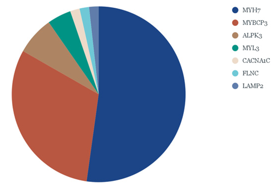
Among all sequenced genes, P, LP, and Variants of Uncertain Significance were identified in 72 (90%) individuals. When considering only P and LP variants, 54 (68%) individuals were classified as having a positive genetic test. For initial characterization, all variants identified are shown in the Central Illustration. Figure 2 illustrates the distribution of P/LP variants in genes strongly or moderately associated with HCM.22 The two most frequently found genes in HCM are highlighted: MYH7 (33%) and MYBPC3 (16%). Additionally, 10% of the sample had no identified variants.

– Distribution of affected genes strongly or moderately related to hypertrophic cardiomyopathy.
Six individuals exhibited HCM-related variants across more than one gene, with no specific pattern identified (Table 2).
To illustrate the importance of a comprehensive genetic and clinical evaluation, we present a detailed and intriguing family case (see Supplementary Material). Variant pathogenicity was classified according to the American College of Medical Genetics and Genomics (ACMG) criteria.23 The distribution of variant pathogenicity classification was as follows: 1) pathogenic (44%) 2) likely pathogenic (34%), and 3) Variant of Unknown Significance (VUS, 22%). All variants identified in MYH7 and MYBP3 were missense, except for one intronic variant in MYH7. A detailed description of all variants can be found in Table 3.
Clinical-genetic characterization of phenotype-positive individuals
To better understand how genetic background influences the HCM phenotype, we described and compared the clinical features of HCM patients with different affected genes (Table 4). This analysis focused exclusively on genes with a well-established correlation to HCM, specifically MYH7 and MYBPC3, based on the available sample size (Table 5). Our comparison of MYBPC3 and MYH7 revealed noteworthy differences in frequencies, although these did not reach statistical significance. Patients with MYH7 variants exhibited a higher frequency of LVOTO compared to those with MYBPC3 variants (35% vs. 18%; p = 0.3). Conversely, patients with MYBPC3 variants presented a more arrhythmogenic phenotype, characterized by a higher prevalence of ventricular arrhythmias (33% vs. 17%, p = 0.3), a higher rate of ICD implantation (27% vs 4%, p = 0.09), more frequent ventricular fibrosis, as identified by LGE on cardiac magnetic resonance (62% vs. 38%, p = 0.5), and a more frequent family history of sudden death (45% vs. 35%, p = 0.4). Additionally, the median age at diagnosis of MYBPC3-affected individuals was lower (37 years versus 46 years; p = 0.07). Other clinical characteristics are detailed in Table 5.
Genotype-positive versus Genotype-negative affected individuals
Among individuals diagnosed with HCM, we compared characteristics of those with identified genetic variants (phenotype-positive/genotype-positive) to those with a negative genetic test (phenotype-positive/genotype-negative). The mean age at HCM diagnosis in genotype-positive individuals was 44 years, while it was 50 years (p = 0.86) in genotype-negative HCM patients. The median left ventricle septum thickness of genotype-positive individuals was 17.5 mm, compared to 21 mm of genotype-negative individuals (p = 0.18). Ventricular arrhythmias were present in 25% of genotype-positive and 12% of genotype-negative individuals (p = 0.77), atrial fibrillation in 23.3% and 12.5% (p = 0.83) and LGE in 48.2% and 0% (p = 0.08) of the genotype-positive and genotype-negative groups, respectively. A family history of SCD was reported in 38% of the genotype-positive group and 25% of the genotype-negative group (p = 0.76). Details are summarized in Table 6.
Discussion
In this study, we found that MYH7 and MYBPC3 were the most prevalent genes in the cohort of HCM patients from southern Brazil, with variants detected in 33% and 16% of the total individuals, respectively. These findings are consistent with previous studies, in which MYH7 and MYBPC3 were identified as the most common genes implicated in HCM, accounting for 30%-50% of cases.2,3,10 However, our study reveals a slightly different distribution. Overall, the frequency of these variants aligns with what has been observed in other populations, although the proportion of cases without identified variants continues to pose a challenge for genetic diagnostics in HCM.
Interestingly, unlike most European and American cohorts, no individuals in our study were found to have variants in TNNT2.24 It is important to note that around 30 genes have been implicated in HCM, with 21 of these showing moderate evidence for gene-disease association. To date, more than 2,000 variants have been documented.9 Most of these encode sarcomeric proteins or are related to the sarcomere structure, which are critical components of the cardiac contractile machinery. Less common genetic variants, also linked to HCM, involve genes encoding sarcomere-related proteins such as ACTN2, ALPK3, CSRP3, FHOD3, JPH2, KLHL24, MT-TI, PLN and TRIM63,22 which in our cohort corresponded to a smaller proportion, as previously described. Observed differences in clinical features between MYH7 and MYBPC3 carriers, such as higher LVOTO in MYH7 and trends toward arrhythmic events and earlier diagnosis in MYBPC3 carriers, did not reach statistical significance. Therefore, these findings should be interpreted as exploratory and hypothesis-generating rather than indicative of causal relationships. Additionally, inclusion of multiple family members may introduce bias, as genetic and environmental factors shared within families could influence observed trends. Future studies with larger cohorts are needed to clarify potential genotype-phenotype correlations.
Unlike many previously reported cohorts, we found a higher proportion of P/LP variants. A meta-analysis by Topriceanu et al.25 reports an overall P/LP variant rate of approximately 30-40% among HCM patients. Our findings could reflect selection bias due to the limited availability of genetic tests, with a preference for selecting patients with a higher likelihood of a positive test (i.e., familial cases). Additionally, individuals with more frequent cardiology visits – often those with more severe phenotypes – may have been more likely to undergo genetic testing.
This first report on a Brazilian HCM cohort with extensive genetic screening aims to clarify genotypic findings, including those related to less-established genes. However, no specific patterns were found, despite some individuals carrying multiple variants. The distribution of variants may differ across generations, and it remains unclear how disease severity is influenced by a single variant compared to multiple variants. Previous reports suggest that modifier genes or additional sarcomeric gene variants may contribute to a worse phenotypic expression of HCM.14,26,31 In our cohort, we observed two VUS in genes with limited evidence for association with HCM, namely ABCC9 and RYR2. Furthermore, we identified a young female football player who exhibited significant left ventricular hypertrophy without fulfilling the criteria for Noonan syndrome. She carried a pathogenic MYH7 variant and a pathogenic LZTR1 variant (associated with Noonan syndrome), which may represent a phenocopy. An intriguing example is also provided by a family in which four individuals carried pathogenic variants in both MYH7 and ALPK3, as detailed in the Supplementary Material.
It is important to emphasize the impact of diagnosing genetic heart diseases, not only on patients but also on at-risk family members. Genetic counseling is indispensable, as it has been shown to reduce anxiety, improve risk perception, enhance disease knowledge, and promote emotional support.31 Additionally, multidisciplinary case discussions are invaluable and essential for the effective management of these patients.
Linking the genetic profile to disease expression in HCM – whether in terms of phenotype or adverse outcomes – remains a major challenge. Current risk prediction tools for SCD primarily rely on established clinical markers such as left ventricular wall thickness, syncope, left ventricular systolic dysfunction, LVOTO, non-sustained ventricular tachycardia, LGE, and family history of SCD.32,33 Previous studies have reported inconsistent results regarding genotype-phenotype correlations in disease severity. Watkins et al.13 demonstrated reduced life expectancy in patients with MYH7 missense variants, while Jansen et al.14 similarly observed poorer clinical outcomes associated with MYH7 mutations. In addition, MYH7 variants have been linked to elevated atrial fibrillation risk.15 However, emerging evidence suggests certain MYH7 variants may correlate with attenuated disease manifestations. Notably, Marsili et al.34 recently identified delayed disease onset with gradual symptom progression, particularly among female carriers of the MYH7 p.Arg1712Gln pathogenic variant. These contrasting findings underscore the importance of more nuanced investigations into genotype-phenotype relationships, accounting for variant-specific effects.
Meanwhile, pathogenic MYBPC3 variants have been linked with later disease onset and higher rates of incomplete penetrance compared to MYH7 variants.14 Conversely, certain TNNT2 variants (Arg92Trp, Arg92Gln, and Ile79Asn) have been associated to increased SCD risk in affected families.27 Although differences among affected individuals in our cohort did not reach statistical significance, it is plausible that patients with positive genotypes experience more severe disease. Importantly, in our study, carriers of MYBPC3 variants showed a higher proportion of features associated with SCD – including more ventricular arrhythmias, higher rates of ICD implantation, greater prevalence of family history of SCD, and earlier age of HCM diagnosis.
On the other hand, we observed greater LVOTO frequency among MYH7 variant carriers, while the prevalence of heart failure appeared similar across groups. While MYH7 mutations disrupt sarcomeric protein interactions, MYBPC3 pathogenicity seems primarily driven by haploinsufficiency – either from nonsense-mediated mRNA decay or the production of defective proteins unable to integrate into the sarcomere.35,36 A Brazilian study by Marsiglia et al. supports this hypothesis by identifying a higher proportion of truncating MYBPC3 variants.17 In our analysis, all MYBPC3 variants were missense; however, this does not necessarily imply a milder phenotype.
Collectively, these findings highlight the need for further investigation into the molecular pathogenic mechanism of MYBPC3. Despite numerous studies, no definitive genotype-phenotype predictors have been established. Disease expression likely varies by variant, rather than by gene alone. Besides traditional cardiovascular risk factors (e.g., hypertension, overweight) influencing HCM development, common genetic variants have been shown to account for a significant portion of disease heritability.37,38 Thus, detailed characterizing specific variants may help refine polygenic risk scores in the future. Ongoing global efforts are likely to drive progress in this area.
Although the relationship between specific genes and disease severity remains controversial, the presence of a positive genotype (regardless of gene) had consistently been associated with worse clinical outcomes. Ho et al.16 identified positive genetic testing as the best independent predictor of adverse events in HCM patients. Similarly, Marsiglia et al.19 observed that Brazilian individuals with positive genotypes had earlier HCM diagnosis and higher ventricular arrhythmia incidence. In our study, there was no difference between groups, even though we observed similar proportions: a higher frequency of ventricular arrhythmias, atrial fibrillation, and left ventricle fibrosis in the genotype-positive patients, along with earlier diagnosis and more positive family histories of SCD. These findings reinforce the importance of recognizing HCM as a genetically driven disease, irrespective of the specific gene involved.
Despite the inherent complexity of HCM, there is substantial evidence supporting the central role of genetics in disease management. The autosomal dominant inheritance pattern, with a 50% chance of transmission, has major implications for the medical care and life decisions of at-risk relatives.1 In Brazil, as in many developing countries, genetics is often underutilized in HCM management due to high cost, limited physician knowledge, and insufficient awareness of its clinical impact. This results in suboptimal patients care. To improve HCM management, it is crucial to promote more comprehensive genetic testing using advanced genomic techniques in larger population samples. We believe that advocating for the integration of genetics into routine HCM management could gradually lower costs and increase access, ultimately improving care for HCM patients in developing countries.
Limitations
The major limitation of our study is the small, locally based sample size. Given the heterogeneity of HCM, a larger cohort including individuals from diverse regions of Brazil would provide a more comprehensive understanding of the genetic characteristics of our population. Moreover, the inclusion of patients primarily from a tertiary center may have introduced selection bias, as these individuals are more likely to represent a severe or specialized subset of HCM cases. This may have influenced both the frequency and classification of the variants identified.
Besides, the inclusion of family members might have inflated the diagnostic yield of genetic testing. The exclusion of phenotype-negative relatives without pathogenic variants also limits our ability to estimate penetrance and assess genotype-phenotype correlations across families. Formal analyses of familial aggregation and genetic segregation were not performed due to the limited sample size and cross-sectional design. In addition, this study did not assess the clinical impact of genetic testing, such as potential changes in patient management or family screening practice, nor did it include longitudinal follow-up to assess clinical outcomes such as sudden death, hospitalization, or progression to heart failure. Expanding the sample to include patients from primary and secondary care centers could help mitigate this bias and provide a more accurate representation of the broader Brazilian HCM population.
Conclusion
In this cohort of southern Brazilian HCM patients and their relatives, MYH7 and MYBPC3 were the most frequently mutated genes, with no pathogenic variants identified in TNNT2. We observed a higher frequency of P/LP variants compared to international cohorts. Hypothesis on the non-significant associations of MYBPC3 variants with markers of disease severity should be explored in further studies. These findings suggest important genetic and phenotypic heterogeneity within this Brazilian HCM cohort and reinforce the clinical value of genetic testing for risk stratification and management.
Supplemental Materials
Electronic Supplemental Material
References
-
1 Semsarian C, Ingles J, Maron MS, Maron BJ. New Perspectives on the Prevalence of Hypertrophic Cardiomyopathy. J Am Coll Cardiol. 2015;65(12):1249-54. doi: 10.1016/j.jacc.2015.01.019.
» https://doi.org/10.1016/j.jacc.2015.01.019 -
2 Lopes LR, Ho CY, Elliott PM. Genetics of Hypertrophic Cardiomyopathy: Established and Emerging Implications for Clinical Practice. Eur Heart J. 2024;45(30):2727-34. doi: 10.1093/eurheartj/ehae421.
» https://doi.org/10.1093/eurheartj/ehae421 -
3 Ommen SR, Ho CY, Asif IM, Balaji S, Burke MA, Day SM, et al. 2024 AHA/ACC/AMSSM/HRS/PACES/SCMR Guideline for the Management of Hypertrophic Cardiomyopathy: A Report of the American Heart Association/American College of Cardiology Joint Committee on Clinical Practice Guidelines. Circulation. 2024;149(23):e1239-311. doi: 10.1161/CIR.0000000000001250.
» https://doi.org/10.1161/CIR.0000000000001250 -
4 Elliott PM, Anastasakis A, Borger MA, Borggrefe M, Cecchi F, Charron P, et al. 2014 ESC Guidelines on Diagnosis and Management of Hypertrophic Cardiomyopathy: The Task Force for the Diagnosis and Management of Hypertrophic Cardiomyopathy of the European Society of Cardiology (ESC). Eur Heart J. 2014;35(39):2733-79. doi: 10.1093/eurheartj/ehu284.
» https://doi.org/10.1093/eurheartj/ehu284 -
5 Maron BJ, Desai MY, Nishimura RA, Spirito P, Rakowski H, Towbin JA, et al. Diagnosis and Evaluation of Hypertrophic Cardiomyopathy: JACC State-of-the-Art Review. J Am Coll Cardiol. 2022;79(4):372-89. doi: 10.1016/j.jacc.2021.12.002.
» https://doi.org/10.1016/j.jacc.2021.12.002 -
6 Ommen SR, Mital S, Burke MA, Day SM, Deswal A, Elliott P, et al. 2020 AHA/ACC Guideline for the Diagnosis and Treatment of Patients with Hypertrophic Cardiomyopathy: Executive Summary: A Report of the American College of Cardiology/American Heart Association Joint Committee on Clinical Practice Guidelines. Circulation. 2020;142(25):e533-57. doi: 10.1161/CIR.0000000000000938.
» https://doi.org/10.1161/CIR.0000000000000938 -
7 Claes GR, van Tienen FH, Lindsey P, Krapels IP, Helderman-van den Enden AT, Hoos MB, et al. Hypertrophic Remodelling in Cardiac Regulatory Myosin Light Chain (MYL2) Founder Mutation Carriers. Eur Heart J. 2016;37(23):1815-22. doi: 10.1093/eurheartj/ehv522.
» https://doi.org/10.1093/eurheartj/ehv522 -
8 Arbelo E, Protonotarios A, Gimeno JR, Arbustini E, Barriales-Villa R, Basso C, et al. 2023 ESC Guidelines for the Management of Cardiomyopathies. Eur Heart J. 2023;44(37):3503-626. doi: 10.1093/eurheartj/ehad194.
» https://doi.org/10.1093/eurheartj/ehad194 -
9 Maron BJ, Maron MS, Semsarian C. Genetics of Hypertrophic Cardiomyopathy after 20 Years: Clinical Perspectives. J Am Coll Cardiol. 2012;60(8):705-15. doi: 10.1016/j.jacc.2012.02.068.
» https://doi.org/10.1016/j.jacc.2012.02.068 -
10 Marian AJ, Braunwald E. Hypertrophic Cardiomyopathy: Genetics, Pathogenesis, Clinical Manifestations, Diagnosis, and Therapy. Circ Res. 2017;121(7):749-70. doi: 10.1161/CIRCRESAHA.117.311059.
» https://doi.org/10.1161/CIRCRESAHA.117.311059 -
11 Roberts R, Sigwart U. New Concepts in Hypertrophic Cardiomyopathies, Part I. Circulation. 2001;104(17):2113-6. doi: 10.1161/hc4201.097429.
» https://doi.org/10.1161/hc4201.097429 -
12 Marian AJ, Roberts R. Molecular Genetic Basis of Hypertrophic Cardiomyopathy: Genetic Markers for Sudden Cardiac Death. J Cardiovasc Electrophysiol. 1998;9(1):88-99. doi: 10.1111/j.1540-8167.1998.tb00871.x.
» https://doi.org/10.1111/j.1540-8167.1998.tb00871.x -
13 Watkins H, Rosenzweig A, Hwang DS, Levi T, McKenna W, Seidman CE, et al. Characteristics and Prognostic Implications of Myosin Missense Mutations in Familial Hypertrophic Cardiomyopathy. N Engl J Med. 1992;326(17):1108-14. doi: 10.1056/NEJM199204233261703.
» https://doi.org/10.1056/NEJM199204233261703 -
14 Jansen M, Brouwer R, Hassanzada F, Schoemaker AE, Schmidt AF, Kooijman-Reumerman MD, et al. Penetrance and Prognosis of MYH7 Variant-Associated Cardiomyopathies: Results from a Dutch Multicenter Cohort Study. JACC Heart Fail. 2024;12(1):134-47. doi: 10.1016/j.jchf.2023.07.007.
» https://doi.org/10.1016/j.jchf.2023.07.007 -
15 Lee SP, Ashley EA, Homburger J, Caleshu C, Green EM, Jacoby D, et al. Incident Atrial Fibrillation is Associated with MYH7 Sarcomeric Gene Variation in Hypertrophic Cardiomyopathy. Circ Heart Fail. 2018;11(9):e005191. doi: 10.1161/CIRCHEARTFAILURE.118.005191.
» https://doi.org/10.1161/CIRCHEARTFAILURE.118.005191 -
16 Ho CY. Genetics and Clinical Destiny: Improving Care in Hypertrophic Cardiomyopathy. Circulation. 2010;122(23):2430-40. doi: 10.1161/CIRCULATIONAHA.110.978924.
» https://doi.org/10.1161/CIRCULATIONAHA.110.978924 -
17 Marsiglia JD, Credidio FL, Oliveira TG, Reis RF, Antunes MO, Araujo AQ, et al. Screening of MYH7, MYBPC3, and TNNT2 Genes in Brazilian Patients with Hypertrophic Cardiomyopathy. Am Heart J. 2013;166(4):775-82. doi: 10.1016/j.ahj.2013.07.029.
» https://doi.org/10.1016/j.ahj.2013.07.029 -
18 Mattos BP, Scolari FL, Torres MA, Simon L, Freitas VC, Giugliani R, et al. Prevalence and Phenotypic Expression of Mutations in the MYH7, MYBPC3 and TNNT2 Genes in Families with Hypertrophic Cardiomyopathy in the South of Brazil: A Cross-Sectional Study. Arq Bras Cardiol. 2016;107(3):257-65. doi: 10.5935/abc.20160133.
» https://doi.org/10.5935/abc.20160133 -
19 Marsiglia JD, Credidio FL, Oliveira TG, Reis RF, Antunes MO, Araujo AQ, et al. Clinical Predictors of a Positive Genetic Test in Hypertrophic Cardiomyopathy in the Brazilian Population. BMC Cardiovasc Disord. 2014;14:36. doi: 10.1186/1471-2261-14-36.
» https://doi.org/10.1186/1471-2261-14-36 -
20 The Share Registry. Sarcomeric Human Cardiomyopathy Registry [Internet]. 2024 [ cited 2026 Feb 08]. Available from: https://www.theshareregistry.org/
» https://www.theshareregistry.org/ -
21 Harris PA, Taylor R, Minor BL, Elliott V, Fernandez M, O'Neal L, et al. The REDCap Consortium: Building an International Community of Software Platform Partners. J Biomed Inform. 2019;95:103208. doi: 10.1016/j.jbi.2019.103208.
» https://doi.org/10.1016/j.jbi.2019.103208 -
22 Hespe S, Waddell A, Asatryan B, Owens E, Thaxton C, Adduru ML, et al. ClinGen Hereditary Cardiovascular Disease Gene Curation Expert Panel: Reappraisal of Genes associated with Hypertrophic Cardiomyopathy. medRxiv. 2024:2024.07.29.24311195. doi: 10.1101/2024.07.29.24311195.
» https://doi.org/10.1101/2024.07.29.24311195 -
23 Richards S, Aziz N, Bale S, Bick D, Das S, Gastier-Foster J, et al. Standards and Guidelines for the Interpretation of Sequence Variants: A Joint Consensus Recommendation of the American College of Medical Genetics and Genomics and the Association for Molecular Pathology. Genet Med. 2015;17(5):405-24. doi: 10.1038/gim.2015.30.
» https://doi.org/10.1038/gim.2015.30 -
24 Ho CY, Day SM, Ashley EA, Michels M, Pereira AC, Jacoby D, et al. Genotype and Lifetime Burden of Disease in Hypertrophic Cardiomyopathy: Insights from the Sarcomeric Human Cardiomyopathy Registry (SHaRe). Circulation. 2018;138(14):1387-98. doi: 10.1161/CIRCULATIONAHA.117.033200.
» https://doi.org/10.1161/CIRCULATIONAHA.117.033200 -
25 Topriceanu CC, Pereira AC, Moon JC, Captur G, Ho CY. Meta-Analysis of Penetrance and Systematic Review on Transition to Disease in Genetic Hypertrophic Cardiomyopathy. Circulation. 2024;149(2):107-23. doi: 10.1161/CIRCULATIONAHA.123.065987.
» https://doi.org/10.1161/CIRCULATIONAHA.123.065987 -
26 Coppini R, Ho CY, Ashley E, Day S, Ferrantini C, Girolami F, et al. Clinical Phenotype and Outcome of Hypertrophic Cardiomyopathy Associated with Thin-Filament Gene Mutations. J Am Coll Cardiol. 2014;64(24):2589-600. doi: 10.1016/j.jacc.2014.09.059.
» https://doi.org/10.1016/j.jacc.2014.09.059 -
27 Moolman JC, Corfield VA, Posen B, Ngumbela K, Seidman C, Brink PA, et al. Sudden Death Due to Troponin T Mutations. J Am Coll Cardiol. 1997;29(3):549-55. doi: 10.1016/s0735-1097(96)00530-x.
» https://doi.org/10.1016/s0735-1097(96)00530-x -
28 Lopes LR, Syrris P, Guttmann OP, O'Mahony C, Tang HC, Dalageorgou C, et al. Novel Genotype-Phenotype Associations Demonstrated by High-Throughput Sequencing in Patients with Hypertrophic Cardiomyopathy. Heart. 2015;101(4):294-301. doi: 10.1136/heartjnl-2014-306387.
» https://doi.org/10.1136/heartjnl-2014-306387 -
29 Wang S, Zou Y, Fu C, Xu X, Wang J, Song L, et al. Worse Prognosis with Gene Mutations of Beta-Myosin Heavy Chain than Myosin-Binding Protein C in Chinese Patients with Hypertrophic Cardiomyopathy. Clin Cardiol. 2008;31(3):114-8. doi: 10.1002/clc.20151.
» https://doi.org/10.1002/clc.20151 -
30 Olivotto I, Girolami F, Ackerman MJ, Nistri S, Bos JM, Zachara E, et al. Myofilament Protein Gene Mutation Screening and Outcome of Patients with Hypertrophic Cardiomyopathy. Mayo Clin Proc. 2008;83(6):630-8. doi: 10.4065/83.6.630.
» https://doi.org/10.4065/83.6.630 -
31 Ingles J, Yeates L, Semsarian C. The Emerging Role of the Cardiac Genetic Counselor. Heart Rhythm. 2011;8(12):1958-62. doi: 10.1016/j.hrthm.2011.07.017.
» https://doi.org/10.1016/j.hrthm.2011.07.017 -
32 O'Mahony C, Jichi F, Pavlou M, Monserrat L, Anastasakis A, Rapezzi C, et al. A Novel Clinical Risk Prediction Model for Sudden Cardiac Death in Hypertrophic Cardiomyopathy (HCM risk-SCD). Eur Heart J. 2014;35(30):2010-20. doi: 10.1093/eurheartj/eht439.
» https://doi.org/10.1093/eurheartj/eht439 -
33 Monda E, Limongelli G. Integrated Sudden Cardiac Death Risk Prediction Model For Patients with Hypertrophic Cardiomyopathy. Circulation. 2023;147(4):281-3. doi: 10.1161/CIRCULATIONAHA.122.063019.
» https://doi.org/10.1161/CIRCULATIONAHA.122.063019 -
34 Marsili L, van Lint FHM, Russo F, van Spaendonck-Zwarts KY, Ader F, Bichon ML, et al. MYH7 p.(Arg1712Gln) is Pathogenic Founder Variant Causing Hypertrophic Cardiomyopathy with Overall Relatively Delayed Onset. Neth Heart J. 2023;31(7):300-7. doi: 10.1007/s12471-023-01798-9.
» https://doi.org/10.1007/s12471-023-01798-9 -
35 Glazier AA, Thompson A, Day SM. Allelic Imbalance and Haploinsufficiency in MYBPC3-Linked Hypertrophic Cardiomyopathy. Pflugers Arch. 2019;471(5):781-93. doi: 10.1007/s00424-018-2226-9.
» https://doi.org/10.1007/s00424-018-2226-9 -
36 Flashman E, Redwood C, Moolman-Smook J, Watkins H. Cardiac Myosin Binding Protein C: Its Role in Physiology and Disease. Circ Res. 2004;94(10):1279-89. doi: 10.1161/01.RES.0000127175.21818.C2.
» https://doi.org/10.1161/01.RES.0000127175.21818.C2 -
37 Daw EW, Chen SN, Czernuszewicz G, Lombardi R, Lu Y, Ma J, et al. Genome-Wide Mapping of Modifier Chromosomal Loci for Human Hypertrophic Cardiomyopathy. Hum Mol Genet. 2007;16(20):2463-71. doi: 10.1093/hmg/ddm202.
» https://doi.org/10.1093/hmg/ddm202 -
38 Tadros R, Zheng SL, Grace C, Jordà P, Francis C, Jurgens SJ, et al. Large Scale Genome-Wide Association Analyses Identify Novel Genetic Loci and Mechanisms in Hypertrophic Cardiomyopathy. medRxiv. 2023:2023.01.28.23285147. doi: 10.1101/2023.01.28.23285147.
» https://doi.org/10.1101/2023.01.28.23285147 -
39 Tavtigian SV, Harrison SM, Boucher KM, Biesecker LG. Fitting a Naturally Scaled Point System to the ACMG/AMP Variant Classification Guidelines. Hum Mutat. 2020;41(10):1734-7. doi: 10.1002/humu.24088.
» https://doi.org/10.1002/humu.24088
-
Study association:
This article is part of the thesis of doctoral submitted by Thais Mariel Andara Beuren, from Universidade Federal do Rio Grande do Sul (UFRGS).
-
Ethics approval and consent to participate:
This study was approved by the Ethics Committee of the Plataforma Brasil under the protocol number CAAE: 554413421.4.1001.5327. Informed consent was obtained from all participants included in the study.
-
Use of Artificial Intelligence:
The authors did not use any artificial intelligence tools in the development of this work.
-
Data Availability Statement:
All datasets supporting the results of this study are available upon request from the corresponding author.
-
Sources of funding:
There were no external funding sources for this study.
-
*Supplemental Materials:
For additional information, please click here.
Edited by
-
Editor responsible for the review:
Natália Olivetti
All datasets supporting the results of this study are available upon request from the corresponding author.

 Frequências dos genes afetados identificados por sequenciamento em paralelo em larga escala.
 Frequencies of affected genes identified by massive parallel sequencing.