 Ontogenetic Characterization of the Mangaba Pericarp (Hancornia speciosa Gomes) (Apocynaceae)
Abstract
The anatomical characterization of mangaba fruit at different developmental stages remains largely unexplored. This study aims to describe the anatomical features of the mangaba pericarp across distinct phenological stages. Conventional anatomical techniques were employed for fruit characterization, along with histochemical assays. Throughout all developmental stages, the exocarp remains uniseriate and bears stomata and papillae; however, only the cuticle undergoes substantial changes, being thin during the early stages and becoming thicker as development progresses. As ripening progresses, the cuticle becomes thicker. The mesocarp is divided into three regions: outer, middle, and inner. In the outer region, the parenchyma cells elongate in the periclinal direction and display a denser organization. Idioblasts containing phenolic compounds are more abundant at the onset of fruit development but decrease as the fruit matures. Articulated and anastomosed laticifers occur throughout the middle mesocarp, along with concentric and collateral vascular bundles, whose vessel elements exhibit a helical pattern. In the endocarp, elongated trichomes develop as the fruit ripens. The pericarp analysis enabled the description of key structures associated with the survival and perpetuation of the species. Moreover, it provides valuable insights for postharvest studies of the fruit.
Keywords:
anatomy; Apocynaceae; Cerrado; Goiás; native fruit
Introduction
Apocynaceae comprises approximately 366 genera and 5,100 species distributed worldwide (Christenhusz & Byng, 2016), including trees, shrubs, and herbs with potential medicinal uses (Islam & Lucky, 2019). The mangabeira (Hancornia speciosa Gomes) is a fruit-bearing tree species that occurs in several regions of Brazil. The known varieties include Hancornia speciosa var. speciosa, H. speciosa var. cuyabensis, H. speciosa var. gardneri, H. speciosa var. pubescens, H. speciosa var. maximilliani, and H. speciosa var. lundii (Monachino, 1945; Collevatti et al., 2018).
According to the national production scenario, Brazil produced 2,560 tons of mangaba in 2023, with the states of Paraíba and Sergipe as the leading producers, yielding 955 tons and 350 tons, respectively. In the same year, the state of Goiás ranked tenth in fruit production, with a total of two tons (IBGE, 2023).
In addition to fresh consumption, the fruits are also used in various processed forms, such as ice creams, jellies, preserves, and candied sweets (Vieira et al., 2017). Mangaba is a multifunctional food with high antioxidant activity and is rich in bioactive compounds, including carotenoids, vitamin C, and vitamin E (Cardoso et al., 2014).
In H. speciosa, the ovary is superior, bicarpellate, and syncarpous, differing from the predominantly apocarpous pattern observed in most Apocynaceae species (Simões & Kinoshita, 2002). The fruits, botanically classified as bacoid (Barroso et al., 1999), exhibit variation in shape and color, depending on the variety of the species, as well as within the same plant (Ganga et al., 2010).
Understanding the organs and reproductive structures of plants is essential for comprehending the life cycle of species, serving as a key element in biodiversity studies as well as in the analysis of taxonomic and phylogenetic relationships among genera and species (Giacomin et al., 2022). The anatomical characterization of fruits contributes significantly to understanding the evolutionary processes related to their development and is highly relevant to research in ecology, biology, and genetic conservation (Thadeo et al., 2015). This knowledge is also fundamental to elucidating seed dispersal mechanisms, thereby supporting biodiversity conservation (Fagundes & Mariath, 2010). Furthermore, the anatomical composition of fruits directly influences their quality and shelf life (Li et al., 2013).
Information on the fruit development of H. speciosa remains limited. Existing studies include investigations of leaf anatomy (Campos et al., 2021), anatomical comparisons among fruit varieties (Abdalla et al., 2021), and anatomical analyses of roots under aluminum stress (Rodrigues et al., 2017; Silva et al., 2020). Therefore, the anatomical description of the pericarp throughout fruit development is needed.
In the study conducted by Abdalla et al. (2021), the pericarp of four mangaba varieties (H. speciosa var. pubescens, H. speciosa var. gardneri, H. speciosa var. speciosa e H. speciosa var. cuyabensis) was characterized (including the exocarp, mesocarp, and endocarp). However, that study examined fruits only at the mature-green stage. It is necessary, therefore, to understand how the pericarp develops throughout fruit growth, as it undergoes structural modifications during ontogeny. A study by Aguiar et al. (2009) on Prestonia riedelii (Müll.Arg.) Markgr. reported cuticle thickening, reduced trichome abundance in the epidermis, increased pecto-cellulosic wall development in the mesocarp, complete differentiation of fibers and phloem, and more distinguishable laticifer canals as fruit ontogeny progresses.
Therefore, the objective of this study was to anatomically characterize the pericarp of H. speciosa var. gardneri occurring in the Brazilian Cerrado throughout its development until full maturity.
Materials and Methods
Mangaba fruits (H. speciosa var. gardneri) were collected from the native plant collection of the Federal University of Goiás, in Goiânia, GO (16°35’38” S, 49°17’23” W). According to the Köppen-Geiger climate classification, the climate is classified as Aw (hot and semi-humid, with a dry season from May to September) (Silva et al., 2017). The soil is classified as a Latossolo Vermelho distrófico with a medium texture (Embrapa, 2013). The air temperature and relative humidity (RH) on the day of fruit collection were 27 °C and 63%, respectively.
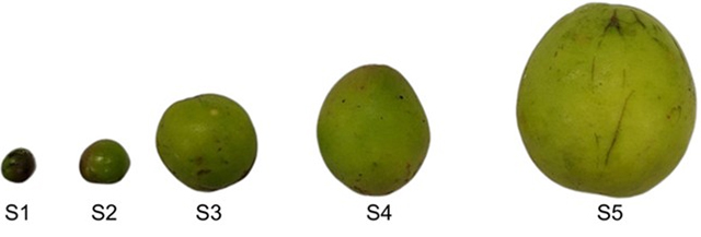
Fruits of H. speciosa var. gardneri were collected at different sizes corresponding to five phenological stages. The stages were determined based on fruit size, considering the length and diameter of 20 fruits randomly collected from four distinct populations (Fig. 1).
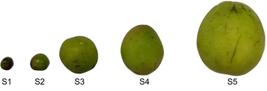
Phenological stages of Hancornia speciosa Gomes var. gardneri. S1: stage 1 (8-11 mm in length and diameter); S2: stage 2 (12-17 mm in length and diameter); S3: stage 3 (24-29 mm in length and diameter); S4: stage 4 (30-38 mm in length and diameter); S5: stage 5 (45-52 mm in length and diameter).
For each phenological stage, four fruits were collected from four H. speciosa individuals. Each fruit was considered one replicate, totaling four replicates. After collection, the fruits were taken to the Plant Anatomy Laboratory at the Institute of Biological Sciences, UFG, for anatomical and histochemical analyses.
Samples were taken from the middle region of fruits at stages S2, S3, S4, and S5. Given their reduced size, samples from stage S1 were considered as whole fruits. The material was fixed in FAA 50 (formaldehyde, propionic acid, 50% ethanol) at a 1:1:18 (v/v) ratio for 24 hours and then stored in 70% ethyl alcohol (Johansen, 1940).
The material was embedded in paraffin, then transverse and longitudinal sections were obtained using a rotary microtome (RM2245, Leica, Germany) at a thickness of 17 μm. After deparaffinization, the sections were stained with 0.5% Astra blue and 1% safranin, each for 15 minutes, to differentiate between non-lignified primary walls and lignified secondary walls, respectively. Permanent slides were mounted using vitral varnish (Paiva et al., 2006).
Histochemical tests to identify phenolic compounds were performed using 10% ferric chloride (Johansen, 1940). For this procedure, freehand transverse sections were obtained from fresh mangaba fruits.
Stomatal visualization was carried out through paradermal sections of the fruit epidermis using a steel blade. The samples were then cleared in a 1% sodium hypochlorite solution for five minutes and subsequently stained with toluidine blue (Abbade et al., 2009). Photomicrographs were obtained using an optical microscope (BX53F2, Olympus, Japan).
Results
Exocarp
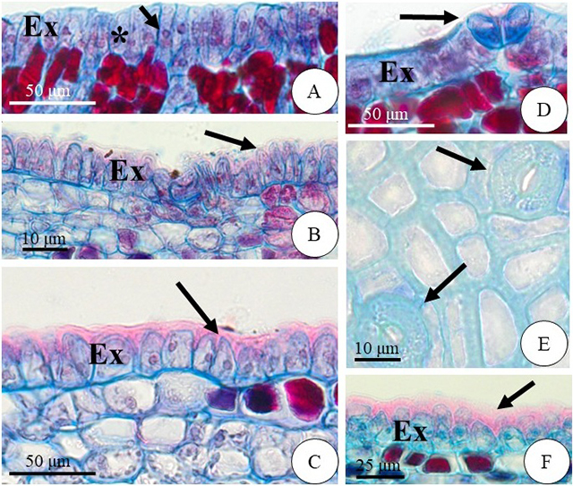
The exocarp of mangaba is uniseriate, with cells elongated primarily in the anticlinal direction (Fig. 2A-D). At the beginning of development, stage S1, the cells exhibit a more prominent nucleus and a visually denser cytoplasm, resulting in a slightly more intense staining (Fig. 2A). From stage S2 onward, the cells display a papillose appearance (Fig. 2B-C). Stomata undergo differentiation at stage S1 (Fig. 2D) and are fully developed by stage S5 (Fig. 2E).
In the early developmental stages (S1 and S2), the exocarp cells are covered by a thin cuticle. However, as growth progresses from stage S3 onward, the cuticle becomes thicker (Fig. 2F). At fruit maturity, the cuticle shows a pronounced thickening, which is clearly evident at stage S5 (Fig. 2F).
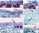
Photomicrographs of transverse (A-B), longitudinal (C, D-F), and paradermal (E) sections of mangaba fruit at various developmental stages. A) Stage S1: exocarp composed of anticlinally elongated cells (*) with a prominent nucleus (arrow). B) Stage S2 and C) Stage S5: exocarp cells exhibiting a papillose appearance (arrow). D) Stomata in development at stage S1 (arrow). E) Fully developed stomata at stage S5 (arrow). F) Thick cuticle at stage S5 (arrow). Ex = exocarp.
Mesocarp
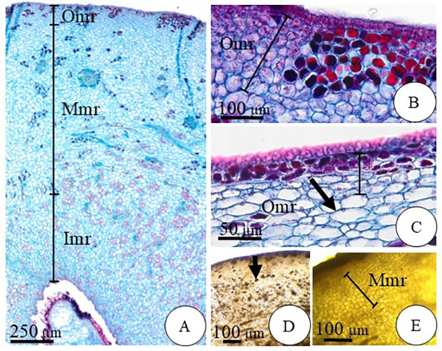
Based on fruit development, the mesocarp of mangaba can be divided into three regions, outer, middle, and inner, across all phenological stages (Fig. 3A).
Outer Region
In stages S1 and S2, the outer mesocarp is composed of up to eight layers of isodiametric parenchyma cells (Fig. 3B), with inconspicuous intercellular spaces. As the fruit develops, the parenchyma cells elongate in the periclinal direction (Fig. 3C), resulting in five densely arranged layers, similar to those observed in stages S3, S4, and S5. In the outer mesocarp, idioblasts containing phenolic compounds are abundant; in stages S1 and S2, they occur closer to the epidermis (Fig. 3D), reaching up to eight layers.
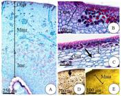
Photomicrographs of transverse sections of mangaba fruit at various developmental stages, highlighting the mesocarp. A) Stage S1: mesocarp regions outer, middle, and inner. B) Stage S2: isodiametric parenchyma cells. C) Stage S5: parenchyma cells elongated in the periclinal direction (arrow). D) Stage S1: parenchyma with idioblasts containing phenolic compounds (arrow). E) Stage S5: parenchyma. Omr = outer mesocarp region; Mmr = middle mesocarp region; Imr = inner mesocarp region.
Middle Region
In the middle mesocarp, the parenchyma cells are visibly larger at all phenological stages (Fig. 3A). However, idioblasts containing phenolic compounds were observed only during the early stages (S1 and S2) (Fig. 3D). In stages S3, S4, and S5, these idioblasts were not detected in the parenchyma (Fig. 3E).
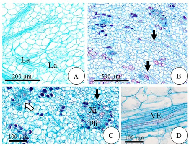
The laticifers are of the articulated and anastomosing type (Fig. 4A) and are distributed throughout the middle region of the mesocarp (Fig. 4B). However, from stage S3 onward, the laticifers appear to be less abundant.
The vascular bundles are of the concentric and collateral types (Fig. 4C), and the predominant vessel elements exhibit a helical pattern across all five phenological stages (Fig. 4D).
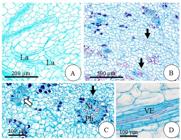
Photomicrographs of transverse (C) and longitudinal (A, B-D) sections of mangaba fruit at various developmental stages, highlighting the middle mesocarp. A) Stage S2: laticifers. B) Stage S1: arrowhead indicating laticifers distributed throughout the region. C) Stage S1: vascular bundles of the concentric type (arrow with black fill) and collateral type (arrow with white fill). D) Stage S5: vessel elements exhibiting a helical pattern. La = laticifers. Xi = xylem. Ph = phloem. VE = vessel elements.
Inner Region
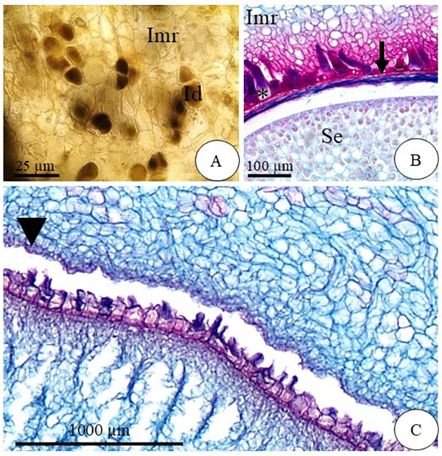
In the inner region of the fruits, idioblasts containing phenolic compounds can also be identified in stages S1 and S2 (Fig. 5A), but they are not observed in later stages. From stage S2 onward, laticifers appear to be less abundant. Furthermore, the cells of the outermost layers of this region are lignified at the S4 stage (Fig. 5B).
Endocarp
The endocarp, the innermost layer of the pericarp, is uniseriate, as observed in stage S2 (Fig. 5C), and remains attached to the seed coat, which bears elongated trichomes (Fig. 5B). These trichomes fit into indentations of the endocarp, which become more developed as the fruit grows, as observed at stage S2 (Fig. 5B).
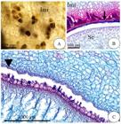
Photomicrographs of transverse (A) and longitudinal (B) sections of mangaba fruit at various developmental stages, highlighting the inner mesocarp and the endocarp. A) Stage S1: idioblasts containing phenolic compounds. B) Stage S4: seed coat (arrow) and trichomes (*). C) Stage S2: uniseriate endocarp (arrowhead); Imr = inner mesocarp region. Id = idioblasts. Se = seed.
Discussion
The uniseriate exocarp observed in mangaba fruits has also been reported in other Apocynaceae species (El-Fiki et al., 2019; Pirolla-Souza et al., 2019; Abdalla et al., 2021), suggesting that this may be a common characteristic of the family. In Prestonia riedelii (Apocynaceae), the exocarp is uniseriate with a thin cuticle during the early developmental stages, becoming progressively thicker as the fruit matures (Aguiar et al., 2009). Species that naturally occur in dry environments tend to develop thick cuticles (Riederer & Müller, 2006; Domínguez et al., 2011), as observed in mangaba (Fig. 2F).
One of the main functions of the cuticle is to reduce excessive water loss (Muller & Riederer, 2005). In mangaba, cuticle thickening plays a crucial role, particularly because mature fruits have a high water content and an uniseriate exocarp that can promote dehydration. Thus, the thick cuticle contributes to maintaining fruit quality (Abdalla et al., 2021).
From stage S2 onward, the cells of the exocarp become papillose, a feature not previously reported in studies of mangaba fruit. Papillae are epidermal cells with an outer, projected periclinal wall (Mantovani et al., 1995) and play a crucial role in reducing excessive transpiration (Toderich et al., 2010; Ribeiro et al., 2012). Additionally, they serve as a crucial physical barrier against the penetration of pathogenic fungi (Sherwood & Vance, 1976).
The growth of mangaba fruits was characterized by an increase in the size of the mesocarp parenchyma cells, a pattern particularly evident between S3 and S5 phenological stages. This increase in volume may be attributed to cell division, cell expansion, or a combination of both processes (Pabón-Mora & Litt, 2011), with cell expansion being directly related to the accumulation of fleshy tissue (Houel et al., 2010; Zhang et al., 2020). In fleshy bacoid-type fruits (Barroso et al., 1999), such as the mangaba, expansion occurs predominantly in the mesocarp, while the exocarp and endocarp remain thin until maturity (Cerri & Reale, 2020; Judkevich et al., 2022). This type of fruit has seeds surrounded by attractive pulp and is directly associated with attracting frugivorous animals (Ridley, 1930), which facilitates its dispersal and the establishment of the species in new environments.
In the early developmental stage, mangaba fruits contain numerous idioblasts with phenolic compounds located in the outer region of the mesocarp. The higher concentration of phenolics during the initial phase of fruit development may be associated with protection against ultraviolet radiation (Inostroza-Blancheteau et al., 2014) and herbivore attack (Singh et al., 2021), serving as a chemical defense strategy (Rodrigues et al., 2017; Jańczak-Pieniążek et al., 2023), as well as protection against high temperatures (Rivero et al., 2001).
The amount of phenolic compounds decreases as the fruit ripens, a pattern also observed in fruits of Butia capitata (Ventura et al., 2022), Rubus ulmifolius (Castro et al., 2023), Malus domestica (Wojdylo & Oszmianski, 2020), and H. speciosa (Freitas, 2013; Morgado et al., 2020). During ripening, the substrates produced are directed primarily toward fruit growth rather than the biosynthesis of phenolic compounds (Seraglio et al., 2018; Pereira et al., 2021). Moreover, as maturation progresses, the concentration of these compounds tends to decrease, resulting in fruits that become less astringent (Freitas, 2013).
The phenolic compounds present in idioblasts are associated with plant defense (Ribeiro et al., 2021). In H. speciosa, the accumulation of these compounds may be related to detoxification strategies against aluminum toxicity in the soil, also functioning as antioxidants under stress conditions (Tolrá et al., 2009; Rodrigues et al., 2017; Coimbra et al., 2023). Considering that Cerrado soils are naturally acidic and rich in aluminum (Ratke et al., 2021), the presence of phenolic compounds becomes essential for the adaptation and survival of mangabeira in this environment.
The presence of laticifer canals is a common feature among species of the Apocynaceae family (Gonçalves et al., 2018; Campos et al., 2021). The latex produced by these structures plays important defensive roles in plants, acting against herbivores and phytopathogens, while also helping to reduce water loss and contributing to wound healing (Tan et al., 2017; Ramos et al., 2019).
In the analyzed fruits of H. speciosa, the laticifers were characterized as articulated and anastomosed. In shoot apices of the same species, Souza et al. (2021) describe laticifers with ramifications originating from anastomoses, which corroborates the results obtained in this study. On the other hand, Abdalla et al. (2021), when analyzing exclusively mature fruits of H. speciosa var. pubescens, described laticifers as non-articulated and branched. This divergence may indicate structural differences between the studied materials, but it may also be related to different criteria of morphoanatomical interpretation and the stage of development considered. In this sense, the importance of ontogenetic studies that evaluate the formation of laticifers in many varieties of the species is reinforced.
The presence of trichomes on the seed coat was also reported by Abdalla et al. (2021) in all H. speciosa varieties analyzed. These structures play an important role in anchoring the seed to the soil, promoting hydration, and regulating internal temperature (Mamut et al., 2014). Mangaba seeds are recalcitrant, naturally exhibiting high moisture content, and maintaining this moisture is essential for ensuring their viability (Nunes et al., 2022). In this context, the increase in trichomes from stage S3 onward contributes significantly to retaining internal moisture.
The vascular system of mangaba fruits is composed of concentric and collateral bundles, which is consistent with other Apocynaceae species that exhibit concentric bundles (Esemann-Quadros et al., 2008; Shewale et al., 2022) and collateral bundles (Aguiar et al., 2009; Neto et al., 2020). This study represents the first characterization of vessel elements in H. speciosa, providing meaningful anatomical evidence that may support future taxonomic refinements within the species.
During mangaba fruit development, an increase in cuticle thickness, the presence of a papillose epidermis, and a reduction in the number of laticifers were observed. These anatomical changes are directly related to the final quality of the fruit. Additionally, the presence of phenolic compounds, which tend to decrease throughout ontogeny, also plays a significant role in this process. The laticifer canals present in the pericarp are of the articulated and anastomosing type, with a significant reduction beginning at stage S3.
The endocarp remains attached to the seed coat, which bears trichomes, a consistent feature across all developmental stages and essential for the species’ perpetuation.
The vascular bundles identified are of the concentric and collateral types, with a predominance of helical vessel elements present at all phenological stages. This study represents the first characterization of vessel elements in H. speciosa Gomes.
Acknowledgements
To the Coordenação de Aperfeiçoamento de Pessoal de Nível Superior (CAPES) for granting the scholarship.
References
-
Abbade LC, Paiva PDO, Paiva R, Castro EM, Centofante AR, Oliveira C. 2009. Anatomia foliar de ipê-branco (Tabebuia roseo-alba (Ridl.) Sand.) - Bignoniaceae, proveniente do cultivo ex vitro e in vitro Acta Scientiarum. Biological Sciences 31: 307-311. doi: 10.4025/actascibiolsci.v31i3.1937
» https://doi.org/10.4025/actascibiolsci.v31i3.1937 -
Abdalla DF, Ferreira INM, Moraes MG, Souza ERB. 2021. Comparative anatomy and histochemistry of fruits of four varieties of Hancornia speciosa Gomes (Apocynaceae). Revista Desafios 8: 31-41. doi: 10.20873/uftv8-10593
» https://doi.org/10.20873/uftv8-10593 -
Aguiar S, Carmello-Guerreiro SM, Kinoshita LS. 2009. Ontogenia e estrutura do pericarpo de Prestonia riedelii (Müll. Arg.) Markgr.) (Apocynaceae). Acta Botânica Brasilica 23: 729-737. doi: 10.1590/S0102-33062009000300012
» https://doi.org/10.1590/S0102-33062009000300012 - Barroso GM, Morim MP, Peixoto AL, Ichaso CLF. 1999. Frutos e sementes: morfologia aplicada à sistemática de dicotiledôneas. Viçosa, Editora UFV.
-
Campos EPCF, Santos DM, Sá RD, Randau KP. 2021. Microscopic analysis applied to the quality control of Hancornia speciosa Gomes. Microscopy and Microanalysis 27: 1226-1233. doi: 10.1017/S1431927621012058
» https://doi.org/10.1017/S1431927621012058 -
Cardoso LM, Reis BL, Oliveira DS, Pinheiro-Sant’ana HM. 2014. Mangaba (Hancornia speciosa Gomes) from the Brazilian Cerrado: nutritional value, carotenoids and antioxidant vitamins. Fruits 69: 89-99. doi: 10.1051/fruits/2013105
» https://doi.org/10.1051/fruits/2013105 -
Castro RI, Vásquez-Rojas C, Cortiella MGI, Parra-Palma C, Ramos P, Morales-Quintana L. 2023. Evolution of the Volatile Organic Compounds, Phenols and Antioxidant Capacity during Fruit Ripening and Development of Rubus ulmifolius Schott Fruits. Horticulturae 9: e13. doi: 10.3390/horticulturae9010013
» https://doi.org/10.3390/horticulturae9010013 -
Cerri M, Reale L. 2020. Anatomical traits of the principal fruits: An overview. Scientia Horticulturae 270: e109390. doi: 10.1016/j.scienta.2020.109390
» https://doi.org/10.1016/j.scienta.2020.109390 -
Christenhusz MJM, Byng JW. 2016. The number of known plants species in the world and its annual increase. Phytotaxa 261: 201-217. doi: 10.11646/phytotaxa.261.3.1
» https://doi.org/10.11646/phytotaxa.261.3.1 -
Coimbra RR, Matos VF, Viana RHO, Ferreira WM, Lólis SF. 2023. Biometry of fruits and seeds and correlation between variables in natural populations of Hancornia speciosa Gomes (Apocynaceae). Diversitas Journal 8: 157-171. doi: 10.48017/dj.v8i1.2357
» https://doi.org/10.48017/dj.v8i1.2357 -
Collevatti RG, Rodrigues EE, Vitorino LC, Lima-Ribeiro MS, Chaves LJ, Telles MPC. 2018. Unravelling the genetic differentiation among varieties of the neotropical savanna tree Hancornia speciosa Gomes. Annals of Botany 122: 973-984. doi: 10.1093/aob/mcy060
» https://doi.org/10.1093/aob/mcy060 -
Domínguez E, Cuartero J, Heredia A. 2011. An overview on plant cuticle biomechanics. Plant Science 181: 77-84. doi: 10.1016/j.plantsci.2011.04.016
» https://doi.org/10.1016/j.plantsci.2011.04.016 -
El-Fiki MA, El-Taher AM, El-Gendy AG, Lila MI. 2019. Morphological and anatomical studies on some taxa of family Apocynaceae. Al-Azhar Journal of Agricultural Research 44: 136-147. doi: 10.21608/ajar.2019.59750
» https://doi.org/10.21608/ajar.2019.59750 - Embrapa - Empresa Brasileira de Pesquisa Agropecuária. 2013. Sistema brasileiro de classificação de solos. 3nd edn. Brasília, Embrapa.
-
Esemann-Quadros K, Mota AP, Kerbauy GB, Guerra MP, Ducroquet JPHJ, Pescador R. 2008. Estudo anatômico do crescimento do fruto em Acca sellowiana Berg. Revista Brasileira de Fruticultura 30: 296-302. doi: 10.1590/S0100-29452008000200005
» https://doi.org/10.1590/S0100-29452008000200005 -
Fagundes NF, Mariath JEA. 2010. Morphoanatomy and ontogeny of fruit in Bromeliaceae species. Acta Botânica Brasilica 24: 765-779. doi: 10.1590/S0102-33062010000300020
» https://doi.org/10.1590/S0102-33062010000300020 - Freitas CCM. 2013. Morfologia e anatomia de frutos e sementes da tribo Willughbeieae (Apocynaceae, Rauvolfioideae). MSc Thesis, Universidade Estadual de Campinas, Brazil.
-
Ganga RMD, Ferreira GA, Chaves LJ, Naves RV, Nascimento JL. 2010. Caracterização de frutos e árvores de populações naturais de Hancornia speciosa Gomes do Cerrado. Revista Brasileira de Fruticultura 32: 101-113. doi: 10.1590/S0100-29452010005000019
» https://doi.org/10.1590/S0100-29452010005000019 -
Giacomin AC, Araújo MGP, Santos PMRS. 2022. Developmental morphoanatomy of the fruit of Isertia hypoleuca Benth. (Rubiaceae). Flora 288: e152006. doi: 10.1016/j.flora.2022.152006
» https://doi.org/10.1016/j.flora.2022.152006 -
Gonçalves MP, Mercadante-Simões MO, Ribeiro LM. 2018. Ontogeny of anastomosed laticifers in the stem apex of Hancornia speciosa (Apocynaceae): A topographic approach. Protoplasma 255: 1713-1724. doi: 10.1007/s00709-018-1262-9
» https://doi.org/10.1007/s00709-018-1262-9 -
Houel C, Bounon R, Chaib J et al 2010. Patterns of sequence polymorphism in the fleshless berry locus in cultivated and wild Vitis vinifera accessions. Plant Biology 10: 284. doi: 10.1186/1471-2229-10-284
» https://doi.org/10.1186/1471-2229-10-284 -
Inostroza-Blancheteau C, Reyes-Díaz M, Arellano A et al 2014. Effects of UV-B radiation on anatomical characteristics, phenolic compounds and gene expression of the phenylpropanoid pathway in highbush blueberry leaves. Plant Physiology and Biochemistry 85: 85-95. doi: 10.1016/j.plaphy.2014.10.015
» https://doi.org/10.1016/j.plaphy.2014.10.015 - Johansen DA. 1940. Plant microtechnique. New York, McGraw-Hill Book Company.
-
Judkevich MD, Salas RM, Gonzalez AM. 2022. Anatomy and development of the edible fruits of Cordiera concolor (Rubiaceae). Annals of the Brazilian Academy of Sciences 94: e20210071. doi: 10.1590/0001-3765202220210071
» https://doi.org/10.1590/0001-3765202220210071 -
IBGE - Instituto Brasileiro de Geografia e Estatística. 2023. Produção de Mangaba. https://www.ibge.gov.br/explica/producao-agropecuaria/mangaba/go 04 Apr 2025.
» https://www.ibge.gov.br/explica/producao-agropecuaria/mangaba/go -
Islam MS, Lucky RAA. 2019. A study on different plants of the Apocynaceae family and their medicinal uses. Universal Journal of Pharmaceutical Research 4: 40-44. doi: 10.22270/ujpr.v4i1.235
» https://doi.org/10.22270/ujpr.v4i1.235 -
Jańczak-Pieniążek M, Cichoński J, Michalik P, Chrzanowski G. 2023. Effect of Heavy Metal Stress on Phenolic Compounds Accumulation in Winter Wheat Plants. Molecules 28: e241. doi: 10.3390/molecules28010241
» https://doi.org/10.3390/molecules28010241 -
Li Z, Yang H, Li P, Liu J, Wang J, Xu Y. 2013. Fruit biomechanics based on anatomy: A review. International Agrophysics 27: 97-106. doi: 10.2478/v10247-012-0073-z
» https://doi.org/10.2478/v10247-012-0073-z -
Mamut J, Tan D-Y, Baskin CC, Baskin JM. 2014. Role of trichomes and pericarp in the seed biology of the desert annual Lachnoloma lehmannii (Brassicaceae). Ecological Research 29: 33-44. doi: 10.1007/s11284-013-1098-x
» https://doi.org/10.1007/s11284-013-1098-x -
Mantovani A, Gomes M, Gomes DMS, Vieira RC. 1995. Anatomia foliar de Rudgea decipiens Müll. Arg. e R. macrophylla Benth. (Rubiaceae). Acta Botanica Brasilica 9: 247-261. doi: 10.1590/S0102-33061995000200005
» https://doi.org/10.1590/S0102-33061995000200005 - Monachino, J. 1945. A revision of Hancornia (Apocynaceae). Lilloa 11: 19-48.
-
Morgado CMA, Lima ACS, Siqueira APS, Souza ERB, Cunha Junior LC. 2020. Bioactive compounds and antioxidant activity of mangaba. Bioscience Journal 36: 473-486. doi: 10.14393/BJ-v36n2a2020-47692
» https://doi.org/10.14393/BJ-v36n2a2020-47692 -
Muller C, Riederer M. 2005. Plant surface properties in chemical ecology. Journal of Chemical Ecology 31: 2621-2651. doi: 10.1007/s10886-005-7617-7
» https://doi.org/10.1007/s10886-005-7617-7 -
Neto ILC, Pace MR, Douglas NA et al 2020. Diversity, distribution, development, and evolution of medullary bundles in Nyctaginaceae. American Journal of Botany 107: 707-725. doi: 10.1002/ajb2.1471
» https://doi.org/10.1002/ajb2.1471 -
Nunes VV, Silva-Mann R, Souza JL, Lima LCDP, Torres MFO, Álvares-Carvalho SV. 2022. Physiological and molecular changes in seeds of Hancornia speciosa Gomes stored in conservative solutions. Journal of Seed Science 44: e202244018. doi: 10.1590/2317-1545v44239482
» https://doi.org/10.1590/2317-1545v44239482 -
Paiva JGA, Fank-de-Carvalho SM, Magalhães MP, Graciano-Ribeiro D. 2006. Verniz vitral incolor 500®: uma alternativa de meio de montagem economicamente viável. Acta Botanica Brasilica 20: 257-264. doi: 10.1590/S0102-33062006000200002
» https://doi.org/10.1590/S0102-33062006000200002 -
Pabón-Mora N, Litt A. 2011. Comparative anatomical and developmental analysis of dry and fleshy fruits of Solanaceae. American Journal of Botany 98: 1415-1436. doi: 10.3732/ajb.1100097
» https://doi.org/10.3732/ajb.1100097 -
Pereira APA, Angolini CFF, Adani HB et al 2021. Impact of ripening on the health-promoting components from fruta-do-lobo (Solanum lycocarpum St. Hill). Food Research International 139: e109910. doi: 10.1016/j.foodres.2020.109910
» https://doi.org/10.1016/j.foodres.2020.109910 -
Pirolla-Souza A, Arruda RCO, Pace MR, Farinaccio MA. 2019. Leaf anatomical characters of Rhabdadenia (Rhabdadenieae, Apocynaceae), their taxonomic implications, and notes on the presence of articulated laticifers in the genus. Plant Systematics and Evolution 305: 797-810. doi: 10.1007/s00606-019-01608-z
» https://doi.org/10.1007/s00606-019-01608-z -
Ramos MV, Demarco D, Souza ICC, Freitas CDT. 2019. Laticifers, latex, and their role in plant defense. Trends in Plant Science 24: 553-567. doi: 10.1016/j.tplants.2019.03.006
» https://doi.org/10.1016/j.tplants.2019.03.006 -
Ratke RF, Rodrigues TS, Cardoso AA, Frazão JJ, Brasil EPF, Zuffo AM. 2021. Propriedades químicas do solo e morfologia radicular da soja sob aplicação de diferentes granulometrias de calcário. Revista em Agronegócio e Meio Ambiente 14: 469-481. doi: 10.17765/2176-9168.2021v14n2e8234
» https://doi.org/10.17765/2176-9168.2021v14n2e8234 - Ridley HN. 1930. The dispersal of plants throughout the world. Ashford, L. Reeve & Co.
-
Ribeiro C, Marinho C, Teixeira S. 2021. Uncovering the neglected floral secretory structures of Rhamnaceae and their functional and systematic significance. Plants 10:736. doi: 10.3390/plants10040736
» https://doi.org/10.3390/plants10040736 -
Ribeiro MNO, Carvalho SP, Pereira FJ, Castro EM. 2012. Leaf anatomy of the cassava as related to potential for tolerance to different environmental conditions. Revista Ciência Agronômica 43: 354-361. doi: 10.1590/S1806-66902012000200019
» https://doi.org/10.1590/S1806-66902012000200019 - Riederer M, Müller C. 2006. Biology of plant cuticle. Oxford, Blackwell Publishing.
-
Rivero RM, Ruiz JM, García PC, López-Lefebre LR, Sánchez E, Romero L. 2001. Resistance to cold and heat stress: Accumulation of phenolic compounds in tomato and watermelon plants. Plant Science 160: 315-321. doi: 10.1016/S0168-9452(00)00395-2
» https://doi.org/10.1016/S0168-9452(00)00395-2 -
Rodrigues AA, Vasconcelos- Filho SC, Rodrigues CA et al 2017. Aluminum influence on Hancornia speciosa seedling emergence, nutrient accumulation, growth and root anatomy. Flora 236: 9-14. doi: 10.1016/j.flora.2017.09.008
» https://doi.org/10.1016/j.flora.2017.09.008 -
Seraglio SKT, Schulz M, Nehring P et al 2018. Nutritional and bioactive potential of Myrtaceae fruits during ripening. Food Chemistry 239: 649-656. doi: 10.1016/j.foodchem.2017.06.118
» https://doi.org/10.1016/j.foodchem.2017.06.118 -
Sherwood RT, Vance CP. 1976. Histochemistry of papillas formed in Reed Canarygrass leaves in response to noninfecting pathogenic fungi. Phytopathology 66: 503-510. doi: 10.1094/Phyto-66-503.
» https://doi.org/10.1094/Phyto-66-503 -
Shewale S, Undale V, Shelar M et al 2022. Morphological and anatomical characterization of Plumeria obtusa L.: An Ayurvedic medicinal plant. Annals of Phytomedicine 11: 787-793. doi: 10.54085/ap.2022.11.2.97
» https://doi.org/10.54085/ap.2022.11.2.97 - Silva SMC, Pires LL, Ribeiro KO, Cruz GHT, Dourado FO. 2017. Caracteres morfológicos de variedades botânicas de Hancornia speciosa Gomes. Revista Mirante 10: 128-145.
-
Silva TAC, Vasconcelos-Filho SC, Rodrigues AA et al 2020. Tolerance of Hancornia speciosa Gomes (Apocynaceae) to potassium fluoride: physiological and anatomical traits. Acta Physiologiae Plantarum 42: 153. doi: 10.1007/s11738-020-03144-7
» https://doi.org/10.1007/s11738-020-03144-7 - Simões AO, Kinoshita LS. 2002. The Apocynaceae S. STR. Of the Carrancas region, Minas Gerais, Brazil. Darwiniana 40: 129-169.
-
Singh S, Kaur I, Kariyat R. 2021. The Multifunctional Roles of Polyphenols in Plant-Herbivore Interactions. International Journal of Molecular Sciences 22: e1442. doi: 10.3390/ijms22031442
» https://doi.org/10.3390/ijms22031442 -
Souza AIRC, Cordeiro KR, Gonçalves MP, Ribeiro LM, Mercadante-Simões MO. 2021. The development of anastomosed laticifers in the stem apical meristem and vascular cambium of Hancornia speciosa (Apocynaceae) is related to climatic seasonality. Trees 35: 1317-1328. doi: 10.1007/s00468-021-02118-7
» https://doi.org/10.1007/s00468-021-02118-7 -
Tan D, Hu X, Fu L et al 2017. Comparative morphology and transcriptome analysis reveals distinct functions of the primary and secondary laticifer cells in the rubber tree. Scientific Reports 7: e3126. doi: 10.1038/s41598-017-03083-3
» https://doi.org/10.1038/s41598-017-03083-3 -
Thadeo M, Hampilos KE, Stevenson DW. 2015. Anatomy of fleshy fruits in the monocots. American Journal of Botany 102: 1757-1779. doi: 10.3732/ajb.1500204
» https://doi.org/10.3732/ajb.1500204 - Toderich KN, Shuyskaya EV, Khujanazarov TM, Ismail S, Kawabata Y. 2010. The Structural and Functional Characteristics of Asiatic Desert Halophytes for Phytostabilization of Polluted Sites. In: Ashraf M, Ozturk M, Ahmad M. (eds.) Plant Adaptation and Phytoremediation. Springer, p. 245-274
-
Tolrá R, Barceló J, Poschenrieder C. 2009. Constitutive and aluminium-induced patterns of phenolic compounds in two maize varieties differing in aluminium tolerance. Journal of Inorganic Biochemistry 103: 1486-1490. doi: 10.1016/j.jinorgbio.2009.06.013
» https://doi.org/10.1016/j.jinorgbio.2009.06.013 -
Ventura LJM, Santos HCM, Simões MOM, Lopes PSN, Ribeiro LM. 2022. Pericarp histogenesis and histochemistry during fruit development in Butia capitata (Arecaceae). Protoplasma 259: 1521-1539. doi: 10.1007/s00709-022-01749-y
» https://doi.org/10.1007/s00709-022-01749-y -
Vieira MC, Souza ERB, Paula MSP, Naves RV, Silva GD. 2017. Mangabeira (Hancornia speciosa Gomes): uma frutífera promissora do Brasil. Scientific Electronic Archives 10: 45-55. doi: 10.36560/1022017354
» https://doi.org/10.36560/1022017354 -
Wojdylo A, Oszmianski J. 2020. Antioxidant activity modulated by polyphenol contents in apple and leaves during fruit development and ripening. Antioxidants 9: e567. doi: 10.3390/antiox9070567
» https://doi.org/10.3390/antiox9070567 -
Zhang W-W, Zhao S-Q, Zhang L-C, Xing Y, Jia W-S. 2020. Changes in the cell wall during fruit development and ripening in Fragaria vesca Plant Physiology and Biochemistry 154: 54-65. doi: 10.1016/j.plaphy.2020.05.028
» https://doi.org/10.1016/j.plaphy.2020.05.028
All data supporting the findings of this study are included in the article.