Essential oil vapor phase approach for antimicrobial evaluation using Hesperozygis myrtoides (A. St.-Hill. ex Benth.) Epling (Lamiaceae) as a model
Abstract
The demand for less hazardous and environmentally friendly surface disinfectants is growing and essential oils may be efficient alternatives. Vapor phase assay is a promising technique which explore the volatility of essential oils for determining the antimicrobial activity. In this study, we modified the conditions of the inverted Petri dish technique to evaluate the minimum inhibitory concentrations (MIC). As a proof of concept, the antimicrobial activity of Hesperozygis myrtoides essential oil (HM) was studied for the first time in the vapor phase. According to the established protocol the oil volatilization dynamics was used to determine the major compounds in the vapor phase. After 270 min, the relative percentage was like that observed in the crude oil analysis. The HM was also evaluated for safety (IC50=921.2 µg ml-1), being considered safe in case of acute oral exposure. The oil vapors completely inhibited the growth of Staphylococcus aureus (MIC=0.39 mg l-1), Candida albicans (MIC=0.83 mg l-1), Aspergillus brasiliensis (MIC=378 mg l-1) and Bacillus subtilis (MIC=704.8 mg l-1). This protocol offers an economical and efficient alternative for antimicrobial activity and safety determination of essential oils, can be used for the evaluation of several applications of essential oils, as disinfectant agent in inhabited environments.
Key words
antimicrobial activity; cytotoxicity for dispersing essential oil; inverted Petri dish method; vaporization dynamics
INTRODUCTION
In recent years, there is a growing consensus that cleaning and disinfecting surfaces in common environments are important to prevent infection spreading. Community-based infections have a strong impact on the most fragile populations, such as children, the elderly and the immunosuppressed. Some potential pathogens include Staphylococcus aureus, Pseudomonas aeruginosa, fecal coliforms, as well as molds, yeasts, and viruses (Scott et al. 2020). Although conventional products, such as bleach, are widely used as sanitizers, the search for new disinfectants that are less hazardous and environmentally friendly is growing (Goodyear et al. 2015).
Essential oils have a recognized range of antimicrobial activities against bacteria, Gram-negative and Gram-positive, and pathogenic fungi (Powers et al. 2019). Some in vitro studies are conducted with essential oils in liquid phase, but their application as a liquid sanitizer requires large amounts. On the other hand, the use of essential oil in its vapor phase, applied as a fumigant, offers advantages, such as the possibility of reaching hard-to-reach areas by manual cleaning, greater antimicrobial potency, since the volatilized components have free binding to microorganisms, and a lower amount of used oil, reducing the cost of its use (Reyes-Jurado et al. 2020).
Many studies have explored the volatility of essential oils to develop methods to determine antimicrobial activity, based on the creating a controlled atmosphere. Some of them use inverted Petri dish (Kloucek et al. 2012a), agar vapor (Inouye et al. 2006), glass or plastic apparatus with essential oil atmosphere (Szczerbanik et al. 2007), microplate with dissolved essential oil (Feyaerts et al. 2017), airtight box, and its variations (Amat et al. 2017, Inouye et al. 2001), allowing the oil volatile components to be released and come into direct contact with the microorganisms wall.
However, most of these assays determine relative antimicrobial potential; it is impossible to compare the results due to variations within each technique applied; use of different units of measurement of minimum inhibitory concentrations (MIC), when the calculation is allowed, showing a limited uniformity in the methodological criteria adopted for the study of this activity (Amat et al. 2017, Reyes-Jurado et al. 2020) and, in many cases, the use of devices that are difficult to access. That is why, it is necessary to modify the testing methodology, making it reproducible and viable.
To develop an effective approach that can be used to evaluate essential oils for disinfection of inhabited environments, we designed a method based on the inverted Petri dish for simple, fast, and low-cost determination of the volatile oils antimicrobial potential in vapor phase accompanied by dynamic volatilization and cytotoxicity assay for essential oil dispersion.
In our investigation, we verified the suitability of our method for evaluating essential oils in the vapor phase using the essential oil from Hesperozygis myrtoides (St.Hill. Ex Benth.), a native Brazilian species. This particular essential oil was chosen due to its demonstrated activity against a spectrum of microorganisms, including yeasts, gram-negative, and gram-positive bacteria such as Candida albicans, Escherichia coli, and Staphylococcus aureus (Martini et al. 2011).
MATERIALS AND METHODS
Chemicals
The analytical solvents were purchased from Synth (Brazil) and Merck (Germany). Other reagents such as (3-(4,5-dimethylthiazol-2-yl)-5-(3-carboxymethoxyphenyl)-2-(4-sulfophenyl)-2H-tetrazolium inner salt)/phenazine methosulfate (MTS/PMS) were obtained from Sigma-Aldrich (USA). Culture media for microorganisms were purchased from Difco (USA) and Dulbecco’s Modified Eagle Medium (DMEM), fetal bovine serum, glutamine for cell cultures from Sigma-Aldrich (USA). The antibiotics nystatin and chloramphenicol were purchased from USP (USA), amikacin and doxorubicin from Sigma-Aldrich (USA), ciprofloxacin and tetracycline from Galena (Brazil).
Plant material and essential oil extraction
Hesperozygis myrtoides was collected in November 2013, in Campos do Jordão, São Paulo, Brazil (22°42’52,8” S, 45°27’42,7” W) (SISBIO No. 2371522). The botanical identification was confirmed by Dr. Inês Cordeiro and the voucher was deposited in herbarium of Institute of Botany, São Paulo, Brazil with reference number PEREIRA 02. This study is registered on the SisGen platform for access to genetic heritage (Registration number AF1FF34), in accordance with the Brazilian Biodiversity Law (13.123/2015).
The essential oil (HM) was extracted from the leaves by hydrodistillation for 4h with a Clevenger type apparatus, in quadruplicate, and stored at -22°C. The yield was calculated based on the dry weight of the plant.
Gas chromatography/mass spectrometry analysis (GC/MS)
The chemical composition of HM (diluted in acetone 1:99, v/v) was determined by gas chromatography coupled to mass spectrometry performed in Agilent 6890 Series GC (Agilent, USA) interfaced with a 5973 series quadrupole MS detector (Agilent, USA), equipped with a DB-5 column (30 x 0.25 mm i.d. x 0.25 µm (Agilent, USA). Injection volume of 1.0 µl. The scan time 0.1 scan/sec and mass range m/z 35-500. Helium was the carrier gas (flow rate of 1 ml min-1). The column temperature was initially held at 40°C for 1 min and subsequently increased to 240°C at 3°C min-1, keeping it at this temperature for 10 min; injection and detection temperature: 250 °C, electron ionization: 70 eV. The components were identified by comparing retention indices (calculated in relation to C5-C28 n-alkanes on DB-5 column) and mass spectra evaluated by analysis with those reported in the literature, software-based library search and visual interpretation of the mass spectra (Adams 2007, NIST 2019).
Headspace GC-MS analysis
The headspace volatile compounds of HM were obtained using a gas chromatograph Agilent 6890 Series GC (Agilent, USA) interfaced with a 5973 series quadrupole MS detector (Agilent, USA), equipped with a CP 8944 VF-5ms column (30 m x 0.25 mm x 0.25 µm, Agilent). In this method, 7 µl (6.3 µg) of HM were added in a 0.062 l sealed flask, with similar headspace volume as a Petri dish (0.054 l). The flask was maintained at 37°C and the evaporation products were collected (1.0 ml) with a Gastight syringe (Hamilton, USA) at 30, 90, 150, 210 and 270 min and injected into the chromatograph. The column temperature was initially held at 60°C and then, subsequently increased to 240°C at 3°C min-1; injection and detection temperature were 220°C and 209°C, respectively; electron ionization: 70 eV; the carrier gas used was helium (flow rate of 1 ml min-1). The compounds identification was carried out by comparing the retention indices and mass spectra with data found in the literature.
Mouse embryo fibroblast BALB/3T3 clone A31 cell cultures
BALB/3T3 clone A31 cell line (ATCC CCL-163) were cultivated in DMEM medium supplemented with 10% fetal bovine serum (v/v), 4 mmol l-1 glutamine and 100 U ml-1 penicillin, 100 mg ml-1 streptomycin and 0.025 mg ml-1 amphotericin (encoded as: DM10 medium). They were incubated at 37°C under 5% CO2 atmosphere in a humidified atmosphere until reaching subconfluency (approximately 80%). The cell detachment was performed with trypsin 0.05% (w/v)/EDTA 0.02% (w/v) in phosphate buffer pH 7.2. The cell suspension was centrifuged, the supernatant discarded, and the pellet suspended in DM10 medium.
Cytotoxicity assay
The cytotoxic effect of HM was measured using the MTS assay (ICCVAM 2006). The HM diluted in DMSO-DM10 was evaluated at a serial concentration in well (14.75 to 0.15 mg ml-1, w/v). In brief, the cells were seeded (2x10⁴ cells/well) in 96-well plate and incubated for 24h. Then, 50 µl of HM dilution were added in each well plus 50 µl of DM10. After 24 h of incubation, the culture medium was removed. The cells were washed with NaCl solution (0.9%, w/v) and 100 µl of MTS/PMS was added and incubated during 3 h. After this period, the supernatant was removed, the cells were again washed with saline and 150 µl of desorption solution (acetic acid-water-ethanol, 1:49:50, v/v/v) were added. DMSO at the maximum concentration used in well (4.67 mg ml-1, w/v) did not affect cell viability. Positive control was given by doxorubicin. All tests were performed in triplicate.
The absorbance was read at 490 nm (Biotek Synergy HT, USA). The cell viability dose-response curves were determined and the IC10 and IC50 values were calculated. The IC50 values were used to determine the medium lethal dose (LD50), according to the following equation 3 (ICCVAM 2001, 2006):
Based on LD50, the toxicity was estimated using the classification by categories established by the United Nations Globally Harmonized System (GSH) for which categories 1 (LD50 <5 mg kg-1 ) and 2 (LD50 5 - 50 mg kg-1) are fatal if ingested; category 3 (LD50 50 - 300 mg kg-1) is toxic if ingested; category 4 (LD50 300 - 2000 mg kg-1) is dangerous if ingested; category 5 (LD50> 5000 mg kg-1) not classified (Bulgheroni et al. 2009).
Microbial cultures
The tests were performed against six microorganism strains recommended for testing surface disinfection (USP 2014). Fungal species were the filamentous fungus Aspergillus brasiliensis (ATCC 16404) and the yeast Candida albicans (ATCC 10231). Gram-positive bacteria included S. aureus (ATCC 6538) and Bacillus subtillis (ATCC 6633). Pseudomonas aeruginosa (ATCC 9027) and Escherichia coli (ATCC 8739) were the Gram-negative bacteria tested. C. albicans and A. brasiliensis cultures were grown at 28°C in a Sabouraud dextrose broth (SDB) for 48h and 96h, respectively. All bacteria cultures were incubated at 37°C for 24 h in tryptic soy broth (TSB). An aliquot from each culture was spread on nutrient agar (Sabouraud dextrose agar, SDA, for fungi; tryptic soy agar, TSA, for bacteria).
A single colony from each plate was suspended in saline (0.85%, m/v). The cell density was estimated by comparing turbidity with a barium sulfate solution according the McFarland scale (Garzoli et al. 2021). The optical density of the scale tube 0.5 McFarland was compared to the measured optical density at 630 nm for the microorganism suspension (spectrophotometer Micronal, USA) and the value was estimated at 1.5 x 10⁸ colony forming units (CFU ml-1). For A. brasiliensis, three slant tubes washing with sterile saline 0.85% (m/v) containing 1% (m/v) polysorbate 80 was used, to avoid aggregation of the spores. Serial dilutions were made with all microorganisms to obtain concentrations from 1.5 x 10⁸ to 1.5 x 10² CFU ml-1. At the end of each standardization, microbial dilution was confirmed by CFU in the agar nutrient.
Minimum inhibitory concentration (MIC) in the broth microdilution method (liquid phase)
The antimicrobial assay was evaluated by microdilution broth method (Moreno et al. 2009). The inoculum was prepared by adjusting the final concentration to 1.5×10³ CFU ml-1 in each well of the 96-well plate. HM was diluted in DMSO-methanol (1:1, v/v) to make a final concentration in the assay of the 151,000-19 mg l-1. Microplates were incubated at 28°C for 48 h for the yeast, at 28°C for 96 h for the fungus and at 37°C for 24 h for the bacteria. The growth inhibition was measured by reading the absorbance at a wavelength of 630 nm, using a multi-well scanning spectrophotometer (Biotek, Synergy HT Multi-Mode, USA); except for A. brasiliensis, where the growth was evaluated only visually. In all assays, nystatin, chloramphenicol, amikacin, ciprofloxacin, and tetracycline were used as positive controls at concentrations of 50 µg ml-1 in the microplate wells. All tests were performed in triplicate.
The lowest concentration of HM that resulted in no microbial growth was considered as the minimum inhibitory concentration (MIC). HM dilutions with no detectable growth in the microplate were transferred into agar nutrient and incubated to determine the lowest lethal concentration (minimum bactericidal/fungicidal concentration, MBC/MFC). HM antimicrobial activity category for surfaces was determined based on the classification proposed by Djabou et al. (2013), where MIC value between 50,000 and 25,000 mg l-1 are considered not active; moderately active for MIC values between 12,500 and 3,000 mg l-1; active for MIC values between 2,000 and 400 mg l-1; and extremely active for MIC values ≤ 200 mg l-1.
Minimum inhibitory concentration (MIC) by the inverted dish method (vapor phase)
The vapor phase method was adapted from literature (Kloucek et al. 2012a, b).
The test was developed in Petri dishes (ø 85 mm) containing 20 ml of appropriate medium (SDA for fungi and TSA for bacteria) previously inoculated with a saline microorganism suspension with cell density between 2.5 - 3.5 ×10² CFU ml-1.
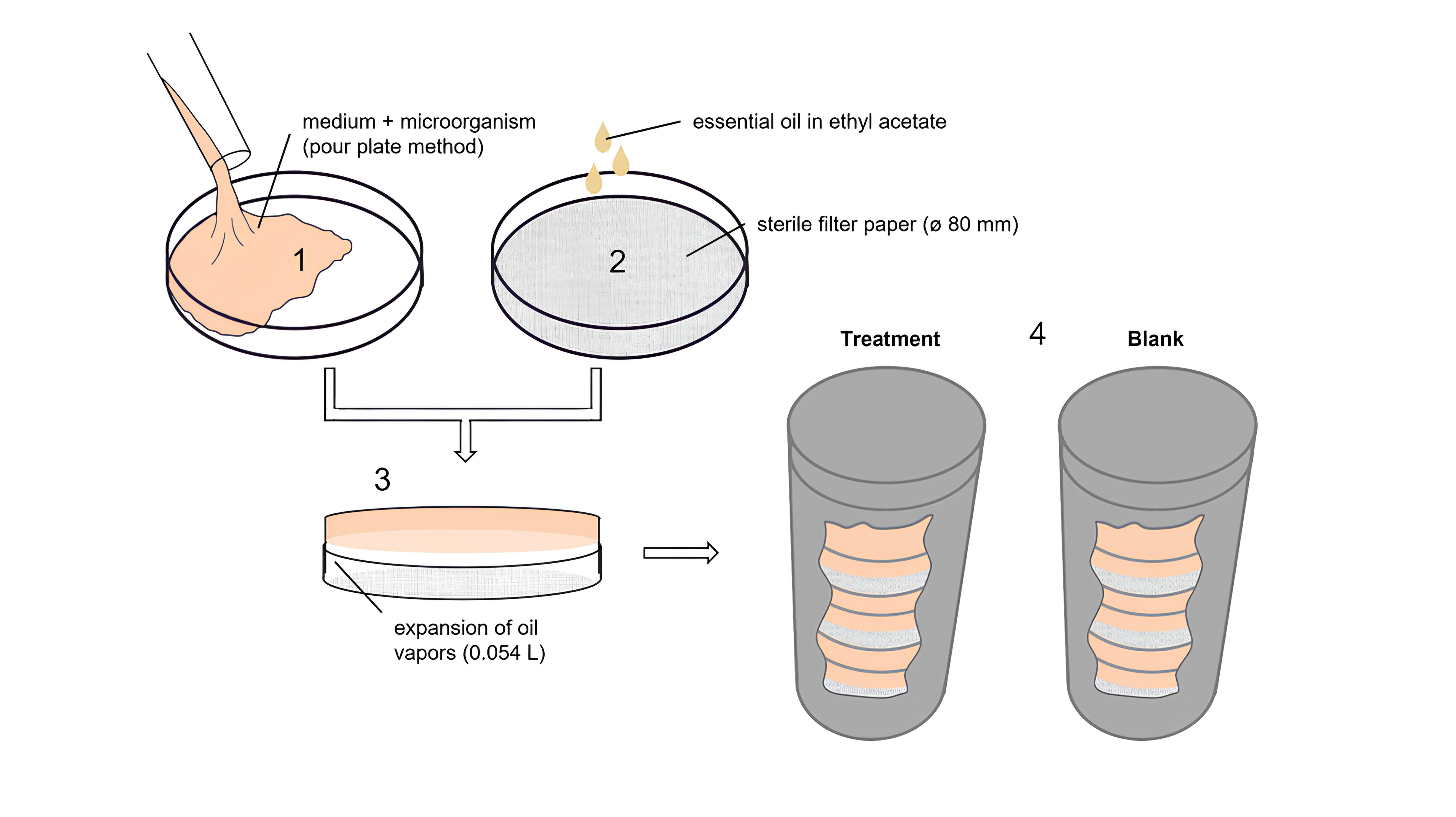
HM was diluted in ethyl acetate at serial concentrations. 500 µl of each HM concentration was distributed on the surface of a sterile blank filter paper (i.d. 80 mm) by micropipette, placed on top of a Petri dish cover (inverted dish method). For total removal of ethyl acetate, the solvent could evaporate for 1 min (standardized time). Using this procedure, ethyl acetate had no effect on the viability of any of the tested microorganism and there was no significant HM loss (data not shown). The dishes were closed with the inoculated bottoms and sealed with Parafilm® to prevent moisture loss. The dishes were placed inside a stainless-steel cylinder, previously disinfected with 70% ethanol (Fig. 1). Plates were incubated following same time and temperature used in broth microdilution method. The growth inhibition was determined by CFU counting were of each dish. Blanks were prepared by adding 500 µl of ethyl acetate to the filters paper and the incubation was carried out in an exclusive incubator. All tests were performed in triplicate.

Schematic representation for the vapor phase antimicrobial assay. The model experiment consists of 1. Petri dish (ø 85 mm) containing 20 ml of culture medium inoculated with the desired microorganism (pour plate method); 2. Sterile filter paper (ø 80 mm) impregnated with essential oil; 3. Inverted plate, with free area for oil expansion of 0.054 l; 4. Stainless-steel cylinder containing the triplicate of samples for each microorganism at concentration tested (treatment) and the blank.
The HM concentration (in mg l-1) in Petri dish headspace and responsible for growth inhibition was calculated according to its expansion volume. For this purpose, the total volume of the Petri dish (Vt, in cm3) (Equation 1) was initially determined and this volume was discounted from the culture medium (Vcm, in cm3) (Equation 2):
Where Vt = Petri dish total volume (cm3), Ve = expansion area volume (cm3 or l), Vcm =culture medium volume (20 cm3), r =diameter (8.5 cm), h =height (1.3 cm), π = 3.14.
MIC values were considered the HM concentrations where no apparent microbial growth in the plates was detected (CLSI/NCCLS 2018). Dishes without colonies were opened in a laminar flow for 5 min (elimination of the residual essential oil vapor) and incubated again. The lowest concentration with no growth of each microorganism was considered as MBC/MFC value. Aqueous solution of formaldehyde, in the concentration of 1 mg l-1, was used as a positive control due to its recognized fumigant activity.
RESULTS AND DISCUSSION
Essential oil composition
HM had an average yield in the extractions of 1.99% (w/v). The total ion chromatogram (TIC) detected at least 15 compounds of which 11 were identified (Table I). The main constituents were the oxygenated monoterpenes pulegone (48.8%) and isomenthone (16.2%). Other constituents were detected in lesser amounts, as isomenthyl acetate (11.1%), neoisomenthol (8.3%), limonene (4.2%) and neoiso-pulegyl acetate (3.1%). The results obtained in this study are consistent with previous works describing the H. myrtoides leaf oil (Castilho et al. 2017).
Volatilization dynamics
The effectiveness of the vapor phase technique is a consequence of the volatility of the compounds present in the tested essential oil, which have a direct contact with the microorganism (Reyes-Jurado et al. 2020). As each compound shows a different volatilization ratio, according to its molecular weight, the volatilization dynamic of HM was performed to know which compounds are released by vaporization and which must be related to antimicrobial activity.
Headspace chemical compositions were measured at 37°C every 60 min during 270 min experiment using a VF-5ms column. The complete analysis is provided in Table II. This test was carried out to simulate the oil evaporation at the temperature conditions of the bacterial incubation. In the initial evaporation steps, the headspace was richer in monoterpene hydrocarbons (α-pinene, β-pinene and limonene), limonene being the most abundant substance. After 90 min, the monoterpene hydrocarbons decreased and oxygenated monoterpenes (isomenthone, neoisomenthol, pulegone and isomenthyl acetate) were the major vapor constituents, with an increasing concentration of pulegone and isomenthone. In the last measurement time (270 min), the relative percentage of the major components pulegone, isomenthone, isomenthyl acetate and neoisomenthol in the headspace was like the percentages obtained for the crude HM.
Changes in the headspace composition during the volatilization process of Hesperozygis myrtoides essential oil.
The oxygenated monoterpene neoisopulegyl acetate and the two sesquiterpenes (β-caryophyllene and its oxide derivative) detected in the crude HM were not detected in the headspace perhaps because the heating temperature used is lower than that required for the volatilization of these compounds during the experimental time used (Hamm et al. 2003). Among the monoterpene hydrocarbons, sabinene was characterized only in the vapor phase. In addition, its relative quantity was approximately constant (2.5 - 1.7%) throughout the analysis, which differed from the tendency towards reduction, observed for the other compounds in the same group. It is suggested that this compound is the result of a thermal degradation process, common for some monoterpene hydrocarbons, such as limonene (McGraw et al. 1999).
Menthone, an oxygenated monoterpene, was also found only in the vapor phase. This compound, present since the first analysis time, followed the general trend of oxygenated monoterpenes, of increasing concentration with the heating time (5.9 - 10.0%). The biosynthetic route of p-menthane monoterpenoids in Lamiaceae shows that menthone precursors are limonene and pulegone (Bergman & Phillips 2021). Thus, the presence of menthone only in the vapor phase suggests that even low temperatures, such as 37 °C, can influence the rearrangement/oxidation processes of these monoterpenes.
Cytotoxicity assay
Most of the essential oils have already been used for a long time. In general, they are safe to be used by inhalation, oral and skin but some of them and their isolated components can have adverse effects (Tisserand & Young 2014). Knowledge about their toxicity in applications as environmental and/or surface disinfectants is extremely important to ensure the user’s safety. Considering that in the H. myrtoides case there are no safety data to be used by humans, HM was evaluated through a validated in vitro cytotoxicity assay against normal mouse embryo fibroblast cell line (BALB/3T3 clone A31), using a specific protocol for dispersing essential oils (Esteves-Pedro et al. 2018). It is a recognized alternative method to the animal use in research activities for estimating the initial doses for acute oral systemic toxicity (ICCVAM 2006). Its applicability is justified because it does not involve the use of animals and at the same time generates the same quality of information as the classic in vivo tests (Esteves-Pedro et al. 2018).
The cytotoxic activity (IC50 value of 921.2 µg ml-1) allowed to estimate the HM LD50 value for acute oral toxicity as 2014.0 mg kg-1. This value places the HM in category 5, according to the United Nations Globally Harmonized System, which represents safety in case of acute oral exposure (GHS 2011).
The European Medicines Agency/Committee on Herbal Medicines Products (EMA/HMPC) reviewed the toxicity of essential oil from Mentha species, taxonomically related to H. myrtoides, and pulegone, concluding that they are non-genotoxic but can be hepatotoxic and carcinogenic with prolonged use (EMA 2016). Moreover, pulegone demonstrated urothelial cytotoxicity in vitro against rat (MYP3) and human (1T1) urothelial cell lines, with LC50 values of 0.27 mM and 0.57 mM, respectively. In vivo studies showed cytotoxic effects when pulegone was administered at high doses (75 mg kg-1 and 150 mg kg-1) to female rats. However, the authors consider it unlikely that humans would be exposed to such high concentrations of pulegone necessary to induce cytotoxicity (Rocha et al. 2012).
Nevertheless, for safe application as a fumigant agent in occupied spaces, HM still needs additional cytotoxic evaluations against lung and brain cell lines as well as assessing its genotoxic potential.
Antimicrobial activity
Modified vapor phase protocol
Inverted Petri dish is a widely used technique for vapor phase assay, mainly due to their low cost and less culture medium manipulation, which reduces the risk of contamination (Reyes-Jurado et al. 2020). In this study, we developed a modified version of this assay to provide qualitative and quantitative measurement of the vapor phase antimicrobial activity, based on previously published protocols (Kloucek et al. 2012a, b).
The microorganisms were incubated using the pour plate technique, which provided homogeneity and distribution, favoring the CFU counting and observation of colony size and development. To standardize the inoculum concentration, we tested densities between 2.0 x 10⁶ - 2.5 x 10² CFU ml-1. The inoculum concentration that showed the best visualization of the colonies formed was 2.5 - 3.5 x 10² CFU ml-1. The medium volume added to the Petri dish was defined as 20 ml, because it generated a satisfactory expansion area. The shape and dimension of the filter paper soaked with essential oil was also important for a homogeneous vapor diffusion. In our case, filter papers with 80 mm diameter (completely covering the top of the plate) improved uniformity of the dish internal atmosphere when compared to 6 - 20 mm, used in previous studies (Andrade et al. 2016, Garzoli et al. 2021). Using a 500 µl of HM solution was enough to cover the whole paper surface. In this study, the absorption of HM in the culture medium and the hermeticity of the system were not considered.
Visualizing the inhibition zones formed, we decided to count the CFUs per plate and compare them with the growth control, which allowed the determination of the MIC (Fig. 2). This procedure also enables to observe the individual colony development with more details and determine whether HM has a biostatic or biocidal effect, after the dish aeration for total removal the vapor HM and subsequent reincubation. At dosages close to MIC, in which growth inhibition was not complete, colonies developed slowly relative to growth control, indicating a delay in development.

Example of a vapor phase assay depicting the effects of different concentrations of Hesperozygis myrtoides essential oil on the growth of Aspergillus brasiliensis after 96 h incubation. (a-c) oil concentrations of 258, 298, and 338 mg l-1, respectively, showing growth inhibition; (d) oil concentration of 378 mg l-1 showing minimum inhibitory concentration (MIC); (e) growth control showing total growth of fungus after 96 h incubation.
Comparison of the H. myrtoides essential oil antimicrobial activity in liquid and vapor phases
After the characterization of the modified conditions to determine the antimicrobial activity by vapor phase, antimicrobial activity of HM against a panel of six microorganisms recommended for testing surface disinfectants was evaluated and compared by exposing them to the liquid and vapor phase methods, confirming the viability of the experimental design against the bacteria and fungi evaluated. Antibiotics and formaldehyde were used as positive controls of microdilution and vapor phase assays, respectively, and all microorganisms were sensitive to them. Once the vapor phase method was developed for colony quantification, the MICs and MBC/MFC of HM were expressed in mg l-1. This was also the unit standardized for expressing the results of the liquid phase, thus making it possible to compare directly the antimicrobial tests performed (Table III).
Antimicrobial activity of Hesperozygis myrtoides essential oil determined by microdilution assay and the modified inverted plate assay.
As reported in study using the agar diffusion method, H. myrtoides essential oil is active against S. aureus, E. coli, Lactobacillus casei, A. brasiliensis and Candida spp. (Martini et al. 2011). Therefore, we chose to use it in our evaluation into antimicrobial activity in the vapor phase.
The antimicrobial activity observed for this oil can be attributed to the monoterpene ketones pulegone and iso-menthone, its main components. In fact, it is known that many compounds in this class have antibacterial and antifungal properties, depending on the concentration used (Badawy et al. 2019). Previously, pulegone in the liquid phase was tested in vitro against eighteen Candida species, showing a broad spectrum of activity, with MIC between 1000 - 2000 mg l-1 (Boni et al. 2016). Staphylococcus aureus (MIC: 385 mg l-1) and Escherichia coli (MIC: 60 mg l-1) were also reported to be sensitive to pulegone in a microdilution assay (Badawy et al. 2019).
Considering the effectiveness of HM, its vapor phase growth inhibitory effect is reported for the first time in this study, being more active about 118 times (C. albicans), 48 times (S. aureus), 4 times (A. niger), 3 times (B. subtillis) and 1 time (P. aeruginosa) than the same essential oil in solution, in microdilution test. Despite the increment in activity for vapor phase has varied between 1 - 118 times, the classify of the category for surface antimicrobials (Djabou et al. 2013) did not change. For the two methods of assessment of antimicrobial activity, HM was considered extremely active for S. aureus and C. albicans, active for B. subtilis and A. brasiliensis, moderately active for P. aeruginosa, and not active for E. coli.
Through the HM volatilization dynamics, it was observed that, pulegone and some monoterpene hydrocarbons with a broad antimicrobial spectrum were released in higher concentrations in the beginning of the process, and these might have contributed to the observed growth inhibition of the evaluated microorganisms. Among these, pulegone (ranged from 48.8 - 48.1%) and limonene (ranged from 28.8 - 3.0%) (Table II) are known for its bacteriostatic and antifungal activities against several microorganisms (Kim et al. 2013, Badawy et al. 2019). α-pinene (ranged from 10.3 - 1.2%) is also active against a variety of microorganisms, acting on increasing membrane permeability or destroying cell integrity (Dai et al. 2013). Additionally, during the HM volatilization there was the formation of menthone (ranged from 5.2 - 10.0%). This oxygenated monoterpene has moderate anticandidal activity (Boni et al. 2016) and may have contributed to increase the HM activity against C. albicans. Still on antimicrobial activity, it is interesting to notice that although S. aureus and B. subtilis are the bacteria more susceptible to HM, this activity cannot be related only to the characteristics of the Gram-positive cell wall, which allows hydrophobic molecules to penetrate the cell (Laird & Phillips 2012). Studies have suggested that essential oil vapors may act by different mechanisms of action from those reported for the oils added directly to the culture medium (microdilution), which may also explain its greater potency (Wu et al. 2019).
Vapor phase technique is described as an interesting tool to evaluate the antimicrobial activity of essential oils as surface disinfectants (Matusiak et al. 2018), natural preservatives (Oliveira Filho et al. 2021) or even for the treatment of respiratory tract infections (Ács et al. 2018), being created the term “vaportherapy” (Inouye et al. 2001). Its application has been carried out using various apparatuses, ranging from simple ones, such as an inverted Petri dish, to others specially designed for this purpose (Reyes-Jurado et al. 2020).
The inverted Petri dish method is quite popular and has the advantages of using commercially available plates at low cost, the possibility of being used for the evaluation of bacteria, yeasts and molds, reduced number of steps and lower risk of cross-contamination, by no transfer of inoculum. Studies describe its use with several commercial oils, applied to a paper disc of varying sizes (generally 6 - 20 mm), where the inhibition zones formed are evaluated (Andrade et al. 2016, Garzoli et al. 2021). However, it is observed that paper size is important for atmosphere homogeneity (Kloucek et al. 2012b). The results of our method, using a paper disc that covers the whole dish lid, allows the distribution of the essential oil on the total agar surface and the consequent uniformity in the inhibition of the microorganism to determine the MIC plus the mode of action of the essential oil, static or cidal, without the need for re-inoculation. Therefore, the test based on the vapor phase of essential oils presented here offers an economical and effective alternative for the determination of antimicrobial activity and can be used for the development of several applications of essential oils, such as fumigant in inhabited environments, medicinal, agricultural and food.
ACKNOWLEDGMENTS
This study was financed in part by the Coordenação de Aperfeiçoamento de Pessoal de Nível Superior – Brazil (CAPES) – Finance Code 001 and by the Conselho Nacional de Desenvolvimento Científico e Tecnológico (CNPq). The authors wish to thank Instituto Nacional de Controle de Qualidade em Saúde (INCQS) da Fundação Oswaldo Cruz (FIOCRUZ, Brazil) for the stock strains donation.
REFERENCES
-
ÁCS K, BALÁZS VL, KOCSIS B, BENCSIL T, BÖSZÖRMÉNYI A & HORVÁTH G. 2018. Antibacterial activity evaluation of selected essential oils in liquid and vapor phase on respiratory tract pathogens. BMC Complement Altern Med 18: 1-9. https://doi.org/10.1186/s12906-018-2291-9.
» https://doi.org/10.1186/s12906-018-2291-9 - ADAMS RP. 2007. Identification of essential oil components by Gas Chromatography/Mas Spectrometry, 4th ed, Allured publishing.
-
AMAT S, BAINES D & ALEXANDER TW. 2017. A vapour phase assay for evaluating the antimicrobial activities of essential oils against bovine respiratory bacterial pathogens. Lett Appl Microbiol 65: 489-495. https://doi.org/10.1111/lam.12804.
» https://doi.org/10.1111/lam.12804 -
ANDRADE BFMT, BARBOSA LN, ALVES FCB, ALBANO M, RALL VLM, SFORCIN JM, FERNANDES AAH & FERNANDES A. 2016. The antibacterial effects of Melaleuca alternifolia, Pelargonium graveolens and Cymbopogon martinii essential oils and major compounds on liquid and vapor phase. J Essent. Oil Res 28: 227-233. https://doi.org/10.1080/10412905.2015.1099571.
» https://doi.org/10.1080/10412905.2015.1099571 -
BADAWY MEI, MAREI GIK, RABEA EI & TAKTAK NEM. 2019. Antimicrobial and antioxidant activities of hydrocarbon and oxygenated monoterpenes against some foodborne pathogens through in vitro and in silico studies. Pestic Biochem Physiol 158: 185-200. https://doi.org/10.1016/j.pestbp.2019.05.008.
» https://doi.org/10.1016/j.pestbp.2019.05.008 -
BERGMAN ME & PHILLIPS MA. 2021. Structural diversity and biosynthesis of plant derived p-menthane monoterpenes. Phytochem Rev 20: 433-459. https://doi.org/10.1007/s11101-020-09726-0.
» https://doi.org/10.1007/s11101-020-09726-0 -
BONI GC, FEIRIA SNB, SANTANA PL, ANIBAL PC, BORIOLLO MFG, BUSO-RAMOS MM, BARBOSA JP, DE OLIVEIRA TR & HÖFLING JF. 2016. Antifungal and cytotoxic activity of purified biocomponents as carvone, menthone, menthofuran and pulegone from Mentha spp. African J Plant Sci 10: 203-210. https://doi.org/10.5897/ajps2016.1454.
» https://doi.org/10.5897/ajps2016.1454 -
BULGHERONI A, KINSNER-OVASKAINEN A, HOFFMAN S, HARTUNG T & PRIETO P. 2009. Estimation of acute oral toxicity using the No Observed Adverse Effect Level (NOAEL) from the 28 day repeated dose toxicity studies in rats. Regul Toxicol Pharmacol 53: 16-19. https://doi.org/10.1016/j.yrtph.2008.10.001.
» https://doi.org/10.1016/j.yrtph.2008.10.001 -
CASTILHO CVV, FANTATTO RR, GAÍNZA YA, BIZZO HR, BARBI NS, LEITÃO SG & CHAGAS ACS. 2017. In vitro activity of the essential oil from Hesperozygis myrtoides on Rhipicephalus (Boophilus) microplus and Haemonchus contortus. Brazilian J Pharmacogn 27: 70-76. https://doi.org/10.1016/j.bjp.2016.08.005.
» https://doi.org/10.1016/j.bjp.2016.08.005 - CLSI/NCCLS - CLINICAL & LABORATORY STANDARDS INSTITUTE/NATIONAL COMMITTEE FOR CLINICAL LABORATORY STANDARDS. 2018. Methods for dilution antimicrobial susceptibility tests for bacteria that grow aerobically, 11th ed., Clinical and Laboratory Standards Institute: Pennsylvania.
-
DAI J, ZHU L & QIU J. 2013. Chemical composition, antioxidant and antimicrobial activities of essential oil from Wedelia prostrata. J Essent Oil-Bearing Plants 12: 479-490. https://doi.org/10.1080/0972060X.2018.1427001.
» https://doi.org/10.1080/0972060X.2018.1427001 -
DJABOU N ET AL. 2013. Phytochemical composition of Corsican teucrium essential oils and antibacterial activity against foodborne or toxi-infectious pathogens. Food Control 30: 354-363. https://doi.org/10.1016/j.foodcont.2012.06.025.
» https://doi.org/10.1016/j.foodcont.2012.06.025 - EMA - EUROPEAN MEDICINES AGENCY. 2016. Public statement on the use of herbal medicinal products containing pulegone and mentophurane. Rev 1 Committee on Herbal Medicinal Products (HMPC), p. 1-24.
- ESTEVES-PEDRO NM ET AL. 2018. Validation cytotoxicity assay for lipophilic substances. Curr Top Med Chem 18: 275-286. 10.2174/1568026618666180410142829.
-
FEYAERTS AF, MATHÉ L, LUYTEN W, TOURNU H, VAN DYCK K, BROEKX L & VAN DIJCK P. 2017. Assay and recommendations for the detection of vapour-phase-mediated antimicrobial activities. Flavour Fragr J 32: 347-353. https://doi.org/10.1002/ffj.3400.
» https://doi.org/10.1002/ffj.3400 -
GARZOLI S, MASCI VL, CARADONNA V, TIEZZI A, GIACOMELLO P & OVIDI E. 2021. Liquid and vapor phase of four conifer-derived essential oils: Comparison of chemical compositions and antimicrobial and antioxidant properties. Pharmaceuticals 14: 1-15. https://doi.org/10.3390/ph14020134.
» https://doi.org/10.3390/ph14020134 - GHS. 2011. Globally harmonized system of classification and labelling of chemicals (GHS) United Nations, 4th ed., New York and Geneva, p. 122-124.
-
GOODYEAR N, BROUILLETTE N, TENAGLIA K, GORE R & MARSHALL J. 2015. The effectiveness of three home products in cleaning and disinfection of Staphylococcus aureus and Escherichia coli on home environmental surfaces. J Appl Microbiol 119: 1245-1252. https://doi.org/10.1111/jam.12935.
» https://doi.org/10.1111/jam.12935 -
HAMM S, LESELLIER E, BLETON J & TCHAPLA A. 2003. Optimization of headspace solid phase microextraction for gas chromatography/mass spectrometry analysis of widely different volatility and polarity terpenoids in olibanum. J Chromatogr A 1018: 73-83. https://doi.org/10.1016/j.chroma.2003.08.027.
» https://doi.org/10.1016/j.chroma.2003.08.027 - ICCVAM. 2001. International Workshop on in vitro methods for assessing acute systemic toxicity. Guidance document on using in vitro data to estimate in vivo starting doses for acute toxicity, NIH: National Institutes of Health: US Public Health Service.
- ICCVAM - International Workshop on in vitro methods for assessing acute systemic toxicity. 2006. Peer Review Panel Report: The Use of In Vitro Basal Cytotoxicity Test Methods for Estimating Starting Doses for Acute Oral Systemic Toxicity Testing. NIH: National Institutes of Health: US Public Health Service.
-
INOUYE S, UCHIDA K, MARUYAMA N, YAMAGUCHI H & ABE S. 2006. A novel method to estimate the contribution of the vapor activity of essential oils in agar diffusion assay. Japanese J Med Mycol 47: 91-98. https://doi.org/10.3314/jjmm.47.91.
» https://doi.org/10.3314/jjmm.47.91 - INOUYE S, UCHIDA K & YAMAGUCHI H. 2001. In-vitro and in-vivo anti-trichophyton activity of essential oils by vapour contact. Mycoses 107: 99-107. doi: 10.1046/j.1439-0507.2001.00618.x.
-
KIM YW ET AL. 2013. Safety evaluation and risk assessment of D-limonene. J Toxicol Environ Heal - Part B Crit Rev 16: 17-38. https://doi.org/10.1080/10937404.2013.769418.
» https://doi.org/10.1080/10937404.2013.769418 - KLOUCEK P, SMID J, FLESAR J, HAVLIK J, TITERA D, RADA V, DRABEK O & KOKOSKA L. 2012a. In vitro inhibitory activity of essential oil vapors against Ascosphaera apis. Nat Prod Commun 7: 253-256.
-
KLOUCEK P, SMID J, FRANKOVA A, KOKOSKA L, VALTEROVA I & PAVELA R. 2012b. Fast screening method for assessment of antimicrobial activity of essential oils in vapor phase. Food Res Int 47: 161-165. https://doi.org/10.1016/j.foodres.2011.04.044.
» https://doi.org/10.1016/j.foodres.2011.04.044 -
LAIRD K & PHILLIPS C. 2012. Vapour phase: a potential future use for essential oils as antimicrobials? Lett Appl Microbiol 54(3): 169-174. https://doi.org/10.1111/j.1472-765X.2011.03190.x.
» https://doi.org/10.1111/j.1472-765X.2011.03190.x - MARTINI MG, BIZZO HR, MOREIRA DL, NEUFELD PM, MIRANDA SN, ALVIANO CS, ALVIANO DS & LEITÃO SG. 2011. Chemical composition and antimicrobial activities of the essential oils from Ocimum selloi and Hesperozygis myrtoides. Nat Prod Commun 6(7): 1027-1030.
-
MATUSIAK K, MACHNOWSKI W, WRZOSEK H, POLAK J, RAJKOWSKA K, ŚMIGIELSKI K, KUNICKA-STYCZYNSKA A & GUTAROWSKA B. 2018. Application of Cinnamomum zeylanicum essential oil in vapour phase for heritage textiles disinfection. Int Biodeterior Biodegrad 131: 88-96. https://doi.org/10.1016/j.ibiod.2017.02.011.
» https://doi.org/10.1016/j.ibiod.2017.02.011 -
MCGRAW GW, HEMINGWAY RW, INGRAM LL, CANADY CS & MCGRAW WB. 1999. Thermal degradation of terpenes: camphene, Δ3-carene, limonene, and α-terpinene. Environ Sci Technol 33: 4029-4033. https://doi.org/10.1021/es9810641.
» https://doi.org/10.1021/es9810641 -
MORENO PRH, LIMA MEL, CARUZO MBR, TORRES DSC, CORDEIRO I & YOUNG MCM. 2009. Chemical composition and antimicrobial activity of the essential oil from Croton heterocalyx Baill. (Euphorbiaceae s.s.) leaves. J Essent Oil Res 21: 190-192. https://doi.org/10.1080/10412905.2009.9700144.
» https://doi.org/10.1080/10412905.2009.9700144 - NIST. 2019. National Institute of Standards and technology - NIST [WWW Document]. US Dep. Commer.
-
OLIVEIRA FILHO, JG, SILVA GC, DE AGUIAR AC, CIPRIANO L, DE AZEREDO HMC, STANISLAU BJ & FERREIRA MD. 2021. Chemical composition and antifungal activity of essential oils and their combinations against Botrytis cinerea in strawberries. Food Measure 15: 1815-1825. https://doi.org/10.1007/s11694-020-00765-x.
» https://doi.org/10.1007/s11694-020-00765-x -
POWERS CN, SATYAL P, MAYO JA, MCFEETERS H & MCFEETERS RL. 2019. Bigger data approach to analysis of essential oils and their antifungal activity against Aspergillus niger, Candida albicans, and Cryptococcus neoformans. Molecules 24: 2868. https://doi.org/100.3390/molecules24162868.
» https://doi.org/100.3390/molecules24162868 -
REYES-JURADO F, NAVARRO-CRUZ AR, OCHOA-VELASCO CE, PALOU E, LÓPEZ-MALO A & ÁVILA-SOSA R. 2020. Essential oils in vapor phase as alternative antimicrobials: A review. Crit Rev Food Sci Nutr 60: 1641-1650. https://doi.org/10.1080/10408398.2019.1586641.
» https://doi.org/10.1080/10408398.2019.1586641 -
ROCHA MS, DODMANE PR, ARNOLD LL, PENNINGTON KL, ANWAR MM, ADAMS BR, TAYLOR SV, WERMES C, ADAMS TB & COHEN SM. 2012. Mode of action of pulegone on the urinary bladder of F344 rats. Tox Sci 128(1): 1-8. https://doi.org/10.1093/toxsci/kfs135.
» https://doi.org/10.1093/toxsci/kfs135 -
SCOTT EA, BRUNING E, NIMS RW, RUBINO JR & IJAZ MK. 2020. A 21st century view of infection control in everyday settings: Moving from the germ theory of disease to the microbial theory of health. Am J Infect Control 48(11): 1387-1392. https://doi.org/10.1016/j.ajic.2020.05.012.
» https://doi.org/10.1016/j.ajic.2020.05.012 -
SZCZERBANIK M, JOBLING J, MORRIS S & HOLFORD P. 2007. Essential oil vapours control some common postharvest fungal pathogens. Aust J Exp Agric 47: 103-109. https://doi.org/10.1071/EA05236.
» https://doi.org/10.1071/EA05236 -
TISSERAND R & YOUNG R. 2014. Essential oil safety: A guide for health care professionals. Churchill Livingstone, p. 69-98. https://doi.org/10.1016/B978-0-443-06241-4.00005-9
» https://doi.org/10.1016/B978-0-443-06241-4.00005-9 - USP 37. 2014. Validation of microbial recovery from pharmacopeial articles. United State Pharmacopeial/National Formul: 1-5.
- WU K, LIN Y, CHAI X, DUAN X, ZHAO X & CHUN C. 2019. Mechanisms of vapor-phase antibacterial action of essential oil from Cinnamomum camphora var. linaloofera Fujita against Escherichia coli. Food Sci Nutr 7: 2546-2555. doi: 10.1002/fsn3.1104.