Polyvinyl alcohol /chitosan membranes with copaiba oil emulsion and silver nanoparticles for possible application in wounds
Abstract
Research on new materials for wound care plays a key role in advancing biomaterials. The present study focused on the elaboration and characterization of polyvinyl alcohol /chitosan (BP) membranes in a ratio of 0.2/0.8 (m/m), with the addition of copaiba oil emulsion and silver nanoparticles (AgNPs), using the casting technique. The nanoparticles were evaluated by UV/Vis spectroscopy and Transmission Electron Microscopy (TEM), while the films were analyzed by Scanning Electron Microscopy (SEM), Fourier transform infrared spectroscopy, and swelling, humidity, and contact angle. UV/Vis findings exhibited the characteristic absorption of AgNPs. The films containing 0.5% copaiba oil emulsion exhibited rough surfaces and few droplets. FTIR spectra indicated the presence of caryophyllene in the membranes at 860 cm-1. The samples demonstrated high fluid absorption capacity, with the BP membrane presenting the highest average hydrophilic contact angle (74.53° in PBS). The humidity content of the BP with 0.5 mL of copaiba oil and AgNPs (BP/AgNPs-0.5) membrane was considerable, with an average of 90.24 %. The results indicate that the BP/AgNPs-0.5 films have great potential for wound care applications.
Key words
Biomaterials; Copaiba Oil; Polymers; Dressings; Blends
INTRODUCTION
The search for materials for biomedical applications, especially in wound treatments, has grown substantially in recent decades, due to the need to develop products that promote efficient and safe healing, reducing infections and accelerating the recovery process (Tu et al. 2022, Rodrigo-Navarro et al. 2021, Montoya et al. 2021, Savencu et al. 2021). Among the biomaterials studied for this purpose, is the use of natural and synthetic polymers stands out, such as polyvinyl alcohol (PVA) and chitosan, which have favorable properties for application in dressings. In addition, the incorporation of bioactive and antimicrobial components, such as essential oils and AgNPs, can leverage the treatment of infections, due to their antimicrobial properties (Lima & Passos 2021).
Polyvinyl alcohol (PVA) is a synthetic polymer widely used in the biomedical field due to its biocompatibility, water solubility, and ability to form films and gels with good mechanical properties and tensile strength (Mallakpour & Mansourzadeh 2018). Chitosan, derived from chitin found in crustaceans, is one of the most investigated biopolymers in biomedical applications. Its antimicrobial and healing properties have been widely studied, making it an excellent option for application in wounds (Rinaudo 2006). In combination with PVA, chitosan not only improves the mechanical strength of the final material but also adds bioactive properties, contributing to the reduction of the proliferation of pathogenic microorganisms in open wounds (Park et al. 2022).
The addition of AgNPs to polymeric matrices has also been shown to be an effective strategy for fighting infections. AgNPs are known for their potent antimicrobial properties against a wide range of bacteria and fungi, being effective even against antibiotic-resistant strains (Bruna et al. 2021, Hamad et al. 2020, Jaswal & Gupta 2023).
Copaiba oil, extracted from trees of the Copaifera genus, is widely recognized for its medicinal properties, especially in traditional medicine in tropical regions of Latin America. Its chemical composition rich in sesquiterpenes and diterpenes is responsible for many of its beneficial effects, including anti-inflammatory, antimicrobial, and healing action (Paranhos et al. 2022, Debone et al. 2019). Research on new dressing formulations with metallic nanoparticles and oil emulsions is scarce in the literature. These studies may reveal unprecedented and important information for the academic community, boosting the investigation of treatment alternatives that have grown in recent decades (Ronaldo Silveira 2022).
In addition to its relevance in the academic field, the search for new materials for dressings also has a strong social justification due to the high cost of treating wounds. Each type of wound requires a specific form of treatment, so there are different types of dressings available. Nevertheless, most of these products have a high cost, making it difficult for the most vulnerable population to access these treatments (Zhang et al. 2020) Lee & Lin (2022) developed a biomaterial composed of PVA/Chitosan nanoparticles, which showed therapeutic efficacy in the treatment of wounds in diabetic rats. This biomaterial presented ionic and sensitive properties, offering functions such as antibacterial, anti-inflammatory, and cell growth promotion, confirmed by in vitro tests (Lee & Lin 2022). Nešović et al. (2019) investigated chitosan/PVA materials with silver nanoparticles (AgNPS), highlighting their excellent physicochemical properties and increased fluid absorption. Biological analyses indicated that the material was non-toxic and demonstrated antimicrobial activity against Staphylococcus aureus and Escherichia coli (Nešović et al. 2019).
However, there are still gaps in knowledge about wound healing and the appropriate materials for each type of wound. Studies exploring the junction between polymers, copaiba oil, and silver nanoparticles are needed. Thus, this study aimed to obtain and characterize polymeric membranes based on chitosan (CS), poly(vinyl alcohol) (PVA), copaiba oil emulsions, and AgNPs, for potential use in the treatment of wounds. The membranes were produced by the casting method and analyzed in terms of morphology, degree of swelling, moisture content, and contact angle, in addition to being examined statistically.
MATERIALS AND METHODS
Materials
The description of the material used in this study is presented in Table I.
Copaiba oil emulsion preparation
The synthesis of the emulsions was carried out using 40 mL of distilled water, 2 mL of polysorbate 80, and 2 mL of Copaiba oil. The polysorbate was dissolved in the oily phase (copaiba oil) and subsequently added to distilled water, with the mixture under magnetic stirring for 20 min. The experiment was based on the methodologies reported in the literature (Sugumar et al. 2015, Paranhos et al. 2022), with some adaptations.
Synthesis of Silver Nanoparticles (AgNPs)
Initially, 125 mL of a 1mM silver nitrate solution was heated in an erlenmeyer flask until boiling. Then, 5 mL of 1% (m/v) sodium citrate solution was added, at a rate of one drop per second, the solution remained boiling until a pale-yellow color was obtained. The erlenmeyer with the particles was cooled under stirring in a thermostatic bath at 22°C. This methodology was based on a previous study (Araújo et al. 2021).
Preparation of membranes
The membranes investigated in our study were obtained by solvent casting method, according to the following description. A solution with 0.2 g chitosan, 0.125 mL of acetic acid, 0.8 g of PVA, and 100 mL of distilled water was obtained by 24 h of stirring at 90 °C. After this step, the previously determined volume of copaiba oil emulsion was added, with the final mixture remaining stirred for 20 min. After this procedure, 10 mL of AgNPs suspension was added and the final mixture was stirred for 30 min. For the membrane formed by only chitosan, a chitosan solution was obtained by adding 1 g of chitosan, 1 mL of glacial acetic acid, and 100 mL of distilled water, with stirring for 24 h at room temperature (RT). The PVA membrane was obtained by adding 1 g of PVA to 100 mL of distilled water, with the solution stirred at 90 C for 10 min. The solutions were placed in petri dishes and dried in an oven at 30°C. The membranes were removed from the petri dishes and neutralized with 0.2% (m/v) sodium hydroxide solution for 2 h. After neutralization, the membranes were dried for 24 h at RT. The methodology used to obtain the membranes is shown schematically in Figure 1. In a summarized way, the acronym and description of the samples are presented in Table II.

Characterization
UV-Vis
Spectrum for AgNPs, in the UV-vis region, was obtained with the model CHEN2000-UV-vis spectrophotometer from Ocean Optics Inc., using quartz cuvettes with an optical path of 1 cm. The measurements were carried out in an aqueous suspension of AgNPs, distilled water was used as a reference.
Fourier transform infrared spectroscopy (FTIR)
The films were analyzed by FTIR and, as a pre-treatment, the samples were dried at 60 °C for 24 h. The infrared spectra of the required samples were obtained by attenuated total reflectance (ATR), using a Thermo spectrometer, model Nicolet iS50 FT-IR, in the spectral region of 4000-400 cm-1, at 100 scans and resolution of 8 cm-1. Data acquisition was performed using the OMNIC software.
Scanning Electron Microscopy (SEM)
Morphological analysis was performed by SEM using a Tescan-Vega 3 analytical microscope (Kohutovice, Czech Republic) equipped with a 5 kV field emission electron source. Before observation, the samples were cut with liquid nitrogen, mounted on a metal smear, and coated with a thin layer of gold palladium.
Transmission electron microscopy (TEM)
The micrograph was obtained using a Jeol electronic microscope, model JEM-2100, with a voltage of 200 kV. The micrograph was transferred to Image J software, version 1.51n (National Institute Health, New York, NY, USA) for particle size measurement.
Degree of intumescence
Membranes were previously weighed and immersed in water and PBS for different time intervals (1, 2, and 3h). After each period, the samples were removed and cleaned with absorbent paper to remove excess liquid adhered to the surface. The degree of intumescence (Di) was calculated as:
Di: is the degree of intumescence (%); Mi: Initial mass before intumescence (g); Mf: Final mass after intumescence (g).
Contact angle
To evaluate the hydrophilicity of the membranes, the sessile drop method was used by applying a drop of distilled water to the samples of the synthesized membranes. The images were captured by a camera and transferred to Image J software, version 1.51n (National Institute Health, New York, NY, USA) for measuring the contact angle.
Humidity test
The percentage of humidity (%U) of the membranes was determined by the gravimetric method through the initial mass (Mi) and the final mass (Mf) after 24 h at 105 °C, according to:
%U = percentage of humidity;
Mi: Initial sample mass (g); Mf: Final sample mass (g).
Statistical analysis
The data obtained for the films were investigated to verify possible statistically significant differences. The measurements were analyzed by analysis of variance (ANOVA) using the Duncan’s test with a significance level of 5% (p < 0.05).
RESULTS AND DISCUSSION
Characterization of AgNPs
UV/Visible (UV/Vis) of AgNPs
In figure 2, the results of UV/Vis spectroscopy for AgNPs are presented. It is possible to infer that AgNPs exhibited a characteristic absorption band for Localized Surface Plasmonic Resonance (LSPR), centered at 426 nm in the UV/Vis spectrum. The presence of this band is attributed to surface electron density. The LSPR band is indicative that there are free electrons in the conduction band of small metallic particles (Nešović et al. 2019). This result indicates the formation of silver particles in nanometric dimensions.

TEM of AgNPs
Figure 3 presents the AgNPs as well as their average particle size, obtained from 50 measurements to calculate the average particle size. Figure 3a shows the presence of spherical and agglomerated particles. Complementarily, the means particle size measurement was performed, resulting in an average of 50.937 nm ± 6.394 (Figure 3b). In addition, a cluster of nanoparticles is observed, on a nanometric scale there is a tendency for agglomeration due to the energy surface resulting from reduced domains (Nešović et al. 2019). These findings confirm the presence of a nanometric material and the success of the synthesis method employed to obtain AgNPs.

Characterization of membranes
SEM
Figure 4 presents the surface morphology of the membranes. CS and PVA displayed a smooth surface (Figure 4a-b). For the BP a dense structure was found as shown in Figure 4c. In contrast, for the membranes BP/AgNPs-0.1 and BP/AgNPs-0.5, dispersed droplets were detected on their surfaces, with a notable increase in the number of droplets for the BP/AgNPs-0.5(Figure 4d-e). These results were assigned to the presence of copaiba oil emulsions. This behavior can be attributed to the presence of hydroxyl groups and primary amines, which confer polarity to the membranes, resulting in repulsion between BP and copaiba oil emulsions, consequently decreasing miscibility (Mata et al. 2022, Antunes et al. 2021, Lamarra et al. 2020). Thus, the SEM images show a non-homogeneous surface for membranes BP/AgNPs-0.1 and BP/AgNPs-0.5 with drops randomly distributed in the BP matrix. This behavior is attributed to the immiscibility between BP and the oil phase.

FTIR
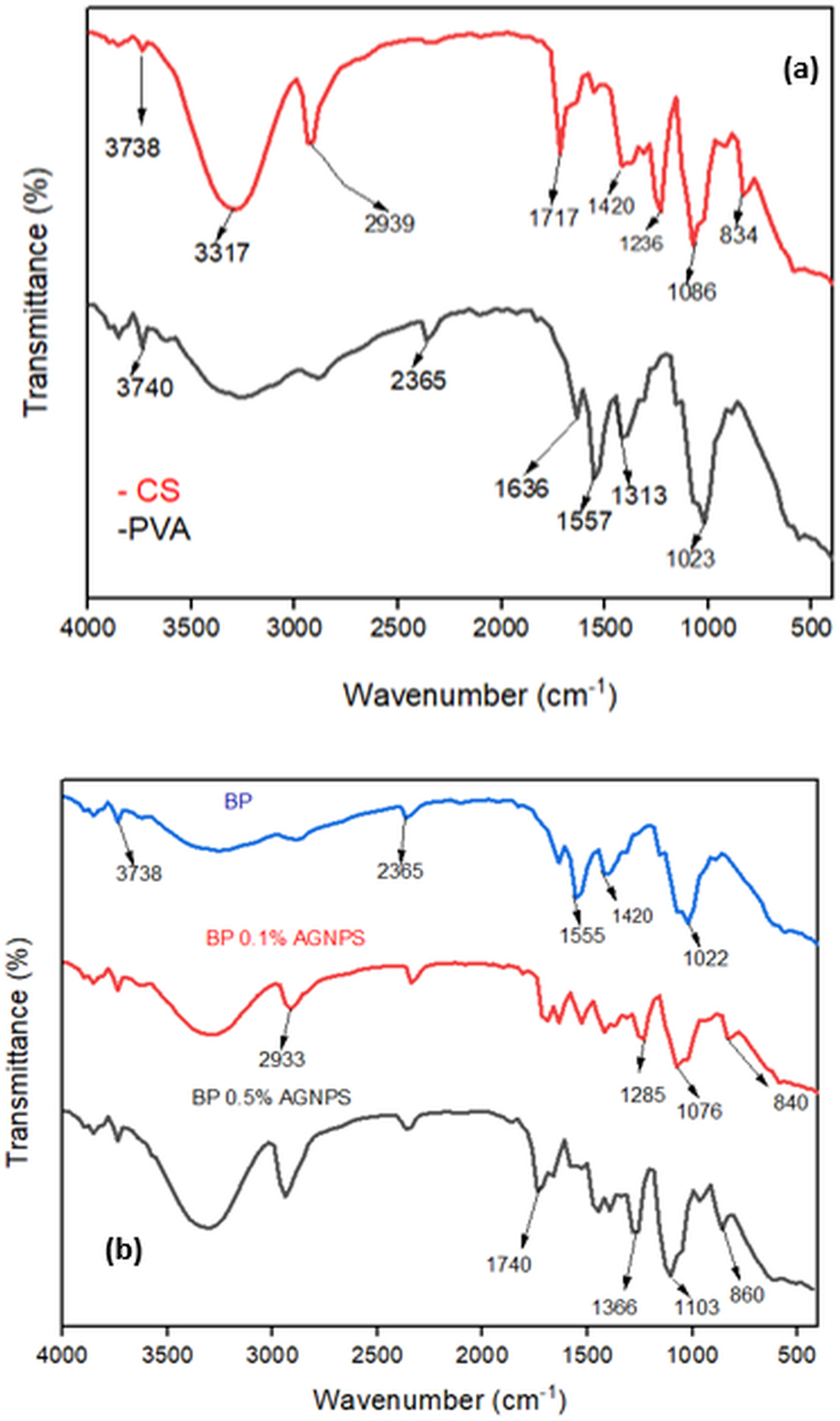
FTIR spectra were acquired to evaluate the vibrational modes of the membranes. FTIR spectra for the membranes are provided in Figure 5. CS shows a broad band at 3738 -3317 cm-1 resulting from the stretching of hydroxyls (O-H), originating from the polymer and adsorbed water (Figure 5a). A band at 2939 cm-1 corresponds to the C-H stretch, and the band at 1717 cm-1 is attributed to the N-H stretching. Furthermore, the band in the range of 1420-1236 cm-1 is assigned to the CH2 functional group. The range of 1086-834 cm-1 is associated with C–O–C C–O C–C bonds, arising from the polysaccharide structure (Sobreira et al. 2020, Braz et al. 2018, Rodrigues et al. 2018). Figure 5a also presents the spectrum of PVA, this spectrum shows an absorption peak at 3740 cm-1, which refers to intermolecular hydrogen bond and O-H stretching vibration (Kalantari et al. 2020). The band at 2365 cm-1 is associated with alkyl C-H stretching, while the peak at 1636-1557 cm-1 is attributed to C=O stretching (Nešović et al. 2019).

BP membrane showed the presence of a broad band around 3738 cm-1, which corresponds to stretching and deformation vibrations of the OH group derived from PVA and CS (Figure 5b). While the band at 1636 cm-1 is attributed to the C=O stretching, the band at 1555 cm-1 refers to the presence of an amino group from CS. At approximately 1420 cm-1 the presence of CH2 occurs, and the band at 1022 cm-1 corresponds to the presence of the -C-O-C group ( Nešović et al. 2019). For BP/AgNPs-0.1 a band at 2933 cm-1 presents greater intensity about the BP sample, indicating the interaction of AgNPs with the polymeric network and the presence of aliphatic CH (Nešović et al. 2019). The band at 1285 cm-1 corresponds to the presence of the C-O functional group derived from carboxylic acids from copaiba oil. The appearance of a band at 840 cm-1 indicates a strong interaction with caryophyllene molecules from copaiba oil, as reported by Paranhos et al. (2022). For BP/AgNPs-0.5 a band at 1740 cm-1 is assigned to the carboxylic groups of copaiba oil (Norcino et al. 2020), and the presence of C-O-C from phenolic groups, is visualized at 1103 cm-1. In addition, the band at 860 cm-1 evidences the presence of caryophyllene in the membrane (Norcino et al. 2020). The FTIR spectra for membranes BP/AgNPs-0.1 and BP/AgNPs-0.5 exhibit the characteristic vibrational modes of their components and also indicate the formation of interactions between AgNPs and the polymer matrix.
Degree of Intumescence
Figure 6a-b shows the degree of intumescence for the membranes in distilled water and phosphate saline buffer solution (PBS) as a function of time. For each membrane, three replicates were performed. The intumescence results were evaluated by the amount of distilled water/PBS absorbed over time. PVA membranes, after being immersed in distilled water/PBS, disintegrated, without conditions for analysis. Regarding CS in the presence of distilled water, there was an increase in the degree of intumescence in the first two hours (49.91 ± 0.62 to 73.45 ± 3.53) and a posterior increase in the last hour of the test (146.42 ± 25.23) (Figure 6a). For the BP membrane, the degree of intumescence in distilled water showed a similar behavior after two hours (84.80 ± 1.63 to 84.80 ± 9.46) and an increase for three hours (115.85 ± 7.68) (Figure 6a). While BP/AgNPs-0.1 had an increase after three hours of immersion (110.33 ± 12.38). BP/AgNPs-0.5 showed a significant increase during two hours, and a reduction after three hours (Figure 6a).

Degree of intumescence in (a) water, and (b) PBS. Means with the same letter have no statistically significant difference for each hour (Duncan’s test, p < 0.05).
CS showed a reduction in PBS for two hours (66.37 ± 2.96 to 31.44 ± 1.99) and an increase after three hours (63.62 ± 5.12). For BP, a slight increase for two hours was observed (70.02 ± 1.91 to 84.80 ± 1.63). While, BP/AgNPs-0.1 in PBS fluid provided a higher growth for three hours (124.54 ± 20.91). However, BP/AgNPs-05 showed a significant increase in absorption after three hours (57.05 ± 2.43, 1 hour; 62.09 ± 2.76, 2 hours; to 128.41 ± 5.40, 3 hours) (Figure 6b). By the results achieved after the immersion in distilled water and PBS, the highest values for the degree of intumescence were 124.54 ± 20.91 for BP/AgNPs-01. (distilled water), and 128.41 ± 5.40 for BP/AgNPS-0.5 (PBS) after three hours. Table 1 shows the degree of intumescence for all membranes available.
The BP/AgNPs-0.1 exhibited stability in water and PBS. The BP/AgNPs-0.1 and BP/AgNPs-0.5 membranes offered a higher absorption, this may be linked to the insertion of AgNPs, which can induce a greater intumescence (Suflet et al. 2021). It is noteworthy that the interaction of polymer chains with AgNPs or by the solvation of nanoparticles alters the absorption of water molecules. Furthermore, structures with higher percentages of copaiba oil (BP/AgNPs-0.5) hinder diffusion, generating greater tortuosity in the system (Hajji et al. 2019). In general, analyzing all samples, BP/AgNPs-0.5 in PBS stands out due to the higher percentage of swelling in PBS, and the PBS has a pH similar to that of the skin (Paranhos et al. 2022). These findings reveal that the integrity of the BP/AgNPs-0.1 and BP/AgNPs-0.5 membranes is preserved after 3 hours of insertion in water and PBS. They also reveal the capacity of fluid absorption by these materials, which is desirable for wound healing.
Contact angle
The values for the contact angle are contained in the bar graph (Figure 7a-b). This technique was performed three times for each sample at room temperature. For this analysis, two different fluids (distilled water and PBS), were used to obtain more details about wettability. The results obtained for CS showed a minimal difference between distilled water (69.28 ± 0.45) and PBS (66.63 ± 0.05). However, for the PVA membranes, a greater difference between the fluids was determined, with a lower value in distilled water (62.50 ± 0.07), and a higher value in PBS (71.32 ± 0.83). BP membranes showed similar values in distilled water (71.36 ± 0.16), and in PBS (74.53 ± 0.16). BP/AgNPs-0.1 provided a significant reduction in the contact angle in distilled water (41.31 ± 0.07) and PBS (48.73 ± 0.06). However, when analyzing the BP/AgNPs-0.5, a difference in values is observed with a higher contact angle in distilled water (55.41 ± 0.02) than in PBS (50.49 ± 0.13). Table 2 shows the contact angle of membranes.

Contact angle in (a)distilled water, and (b) PBS. Means with the same letter have no statistically significant difference (Duncan’s test, p < 0.05).
According to Severo et al. (2022) wettability is divided into certain categories: superhydrophilic samples with contact angles smaller than 40°, hydrophilic samples between 40 and 90°, hydrophobic samples between 90 and 120°, and superhydrophobic samples with angles greater than 120°. Ferreira et al. (Ferreira et al. 2022) describe that ideal dressings should have a contact angle <90° due to their greater ability to absorb exudates from the wound. Comparing our findings with the literature all the membranes had contact angle values, in the range of 40 to 90°. Thus, the hydrophilic character of all membranes is noted (Mhatre et al. 2021). Our results for the contact angle support the use of BP/AgNPs-0.1 and BP/AgNPs-0.5 as dressings due to their hydrophilic nature, which favors the absorption of fluids such as those exuded from wounds.
Humidity
Figure 8 shows the humidity (%) of the membranes. For this analysis, three replicates were performed for each sample. The CS presented a lower humidity when compared to the PVA. These results can be explained by the fact that chitosan films have deacetylated groups, which spontaneously aggregate to hydroxyl and amino groups, characterized by a strong affinity with water molecules, however, this mutual influence seems to be less hydrophilic than PVA. On the other hand, the hydrophilic nature of PVA is attributed to the hydroxyl groups that promote humidity absorption (Severo et al. 2022).

Humidity for the membranes. Means with the same letter have no statistically significant difference (Duncan’s test, p < 0.05).
For the BP membrane, there was a reduction in the humidity when compared to BP/AgNPs-0.1 and BP/AgNPs-0.5. We can relate these results to the lack of addition of emulsion and AgNPs, making absorption difficult (Severo et al. 2022, Mhatre et al. 2021). Similar results were obtained by Alvarenga et al. (2020) in their work with the addition of copaiba oil, directly and microencapsulated. In this context, membranes with emulsions and AgNPs, in particular (0.5% v/v), should be preferred in obtaining biomaterials for wound treatment because they have a high humidity (%). Thus, the humidity results associated with those found for the contact angle and intumescence reveal the promising character for the use of BP/AgNPs-0.5 as a dressing for wound healing. In addition, BP/AgNPs-0.5 has additives in higher content with antibacterial potential. Table 3 shows the humidity of membranes.
Polymeric membranes were successfully developed using the casting method, with potential for application in wound treatment. These membranes were shown to be suitable for different types of exudative wounds, with the BP/AgNPs-0.5 formulation being more suitable for wounds with greater exudate production. The analysis of the structure revealed interfacial matrices with favorable porosity and contact angle, in addition to improved fluid absorption, directly influenced by the oil concentrations. The results suggest that the presence of copaiba oil in the polymeric matrix may favor the healing process. However, additional studies like MTT (3-(4,5-dimethylthiazol-2-yl)-2,5-diphenyltetrazolium bromide, a tetrazole) assay and antimicrobial (antibacterial and antifungal) testing, required to validate the biocompatibility and effectiveness of these membranes as potential biomaterials.
CONCLUSIONS
-
Polymeric blends were manufactured with CS, PVA, copaiba oil emulsion, and AgNPs.
-
UV-Vis spectrum for AgNPs showed the behavior of metallic nanoparticles.
-
TEM image showed the presence of spherical particles in nanometric dimensions.
-
SEM findings revealed a porous and smooth surface for membranes with and without copaiba oil emulsion, respectively.
-
FTIR results showed the insertion of new bands after the insertion of copaiba oil emulsion and AgNPs when compared with the control samples of CS and PVA.
-
In addition, the swelling showed good fluid absorption for the fluids (distilled water and PBS), which are essential for application in dressings. Contact angle tests demonstrated that the BP film has a greater hydrophilic character in PBS and PVA in distilled water, which is indicative of a strong interaction between these materials and the fluids. The percentage of humidity was influenced by the copaiba oil emulsions and AgNPs.
-
Our findings demonstrate that BP/AgNPs-0.5 membranes are recommended for application in the treatment of wound healing. Because they have characteristics that compose a biomaterial, such as absorption capacity, fluid retention, and hydration capacity, more studies are needed on these materials, mainly in vitro and in vivo investigations.
Acknowledgements
This article was funded by the Dean of Research and Graduate Studies (Propesp) of the Federal University of Pará, process number 008.033/2023. The authors declare no conflict of interest.
References
- ALVARENGA MOP, BITTENCOURT LO, MENDES PFS, RIBEIRO JT, LAMEIRA OA, MONTEIRO MC, BARBOZA CAG, MARTINS MD & LIMA RR. 2020. Safety and Effectiveness of Copaiba Oleoresin (C. reticulata Ducke) on Inflammation and Tissue Repair of Oral Wounds in Rats. Int J Mol Sci 21: 3568.
- ANTUNES JC, TAVARES TD, TEIXEIRA MA, TEIXEIRA MO, HOMEM NC, AMORIM MTP & FELGUEIRAS HP. 2021. Eugenol-Containing Essential Oils Loaded onto Chitosan/Polyvinyl Alcohol Blended Films and Their Ability to Eradicate Staphylococcus aureus or Pseudomonas aeruginosa from Infected Microenvironments. Pharmaceutics 13: 195.
- ARAÚJO IRD, NEVES RCS, LIMA GMS, CÂMARA CAGD, PAULA MVS & ALVES JUNIOR S. 2021. Avaliação da atividade antimicrobiana do fluido AGNPS-S.AROMATICUM@ALGINATO. In: Engenharia de Produtos Naturais: planejamento, experimentação, obtenção de produtos e purificação, Editora Científica Digital, p. 408-424.
- BRAZ EMA, SILVA SCCC, DA SILVA DA, CARVALHO FAA, BARRETO HM, SANTOS JÚNIOR LS & DA SILVA FILHO EC. 2018. Modified chitosan-based bioactive material for antimicrobial application: Synthesis and characterization. Int J Biol Macrom 117: 640-647.
- BRUNA T, MALDONADO-BRAVO F, JARA P & CARO N. 2021. Silver Nanoparticles and Their Antibacterial Applications. Int J Mol Sci 22: 7202.
- DEBONE HS, LOPES PS, SEVERINO P, YOSHIDA CMP, SOUTO EB & DA SILVA CF. 2019. Chitosan/Copaiba oleoresin films for would dressing application. Int J Pharm 555: 146-152.
- FERREIRA ES, PARANHOS SB, DA PAZ SPA, CANELAS CAA, NASCIMENTO LAS, PASSOS MF, DA SILVA ACR, MONTEIRO SN, PAULA MVS & CANDIDO VS. 2022. Synthesis and Characterization of Natural Polymeric Membranes Composed of Chitosan, Green Banana Peel Extract and Andiroba Oil. Polymers 14: 1105.
- HAJJI S, KHEDIR SB, HAMZA-MNIF I, HAMDI M, JEDIDI I, KALLEL R, BOUFI S & NASRI M. 2019. Biomedical potential of chitosan-silver nanoparticles with special reference to antioxidant, antibacterial, hemolytic and in vivo cutaneous wound healing effects. Biochimica et Biophysica Acta (BBA) - General Subjects 1863: 241-254.
- HAMAD A, KHASHAN KS & HADI A. 2020. Silver Nanoparticles and Silver Ions as Potential Antibacterial Agents. J Inorg Organomet Polym 30: 4811-4828.
- JASWAL T & GUPTA J. 2023. A review on the toxicity of silver nanoparticles on human health. Materials Today: Proceedings 81: 859-863.
- KALANTARI K, MOSTAFAVI E, SALEH B, SOLTANTABAR P & WEBSTER TJ. 2020. Chitosan/PVA hydrogels incorporated with green synthesized cerium oxide nanoparticles for wound healing applications. Eur Polym J 134: 109853.
- LAMARRA J, CALIENNI MN, RIVERO S & PINOTTI A. 2020. Electrospun nanofibers of poly(vinyl alcohol) and chitosan-based emulsions functionalized with cabreuva essential oil. Int J Biol Macromol 160: 307-318.
- LEE Y-H & LIN S-J. 2022. Chitosan/PVA Hetero-Composite Hydrogel Containing Antimicrobials, Perfluorocarbon Nanoemulsions, and Growth Factor-Loaded Nanoparticles as a Multifunctional Dressing for Diabetic Wound Healing: Synthesis, Characterization, and In Vitro/In Vivo Evaluation. Pharmaceutics 14: 537.
- LIMA TPL & PASSOS MF. 2021. Skin wounds, the healing process, and hydrogel-based wound dressings: a short review. J Biomater Sci Polym Ed 32: 1910-1925.
- MALLAKPOUR S & MANSOURZADEH S. 2018. Sonochemical synthesis of PVA/PVP blend nanocomposite containing modified CuO nanoparticles with vitamin B1 and their antibacterial activity against Staphylococcus aureus and Escherichia coli. Ultrasonics Sonochemistry 43: 91-100.
- MATA GC, MORAIS MS, OLIVEIRA WP & AGUIAR ML. 2022. Composition Effects on the Morphology of PVA/Chitosan Electrospun Nanofibers. Polymers 14: 4856.
- MHATRE A, BHAGWAT A, BANGDE P, JAIN R & DANDEKAR P. 2021. Chitosan/gelatin/PVA membranes for mammalian cell culture. Carbohydr Polym Technol Appl 2: 100163.
- MONTOYA C, DU Y, GIANFORCARO AL, ORREGO S, YANG M & LELKES PI. 2021. On the road to smart biomaterials for bone research: definitions, concepts, advances, and outlook. Bone Res 9: 1-16.
- NEŠOVIĆ K, JANKOVIĆ A, RADETIĆ T, VUKAŠINOVIĆ-SEKULIĆ M, KOJIĆ V, ŽIVKOVIĆ L, PERIĆ-GRUJIĆ A, RHEE KY & MIŠKOVIĆ-STANKOVIĆ V. 2019. Chitosan-based hydrogel wound dressings with electrochemically incorporated silver nanoparticles – In vitro study. Eur Polym J 121: 109257.
- NORCINO LB, MENDES JF, NATARELLI CVL, MANRICH A, OLIVEIRA JE & MATTOSO LHC. 2020. Pectin films loaded with copaiba oil nanoemulsions for potential use as bio-based active packaging. Food Hydrocolloids 106: 105862.
- PARANHOS SB, FERREIRA ES, CANELAS CAA, DA PAZ SPA, PASSOS MF, DA COSTA CEF, DA SILVA ACR, MONTEIRO SN & CANDIDO VS. 2022. Chitosan Membrane Containing Copaiba Oil (Copaifera spp.) for Skin Wound Treatment. Polymers 14: 35.
- PARK KJ, LEE J-S, JO HJ, KIM ES & LEE HG. 2022. Antimicrobial and indicator properties of edible film containing clove bud oil-loaded chitosan capsules and red cabbage for fish preservation. Int J Biol Macromol 196: 163-171.
- RINAUDO M. 2006. Chitin and chitosan: Properties and applications. Progress in Polymer Science 31: 603-632.
- RODRIGO-NAVARRO A, SANKARAN S, DALBY MJ, DEL CAMPO A & SALMERON-SANCHEZ M. 2021. Engineered living biomaterials. Nat Rev Mater 6: 1175-1190.
- RODRIGUES IA, RAMOS AS, FALCÃO DQ, FERREIRA JLP, BASSO SL, SILVA JR A & AMARAL ACF. 2018. Development of Nanoemulsions to Enhance the Antileishmanial Activity of Copaifera paupera Oleoresins. BioMed Res Int 2018: 9781724.
-
RONALDO SILVEIRA EA. 2022. Nanopartículas de prata: síntese, atividade antibacteriana e comparativo com um desinfetante comum. Tecnol Metal Mater Min 19: e2638. http://dx.doi.org/10.4322/2176-1523.20222638.
» https://doi.org/10.4322/2176-1523.20222638 - SAVENCU I, IURIAN S, PORFIRE A, BOGDAN C & TOMUȚĂ I. 2021. Review of advances in polymeric wound dressing films. React Funct Polym 168: 105059.
- SEVERO AMC, FOOK MVL & LEITE IF. 2022. Hidrogéis de PVA/quitosana funcionalizados com óleo de melaleuca visando aplicação como curativos. Matéria (Rio J) 27: e13149.
- SOBREIRA TGP, SILVA LA, MENEZES FD, FRANÇA EJ & AQUINO KAS. 2020. Structural aspects of chitosan/pva spheres cross-linked with glutaraldehyde and submitted to different thermal treatments. Quím Nova 43: 1251-1257.
- SUFLET DM, POPESCU I, PELIN IM, ICHIM DL, DARABA OM, CONSTANTIN M & FUNDUEANU G. 2021. Dual Cross-Linked Chitosan/PVA Hydrogels Containing Silver Nanoparticles with Antimicrobial Properties. Pharmaceutics 13: 1461.
- SUGUMAR S, MUKHERJEE A & CHANDRASEKARAN N. 2015. Eucalyptus oil nanoemulsion-impregnated chitosan film: antibacterial effects against a clinical pathogen, Staphylococcus aureus, in vitro. Int J Nanomedicine 10: 67-75.
- TU Z, ZHONG Y, HU H, SHAO D, HAAG R, SCHIRNER M, LEE J, SULLENGER B & LEONG KW. 2022. Design of therapeutic biomaterials to control inflammation. Nat Rev Mater 7: 557-574.
- ZHANG X, SHU W, YU Q, QU W, WANG Y & LI R. 2020. Functional Biomaterials for Treatment of Chronic Wound. Front Bioeng Biotechnol 8.
Not applicable.