Bergeyella zoohelcum strain involved in chronic canine rhinitis
Abstract
The pathogenesis of dog’s bacterial rhinitis is poorly understudied. Bergeyella zoohelcum was isolated from an animal with chronic rhinitis as primary agent. The bacterium identification was performed by both MALDI-TOF and 16S rDNA gene sequencing. The phylogenetic relationship of the strain inside de genera was demonstrated, as well as the antimicrobial susceptibility profile and the ability of biofilm formation. We highlight the virulence profile of the B. zoohelcum in, to the best of our knowledge, the first identification of B. zoohelcum as causal agent of bacterial rhinitis in dogs.
Key words
phylogenetic relationship; nasal infection; zoonotic; Weeksella zoohelcum
INTRODUCTION
The pathogenesis of dog’s bacterial rhinitis is poorly understudied. A low number of bacteria species are recognized as primary agents. Moreover, bacterial infections in dogs’ nasal cavity might be secondary to dental diseases (Windsor & Johnson 2006). Nevertheless, despite the role of dogs’ upper respiratory microbial communities in nasal diseases is not well established yet, commensal bacteria may interfere with the disease’s progress (Tress et al. 2017).
Bergeyella zoohelcum is part of the healthy microbiota of the upper respiratory tract of dogs, cats, and other mammals (Sharma et al. 2019, Chen et al. 2017), which can cause diseases under imbalance conditions (Arriba et al. 2018). B. zoohelcum is an aerobic, non-motile, rod-shaped, Gram-negative bacteria, and a zoonotic pathogen, as result of dogs’ or cats’ bites. There are reports of B. zoohelcum related to humans, cellulitis (Lin et al. 2007), bacteremia (Sharma et al. 2019), septicemia (Kivinen et al. 2003), leg abscess (Reina & Borrell 1992), meningitis (Bracis et al. 1979), and infectious endocarditis (Clark et al. 2017).
Considering the scarcity of reports about bacterial rhinitis in dogs, especially with B. zoohelcum implication, this report presents the characterization of a B. zoohelcum strain related to a rare case of canine bilateral purulent rhinitis.
MATERIALS AND METHODS
We investigated a case of a female canine, 10 years old, with a history of non-specific rhinitis and non-specific respiratory clinical signs, including sneezing and purulent secretion, which was not responsive to a large-term clindamycin treatment. During the clinical evaluation, the veterinarian observed that the animal presented regions with dental tartar, without the establishment of periodontal disease.
Microorganism characterization
A nasal secretion sample was collected using a sterile swab and submitted to microbiological analysis. The sample was inoculated in Blood Agar Base (Kasvi, Brazil) supplemented with 5% sheep blood and MacConkey agar (IonLab, Indian), then incubated at 37°C for 48 h under aerobic conditions.
Bacterial identification was performed by Matrix-Assisted Laser Desorption and Ionization Time of Flight Mass Spectrometry (MALDI-TOF; Bruker Corporation, USA) using extraction method, following the manufacturer’s recommendations.
Total DNA extraction and Polymerase Chain Reaction (PCR)
To confirm the bacterial identification, sequencing of partial 16S-rDNA gene was performed. Total DNA was extracted by Purelink Genomic DNA Minikit (Invitrogen, Thermo Fischer Scientific, USA) and the partial 16S-rDNA was amplified by PCR using universal primers 27F (5’-AGAGTTTGATCATGGCTCAG-3)’ e 1492R (5’-TACGGYTACCTTGTTACGACTT-3’), in 25 µl reaction using 1 U GoTaq DNA polymerase (Promega, USA), 1 X GoTaq buffer, 10 mM dNTP, 10 pmol of each primer and 30 ng of the DNA. PCR conditions were: an initial cycle at 94°C for 5 min, followed by 35 cycles of 94°C for 1 min, 52°C for 1 min and 72°C for 1 min, and a final extension at 72°C for 5 min (Breyer et al. 2023). PCR products were purified using the Purelink Quick PCR Purification Kit (Invitrogen, Thermo Fischer Scientific), quantified by NanoDrop Lite Spectrophotometer (Thermo Fischer Scientific, USA), and sequenced using Sanger method at ABI-PRISM 3500 Genetic Analyzer (Applied Biosystems Inc. Foster City, USA). Sequenced DNA fragments from overlapping strands generated a 1,398 nt consensus sequence using Geneious Prime 2024.0.3, which was deposited on GenBank database (Accession Number PP936189). The partial 16S-rDNA gene sequence was analyzed using BLASTn tool against GenBank database.
Phylogenetic analysis
To assess the evolutionary distance of B. zoohelcum LBV051/22 from other Bergeyella spp., we performed a phylogenetic analysis. A total of 10 16S-rDNA sequences of Bergeyella spp. were retrieved from GenBank database and used in the study: five B. zoohelcum clinical strains from the oral cavity of canines (JN713353.1, LC460808.1, LC460822.1, LC460806.1, and LC460809.1); the referential B. zoohelcum ATCC 43767 (MH789409.1); and four Bergeyella porcorum clinical strains from the oral cavity of swine (N886702.2, OR493469.1, MT760315.1, and LN886700.1). In addition, Riemerella columbina RCAD0985 (MT804608.1) was used to root the tree. Sequences were aligned using ClustalW in MEGA11 (Thompson et al. 1994). The phylogenetic tree was constructed using Maximum Likelihood (bootstrap=1,000; Kimura-2-parameter model with Gamma distribution of 0.05) by Mega11 software (Tamura et al. 2021).
Antimicrobial susceptibility test
To provide a more robust understanding of the rare B. zoohelcum identified a phenotypic characterization of the strain was performed, including the antimicrobial susceptibility profile and the biofilm production ability, aiming to determine the most adequate treatment to be used for the recurring nasal infection and the virulent potential of the strain, respectively.
In detail, the isolate was subjected to in vitro antimicrobial susceptibility test based on the Kirby-Bauer disk diffusion method in Mueller Hinton sheep blood agar (Sigma-Aldrich, Brazil), against 13 antimicrobials (Table I).
Susceptibility antimicrobial profile of Bergeyella zoohelcum isolated from canine nasal cavity.
Biofilm formation assay
The ability of biofilm formation was evaluated as described by Stepanović et al. (2000). The isolate was cultured in sheep blood agar at 37°C for 24 h. Bacterial growth was suspended in saline solution up to 0.5 OD McFarland scale, then 20 µL of the suspension was placed into sterile 96-well plate with 180 µL Tryptic Soy Broth (TSB; Merck®, Germany) and incubated at 37°C for 72 h. Biofilm was fixed with methanol (PA) and stained with violet crystal solution 0.5%. The plate was rinsed with ethanol (95%) and the absorbance was measured at 550 nm in Multiskan FC microplate photometer (Thermo Fischer Scientific, USA). Three biological replicates were performed with two technical replicates. Staphylococcus aureus ATCC 25923 and Enterococcus sp. FC1A were used as reference controls for strong biofilm formation, while wells containing only TSB were used as negative controls.
RESULTS
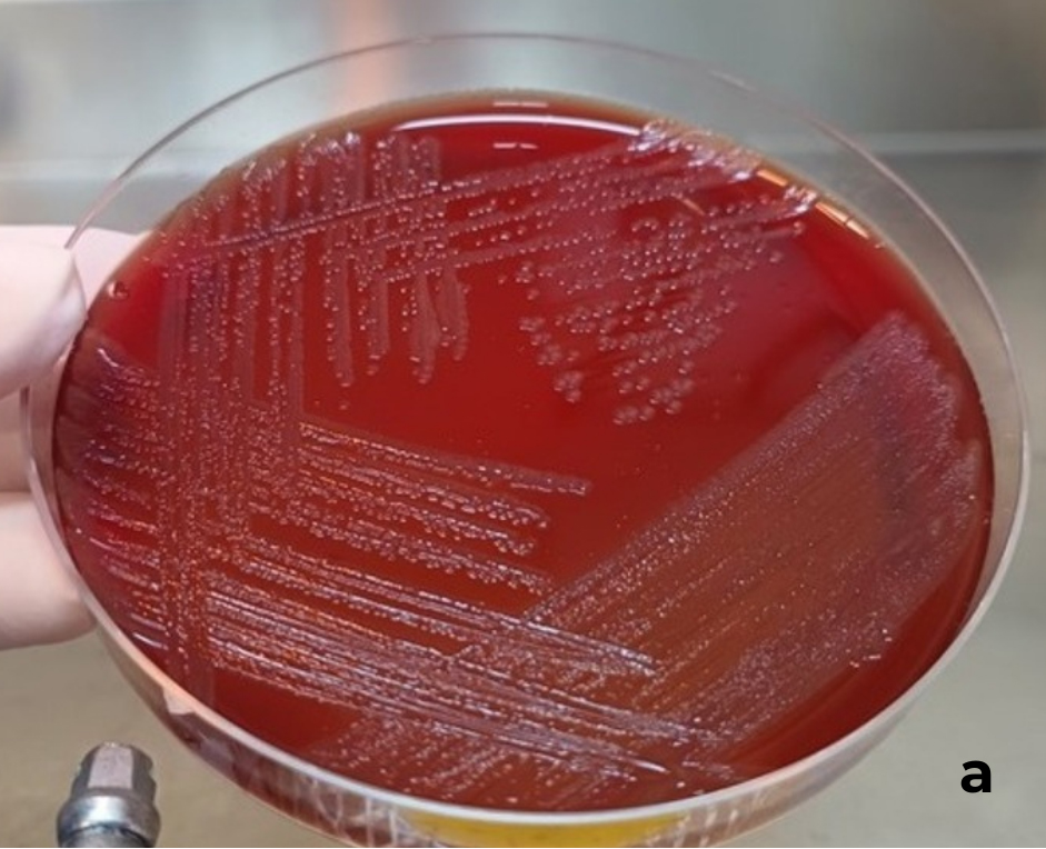
After incubation, pure growth of small, shiny, mucoid, and non-hemolytic colonies was observed on blood agar plate (Fig. 1a), whereas no growth was identified on the MacConkey agar. Gram staining showed Gram-negative rod-shaped bacteria (Fig. 1b). The strain was identified by MALDI-TOF as Bergeyella zoohelcum (score 2.182). The partial 16S-rDNA gene sequence was analyzed using BLASTn tool against GenBank database, confirming the bacterial identification as B. zoohelcum (97.2% identity and 99.86% coverage).
(a) Bergeyella zoohelcum culture in blood agar showing small, shiny, mucoid colonies with non-hemolytic activity after 48h incubation at 37 °C. (b) Gram staining was performed on bacteria from colonies grown on blood agar. Gram-negative rod-shaped in detached.
Evolutionary distances between the isolate and the 11 strains were evaluated, showing that B. zoohelcum LBV051/22 clustered together with other canine B. zoohelcum, and exhibited a clear evolutionary distance from the porcine B. porcorum included in the analysis (Fig. 2). B. zoohelcum exhibited large inhibition zone (millimeters) for 12 antimicrobials tested, and resistance only to sulfonamide, showing an absence of inhibition zone (Table I). Based on the absorbance, the strain was classified as moderate biofilm producer, as proposed by Stepanović et al. (2000).
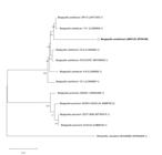
Phylogenetic analysis of partial 16S-rDNA Bergeyella zoohelcum LBV051/22 strain and other Bergeyella spp. strains. Evolutionary analyses were inferred using Maximum Likelihood (bootstrap=1,000; Kimura-2-parameter model with Gamma distribution of 0.05) by MEGA11 software.
DISCUSSION
The animal showed a chronic bacterial rhinitis associated with allergic rhinitis with the presence of tartar, which allows the exacerbated growth of opportunistic bacteria and ascension into the nasal cavity. In general, the microbiological investigation of the pathogens involved in bacterial rhinitis in dogs is highly recommended, as there is no clear bacterial etiologic agent for such disease, and in order to ensure the accurate diagnosis and the application of the most appropriate treatment for the animal. An inadequate treatment increases the risk of diseases chronicity and favors the development of bacterial resistance, being a concern for One Health.
B. zoohelcum LBV051/22 showed a moderate biofilm formation, proven its mild persistence after 72 h of incubation in TSB. In a previous study, Arriba et al. (2018) demonstrated that B. zoohelcum and B. porcorum isolated from nasal cavities of piglets showed poor ability to form biofilm. Similarly, although abundant in canine tartars, B. zoohelcum from canine dental samples also showed weak biofilm formation in vitro, suggesting that co-infections might increase the adhesion and formation of dental biofilm (Holcombe et al. 2014).
Regarding B. zoohelcum susceptibility to antimicrobials, we tested 13 antibiotics from 10 classes, and the isolate demonstrated to be resistant to sulfonamide and susceptible to the other groups of antimicrobials. Previous studies have determined the susceptibility of B. zoohelcum to beta-lactams, fluoroquinolones (Hu et al. 2021) and chloramphenicol (Montejo et al. 2001). Therefore, the therapeutic treatment was based on enrofloxacin and anti-inflammatory agents, which have provided good results in the animal. Is noteworthy that previously to the bacterial culture and antimicrobial test, the animal was treated with clindamycin without positive results. Discrepancies between in vitro antimicrobial tests and in vivo treatment responses could be related to a variability of factors, including the inability of the in vitro assay to reproduce the real in vivo condition.
CONCLUSIONS
To the best of our knowledge, this is the first report of B. zoohelcum as a causative agent of dogs’ rhinitis. The recurrence of clinical signs was managed based on treatment according to the antimicrobial susceptibility test. Therefore, both microbiological analysis and susceptibility tests are crucial in cases of canine rhinitis. Furthermore, considering that B. zoohelcum is zoonotic, this finding underscores the importance of addressing potential public health implications.
REFERENCES
-
ARRIBA ML, LOPEZ-SERRANO S, GALOFRE-MILA N & ARAGON V. 2018. Characterization of Bergeyella spp. isolated from the nasal cavities of piglets. Vet J 234: 1-6. https://doi.org/10.1016/j.tvjl.2018.01.004.
» https://doi.org/10.1016/j.tvjl.2018.01.004 - BRACIS R, SEIBERS K & JULIEN RM. 1979. Meningitis caused by group II J following a dog bite. West J Med 131(5): 438.
-
CHEN Y, LIAO K, AI L, GUO P, HUANG H, WU Z & LIU M. 2017. Bacteremia caused by Bergeyella zoohelcum in an infective endocarditis patient: case report and review of literature. BMC Infect Dis 17: 1-4. https://doi.org/10.1186/s12879-017-2391-z.
» https://doi.org/10.1186/s12879-017-2391-z - CLARK L, PARKINS MD, CHOW BL, LYNCH T & CHURCH D. 2017. Endocarditis caused by an oral taxon species of Bergeyella identified by partial 16S rDNA sequencing: Case report and review of the literature. Can J Infect Dis Med Microbiol 2(1): 56-63. DOI: 10.3138/jammi.2.1.008.
-
BREYER GM, SILVA MERJ, SLAVIERO M, ALMEIDA BA, SILVA EMS, SCHMIDT VRQ, ALIEVI M, MABONI G, PAVARINI SP & SIQUEIRA FM. 2023. Genotypic characterization of Streptococcus didelphis causative of fatal infection in the white-eared opossums. Lett Appl Microbiol 76(12): ovad131. https://doi.org/10.1093/lambio/ovad131.
» https://doi.org/10.1093/lambio/ovad131 -
HOLCOMBE LJ, PATEL N, COLYER A, DEUSCH O, O’FLYNN C & HARRIS S. 2014. Early canine plaque biofilms: Characterization of key bacterial interactions involved in initial colonization of enamel. PLoS ONE 9: e113744. https://doi.org/10.1371/journal.pone.0113744.
» https://doi.org/10.1371/journal.pone.0113744 -
HU M, LI H, HU T, ZHOU SY, YANG ZH, FAN CL, BAO R, SHI JG & ZHAO KB. 2021. The Detection of Bergeyella zoohelcum in Young Children after Cat Bite. BioRxiv: 2021-2023. https://doi.org/10.1101/2021.03.26.437298
» https://doi.org/10.1101/2021.03.26.437298 - KIVINEN PK, LAHTINEN MR, RUOTSALAINEN E, HARVIMA IT & KATILA ML. 2003. Bergeyella zoohelcum septicemia of a patient suffering from severe skin infection. Acta Derm Venereol 83(1): 74-75.
- LIN WR, CHEN YS & LIU YC. 2007. Cellulitis and bacteremia caused by Bergeyella zoohelcum. J Formos Med Assoc 106(7): 573-576. DOI: 10.1016/S0929-6646(07)60008-4.
- MONTEJO M, AGUIRREBENGOA K, UGALDE J, LOPEZ L, SAEZ NIETO JA & HERNÁNDEZ JL. 2001. Bergeyella zoohelcum bacteremia after a dog bite. Clin Infect Dis 33(9): 1608-1609. DOI: 10.1086/322724. PMID: 11588704.
- REINA J & BORRELL N. 1992. Leg abscess caused by Weeksella zoohelcum following a dog bite. Clin infect Dis 14(5): 1162-1163.
- SHARMA S, SALAZAR H, SHARMA S, NASSER MF & DAHDOUH M. 2019. Bergeyella zoohelcum Bacteremia from Therapy Dog Kisses. Cureus 11(4): e4494. DOI: 10.7759/cureus.4494. PMID: 31259112; PMCID: PMC6581327.
-
STEPANOVIĆ S, VUKOVIĆ D, DAKIĆ I, SAVIĆ B & ŠVABIĆ-VLAHOVIĆ M. 2000. A modified microtiter-plate test for quantification of staphylococcal biofilm formation. J Microbiol Methods 40(2): 175-179. https://doi.org/10.1016/S0167-7012(00)00122-6.
» https://doi.org/10.1016/S0167-7012(00)00122-6 - TAMURA K, STECHER G & KUMAR S. 2021. MEGA11: molecular evolutionary genetics analysis version 11. Mol Biol Evol 38(7): 3022-3027.
-
THOMPSON JD, HIGGINS DG & GIBSON TJ. 1994. CLUSTAL W: improving the sensitivity of progressive multiple sequence alignment through sequence weighting, position-specific gap penalties and weight matrix choice. Nucleic Acids Res 22(22): 4673-4680. https://doi.org/10.1093/nar/22.22.4673.
» https://doi.org/10.1093/nar/22.22.4673 -
TRESS B, DORN ES, SUCHODOLSKI JS, NISAR T, RAVINDRAN P, WEBER K, HARTMANN K & SCHULZ BS. 2017. Bacterial microbiome of the nose of healthy dogs and dogs with nasal disease. PLoS ONE 12(5): e0176736. https://doi.org/10.1371/journal.pone.0176736.
» https://doi.org/10.1371/journal.pone.0176736 -
WINDSOR RC & JOHNSON LR. 2006. Canine chronic inflammatory rhinitis. Clin Tech Small Anim Pract 21(2): 76-81. https://doi.org/10.1053/j.ctsap.2005.12.014.
» https://doi.org/10.1053/j.ctsap.2005.12.014