
 Molecular assays and point-of-care tools: a review of amplification methods and nucleic acid detection systems
Abstract
Molecular diagnosis is crucial for combating infectious diseases due to its high sensitivity and precision. However, its application in point-of-care testing (POCT) faces challenges, particularly in remote areas and developing countries. This review, based on searches of indexed publication databases, examined major nucleic acid amplification methods, detection techniques and miniaturization strategies for POCT platform development, focusing on the period 2015–2025. The need for regulatory standardization and the advantages and disadvantages of amplification techniques were discussed. Microfluidic approaches—reaction chambers, continuous flow, and droplets—were analyzed as the basis for functional device integration, emphasizing their role in reducing volumes, energy consumption and processing time. The sensitivity, cost and instrumentation complexity of fluorescence-, colorimetry- and electrochemistry-based detection systems were addressed. The practical feasibility of integrating these into miniaturized POCT platforms underscores the structural role of microfluidics in advancing portable molecular diagnostics. Despite progress, miniaturization complexity and reagent costs remain significant technological barriers.
Key words
Amplification; Infectious Diseases; Molecular Diagnostics; Nucleic Acid; Pathogen Detection; Point-of-care Testing
INTRODUCTION
Current research into the molecular detection of pathogens is focused on developing rapid and reliable strategies adapted to the critical demands of emergency medical care, which include characteristics such as the ease of sample preparation and reading of tests (Krampa et al. 2020, Plebani et al. 2024).
In this context, the development of ultra-sensitive, specific and quantitative analytical POCT (point-of-care testing) tools for the detection of diseases using reduced sample volumes represents an important advance in global public health, as it facilitates timely and rapid disease surveillance, control and epidemiological mapping (ECDC 2022).
The POCT tools should ideally be reliable, specific, sensitive, portable, easy to use and capable of detecting many unprocessed samples in a short amount of time (Larkins & Thombare, 2025). When based on molecular diagnostics, POCT tools allow the determination of the number of copies of the target nucleic acid per sample with precision, providing the detection and quantification of the target pathogen and allowing the definition of the most appropriate treatment for patients (Gavina et al. 2023).
In this review, the conventional, modern and alternative molecular methods used in nucleic acid amplification, such as PCR (polymerase chain reaction), LAMP (loop-mediated isothermal amplification) and RPA (recombinase polymerase amplification), are initially addressed. Next, the detection systems and their challenges for the development of new POCT tools are presented. These topics seek to guide the acquisition of low-cost solutions for use in health facilities, with the aim of presenting the different methodologies for miniaturization of devices and reducing the cost of diagnostic kits in molecular assays.
The bibliographic survey of indexed publications was conducted using the PUBMED, Google Scholar, SciELO, and CAPES Periodicals databases. Only publications in English, primarily from 2015 to 2025, were included. The keywords used in the search were “Amplification”, “Infectious Diseases”, “Molecular Diagnostics”, “Nucleic Acid”, “Pathogen Detection, “Point-of-care Testing”, “Electrochemistry”, “Polymerase Chain Reation”, “Loop-Mediated Isothermal Amplification”, “Recombinase Polymerase Amplification” and “Microfluidic”.
NUCLEIC ACID AMPLIFICATION METHODS IN MOLECULAR DIAGNOSTICS
The following section highlights the main molecular amplification techniques that have been fundamental in molecular diagnostics, biomedical research, and various biotechnological applications over the past decade. Among these methodologies, notable examples include both polymerase chain reaction (PCR)-based techniques and their variations, which have been widely employed since their development, as well as newer and more innovative methods such as loop-mediated isothermal amplification (LAMP) and recombinase polymerase amplification (RPA). These techniques differ in their operating principles, equipment requirements, reaction time, and sensitivity, enabling their use in contexts ranging from laboratories with sophisticated infrastructure to field situations with limited resources. To understand how nucleic acid detection strategies have evolved and how they may change in the future, it is essential to compare their characteristics.
Polymerase chain reaction (PCR)
Since its invention in the early 90s, the polymerase chain reaction (PCR) has become an indispensable tool in many fields of molecular diagnostics, including the determination of viral or bacterial loads in clinical samples, identification and quantification of microorganisms in food, diagnosis of tumors, gene expression analysis and forensic investigations (Khehra et al. 2023).
PCR is the standard method of molecular diagnosis recognized by health agencies in the detection of pathogens, due to its high sensitivity and specificity, especially in cases of patients with low antigen concentrations and/or low parasitic loads, mixed infections and/or in the identification of drug resistance. These characteristics are extremely important, especially in neglected diseases, and PCR is therefore considered as the main tool in the process of eradicating these diseases (Snounou et al. 1993).
Although several studies have shown that conventional PCR has limitations that may affect the applicability of the method in the field when compared to examinations using light microscopes and RDTs (rapid diagnostic tests), PCR has a greater efficiency (Mixson-Hayden et al. 2010).
In the traditional PCR method, molecular diagnosis is performed by analyzing the sample through the electrophoresis technique, after a certain number of thermal cycles necessary for amplification of the target nucleic acid. This method has a wide application due to the low cost and simplicity of the diagnostic kit and equipment. However, in this process, the molecular diagnosis is performed after amplification of the target nucleic acid and requires a controlled environment to perform the amplification reaction in order to avoid contamination of the sample. Moreover, in this traditional amplification format, it is not possible to determine the target concentration within the sample, making it a purely qualitative methodology.
The PCR process is thermostable. In the microtube where the reaction will occur, the target DNA, thermostable enzymes such as Taq DNA polymerase, triphosphate deoxyribonucleotides (dNTPs), the reaction buffer, ionic cofactors such as Mg2⁺ and primers are mixed. Primers are synthetic primers (sense and anti-sense primers), which comprise small DNA sequences that must be complementary to the sequences of the target nucleic acid to be amplified. The thermocycling profile in the amplification of the target nucleic acid in the PCR method is divided into three steps:
Denaturation – the temperature is raised to 92/95 °C, and a rearrangement of the nucleic acid molecule occurs, resulting in the breakdown of the hydrogen bonds that hold the double strand together, and exposes the nucleic acid strand to the template that is the target nucleic acid to be amplified.
Annealing – the temperature is reduced to 50-65 °C, according to the primers used, allowing the hybridization process, and the primers (specific oligonucleotides) bind to the nucleic acid template in a complementary way. Primers cannot anneal to each other, and their annealing sites must be sufficiently distant to allow subsequent synthesis of new amplifications.
Extension – a temperature increase to 72 °C occurs, facilitating the action of the polymerase on the nucleic acid. The polymerase extends the primers from the 3’ terminus of each primer and synthesizes the complementary strands in the 5’ to 3’ direction along the template of the target nucleic acid. This process generates new strands of the target nucleic acid, at around 24 nucleotides per second, and after 20 cycles or more, an order of picograms or nanograms can be amplified millions of times (Eisenstein 1990).
The sequence of the steps used to perform the molecular diagnosis via the PCR method, as well as the thermal profile with the typical values of temperature and time, can be seen in Figure 1.

Steps for molecular diagnosis using PCR. (a) collection of the biological sample containing the genetic material of interest. (b) PCR reaction preparation, including the main components: template DNA, primers, nucleotides and DNA polymerase. (c) amplification of the DNA in the thermocycler, following three main steps: (1) denaturation: the double-stranded DNA is heated to a temperature of 92/95 ºC (2) annealing of the primers and (3) extension of the new DNA strand.
Other PCR methodologies have been developed to increase reliability and reduce the length of time needed for molecular diagnosis. Thus, qPCR (quantitative real time-PCR) stands out for allowing confirmatory diagnosis by analyzing the concentration of the target pathogen in real time, using one or two molecular probes labeled with fluorescence. qPCR continuously detects and monitors amplicon formation throughout the reaction, without the need for further analysis.
In qPCR, nucleic acid sequences can be accurately detected in a short time, with quantitative and specific results enabled by a gene-specific probe that binds within the target sequence. The probe is labeled with a fluorescent reporter and a quencher, allowing quantification of the target based on fluorescence emission. Additionally, the risk of cross- contamination is reduced, as the reaction occurs in a closed tube and does not require post-amplification handling. Therefore, this method allows the absolute quantification of infections in clinical trials where accuracy is critical (Farcas et al. 2004, Perandin et al. 2004, Rougemont et al. 2004, Hu & Hu 2016, Almeida-de-Oliveira et al. 2019, Artika et al. 2022).
However, to perform quantitative detection of the target pathogen, it is necessary to include a fluorescent dye in the diagnostic kit, which binds to the target nucleic acid according to the amplification process, correlating the increase in fluorescent signal intensity with the level of infection by the pathogen. The inclusion of fluorescent dyes increases the cost of the diagnostic kit and the most modern methods employ RT-qPCR, which makes the cost of the diagnostic kit cheaper because it involves less manipulation of samples, facilitating the preparation of the solution for amplification of the target nucleic acid and its quantification, even with the inclusion of fluorescent dye (Ho-Pun-Cheung et al. 2009).
Alternative methods of nucleic acid amplification eliminate thermocycling, known as isothermal methods, and simplify the instrumentation required for the molecular assay, but the reagents used in the solution are expensive and have their marketing rights reserved to specific manufacturers. Despite these obstacles that make isothermal methods more costly, many laboratories have invested in the development of kits for amplification of nucleic acids via these methods. In the development of POCT tools, isothermal methods allow the use of heating systems that require simpler electronic circuits, facilitating miniaturization and reducing the final cost of the device. However, the WHO (World Health Organization) recognizes isothermal methods as valid methodologies in molecular diagnosis only in specific cases, such as tuberculosis (Isaiah et al. 2024). However, the efficiency of the LAMP (loop-mediated isothermal amplification) methodology in the determination of pathogens has been demonstrated by several research groups (Glökler et al. 2021).
The LAMP nucleic acid amplification method is the most studied among the isothermal methods, especially in the detection of neglected tropical diseases (Garg et al. 2022). The description of the LAMP method for nucleic acid amplification is presented below.
Loop-mediated isothermal amplification (LAMP)
Assays based on the LAMP method and other similar methods of isothermal amplification have recently emerged as alternatives to PCR, due to simplifications in the constructive requirements of the equipment. Although these methods still have limited multiplexing capacity, due to the complex steps required to prepare the samples, the amplification of nucleic acids occurs at constant temperature, in the case of the LAMP method around 62 °C to 65 °C (Cook et al. 2015, Zhao et al. 2015).
LAMP is characterized by amplification of the target sequence at a constant temperature of 60–65 °C using the Bst polymerase enzyme, which is capable (through a mechanism not yet fully understood) of performing strand displacement, separating the DNA duplex and displacing the upstream strand during DNA synthesis. This activity allows elongation to continue without the pause caused by the 5’ end of the DNA (Notomi et al. 2000, Oscorbin & Filipenko 2023). Between four and six different primers are used, which are specifically designed to recognize six to eight distinct regions of the target gene. The forward inner primer (FIP) consists of two regions: F2, located at the 3’ end, and F1c, at the 5’ end. The F2 region is complementary to the F2c region of the target sequence (fitting together like a zipper), while F1c is identical to the F1c region of the target sequence. The forward outer primer (FOP or F3) contains only the F3 region, which is complementary to the F3c region of the target sequence. This primer is shorter and used at a lower concentration than the FIP. Similarly, the backward inner primer (BIP) is composed of the B2 region (at the 3’ end) and the B1c region (at the 5’ end). The B2 region is complementary to the B2c region of the target sequence, while the B1c region is identical to the B1c region of the target sequence. Finally, the backward outer primer (BOP or B3) contains the B3 region, which is complementary to the B3c region of the target (Notomi et al. 2000). The nucleic acid amplification sequence using the LAMP method can be seen in Figure 2.

LAMP amplification mechanism. The FIP and BIP primers initiate DNA synthesis. This mechanism results in the formation of loop structures (F-loop and B-loop), which serve as targets for cyclic amplification. Exponential amplification occurs continuously, generating multiple copies of the DNA in loop structures.
Since its first description, the LAMP technique has proven to be a highly sensitive and rapid method, and has become a forerunner in isothermal molecular diagnostic technology for pathogen identification (Mori et al. 2001, Oriero et al. 2015). The molecular diagnosis using the LAMP technique meets the requirements of sensitivity and specificity in the investigation of pathogens, allowing different formats and variants of assays such as RT-LAMP (reverse transcription loop-mediated isothermal amplification), which uses the reverse transcriptase enzyme to create a complementary DNA molecule from an RNA sequence, and multiplex LAMP, which is capable of detecting multiple genes in a single assay using a set of primers or a combination of primers and probes, which seek to reduce cost and time in the amplification of the target nucleic acid (Agel & Altın 2024, Jang et al. 2021, Talap et al. 2022). The LAMP technique also presents satisfactory results in identifying the concentration level of the target pathogen via different detection systems, as can be seen in the studies presented by several research groups (He et al. 2020, Yu et al. 2022, Papadakis et al. 2022).
Nonetheless, the temperature required for molecular diagnosis in the LAMP method is still considered high, requiring robust thermal systems to maintain the temperature level of the amplification of the target nucleic acid stable during the assay, as temperature variation can cause errors in molecular diagnosis. Therefore, isothermal methods with amplification of the target nucleic acid close to room temperature can facilitate the development of POCT tools.
Among these methods, RPA (recombinase polymerase amplification), which occurs around 40 °C, has been presented as the main option in the development of new POCT tools using molecular diagnostics (Liu et al. 2024b). The following is a description of the RPA method used in the amplification of nucleic acid.
RPA - Recombinase polymerase amplification
The RPA method performs the amplification of nucleic acids through an isothermal profile close to room temperature, between 37 and 45 °C, and variations of +/- 1 °C do not influence the result of the molecular diagnosis. The preparation of the solution for sample analysis can be carried out simply and the RPA method is capable of amplifying a low number of copies, such as 1 to 10 copies of the target nucleic acid in less than 20 minutes.
This method can be applied to amplify diverse targets such as RNA, miRNA, ssDNA and dsDNA from a wide variety of organisms and samples. The RPA amplification process begins when the recombinase protein binds to primers to form a recombinase-primer complex (Lobato & O’Sullivan 2018).
The resulting complex searches for homologous sequences in double-stranded DNA and, by identifying them, promotes the invasion of the strand by the primer at the cognate site. To prevent ejection of the inserted primer, the displaced DNA strand is stabilized by SSB proteins (single-stranded DNA-binding proteins) (Li et al. 2018). The recombinase then detaches from the DNA filament, leaving the 3’-OH end of the primer free and accessible for DNA polymerase to initiate synthesis. The cyclic repetition of this process results in exponential amplification (Figure 3a). In the RPA method, amplification can be performed in solid phase, solution phase, as well as in a bridged amplification format.

Amplification by RPA and its different application platforms. (a) mechanism of amplification by RPA resulting in amplification of double-stranded DNA. (b) platform based on solid phase amplification, in which microplates or membranes and modified primers are used for the detection of target DNA. (c) platform based on solution-phase amplification in which amplification occurs in a liquid medium, using free reagents to facilitate the reaction. (d) platform based on bridge amplification using immobilized primers that interact with the target DNA, commonly used with other technologies.
In solid phase RPA, a modified primer is covalently bonded to a solid surface. This amplification format has the inherent advantage of being highly adaptable to multiplexed amplification, particularly when detection is facilitated by the surface on which amplification occurs, for example, microplates, membranes, electrodes, plastic slides and glass slides, among others (del Río et al. 2017) (Figure 3b).
The solution-phase RPA is the traditional format of the technique, in which the entire reaction takes place in solution, i.e., the primers, the recombinase enzyme, the SSB protein, the DNA polymerase and the target DNA are all mixed in the same liquid medium, without any immobilized components (Lobato & O’Sullivan 2018) (Figure 3c).
In the bridge amplification format, both the forward and reverse primers are immobilized forming a type of “bridge” as the polymerase extends the strand and the new strand bends. Bridged amplification also has the potential for multiplexed amplification and several approaches can be explored to improve the attainable detection limit (Santiago-Felipe et al. 2016) (Figure 3d).
RPA has already been successfully integrated with different detection strategies, from endpoint lateral flow strips to real-time fluorescent detection (Lobato &O’Sullivan 2018). Other isothermal methods that present promising data in molecular assays are helicase- dependent amplification (HDA), nucleic acid sequence-based amplification (NASBA), rolling circle amplification (RCA) and multiple displacement amplification (MDA), which basically use different reagents and temperature levels to detect and quantify the target pathogen (Zanoli & Spoto 2012). The amplification method of the target nucleic acid represents the main role in molecular diagnosis, but the pathogen detection technique represents a critical point in determining the type of infection, the level of infection and the optimal treatment to the patient, according to the analyzed sample. Therefore, in the development of POCT tools, the detection system must be reliable and have high sensitivity and specificity, using robust electronic components and easy integration and miniaturization, in order to reduce the possibility of human error in the execution of the method and availability of the result. In this sense, we present below the main detection systems applied in molecular diagnostics.
DETECTION SYSTEMS IN MOLECULAR DIAGNOSTICS
The detection system represents one of the main challenges in the development of POCT tools due to the complexity in the integration between the components of the heating system and the realization of the determination and quantification of pathogen infection during or shortly after molecular assays for amplification of the target nucleic acid. In this section of the review, the detection systems currently applied in molecular diagnostics and under development for application in POCT tools will be described.
Molecular diagnostics via electrophoresis in POCT tools
Electrophoresis is the detection system used for the determination of the pathogen in the amplification of the target nucleic acid via the conventional PCR method. Agarose gel electrophoresis is considered one of the cheapest and simplest techniques to use, and it consists of separating the target DNA according to its number of base pairs in an agarose matrix. By adding a dye or an intercalating agent such as ethidium bromide (EtBr), these fragments can be visualized under ultraviolet light (Wittmeier & Hummel 2022).
Although gel electrophoresis is a highly reliable detection technique, its application in POCT tools is considered unfeasible due to its prolonged analysis time, the need for specialized equipment, the limitations in resolution, and its use of potentially hazardous reagents (Valones et al. 2009).
Thus, in the search for the development of rapid, portable and automated tests, microcapillary electrophoresis (µCE) was developed, especially considering its application in microfluidic cartridges, which makes the application of electrophoresis viable for the development of POCT tools (Nguyen et al. 2022). POCT tools that use the µCE in the detection system require a contrast substance to be added in the diagnostic kit, allowing one to obtain an adequate performance in the detection of the pathogen and quantification of the infection after the amplification step of the target nucleic acid.
Because their application has been validated in molecular diagnostic kits, fluorescent dyes are the main choice for use in POCT tools with µCE detection. However, the detection system must contain the optical components necessary to perform the fluorescence intensity reading and the components to apply the electrical potential necessary for the band pairs of the nucleic acid, which has a negative charge, to be displaced in the microfluidic cartridge in the region of the agarose gel.
Fluorescence is the technique applied in current qPCR equipment and there are already commercial POCT tools using this detection system in molecular diagnostics, but they still need improvements to achieve high reliability (Petralia & Conoci 2017). The following fluorescence detection systems and their application in POCT tools are described.
Molecular diagnostics via fluorescence spectroscopy in POCT tools
Fluorescence spectroscopy is a type of electromagnetic spectroscopy that analyzes the sample based on its fluorescent properties. The fluorescence detection system performs the analysis by emitting a beam of light with a fixed wavelength, which excites the electrons in the sample, and the luminescence generated by the decay of the excitation energy is directed to a filter and to a detector, whereby the measurement and identification of changes in the sample occur through the graph of intensity versus emission wavelength (Nath et al. 2023).
In the fluorescence detection system, the emission wavelength is fixed and the excitation and absorption spectra are specific, allowing an observation analogous to the absorbance spectrum, but much more sensitive in terms of detection limits and molecular specificity (Xu et al. 2021). For this reason, the fluorescence detection system in molecular diagnostics is considered complex, because the source of excitation of the electrons in the sample to be analyzed needs high energy at wavelengths close to the maximum absorption wavelength of the fluorescent dye used in the diagnostic kit (Guo et al. 2014, Buultjens et al. 2021).
In molecular diagnostics, the observation of the increased concentration of the pathogen during amplification of the target nucleic acid is carried out by increasing the intensity of the fluorescence peak. To obtain this type of response in molecular diagnostics, it is necessary that a fluorescent molecule be inserted into the nucleic acid amplification kit, which are usually fluorescence dyes such as SYBR green and EVA green or a fluorogenic probe, such as TaqMan (Koo et al. 2021).
Currently, only fluorescence spectroscopy detection systems are applied to qPCR, which is the only real-time molecular diagnosis approved by regulatory bodies (Ma et al. 2021a). Figure 4 presents a fluorescence detection scheme for analysis of the concentration of the target pathogen in molecular diagnostics.
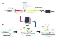
Fluorescence detection in molecular diagnostics. (a) the laser emits light that passes through a collimator lens and an excitation filter before reaching the sample. The light emitted by the sample passes through an emission filter and a condensing lens before being detected. b) steps of the qPCR process.
However, considering the high costs and complexity of the diagnostic kit and the fluorescence detection system for the development of POCT tools, colorimetry systems present themselves as an interesting option and are easy to adapt to existing systems. Prices in Brazil for fluorescent probes for qPCR, for example, can be around USD 276, while a vial of reagent for performing a colorimetric reaction may cost less than USD 92 (these prices can be verified directly with companies specializing in the sale of these reagents), which allows a much greater number of assays to be performed. In the next section, the colorimetry detection system applied to molecular diagnostics for the development of POCT tools will be described.
Molecular diagnostics via colorimetry in POCT tools
Colorimetry is the measurement of the wavelength and intensity of electromagnetic radiation in the region of the visible spectrum. This is extensively applied for the identification and determination of concentrations of substances that absorb light. Colorimetric analysis is a method for determining the concentration of a chemical element or chemical compound in a solution with the aid of a color reagent, and is applicable to both organic compounds and inorganic compounds and can be used with or without an enzymatic step (Jaroenram et al. 2022).
The colorimeter, also known as a spectrophotometer, is an analytical system in which specific solutions absorb a certain wavelength of light emitted through the sample as a way of calculating the concentration of a solution via the Beer-Lambert Law. The spectrophotometer can determine the intensity or, more precisely, the wavelength of light as a function of color by measuring the amount of light reflected through the sample, according to the diagnostic kit and the wavelength used by the emitting source (Lim et al. 2019). This process, known as absorbance, is a unit measure of the amount of light passing through a given volume of liquid, the distance between the specific wavelength emitter and the detector must be fixed, as well as the volumetric area of the solution to be analyzed. The accuracy of the detector is related to its reading capacity, in frequency or voltage level, and the applied converter circuits (Eom & Dasgupta 2006, Gong et al. 2009).
In the molecular diagnostic kit, dyes with the ability to change color according to the amplification of the target nucleic acid should be added, such as hydroxynaphthol blue (HNB) dyes changing the color from violet to blue, phenol red (PSP) changing the color from pink to yellow, and neutral red (NR) changing the color from orange to pink. The steps and process for performing molecular diagnosis by colorimetry are represented in Figure 5. Currently, detection using the colorimetric technique is more often applied to the LAMP methods of molecular diagnosis; however, many studies have been developed using the PCR method (Lau et al. 2015).

Detection via colorimetry in molecular diagnostics. (a) sample collection and DNA extraction. (b) amplification process of the target nucleic acid. (c) use of indicator dyes to monitor the presence of the target pathogen and its concentration.
Fluorescence and colorimetric detection systems perform an indirect measurement of the sample. In order to develop a technology for the direct detection and analysis of nucleic acid amplification of the target in the diagnostic kit solution in POCT tools, the electrochemical detection system has emerged as a promising alternative. Detection via electrochemistry allows integration with the thermal transfer system in a more appropriate way, which is less costly to manufacture and easy to handle. Therefore, the electrochemical detection system allows a direct reading of the sample, and the use of redox species, for addition in the diagnostic kit solution. Compared to the dyes used in fluorescence detection systems and colorimetry, these have a lower cost. The following developments of electrochemical techniques in detection systems for the development of POCT tools are described.
Molecular diagnostics via electrochemical techniques in POCT tools
Electrochemistry is one of the main areas of analytical chemistry, and involves the measurement of electrical properties under conditions that, directly or indirectly, allow an association between the magnitude of the measured property and the concentration of certain specific chemical species (Hilt 2020).
Electroanalytical methods are a class of techniques that make use of measurable electrical properties such as electric current, differences in potential and the accumulation of interfacial charges from phenomena in which a redox species interacts physically and/or chemically with the solution under analysis (Lee et al. 2003, Zhang et al. 2011, Sousa-Pereira et al. 2013, Zhou et al. 2023).
In the development of molecular diagnostic kits coupled to an electrochemical detection system, it is necessary to use an intercalating probe, which binds to the target nucleic acid. As nucleic acid amplification occurs, it is possible to register changes in electrical properties which are related to the level of infection by the pathogen (Amor-Gutiérrez et al. 2020).
The main non-specific electroactive mediators for nucleic acid detection of the target can be transition metal complexes, for example, osmium and ruthenium or organic species such as methylene blue and Hoechst 33258, which are cheaper than the dyes used in fluorescence or colorimetric detection systems (Kobayashi et al. 2004).
Redox intercalating probes have an important analytical role in electrochemical measurements, whereby in the absence of the target they must provide a stable baseline response, and in the presence of the target provide detectable variations in signal amplitude. The amplicon (nucleic acid amplification reaction target) performs a binding interaction with the redox intercalating probes, and the molecules of the probe are freely ionized in solutions and are available for electrocatalysis. In other words, as amplification occurs, the amplicons that are produced bind to the intercalating probes, forming an electrochemically inactive probe- amplicon complex, which reduces the electric current values (Chen et al. 2019).
However, the concentration of the redox species applied in the development of the diagnostic kit for detection through electrochemical systems is a critical point, since the relationship of greater response of the electrochemical signal must be found without inhibiting the amplification process of the target nucleic acid, due to the excess intercalating probes (Moreau et al. 2017).
Qualitative or quantitative responses of a given analyte in electrochemical systems are observed when controlled perturbations are applied, such as, for example, a potential difference between the electrodes of the electrochemical cell to analyze the chemical reactivity of a surface or a solution. The electroanalytical signals can be related to some intrinsic chemical parameter of the species through the measurement of the potential difference or current related to the analyte in an electrochemical cell (Goda et al. 2015, Henihan et al. 2016, Tsaloglou et al. 2018).
The oxidation-reduction reactions are controlled and measured in the electrochemical cell in an electrolytic medium by a potentiostat. One of the main electrochemical techniques of direct measurement in the solution is voltammetry, which applies a potential scan, whose range depends on the redox species used, and the electrochemical cell can be composed of a system of three electrodes. In practical terms, this method is non-destructive, since only a very small amount of the analyte is consumed on the two-dimensional surface of the working electrodes and counter electrode (Rosario & Mutharasan 2014, Santhanam et al. 2020, Nunez-Bajo et al. 2020).
Voltammetry performs the kinetic and thermodynamic analysis of electron addition (reduction) and electron loss (oxidation), which can be observed through different types of potential applied by the potentiostat, such as linear scanning voltammetry (LSV), cyclic voltammetry (CV), differential pulse voltammetry (DPV) and square wave voltammetry (SWV). The most used technique in the development of detection systems using electrochemistry in molecular diagnostics is cyclic voltammetry, which permits the study of the redox properties of compounds and interfacial structures in solution (Shah et al. 2013, Temerk et al. 2015, Radi et al. 2013).
The structure of POCT tools for molecular diagnosis using the electrochemical detection system can be seen in Figure 6.
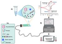
A system of detection by electrochemical techniques in real time. a) amplification occurs in an electrode connected to a potentiostat. b) in real time, the system monitors the amplification of DNA by means of a redox test.
Yeung et al. (2006) were the first to demonstrate the possibility of electrochemically monitoring the amplification of target nucleic acid via the PCR method. The strategy was based on the immobilization of a redox-labeled base on the surface of the working electrode, where the electrochemical detection system performed the analysis of the amplification process of the target nucleic acid in molecular diagnostics using the solid phase PCR method. However, this approach showed little detection efficiency after amplification of the target nucleic acid, obtaining a linear signal growth, instead of an exponential one, as expected by the PCR method. Therefore, the detection of low concentrations in the ng/L range was not possible, even with the immobilization of target pathogen receptor probes on the working electrode, hindering its application in complex fluid matrices such as target nucleic acid amplification solutions (Attoye et al. 2021, Bukkitgar et al. 2021).
Despite the fact that the modification of the surface of the working electrodes allows one to increase the selectivity to a specific part of the pathogen, during amplification of the target nucleic acid, the accumulation of the analyte occurs in the receptor probes, which are kept attached to the electrode by covalent bonds, ion exchange and other electrostatic bonds. As in every adsorption process, the amount of analyte accumulated on the electrode surface is a function of many factors, such as solvent, electrode material, ionic strength, pH, mass transport, potential or temperature, making it extremely difficult to obtain a reproducible process in detecting specific parts of a particular pathogen (Martin et al. 2016). Since the modification of the working electrode is a procedure that involves the formation of a surface layer, the loss of linearity at relatively high concentrations (around 10-6 mol L-1) is common (West 2020, Han et al. 2022, Safitri et al. 2022).
The process of immobilizing a redox-labeled base on the surface of the working electrode is quite laborious and this process can cause phenomena such as memory effects or instability in the electrodes during the amplification process of the target nucleic acid, restricting the number of measurements. In other words, the adsorption saturation of the target nucleic acid on the surface of the working electrode forms a passivation layer, limiting the measurement capability of the detection system to a few cycles of oxidation reduction (Barek 2021).
As an alternative strategy for reducing the influence of the modification of the working electrode, an electrochemical detection process was developed based on reading the polymerization of the target nucleic acid in the solution instead of using the surface of the working electrode (Yeung et al. 2007). In this alternative method, the electrochemical detection is performed through the exponential consumption of free nucleoside triphosphate during the PCR reaction, being possible to monitor the DNA amplification in each PCR cycle in a multiplexed array of hermetically sealed electrochemical cells, with a volume of less than 50 µL of PCR solution per cell, allowing one to obtain a complete kinetic curve of the amplification cycles of the target nucleic acid. The principle of electrochemical detection of exponential consumption of free nucleoside triphosphate is analogous to that of fluorescent intercalant detection of ds-DNA, except that the signal from the redox intercalator is decreased exponentially as the amount of the amplified target nucleic acid increases with the number of PCR cycles. However, compared to fluorescent qPCR, the electrochemical strategy showed lower sensitivity and specificity (Deféver et al. 2009). Therefore, to overcome these limitations, Deféver et al. (2011) proposed an alternative strategy based on the use of a DNA intercalating redox probe, Os[(bpy) (2) DPPZ](2+), which after binding to double-stranded DNA (ds-DNA) becomes electrochemically detectable and has less effect of inhibiting the amplification process of the target nucleic acid via the PCR method.
This methodology is comparable to qPCR based on optical detection, offering the same advantages as fluorescent qPCR using SYBR green, but with the additional advantage of presenting a lower cost in the development of the diagnostic kit and being simpler to integrate into miniaturized systems, such as in the microchip platform and or in microfluidic cartridges. However, the electrochemical reaction is a thermodynamic process and PCR methods use different temperature levels during the amplification cycles of the target nucleic acid, causing significant disturbances in the diffusion coefficient, i.e., change in the adsorption rate of the target probe on the working electrode, causing a change in the rate of consumption of free nucleoside triphosphate.
In this case, the LAMP method presents itself as an interesting alternative, since the amplification reaction of the target nucleic acid is carried out by an isothermal profile, which eliminates the problem of disturbances in the diffusion coefficient on the working electrode, which are caused by thermal instabilities during measurements. This allows one to obtain satisfactory results through real-time electrochemical detection with analytical performance that is comparable to those obtained in real-time fluorescence LAMP and real-time fluorescence PCR (Martin et al. 2016, Marangoni et al. 2023).
The application of CRISPR in diagnostic kits has shown great potential for increasing the performance of pathogen detection in several nucleic acid amplification methods and using different real-time detection systems (Dong et al. 2023, Mendes et al. 2024, Hassan et al. 2025).
Recently, the ability to detect with a high level of specificity and the ability to detect low levels of infection using CRISPR/Cas with electrochemical systems for detection without immobilization of the receiving probe on the working electrode has been observed (Zhou et al. 2023, Carlo et al. 2024, Lakshmanan & Liu 2025).
However, the cost is still higher than the standard diagnostic kits of the qPCR method. Immobilization of the receiving probe on the working electrode represents an important tool in the selectivity of molecular diagnostics using electrochemical detection systems. In this process, electrochemical impedance spectroscopy (EIS) is the technique used for the analysis of interfacial properties related to bio-recognition events that occur on the electrode surface, such as antibody-antigen recognition, substrate-enzyme interaction or capture of whole cells (Magar et al. 2021).
In electrochemical impedance spectroscopy, an alternating current signal of small amplitude scans an electrochemical cell over a wide frequency range, permitting the study of capacitive, inductive and diffusion processes that occur in the electrochemical cell (Lim et al. 2021, Biswas et al. 2022). Thus, EIS is considered to be the main tool in the evaluation of selectivity in the recognition of the target pathogen in electrochemical sensors applied in molecular diagnostics (Lopes et al. 2022).
Therefore, despite the difficulties presented, the electrochemical detection system is the most suitable system for performing direct monitoring of the solution during the amplification process of the target nucleic acid quantitatively in real time, and the simplest technique to obtain a molecular diagnostic microchip in the development of portable POCT tool (Safavieh et al. 2016, Tsaloglou et al. 2018, Shabani et al. 2020, Tamiya 2022).
Electroanalytical techniques are promising as alternatives aimed at achieving this goal, since they require minimal instrumentation, low energy consumption and electrochemical cells can be easily integrated through microelectronic processes or other printing techniques in miniaturized format, together with the reservoirs or reaction chambers where the molecular diagnostic solution must be contained. Below, we present some formats and applications of microfluidic systems that use electrochemical, fluorometric or colorimetric detection, each with specific advantages in sensitivity, specificity, multiplexing, simplicity and cost, thus allowing adaptation to different diagnostic and monitoring scenarios.
MINIATURIZATION AND MICROFLUDICS
Portable molecular diagnostic POCT tools require the incorporation of sample preparation, reaction and detection into small and accessible platforms. Therefore, the main miniaturization strategy is based on functional integration through microfluidics, which allows the use of reduced volumes and low energy consumption (John & Price 2014, Sachdeva et al. 2021). The three structural concepts of microfluidics in molecular diagnostics are reaction chambers, continuous flow and droplets (Ma et al. 2021b, Vladisaljević 2024, Dorfman et al. 2005).
Microfluidic reaction chamber structures (Figure 7b) allow the use of small volumes, the integration of dry reagents, and easy incorporation of optical or electrochemical detection systems; however, they present limitations in high-throughput applications or those requiring efficient reagent mixing (Wong et al. 2024, Cai et al. 2023, Damiati et al. 2022, Nguyen et al. 2024, Mendes et al. 2019).

Schematic representation of an integrated platform for molecular POCT based on nucleic acid amplification via microfluidics. (a) Biological material containing pathogens is collected and the sample is processed and purified; then, nucleic acids are amplified in an integrated platform with a temperature control system and a detection system. (b) Main microfluidic concepts in molecular diagnostics: reaction chambers, continuous flow and droplet-based systems. (c) Nucleic acid amplification methods with applicable temperature control systems for PCR, LAMP and RPA. (d) Detection mechanisms that can be integrated into the platform: fluorescence, colorimetric detection and electrochemical detection.
Continuous flow is applied in sequential steps of sample preparation or purification, but its use in ultracompact systems is limited due to the complexity of flow and mixing control (Hernández-Neuta et al. 2018, Kwon et al. 2019, Wang et al. 2021).
In droplet microfluidics, each droplet acts as a microreactor, making it ideal for detecting rare events in high-throughput assays, such as digital PCR (dPCR); however, it requires precise fluid control and the integration of sophisticated optical modules (Wei et al. 2022, Abate et al. 2013, Cui et al. 2017). The choice between droplet microfluidics, continuous flow, reaction chambers, or hybrid structures depends on the diagnostic application, sensitivity requirements and processing rate, as well as practical factors such as cost and ease of use.
The manipulation of small volumes allows extremely rapid heating and cooling rates through the use of thick-film heaters, printed by screen printing, combined with thermoelectric coolers (TEC) in the thermal control system, significantly reducing nucleic acid amplification time, despite the complexity of integrating these components into the system (Dong et al. 2021, An et al. 2023, Sun et al. 2024). Thin-film deposition technology enables on-chip integration of the thermal control system, which, when combined with heat sinks and forced-air flow systems, enhances heat dissipation during device operation (Dos-Reis-Delgado et al. 2023, Veltkamp et al. 2020).
The detection system integrated into miniaturized platforms for molecular diagnostics varies according to factors such as sensitivity and cost. Fluorescence detection offers high sensitivity but requires more complex optical systems compared to colorimetric detection, which is better suited for quantitative and semi-quantitative tests (Fang et al. 2022, Măriuţa et al. 2020, Wu et al. 2022). Electrochemical detection is low cost and is compatible with on-chip integration without the need for external devices, unlike optical detection systems (Marchlewicz et al. 2020). A schematic representation of miniaturized POCT devices can be seen in Figure 7.
An ideal miniaturized POCT system should incorporate a polymeric microfluidic cartridge with integrated reaction chambers, potentially including paper elements for sample preparation and capillary-driven flow generation (Chu et al. 2025, Cinti et al. 2018, Mahardika et al. 2023). However, hybrid approaches, which combine different miniaturization strategies, show promising results in CFD (computational fluid dynamics) simulations and are likely to be the most promising for the next generation of POCT devices (Waqas et al. 2024).
Over the past five years, several studies have aimed at developing microfluidic platforms for molecular pathogen detection, as summarized in Table I. In this table, a predominance of articles targeting SARS-CoV-2 can be observed, due to the pandemic caused by this pathogen, which began in 2020.
Summary of the studies from the past five years that aimed at developing molecular amplification assays for pathogen detection using microfluidic strategies.
Additionally, from the analysis of Table I, it is possible to conclude that studies more frequently employed the microfluidic reaction chamber approach combined with fluorescence detection, likely because these strategies are already well established for new molecular detection platforms. However, although most studies used PCR as the amplification methodology, a similar number of articles employed LAMP, probably because LAMP has fewer thermal requirements, being an isothermal technique.
Within the articles reviewed in the aforementioned table, different detection limits were reported for each of the developed diagnostic platforms, showing, for example, ranges from 10⁴ to 500 copies/mL for virus detection (Wang et al. 2024, Liu et al. 2024c). In bacterial detection, the lowest detection limit reported was 25 bacteria (Xiao et al. 2024).
Thus, the selection of microfluidic strategies and detection methods directly affects the sensitivity of POCT platforms. This is essential for achieving detection limits that meet clinical requirements, especially in situations of early diagnosis and monitoring of pathogens with low viral loads.
CONCLUSIONS
In this review, the main nucleic acid amplification methods and detection systems applicable to the development of POCT tools were presented and discussed. The growing demand for fast, accurate, sensitive and affordable POCT tools in regions with limited infrastructure drives innovation in this field. PCR-based methods and their variants remain the gold standard, but isothermal methods such as LAMP and RPA gain prominence for allowing simpler operation and potential miniaturization. Among detection systems, fluorescence is the most widespread system in laboratory applications, while colorimetry offers an accessible means of visualization, especially in rapid screening contexts.
Electrochemistry, in turn, emerges as a promising alternative for POCT applications, as it combines sensitivity, low energy consumption and ease of integration with microfluidic platforms. Despite the advances, important challenges remain, such as the need to standardize protocols, reduce reagent and device costs, increase field robustness, and improve selectivity and sensitivity in complex arrays. Electrochemical techniques, although less used, have proven themselves to be increasingly viable, especially when combined with strategies such as the use of intercalating redox species, microchip platforms and CRISPR-based systems.
Thus, the convergence between isothermal amplification, electrochemical detection and microfluidic integration technologies represents a promising direction for the advancement of POCT tools. The development of modular, reproducible, scalable and low-cost devices is the key to making decentralized molecular diagnostics a viable reality on a large scale.
Acknowledgements
We would like to thank the institutions Fundação de Amparo à pesquisa do Amazonas (FAPEAM), Coordenação de Aperfeiçoamento de Pessoal de Nível Superior (CAPES, Brazil) and Conselho Nacional de Desenvolvimento Científico e Tecnológico (CNPq) for the financial support provided for this study.
References
- ABATE AR, HUNG T, SPERLING RA, MARY P, ROTEM A, AGRESTI JJ, WEINER MA & WEITZ DA. 2013. DNA sequence analysis with droplet-based microfluidics. Lab Chip 13: 4864.
- ABDELFATAH T ET AL. 2023. Nanoplasmonic amplification in microfluidics enables accelerated colorimetric quantification of nucleic acid biomarkers from pathogens. Nature Nanotechnology 18(8): 922-932.
- AGEL E & ALTIN KH. 2024. Field-applicable simultaneous multiplex LAMP assay for screening HBV and HCV co-infection in a single tube. BMC Infect Dis 24: 1-13.
- ALMEIDA-DE-OLIVEIRA NK, MOREIRA OC, DE LAVIGNE AR, MENDONÇA-LIMA L, WERNECK GL, DANIEL-RIBEIRO CT & FERREIRA-DA-CRUZ MDF. 2019. Analytical validation of real-time quantitative PCR assays for optimum diagnosis of vivax malaria. Mem Inst Oswaldo Cruz 114: e180350.
- AMOR-GUTIÉRREZ O, SELVOLINI G, FERNÁNDEZ-ABEDUL TM, DE LA ESCOSURA-MUÑIZ A & MARRAZZA G. 2020. Folding-Based Electrochemical Aptasensor for the Determination of β-Lactoglobulin on Poly-L-Lysine Modified Graphite Electrodes. Sensors 20: 2349.
- AN YQ ET AL. 2023. Ultrafast Microfluidic PCR Thermocycler for Nucleic Acid Amplification. Micromachines 14: 658.
- ARTIKA IM, DEWI YP, NAINGGOLAN IM, SIREGAR JE & ANTONJAYA U. 2022. Real-Time Polymerase Chain Reaction: Current Techniques, Applications, and Role in COVID-19 Diagnosis. Genes (Basel) 13.
- ATTOYE B, BAKER MJ, THOMSON F, POU C & CORRIGAN DK. 2021. Optimisation of an Electrochemical DNA Sensor for Measuring KRAS G12D and G13D Point Mutations in Different Tumour Types. Biosensors (Basel) 11: 42.
- BAREK J. 2021. How to Improve the Performance of Electrochemical Sensors via Minimization of Electrode Passivation. Chemosensors 9: 12.
- BISWAS GC, CHOUDHURY S, RABBANI MM & DAS J. 2022. A Review on Potential Electrochemical Point-of-Care Tests Targeting Pandemic Infectious Disease Detection: COVID-19 as a Reference. Chemosensors 10: 269.
- BUKKITGAR SD, SHETTI NP & AMINABHAVI TM. 2021. Electrochemical investigations for COVID-19 detection-A comparison with other viral detection methods. Chem Eng J 420: 127575.
- BUULTJENS AH, VANDELANNOOTE K, SHARKEY LK, HOWDEN BP, MONK IR, LEE JYH & STINEAR TP. 2021. Low-Cost, Open-Source Device for High-Performance Fluorescence Detection of Isothermal Nucleic Acid Amplification Reactions. ACS Biomater Sci Eng 7: 4982-4990.
- CAI S, JIN Y, LIN Y, HE Y, ZHANG P, GE Z & YANG W. 2023. Micromixing within microfluidic devices: Fundamentals, design, and fabrication. Biomicrofluidics 17.
- CARLO D, RESEARCH SB, CARLO E Di & SORRENTINO C. 2024. State of the art CRISPR-based strategies for cancer diagnostics and treatment. Biomarker Res 12: 1-20.
- CHELLANI HH ET AL. 2025. Volumetric, Microfluidic Plasmonic RT-PCR. Small Methods 9.
- CHEN BJ, MANI V, HUANG ST, HU YC & SHAN HCP. 2019. Bisintercalating DNA redox reporters for real-time electrochemical qLAMP. Biosens Bioelectron 129: 277-283.
- CHEN X, LIU Y, ZHAN X, GAO Y, SUN Z, WEN W & ZHENG W. 2022. Ultrafast PCR Detection of COVID-19 by Using a Microfluidic Chip-Based System. Bioengineering 9: 548.
- CHU PY, HUANG PS, CHEN CY, TSAI KY, CHIU SY, FAN LW, CHENG YC, LIN CJ, HSIEH CH & WU MH. 2025. Development of a point-of-care testing (POCT)-use paper-based device for recombinase polymerase amplification (RPA)-based bioassays-: Demonstration of the detection of Neisseria gonorrhoeae. Sens Act Rep 9: 100307.
- CINTI S, PROIETTI E, CASOTTO F, MOSCONE D & ARDUINI F. 2018. Paper-Based Strips for the Electrochemical Detection of Single and Double Stranded DNA. Anal Chem 90: 13680-13686.
- COOK D, PFISTER JA, CONSTANTINO JR, ROPER JM, GARDNER DR, WELCH KD, HAMMOND ZJ & GREEN BT. 2015. Development of a PCR-based method for detection of delphinium species in poisoned cattle. J Agric Food Chem 63: 1220-1225.
- CUI M, FENG H, GUO D, WANG D & ZHENG B. 2017. A droplet-based microfluidic platform for kinetics-based detection of single nucleotide variation at room temperature with large discrimination factors. Sens Actuators B Chem 253: 731-737.
- DAMIATI LA, EL-YAAGOUBI M, DAMIATI SA, KODZIUS R, SEFAT F & DAMIATI S. 2022. Role of Polymers in Microfluidic Devices. Polymers 2022, Vol 14, Page 5132 14: 5132.
- DEFÉVER T, DRUET M, EVRARD D, MARCHAL D & LIMOGES B. 2011. Real-time electrochemical PCR with a DNA intercalating redox probe. Anal Chem 83: 1815-1821.
- DEFÉVER T, DRUET M, ROCHELET-DEQUAIRE M, JOANNES M, GROSSIORD C, LIMOGES B & MARCHAL D. 2009. Real-time electrochemical monitoring of the polymerase chain reaction by mediated redox catalysis. J Am Chem Soc 131: 11433-11441.
- DEL RÍO JS, LOBATO IM, MAYBORODA O, KATAKIS I & O’SULLIVAN CK. 2017. Enhanced solid-phase recombinase polymerase amplification and electrochemical detection. Anal Bioanal Chem 409: 3261-3269.
- DONG J, WU X, HU Q, SUN C, LI J, SONG P, SU Y & ZHOU L. 2023. An immobilization-free electrochemical biosensor based on CRISPR/Cas13a and FAM-RNA-MB for simultaneous detection of multiple pathogens. Biosens Bioelectron 241: 115673.
- DONG X, LIU L, TU Y, ZHANG J, MIAO G, ZHANG L, GE S, XIA N, YU D & QIU X. 2021. Rapid PCR powered by microfluidics: A quick review under the background of COVID-19 pandemic. Trends in Analytical Chemistry 143: 116377.
- DORFMAN KD, CHABERT M, CODARBOX JH, ROUSSEAU G, DE CREMOUX P & VIOVY JL. 2005. Contamination-free continuous flow microfluidic polymerase chain reaction for quantitative and clinical applications. Anal Chem 77: 3700-3704.
- DOS-REIS-DELGADO AA, CARMONA-DOMINGUEZ A, SOSA-AVALOS G, JIMENEZ- SAAIB IH, VILLEGAS-CANTU KE, GALLO-VILLANUEVA RC & PEREZ-GONZALEZ VH. 2023. Recent advances and challenges in temperature monitoring and control in microfluidic devices. Electrophoresis 44: 268-297.
- EISENSTEIN BI. 1990. The polymerase chain reaction. A new method of using molecular genetics for medical diagnosis. N Engl J Med 322(3): 178-183.
- EOM IY & DASGUPTA PK. 2006. Frequency-selective absorbance detection: Refractive index and turbidity compensation with dual-wavelength measurement. Talanta 69: 906-913.
- ESTRELA PFN, DE SOUZA JÚNIOR MN, DE OLIVEIRA KG & DUARTE GRM. 2025. Stick-and-test: simple tape-based devices for SARS-CoV-2 isothermal molecular detection. Anal Bioanal Chem.
- ECDC - EUROPEAN CENTRE FOR DISEASE PREVENTION AND CONTROL. 2022. Assessment of point of care testing devices for infectious disease surveillance, prevention and control - a mapping exercise. Stockholm.
- FANG Y, WANG Y, SU X, LIU H, CHEN H, CHEN Z, JIN L & HE N. 2022. A miniaturized and integrated dual-channel fluorescence module for multiplex real-time PCR in the portable nucleic acid detection system. Front Bioeng Biotechnol 10: 996456.
- FARCAS GA, ZHONG KJY, MAZZULLI T & KAIN KC. 2004. Evaluation of the RealArt Malaria LC Real-Time PCR Assay for Malaria Diagnosis. J Clin Microbiol 42: 636-638.
- GARG N, AHMAD FJ & KAR S. 2022. Recent advances in loop-mediated isothermal amplification (LAMP) for rapid and efficient detection of pathogens. Curr Res Microb Sci 3: 100120.
- GAVINA K, FRANCO LC, KHAN H, LAVIK JP & RELICH RF. 2023. Molecular point-of-care devices for the diagnosis of infectious diseases in resource-limited settings - A review of the current landscape, technical challenges, and clinical impact. J Clin Virol 169: 105613.
- GLÖKLER J, LIM TS, IDA J & FROHME M. 2021. Isothermal amplifications-a comprehensive review on current methods. Crit Rev Biochem Mol Biol 56: 543-586.
- GODA T, TABATA M & MIYAHARA Y. 2015. Electrical and electrochemical monitoring of nucleic acid amplification. Front Bioeng Biotechnol 3: 136057.
- GONG W, KRAFT M, MORGAN H & MOWLEM M. 2009. A Simple, Low-Cost Double Beam Spectrophotometer for Colorimetric Detection of Nitrite in Seawater. IEEE Sens J 9: 862-869.
- GUO N, CHEUNG K, WONG HT & HO D. 2014. CMOS time-resolved, contact, and multispectral fluorescence imaging for DNA molecular diagnostics. Sensors (Switzerland) 14: 20602-20619.
- HAN H, NOBUSAWA K & YAMASHITA I. 2022. Anomalous Enhancement of Electrochemical Charge Transfer by a Ru Complex Ion Intercalator. Anal Chem 94: 571-576.
- HASSAN YM, MOHAMED AS, HASSAN YM & EL-SAYED WM. 2025. Recent developments and future directions in point-of-care next-generation CRISPR-based rapid diagnosis. Clin Exp Med 25: 33.
- HE D, YANG J, JIANG X, LIN Y, CHEN H, TANG Y & DIAO Y. 2020. A quantitative loop-mediated isothermal amplification assay for detecting a novel goose astrovirus. Poult Sci 99: 6586-6592.
- HENIHAN G ET AL. 2016. Label- and amplification-free electrochemical detection of bacterial ribosomal RNA. Biosens Bioelectron 81: 487-494.
- HERNÁNDEZ-NEUTA I, PEREIRO I, AHLFORD A, FERRARO D, ZHANG Q, VIOVY JL, DESCROIX S & NILSSON M. 2018. Microfluidic magnetic fluidized bed for DNA analysis in continuous flow mode. Biosens Bioelectron 102: 531-539.
- HILT G. 2020. Basic Strategies and Types of Applications in Organic Electrochemistry. ChemElectroChem 7: 395-405.
- HO-PUN-CHEUNG A, BASCOUL-MOLLEVI C, ASSENAT E, BOISSIÈRE-MICHOT F, BIBEAU F, CELLIER D, YCHOU M & LOPEZ-CRAPEZ E. 2009. Reverse transcription- quantitative polymerase chain reaction: Description of a RIN-based algorithm for accurate data normalization. BMC Mol Biol 10.
- HU Y & HU Y. 2016. Regulatory Concern of Polymerase Chain Reaction (PCR) Carryover Contamination. Polymerase Chain Reaction for Biomedical Applications.
- ISAIAH Z, SHEHU A, UMAR MM & MUSA MI. 2024. Incidence of Other Bacterial Pathogens among Patients suspected with Pulmonary Tuberculosis attending Infectious Diseases Hospital, Kano.
- JANG WS ET AL. 2021. Development of a multiplex Loop-Mediated Isothermal Amplification (LAMP) assay for onsite diagnosis of SARS CoV-2. PLoS One 16.
- JAROENRAM W ET AL. 2022. One-step colorimetric isothermal detection of COVID-19 with AI-assisted automated result analysis: A platform model for future emerging point-of-care RNA/DNA disease diagnosis. Talanta 249: 123375.
- JOHN AS & PRICE CP. 2014. Existing and Emerging Technologies for Point-of-Care Testing. Clin Biochem Rev 35: 155.
- KHEHRA N, PADDA IS & SWIFT CJ. 2023. Polymerase Chain Reaction (PCR). StatPearls.
- KOBAYASHI M, KUSAKAWA T, SAITO M, KAJI S, OOMURA M, IWABUCHI S, MORITA Y, HASAN Q & TAMIYA E. 2004. Electrochemical DNA quantification based on aggregation induced by Hoechst 33258. Electrochem commun 6: 337-343.
- KOO SBN, CHI HG, KIM JD, KIM YS, PARK JS, PARK CY & LEE DJ. 2021. Multiple Compact Camera Fluorescence Detector for Real-Time PCR Devices. Sensors 21: 7013.
- KRAMPA FD, ANIWEH Y, KANYONG P & AWANDARE GA. 2020. Recent Advances in the Development of Biosensors for Malaria Diagnosis. Sensors (Basel) 20.
- KWON K, GWAK H, HYUN KA, KWAK BS & JUNG H Il. 2019. High-throughput microfluidic chip for magnetic enrichment and photothermal DNA extraction of foodborne bacteria. Sens Actuators B Chem 294: 62-68.
- LAKSHMANAN K & LIU BM. 2025. Impact of Point-of-Care Testing on Diagnosis, Treatment, and Surveillance of Vaccine-Preventable Viral Infections. Diagnostics 15.
- LARKINS MC & THOMBARE A. 2025. Point-of-Care Testing. May 29. In: StatPearls [Internet]. Treasure Island (FL): StatPearls Publishing; Jan-. PMID: 37276307
- LAU YL, LAI MY, TEOH BT, ABD-JAMIL J, JOHARI J, SAM SS, TAN KK & ABUBAKAR S. 2015. Colorimetric Detection of Dengue by Single Tube Reverse-Transcription-Loop- Mediated Isothermal Amplification. PLoS One 10: e0138694.
- LEE TMH, CARLES MC & HSING IM. 2003. Microfabricated PCR-electrochemical device for simultaneous DNA amplification and detection. Lab Chip 3: 100-105.
- LI J, MACDONALD J & VON STETTEN F. 2018. Review: a comprehensive summary of a decade development of the recombinase polymerase amplification. Analyst 144: 31-67.
- LI X, ZHAO X, YANG W, XU F, CHEN B, PENG J, HUANG J & MI S. 2021. Stretch-driven microfluidic chip for nucleic acid detection. Biotechnol Bioeng 118: 3559-3568.
- LIM DYS, SEO MJ & YOO JC. 2019. Optical temperature control unit and convolutional neural network for colorimetric detection of loop-mediated isothermal amplification on a Lab- on-A-disc platform. Sensors (Switzerland) 19.
- LIM J, KIM J, LEE E, PARK H & LEE JH. 2021. An analysis method to detect the presence of DNA using electrochemical impedance spectroscopy (EIS) for real-time PCR. Microsystem Technologies 27: 3211-3217.
- LIU J, ZENG Z, LI F, JIANG B, NIE Y, ZHANG G, PANG B, SUN L & HAO R. 2024a. Portable and simultaneous detection of four respiratory pathogens through a microfluidic LAMP and real-time fluorescence assay. Analyst 149: 5091-5100.
- LIU Y, CHAO Z, DING W, FANG T, GU X, XUE M, WANG W, HAN R & SUN W. 2024b. A multiplex RPA-CRISPR/Cas12a-based POCT technique and its application in human papillomavirus (HPV) typing assay. Cell Mol Biol Lett 29: 1-24.
- LIU Y, HU Z, YANG S, XU N, SONG Q, GAO Y & WEN W. 2024c. Fabrication of Cost-Effective Microchip-Based Device Using Sandblasting Technique for Real-Time Multiplex PCR Detection. Micromachines (Basel) 15.
- LOBATO IM & O’SULLIVAN CK. 2018. Recombinase polymerase amplification: Basics, applications and recent advances. TrAC - Trends in Analytical Chemistry 98: 19-35.
- LOPES LC, SANTOS A & BUENO PR. 2022. An outlook on electrochemical approaches for molecular diagnostics assays and discussions on the limitations of miniaturized technologies for point-of-care devices. Sensors and Actuators Reports 4: 100087.
- MA H, BELL KN & LOKER RN. 2021a. qPCR and qRT-PCR analysis: Regulatory points to consider when conducting biodistribution and vector shedding studies. Mol Ther Methods Clin Dev 20: 152-168.
- MA J, TRAN G, WAN AMD, YOUNG EWK, KUMACHEVA E, ISCOVE NN & ZANDSTRA PW. 2021b. Microdroplet-based one-step RT-PCR for ultrahigh throughput single-cell multiplex gene expression analysis and rare cell detection. Scientific Reports 2021 11:1 11: 1-18.
- MA Z, MA M, CAO X, JIANG Y & GAO D. 2024. Droplet digital molecular beacon-LAMP assay via pico-injection for ultrasensitive detection of pathogens. Microchimica Acta 191.
- MAGAR HS, HASSAN RYA & MULCHANDANI A. 2021. Electrochemical impedance spectroscopy (Eis): Principles, construction, and biosensing applications. Sensors 21.
- MAHARDIKA IH, NAORUNGROJ S, KHAMCHAROEN W, KIN S, RODTHONGKUM N, CHAILAPAKUL O & SHIN K. 2023. Point-of-Care Testing (POCT) Devices for DNA Detection: A Comprehensive Review. Adv Nanobiomed Res 3: 2300058.
- MARANGONI JM, NG KKS & EMADI A. 2023. Strategies for the Voltammetric Detection of Loop-Mediated Isothermal Amplification. Micromachines 2023, Vol 14, Page 472 14: 472.
- MARCHLEWICZ K, IGA O, ZUZANNA I-S, ROBERT Z, KAMIL Ż, ELŻBIETA J, ZBIGNIEW B & ELŻBIETA M. 2020. Miniaturized device for performing PCR, integrated with an electrochemical DNA biosensor for detection of corynebacterium diphtheriae. In: MicroTAS 2020 - 24th International Conference on Miniaturized Systems for Chemistry and Life Sciences, San Diego: Chemical and Biological Microsystems Society, p. 1183-1184.
- MĂRIUŢA D, COLIN S, BARROT-LATTES C, LE CALVÉ S, KORVINK JG, BALDAS L & BRANDNER JJ. 2020. Miniaturization of fluorescence sensing in optofluidic devices. Microfluidics and Nanofluidics 24: 1-28.
- MARTIN A, BOUFFIER L, GRANT KB, LIMOGES B & MARCHAL D. 2016. Real-time electrochemical LAMP: a rational comparative study of different DNA intercalating and non- intercalating redox probes. Analyst 141: 4196-4203.
- MENDES GM, OLIVEIRA KG, BORBA JC, OLIVEIRA TS, FIACCADORI FS, NOGUEIRA ML, BAILÃO AM, SOARES CMA, CARRILHO E & DUARTE GRM. 2019. Molecular Diagnostics of Dengue by Reverse Transcription-Loop Mediated Isothermal Amplification (RT- LAMP) in Disposable Polyester-Toner Microdevices. J Braz Chem Soc 30: 1841-1849.
- MENDES LMC ET AL. 2024. CRISPR-Cas9 gene editing and its therapeutic applications: A literature review. Res Soc Develop 13: e11513846681-e11513846681.
- MIXSON-HAYDEN T, LUCCHI NW & UDHAYAKUMAR V. 2010. Evaluation of three PCR-based diagnostic assays for detecting mixed Plasmodium infection. BMC Res Notes 3.
- MOREAU M, DELILE S, SHARMA A, FAVE C, PERRIER A, LIMOGES B & MARCHAL D. 2017. Detection of a few DNA copies by real-time electrochemical polymerase chain reaction. Analyst 142: 3432-3440.
- MORI Y, NAGAMINE K, TOMITA N & NOTOMI T. 2001. Detection of loop-mediated isothermal amplification reaction by turbidity derived from magnesium pyrophosphate formation. Biochem Biophys Res Commun 289: 150-154.
- NATH P, MAHTABA KR & RAY A. 2023. Fluorescence-Based Portable Assays for Detection of Biological and Chemical Analytes. Sensors 23: 5053
- NGUYEN VD, NGUYEN HQ, BUI HK, KANG YJ & SEO TS. 2024. A smartphone-controllable molecular diagnostic platform for SARS-CoV-2 detection by reverse-transcription recombinase polymerase amplification. Sens Actuators B Chem 398: 134728.
- NGUYEN VD, NGUYEN HQ, BUI KH, KO YS, PARK BJ & SEO TS. 2022. A handheld-type total integrated capillary electrophoresis system for SARS-CoV-2 diagnostics: Power, fluorescence detection, and data analysis by smartphone. Biosens Bioelectron 195.
- NOTOMI T, OKAYAMA H, MASUBUCHI H, YONEKAWA T, WATANABE K, AMINO N & HASE T. 2000. Loop-mediated isothermal amplification of DNA. Nucleic Acids Res 28.
- NUNEZ-BAJO E, SILVA PINTO COLLINS A, KASIMATIS M, COTUR Y, ASFOUR T, TANRIVERDI U, GRELL M, KAISTI M, SENESI G, STEVENSON K & GÜDER F. 2020. Disposable silicon-based all-in-one micro-qPCR for rapid on-site detection of pathogens. Nature Communications 11: 1-10.
- ORIERO EC, OKEBE J, JACOBS J, VAN GEERTRUYDEN JP, NWAKANMA D & D’ALESSANDRO U. 2015. Diagnostic performance of a novel loop-mediated isothermal amplification (LAMP) assay targeting the apicoplast genome for malaria diagnosis in a field setting in sub-Saharan Africa. Malar J 14.
- OSCORBIN I & FILIPENKO M. 2023. Bst polymerase — a humble relative of Taq polymerase. Comput Struct Biotechnol J 21: 4519-4535.
- PAPADAKIS G ET AL. 2022. Portable real-time colorimetric LAMP-device for rapid quantitative detection of nucleic acids in crude samples. Sci Rep 12: 1-15.
- PARK YM, PARK J, LIM SY, KWON Y, BAE NH, PARK JK & LEE SJ. 2021. Integrated pumpless microfluidic chip for the detection of foodborne pathogens by polymerase chain reaction and electrochemical analysis. Sens Actuators B Chem 329: 129130.
- PERANDIN F ET AL. 2004. Development of a Real-Time PCR Assay for Detection of Plasmodium falciparum, Plasmodium vivax, and Plasmodium ovale for Routine Clinical Diagnosis. J Clin Microbiol 42: 1214-1219.
- PETRALIA S & CONOCI S. 2017. PCR technologies for point of care testing: Progress and perspectives. ACS Sens 2: 876-891.
- PLEBANI M ET AL. 2024. Point-of-care testing: State-of-the art and perspectives. Clin Chem Lab Med 63.
- RADI AE, NASSEF HM & EISSA A. 2013. Voltammetric and ultraviolet-visible spectroscopic studies on the interaction of etoposide with deoxyribonucleic acid. Electrochim Acta 113: 164-169.
- ROSARIO R & MUTHARASAN R. 2014. Nucleic acid electrochemical and electromechanical biosensors: A review of techniques and developments. Rev Anal Chem 33: 213-230.
- ROUGEMONT M, VAN SAANEN M, SAHLI R, HINRIKSON HP, BILLE J & JATON K. 2004. Detection of four Plasmodium species in blood from humans by 18S rRNA gene subunit- based and species-specific real-time PCR assays. J Clin Microbiol 42: 5636-5643.
- SACHDEVA S, DAVIS RW & SAHA AK. 2021. Microfluidic Point-of-Care Testing: Commercial Landscape and Future Directions. Front Bioeng Biotechnol 8: 602659.
- SAFAVIEH M, KANAKASABAPATHY MK, TARLAN F, AHMED MU, ZOUROB M, ASGHAR W & SHAFIEE H. 2016. Emerging Loop-Mediated Isothermal Amplification-Based Microchip and Microdevice Technologies for Nucleic Acid Detection. ACS Biomater Sci Eng 2: 278-294.
- SAFITRI E, HENG LY, AHMAD M, TAN LL, NAZARUDDIN N, SUHUD K, CHIANG CP & IQHRAMMULLAH M. 2022. Electrochemical DNA Biosensor Based on Mercaptopropionic Acid-Capped ZnS Quantum Dots for Determination of the Gender of Arowana Fish. Biosensors 12: 650.
- SANTHANAM M, ALGOV I & ALFONTA L. 2020. DNA/RNA Electrochemical Biosensing Devices a Future Replacement of PCR Methods for a Fast Epidemic Containment. Sensors 20: 4648.
- SANTIAGO-FELIPE S, TORTAJADA-GENARO LA, PUCHADES R & MAQUIEIRA Á. 2016. Parallel solid-phase isothermal amplification and detection of multiple DNA targets in microliter-sized wells of a digital versatile disc. Microchimica Acta 183: 1195-1202.
- SEOK Y, YIN Q, LI R, MAUK MG, BAI H & BAU HH. 2022. Manually-operated, slider cassette for multiplexed molecular detection at the point of care. Sens Actuators B Chem 369.
- SHABANI E, DOWLATSHAHI S & ABDEKHODAIE MJ. 2020. Laboratory detection methods for the human coronaviruses. Eur J Clin Microb Infect Dis 40: 225.
- SHAH A, NOSHEEN E, MUNIR S, BADSHAH A, QURESHI R, REHMAN ZU, MUHAMMAD N & HUSSAIN H. 2013. Characterization and DNA binding studies of unexplored imidazolidines by electronic absorption spectroscopy and cyclic voltammetry. J Photochem Photobiol B 120: 90-97.
- SNOUNOU G, VIRIYAKOSOL S, XIN PING ZHU, JARRA W, PINHEIRO L, DO ROSARIO VE, THAITHONG S & BROWN KN. 1993. High sensitivity of detection of human malaria parasites by the use of nested polymerase chain reaction. Mol Biochem Parasitol 61: 315-320.
- SOUSA-PEREIRA D, GOULART CM, DOS REIS CM & ECHEVARRIA A. 2013. Synthesis and evaluation of the anti-corrosion activity of thiosemicarbazide and thiosemicarbazone 4-N- (p-methoxyphenyl) substituted. Revista Virtual de Quimica 5: 770-785.
- SUKUMAR P, SALEH A, DELIORMAN M & QASAIMEH MA. 2025. Single-Layer Radially Compartmentalized Paper Chip (RCP-Chip) for Rapid Isothermal Multiplex Detection of SARS-CoV-2 Gene Targets. Advanced Sensor Research 4: 70010.
- SUN X ET AL. 2024. General strategy for developing thick-film micro-thermoelectric coolers from material fabrication to device integration. Nat Commun 15: 3870.
- TALAP J, SHEN M, YU L, ZENG S & CAI S. 2022. RT-LAMP assay combining multi- fluorescent probes for SARS-CoV-2 RNA detection and variant differentiation. Talanta 248.
- TAMIYA E. 2022. Portable Electrochemical DNA Sensors Based on Gene Amplification Reactions to Screen and Identify Pathogen and SNPs. Sensors 22: 1865.
- TEMERK Y, IBRAHIM M, IBRAHIM H & KOTB M. 2015. Interactions of an anticancer drug Formestane with single and double stranded DNA at physiological conditions. J Photochem Photobiol B 149: 27-36.
- TSALOGLOU MN, NEMIROSKI A, CAMCI-UNAL G, CHRISTODOULEAS DC, MURRAY LP, CONNELLY JT & WHITESIDES GM. 2018. Handheld isothermal amplification and electrochemical detection of DNA in resource-limited settings. Anal Biochem 543: 116-121.
- VALONES MAA, GUIMARÃES RL, BRANDÃO LAC, DE SOUZA PRE, DE ALBUQUERQUE TAVARES CARVALHO A & CROVELA S. 2009. Principles andapplications of polymerase chain reaction in medical diagnostic fields: a review. Brazilian Journal of Microbiology 40: 1-11.
- VELTKAMP HW, MONTEIRO FA, SANDERS R, WIEGERINK R & LÖTTERS J. 2020. Disposable DNA Amplification Chips with Integrated Low-Cost Heaters †. Micromachines 11: 238.
- VLADISALJEVIĆ GT. 2024. Droplet Microfluidics for High-Throughput Screening and Directed Evolution of Biomolecules. Micromachines 15: 971.
- WANG CH, CHANG JR, HUNG SC, DOU HY & LEE GB. 2022. Rapid molecular diagnosis of live Mycobacterium tuberculosis on an integrated microfluidic system. Sens Actuators B Chem 365: 131968.
- WANG H, XU J, LI S, WANG X, LIU G, YANG S, ZHAO F, LIU Q, CHEN X, HE C & LI M. 2023. An integrated dual-layer microfluidic platform for multiple respiratory viruses screening. Anal Chim Acta 1242: 340812.
- WANG S ET AL. 2024. Rapid, multiplex and automated detection of bacteria and fungi in endophthalmitis via a microfluidic real-time pcr system. J Ophthalmic Inflamm Infect 14: 1-12.
- WANG Y, QI W, WANG L, LIN J & LIU Y. 2021. Magnetic Bead Chain-Based Continuous- Flow DNA Extraction for Microfluidic PCR Detection of Salmonella. Micromachines 12: 384.
- WAQAS M, JANUSAS G, NAGINEVIČIUS V & PALEVICIUS A. 2024. The Design and Investigation of Hybrid a Microfluidic Micromixer. Applied Sciences 14: 5315.
- WEI C, YU C, LI S, MENG J, LI T, CHENG J & LI J. 2022. A droplet-based multivolume microfluidic device for digital polymerase chain reaction. Sens Actuators B Chem 371: 132473.
- WEST RM. 2020. Review—Electrical Manipulation of DNA Self-Assembled Monolayers: Electrochemical Melting of Surface-Bound DNA. J Electrochem Soc 167: 037544.
- WITTMEIER P & HUMMEL S. 2022. Agarose gel electrophoresis to assess PCR product yield: comparison with spectrophotometry, fluorometry and qPCR. Biotechniques 72: 155-158.
- WONG WD, MAJNIS MF, LAI CW, SAGADEVAN S & MUHD JULKAPLI N. 2024. Enhancement of mixing and reaction efficiency of various fluids applications at different microfluidic configuration and design. Chem Eng Proc - Process Intensification 198: 109729.
- WU Y, BAI L, YE C, YUHONG GUAN, KUNMING YAN, CHEN H & JIANG Z. 2022. Novel miniaturized fluorescence loop-mediated isothermal amplification detection system for rapid on-site virus detection. Front Bioeng Biotechnol 10: 964244.
- XIAO Y ET AL. 2024. Fully integrated and automated centrifugal microfluidic chip for point-of-care multiplexed molecular diagnostics. Biosens Bioelectron 255: 116240.
- XU Z, WANG Y, ZHANG J, SHI C & YANG X. 2021. A highly sensitive and selective fluorescent probe using mpa-inp/zns qds for detection of trace amounts of cu2+ in water. Foods 10: 2777.
- YEUNG SSW, LEE TMH & HSING IM. 2006. Electrochemical real-time polymerase chain reaction. J Am Chem Soc 128: 13374-13375.
- YEUNG SSW, LEE TMH & HSING IM. 2007. Electrochemistry-Based Real-Time PCR on a Microchip.
- YU Y, ZHOU JXY, LI B, JI M, WANG Y, CARNABY E, ANDERSSON MI, HUANG WE & CUI Z. 2022. A quantitative RT-qLAMP for the detection of SARS-CoV-2 and human gene in clinical application. Microb Biotechnol 15: 2619.
- ZANOLI LM & SPOTO G. 2012. Isothermal Amplification Methods for the Detection of Nucleic Acids in Microfluidic Devices. Biosensors (Basel) 3: 18.
- ZHANG Y, CHEN D, TANG X, XU T, LI S, ZHAO X, MIAO Z, ZHANG Y, ZHOU H, LI Y, LI Y, CHEN P & LIU BF. 2025. Deep Learning-Enhanced Hand-Driven Spatial Encoding Microfluidics for Multiplexed Molecular Testing at Home. ACS Nano.
- ZHANG Y, WANG Z, WANG Y, HUANG L, JIANG W & WANG M. 2011. Electrochemical Detection of Sequence-Specific DNA with the Amplification of Gold Nanoparticles. International Journal of Electrochemistry 2011: 619782.
- ZHAO Y, CHEN F, LI Q, WANG L & FAN C. 2015. Isothermal Amplification of Nucleic Acids. Chem Rev 115: 12491-12545.
- ZHOU J, REN XM, WANG X, LI Z & J XIAN C. 2023. Recent advances and challenges of the use of the CRISPR/Cas system as a non-nucleic acid molecular diagnostic. Heliyon 9.
Edited by
-
Handling editor
Yraima Cordeiro
No new data were generated or analyzed during this study.


