Revisiting Diplococcium peruamazonicum Matsush. (Ascomycota) 31 years after its description
Abstract
Diplococcium peruamazonicum Matsush. (Helotiales, Ascomycota) is being reported for the first time from Brazil and for the second time worldwide, 31 years after its original description in Peru. This rare ascomycete was found on Marajó Island (Brazilian Amazon) as a saprobic fungus on leaf litter of Astrocaryum vulgare Mart. A detailed morphological description, illustrations and taxonomic comments are provided here, as well as a key to the Diplococcium species recorded in Brazil. This report expands the geographic distribution of D. peruamazonicum in the Amazon.
Key words
Amazon; Astrocaryum; new record; palm tree; taxonomy
INTRODUCTION
Diplococcium was proposed by Grove (1885) to accommodate the type species D. spicatum Grove, found on decaying wood in Britain. This genus is characterized by macronematous, simple or branched conidiophores with polytretic, terminal or intercalary conidiogenous cells, forming 0–12-euseptate, smooth or verrucose, brown to dark brown conidia in acropetal chains, with schizolytic secession (Hernández-Restrepo et al. 2012).
Twenty-nine species are currently accepted in Diplococcium (Goh & Hyde 1998, Hernández-Restrepo et al. 2012, Ma et al. 2012, Silva et al. 2014, Almeida et al. 2015), being delimited mainly by the shape, size, septation, pigmentation and ornamentation of the conidia and the presence or absence of branched conidiophores (Hernández-Restrepo et al. 2012). Diplococcium species are primarily saprobes, occurring in decaying bark, leaves, or branches of various plants, but may also act as mycoparasites (Holubová-Jechová 1982).
Diplococcium peruamazonicum was collected during an investigation of asexual morph of saprobic ascomycetes on leaf litter of Astrocaryum vulgare Mart. (common name: tucumã-do-Pará) in the Eastern Amazon (Pará). This is the first register of this species in Brazil. In this work, we provide the description, illustrations and taxonomic comments of the specimens found, and a key to the Diplococcium species recorded in the country.
MATERIALS AND METHODS
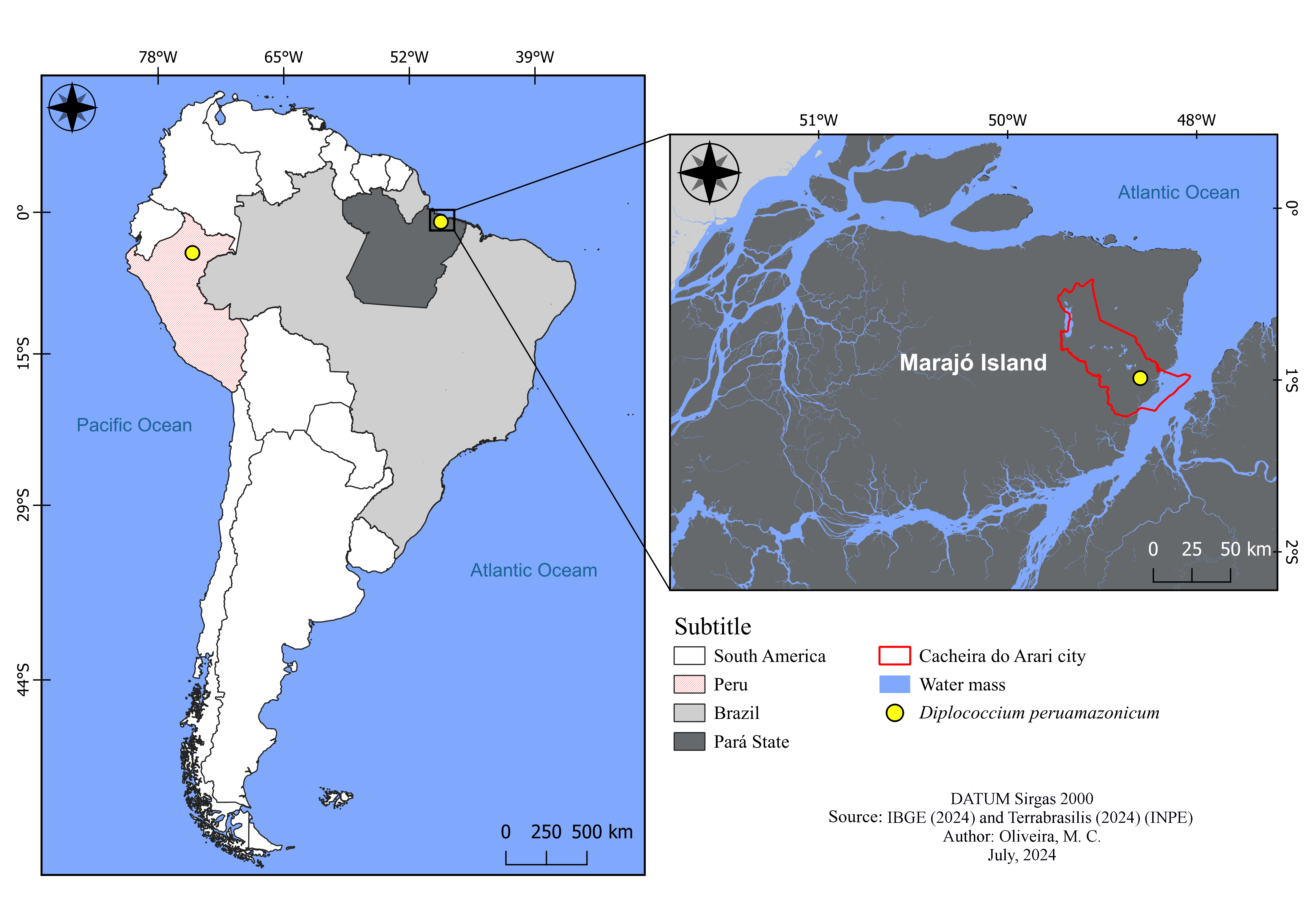
Collections of fungal specimens were carried out in July 2023 in the municipality of Cachoeira do Arari (Figure 1) (1°0’16’’S, 48°57’27’’W), belonging to the mesoregion of Marajó and microregion of Arari, located east of Marajó Island (Cidade-Brasil 2023). Decomposing leaflet samples of A. vulgare were collected and processed following the procedures outlined by Castañeda-Ruiz et al. (2016). The substrates, kept under incubation, were examined periodically for 30 days with the aid of a stereomicroscope. Semi-permanent microscope slides of the fungal specimens were prepared using lactoglycerol. Reproductive structures were observed, measured and photographed under an optical microscope (Leica DM6B) equipped with a digital camera (Leica DFC7000 T). For identification, keys and descriptions available in the literature were used. Semi-permanent slides and samples of D. peruamazonicum on dry substrate were deposited at the João Murça Pires Herbarium (MG) of the Emílio Goeldi Museum of Pará, Belém (Pará).Figure 2

Location of specimen collection of Diplococcium peruamazonicum in the municipality of Cachoeira do Arari, Pará, Brazil (study area) and the location of the holotype in Peru.

Diplococcium peruamazonicum. a-b) Conidiophores with conidium. c-g) Conidia. Bars: a-e (20 μm); f-g (10 μm).
RESULTS
Diplococcium peruamazonicum Matsush., Matsush. Mycol. Mem. 7: 49 (1993).
Conidiophores macronematous, mononematous, single, simple, erect, straight or slightly flexuous, cylindrical, septate, smooth, brown, 62.5–175 × 5–5.5 µm. Conidiogenous cells enteroblastic, polytretic, 1–3 pores, integrated, terminal, determinate, cylindrical, rounded apex, smooth, brown, 12.5–22.5 × 5–5.5 µm. Conidial secession schizolytic. Conidia catenate, acropleurogenous, broadly fusiform, ovoid to ellipsoid, acute at the ends, 1-septate, thick and darkly pigmented septum, smooth, dark brown, 12.5–25 × 10–11 µm.
Examined material: BRAZIL. Pará: Marajó Island, Cachoeira do Arari, Vila da Sé, 0°59’21.599”S, 48°43’40.22”W, on decaying leaflet of Astrocaryum vulgare Mart. (Arecaceae), 14 July 2023, E.G. Nascimento 01 (MG 249960).
Habitat: saprobes occurring on petioles of unidentified palms (Matsushima 1993) and leaflets of A. vulgare (Arecaceae).
Known distribution: Brazil (this study), Peru (Matsushima 1993).
Comments: Diplococcium peruamazonicum is morphologically comparable to D. dendrocalami Goh, K.D. Hyde & Umali (Goh et al. 1998), and D. clavariarum (Desm.) Hol.-Jech. (Holubová-Jechová 1982), due to the shape and septation of the conidia. However, in D. dendrocalami, the conidia are oblong to ellipsoidal, 1(–2)-septate, rarely 3-septate, measuring 12–22 × 6–9 μm, with conidiophores that sometimes regenerate percurrently. Diplococcium clavariarum presents ellipsoidal to obclavate conidia, 0–1-septate, measuring 13–29 × 6–9 μm, with no regeneration in the conidiophores, occurring as a mycoparasite on Clavaria species (Basidiomycota). In D. peruamazonicum, the conidia are broadly fusiform, ovoid to ellipsoidal, with one thick and dark septum, rarely 2-septate, measuring 12–34 × 7–10 μm, with conidiophores that do not regenerate. The characteristics of the Brazilian specimens agree with the description of the holotype (Matsushima 1993), differing only in the size of the conidiophores, which were smaller than the Peruvian specimen (62.5–175 × 5 µm vs. 130–500 × 4–6 µm).
The material from Marajó represents the first record of D. peruamazonicum from Brazil and the second worldwide, 31 years after the original description of the species. In Brazil, seven species of this genus have already been recorded in the Amazon (Pará) and Caatinga (Bahia) biomes (CRIA 2024). The presence of D. peruamazonicum in the Brazilian Amazon suggests that this species may have a broader distribution, underscoring the importance of conducting more comprehensive fungal inventories in this biome.
Key to species of Diplococcium occurring in Brazil
1. Conidia verruculose, oblong to cylindrical, 9–25 × 4.5–6 µm, 1–4-septate, pale brown; conidiophores simple, 93–215 × 4–5 µm.........................................................................................D. verruculosum
1’. Conidia smooth..............................................................2
2. Conidia versicolor, subellipsoid, 2(–3)-septate, central cells dark brown, end cells paler, 17–25 × 7–9 µm; conidiophores simple, 170–250 × 7–12 µm........................................................................D. laxusporum
2’. Conidia concolor, with all the cells of the same color..........................................................................................3
3. Conidiophores simple or branched......................4
3’. Conidiophores always simple...................................6
4. Conidia aseptate, limoniform to ovoid, pale brown, reddish to dark brown, 7.5–22 × 5–12 µm; conidiophores 135−189 × 12−16.5 µm.....................................................................................................D. variegatum
4’. Conidia septate...............................................................5
5. Conidia 1-septate, constricted at the septa, ellipsoidal, subcylindrical, 6–9 × 3–4 µm; conidiophores 200−400 × 2.5−4 µm........................................................................................................ D. spicatum
5’. Conidia 1–5-septate, not or slightly constricted at the septa, cylindrical to ellipsoidal, 11–55 × 5–15 µm; conidiophores up to 250 × 4−6 µm..............................................................................D. varieseptatum
6. Conidia 0–12-septate, mostly 1-septate, constricted at the septa, cylindrical, ellipsoid to oblong, 10–77 × 6–10 µm; conidiophores 120–300 × 4.5–9 µm..…....................................D. heteroseptatum
6’. Conidia 1–3-septate......................................................7
7. Conidia mostly 1–2-septate but occasionally 3-septate, oblong to ellipsoid, broadly rounded at both ends, 12–22 × 6–9 µm; conidiophores 120–280 × 7.5–9 µm.......................................D. dendrocalami
7’. Conidia mostly 1–2-septate, broadly fusiform, ovate to ellipsoid, 12–34 × 7–10.5 µm; conidiophores 130–500 × 4–6 µm.............................................................................................D. peruamazonicum
ACKNOWLEDGMENTS
We thank the Universidade Federal Rural da Amazônia and the Museu Paraense Emílio Goeldi for the infrastructure supplied. This study was supported by the Coordenação de Aperfeiçoamento de Pessoal de Nível Superior (CAPES) – financing code 001.
REFERENCES
- ALMEIDA DAC, SANTA-IZABEL TS, GUSMÃO LFP & CASTAÑEDA-RUIZ RF. 2015. A new species of Diplococcium from the Brazilian semi-arid region. Mycotaxon 130: 495-498.
- CASTAÑEDA-RUIZ RF, HEREDIA G, GUSMÃO LFP & LI DW. 2016. Fungal diversity of Central and South America. In: LI DW (Ed), Biology of Microfungi, Switzerland: Springer International Publishing, p. 197-217.
-
CIDADE-BRASIL. 2023. Available at: https://www.cidade-brasil.com.br/ Accessed on December 04, 2023.
» https://www.cidade-brasil.com.br/ -
CRIA – CENTRO DE REFERÊNCIA E INFORMAÇÃO AMBIENTAL. 2024. Specieslink. Available at: http://www.splink.org.br/index Accessed on March 04, 2024.
» http://www.splink.org.br/index - GOH TK & HYDE KD. 1998. A synopsis of and a key to Diplococcium species, based on the literature, with a description of a new species. Fungal Divers 1: 65-83.
- GOH TK, HYDE KD & UMALI TE. 1998. Two new species of Diplococcium from the Tropics. Mycologia 90: 514-517.
- GROVE WB. 1885. New or noteworthy fungi. II. J Bot Br Foreign 23: 155-168.
- HERNÁNDEZ-RESTREPO M, SILVERA-SIMÓN C, MENA-PORTALES J, MERCADO-SIERRA Á, GUARRO J & GENÉ J. 2012. Three new species and a new record of Diplococcium from plant debris in Spain. Mycol Prog 11: 191-199.
- HOLUBOVÁ-JECHOVÁ V. 1982. Lignicolous hyphomycetes from Czechoslovakia 6. Spadicoides and Diplococcium. Folia Geobot Phytotax 17: 295-327.
-
IBGE – INSTITUTO BRASILEIRO DE GEOGRAFIA E ESTATÍSTICA. 2024. Available at: http://www.ibge.gov.br Accessed on March 22, 2024.
» http://www.ibge.gov.br - MA LG, MA J, ZHANG YD & ZHANG XG. 2012. Spadicoides camelliae and Diplococcium livistonae, two new hyphomycetes on dead branches from Fujian Province, China. Mycoscience 53: 25-30.
- MATSUSHIMA T. 1993. Matsushima Mycological Memoirs nº 7. Kobe: Published by the author, 141 p.
- SILVA SS, CRUZ ACR, GUSMÃO LFP & CASTAÑEDA-RUIZ RF. 2014. Diplococcium variegatum, a new conidial fungus from the semi-arid Caatinga biome of Brazil. Mycotaxon 127: 59-62.
-
TERRABRASILIS. 2024. Instituto Nacional de Pesquisas Espaciais (INPE), Available at: http://www.terrabrasilis.dpi.inpe.br Accessed on March 22, 2024.
» http://www.terrabrasilis.dpi.inpe.br