First report of Penicillium brevicompactum causing disease in Pleurotus ostreatus
Abstract
Oyster mushroom (Pleurotus ostreatus) is the second most produced mushroom globally, but increased production is linked to new diseases. This study reports the first occurrence of Penicillium brevicompactum on P. ostreatus mushrooms. Symptoms, identified in the primordia, include atrophy, malformation, drying, and sporulation, causing up to 100% losses. The pathogen was confirmed with 99% identity via ITS region analysis and phylogenetic comparison. The research highlights P. brevicompactum’s infection of mushroom primordia, emphasizing the need for pathogen identification and preventive measures to mitigate production losses.
Key words
mushroom cultivation; oyster mushroom disease; pathogenicity; yield loss
Pleurotus ostreatus (Jacq.) Kumm. is an edible basidiomycete commonly known as oyster mushroom or shimeji mushroom (Roberti et al. 2019, Zied & Pardo-Giménez 2017). It constitutes the second most widely produced genus of edible mushrooms worldwide, accounting for 19% of the production (Zied & Pardo-Giménez 2017), assuming significant importance for global mushroom cultivation. The growth in mushroom cultivation and consumption is accompanied by an increase in research that highlights the medicinal and nutritional value that mushrooms possess (Vaishnavi et al. 2022). The increase in mushroom production can also be associated with the rise in the occurrence of new diseases. Similar to other agricultural crops, commercial-scale mushroom production can create favorable conditions for the emergence and spread of diseases, due to factors such as population density, monoculture, improper management practices, use of contaminated substrates, among others (Petre 2016). Among them, the incidence of diseases related to the production system. Several producers have already reported contamination occurrences in production, with potential losses of up to 100%.
One of the diseases associated with the cultivation of Pleurotus ostreatus mushrooms is characterized by the atrophy of fruiting primordia, with no growth and spread of the disease and spores in atrophied primordia (Figures 1a, b).

a) Symptoms caused in oyster mushroom (Pleurotus ostreatus) primordia. b) Atrophy, malformation, drying, and abundant fasciation caused by Penicillium brevicompactum.
The pathogenicity and virulence of pathogens can vary depending on the host, especially when morphological and environmental characteristics vary significantly (Sacristán & García-Arenal 2008). Therefore, when pathogens do not exhibit much specificity, infecting various hosts, and with the increase in the number of mushroom producers, they may become potential issues for production. When cultivated on growth media, it gradually gives rise to light-green colonies, approximately 20 mm (0.8 inches) in diameter per week, at a temperature of 25°C. The fruiting structures (penicilli) exhibit a brush-like appearance, being large and typically featuring two conidia-bearing branches. The conidia (asexual spores) measure 2.5–3.5 μm in length, mostly smooth, and have an ellipsoidal shape. P. brevicompactum exhibits optimal growth at 23°C (73.4°F), with a temperature range spanning from –2°C (28.4°F) to 30°C (86°F) (Pitt 2006).
Despite what has been reported by other authors regarding the occurrence of Penicillium brevicompactum contamination in adjacent mushroom primordia (Tian et al. 2017), which was observed few contaminations in nearby primordia in Pleurotus ostreatus mushroom cultivation. It was observed that the pathogen only infects mushroom primordia where there is no differentiation of mycelial tissue or in initial stages of differentiation.
The pathogen spores were collected using a platinum loop from primordia exhibiting symptoms like those in Figures 1a, b and dispersed in an “S” pattern on Petri dishes containing potato dextrose agar (PDA) medium for colony growth observation. After colony growth, slides were prepared with dye stain to facilitate visualization of the pathogen’s structures under an optical microscope with a 10x objective.
To identify the pathogen at the species level, a pure colony was isolated on BDA medium, and DNA was extracted from approximately 150 mg of mycelium using the Wizard Magnetic DNA Purification System kit. The DNA was quantified and assessed for purity with a Nanodrop 2000 spectrophotometer. Conventional PCR was conducted on 20 ng of purified genomic DNA using a Veriti thermocycler, with thirty-two cycles for the ITS region involving denaturation, primer annealing, and product extension. The PCR products were analyzed via 1% agarose gel electrophoresis and then enzymatically purified with ExoI/SAP. Sanger sequencing was performed on the purified PCR products using BigDye v3.1, involving thirty-five cycles of denaturation, primer annealing, and product extension. The labeled products were precipitated with ammonium acetate and ethanol, then resuspended in HiDi-formamide for sequencing on a Genetic Analyzer 3500xL. The resulting electropherograms were converted to base sequences using Sequencing Analysis v5.4 software. The primers used were specific for the internal transcribed spacer (ITS) region, with the following sequences: ITS1 - TCCGTAGGTGAACCTGCGG and ITS4 – TCCTCCGCTTATTGATATGC (Aatsinki 1997), respectively.
After species identification, a 99% identity match was found with the species Penicillium brevicompactum (497/502 ITS bases). A phylogenetic tree (Figure 2) was constructed using the same primer sequences: The sequence alignment was performed using the MAFFT program (Nakamura et al. 2018). Sequences extracted from a scientific article reporting the occurrence of Penicillium brevicompactum in the cultivation of a mushroom known as bunashimeji or beech mushroom, Hypsizygus marmoreus (Kim et al. 2019), were used for comparison and construction of the phylogenetic tree. The sequence of Aspergillus niger, with the code NR_111348, was used as the outgroup for the analyses. Subsequently, the ends of the sequences were trimmed using the AliView program (Larsson 2014), ensuring that the sequences started and ended at the same point as the “17.8-0993.HEIC_P._brevicompactum” sequence. The tree was constructed using the maximum likelihood method with 1000 bootstraps, performed with the IQTree program (Nguyen et al. 2014), using the “TIM2+F+I+G4” model, which was identified by IQTree as the best model for the analyzed sequences. Finally, the tree visualization was done using the ITol tool (Letunic & Bork 2021). The triangles indicated on the tree represent branches supported by at least 70% of the bootstraps, with larger triangles indicating a higher tendency for 100% compatibility.
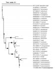
Phylogenetic tree constructed using the maximum likelihood method, comparing the internal transcribed spacer (ITS) sequences of Penicillium brevicompactum obtained in this study, highlighted in bold. The branch lengths are proportional to the phylogenetic distance, represented by the scale bar. The bold triangles represent the similarity with other possible phylogenetic trees evaluated, with the smallest triangle representing 70% compatibility. Larger triangles indicate compatibility trending towards 100%. The bolded strain represents the one isolated and described in this study. The final sequence obtained in the study is represented in bold.
To confirm the pathogenicity, Koch’s postulates were conducted as follows: three Pleurotus ostreatus cultivation bags were sprayed with 1 mL of conidial suspension (106 conidia/mL) obtained from the pure colony isolates on BDA medium, while another three bags were sprayed with sterilized water as a control. All treatments were kept in the same mushroom cultivation room at 20-22°C and 85-90% relative humidity.
It was observed that after reinoculation of the pathogen onto the primordia of Pleurotus ostreatus mushrooms, symptoms began with drying and covering of the primordia with conidiophores and a large mass of green-grayish spores (Figures 1a, b).
In addition to causing diseases in Pleurotus ostreatus mushroom cultivation, Penicillium brevicompactum has also been reported as a causative agent of diseases in the cultivation of Hypsizygus marmoreus (Kim et al. 2020), Inonotus obliquus (Min et al. 2019), and Grifola frondosa (Tian et al. 2017) mushrooms. Besides mushrooms, P. brevicompactum has been reported to contaminate various other products, such as garlic (Valdez et al. 2009), apples, and pears (Louw & Korsten 2014).
Therefore, Penicillium brevicompactum is a pathogen that causes direct damage to Pleurotus ostreatus mushrooms by inhibiting their development or causing deformities. This is the first report of Penicillium brevicompactum causing disease in P. ostreatus cultivation.
ACKNOWLEDGMENTS
We would like to thank Coordenação de Aperfeiçoamento de Pessoal de Nível Superior (CAPES) - Brazil for funding the research, without which it would not have been possible to obtain the results.
REFERENCES
-
AATSINKI JT. 1997. Coupled One-Step Reverse Transcription and Polymerase Chain Reaction Procedure for Cloning Large cDNA Fragments. In: White BA (Ed), PCR Cloning Protocols, Methods in Molecular Biology™, vol. 67, Humana Press, Totowa, NJ. https://doi.org/10.1385/0-89603-483-6:55
» https://doi.org/10.1385/0-89603-483-6:55 - KIM JY, KWON HW, LEE DH, KO HK & KIM SH. 2019. Isolation and Characterization of Airborne Mushroom Damaging Trichoderma spp. from Indoor Air of Cultivation Houses Used for Oak Wood Mushroom Production Using Sawdust Media. Plant Pathol J 35: 674-683.
- KIM M-K, SIM S-A, KIM A-Y, KWON J-H & CHANG Y-H. 2020. Characterization of Green Mold Contamination caused by Penicillium brevicompactum in Hypsizygus marmoreus. Korean J Mycol 48: 397-405.
- LARSSON A. 2014. AliView: a fast and lightweight alignment viewer and editor for for large datasets. Bionfirmatics 30(22): 3276-3278.
- LETUNIC I & BORK P. 2021. Interactive Tree Of Life (iTOL) v5 an online tool for phylogenetic tree display and annotation.pdf. Nucleic Acids Res 49: 293-296.
- LOUW JP & KORSTEN L. 2014. Pathogenic Penicillium spp. on Apple and Pear. Plant Dis 98: 590-598.
- MIN C, DONG H, LIU X & ZHANG Z. 2019. Screening and identification of a Penicillium brevicompactum strain isolated from the fruiting body of Inonotus obliquus and the fermentation production of mycophenolic acid. Ann Mirobiology 69: 1351-1360.
- NAKAMURA T, YAMADA KD, TOMII K & KATOH K. 2018. Parallelization of MAFFT for large-scale multiple sequence alignmentes. Bioinformatics 34: 2490-2492.
- NGUYEN L-T, SCHMIDT HA, VON HAESELER A & MINH BQ. 2014. IQ-TREE a fast and effective stochastic algorithm for estimating maximum-likelihood phylogeneis. Mol Biol Evol 32: 268-274.
- PETRE M. 2016. Mushroom Biotechnology: Developments and applications, 1st ed. Londres, 244 p.
- PITT JI. 2006. Food Spoilage Microorganisms. PartIV: Spoliage moulds. 16.5 Penicillium species causing foods spoilage, p. 438-450.
- ROBERTI R, DI FRANCESCO A, INNOCENTI G & MARI M. 2019. Potential for biocontrol of Pleurotus ostreatus green mould disease by Aureobasidium pullulans De Bary (Arnaud). Biol Control 135: 9-15.
- SACRISTÁN S & GARCÍA-ARENAL F. 2008. The evolution of virulence and pathogenicity in plant pathogen populations. Mol Plant Pathol 9: 369-384.
- TIAN FH, LI CT & LI Y. 2017. First report of Penicillium brevicompactum causing blue mold disease of Grifola frondosa in China. Plant Dis 101: 1549.
- VAISHNAVI MDP, SHARMA A, TIWARI J, SINGH S & SHARMA S. 2022. View of Production Of Edible Mushrooms To Meet The Food Security. Available on: https://journalppw.com/index.php/jpsp/article/view/7154/4879. J Posit Sch Pyschology 6: 4316-4325.
- VALDEZ JG, MAKUCH MA, ORDOVINI AF, FRISVAD JC, OVERY DP, MASUELLI RW & PICCOLO RJ. 2009. Identification, pathogenicity and distribution of Penicillium spp. isolated from garlic in two regions in Argentina. Plant Pathol 58: 352-361.
- ZIED DC & PARDO-GIMÉNEZ A. 2017. Edible and Medicinal Mushrooms, 1st ed. 585 p.