
 Evaluation of cardiovascular effects in the male offspring and dams of rats submitted to a high-fat diet with flaxseed oil during pregnancy and lactation
Abstract
A maternal diet high in saturated fat during critical periods of development is associated with an increased risk of chronic diseases in adulthood. We investigated whether cardiovascular effects in adulthood can be altered by the maternal high-fat diet with flaxseed oil (omega-3) as well as the effects of this diet in dams. Wistar rats were fed during pregnancy and lactation according to the following: control (CT) – standard diet; high-fat (HL) – diet rich in saturated fatty acids; and high-fat with omega-3 (HLꞶ3) – diet rich in omega-3 from flaxseed oil. Body mass, lipid peroxidation, biochemical profile, heart and kidney weights and cardiac hypertrophy index (CHI) were measured in dams. In the offspring, body mass, murine parameters, dietary and water control, systolic and diastolic blood pressure at 60 and 90 days, biochemical parameters, heart and kidney weights, CHI and vascular reactivity were evaluated. In dams, the HLꞶ3 diet delayed the increase in body mass in lactation and reversed oxidative damage in the heart. Therefore, our data suggest that HL diet promotes adverse cardiometabolic effects and the HLꞶ3 diet attenuated the weight gain and oxidative stress in dams, while in the offspring the HLꞶ3 prevented the increase in the Phe-induced contraction.
Key words
Blood pressure; fatty acids; omega-3; oxidative stress; vasorelaxation
INTRODUCTION
Obesity is a growing health concern in the world, being considered one of the biggest global public health challenges (WHO 2018). The changes observed in the last decades of the 20th century, such as the Western dietary pattern and the population’s sedentary lifestyle are part of the so-called nutritional transition and are associated with the accelerated growth in obesity and consequently the increased risk of associated metabolic diseases (Kac & Pérez -Escamilla 2003, Rakhra et al. 2020). The Western diet is widely consumed in the West, including by pregnant women (Musial et al. 2017) and its characteristics are low intake of omega-3, high consumption of foods rich in saturated fat, sugar and salt instead of fruits and vegetables (Malesza et al. 2021, Rakhra et al. 2020).
Excess weight represents the sixth most important risk factor for the global burden of chronic non-communicable diseases (NCDs) with cardiovascular diseases being the main contributors to this burden. In Brazil, the cardiovascular diseases are the main cause of death among the NCDs (INCA 2019, WHO 2020, Oliveira et al. 2020). In addition, excess of body weight and its complications generate an economic worry for nations, overloading health systems (Withrow & Alter 2011, Bahia et al. 2012, Mazzoccante et al. 2013).
Phenotypic plasticity is the ability of a sensitive and plastic organism to produce distinct phenotypes from a single genotype, in response to environmental variations (Sommer 2020). In this context, epidemiological, clinical and experimental observations have led to the increasingly solid theory in the scientific literature that the risk of developing some NCDs in adulthood, including obesity and its metabolic complications such as diabetes and high blood pressure, is influenced by environmental variables that act early in life and not just due to genetic and adult lifestyle factors (Safi-Stibler & Gabory 2020, Hoffman et al. 2021).
Maternal nutrition is one of the most documented extrinsic environmental factors that interfere with phenotypic plasticity. Several experimental studies demonstrate the long-term effects of maternal dietary imbalance during critical periods of development, such as pregnancy and/or lactation, contributing to the development of diseases in offspring (Jiménez-Chillarón et al. 2012, Ramalingam et al. 2021). Maternal nutrition rich in saturated fat, for instance, has been shown to induce weight gain and increased levels of blood glucose, cholesterol and triglycerides (Volpato et al. 2012, Alves-de-Oliveira et al. 2022), causing endothelial dysfunction and oxidative stress in adult offspring (Torrens et al. 2012, Bloise 2019).
Maternal high-fat diet (HFD) feeding in animal models (rats and mice) have shown definitive effects on body composition and metabolism in the offspring, leading to alterations in blood pressure (BP) at different moments throughout the offspring’s life (Khan et al. 2005, Elahi et al. 2009, Prior et al. 2014). Studies have demonstrated that HFD or obesity induce BP changes trough diverse mechanisms, including adipose tissue increasing, stimulating leptin dysregulation (Otero et al. 2006, Barbosa et al. 2020), inflammation and sympathetic overactivity, besides oxidative stress involvement (Hall et al. 2015, Zhang et al. 2018).
On the other hand, experimental studies in rodents have revealed the ability of omega-3 to mitigate the harmful effects of consuming a maternal diet rich in saturated fatty acids on offspring (Albert et al. 2017, Ramalingam et al. 2021). However, the use of fish oil as a source of omega-3 can sometimes limit its consumption by humans (Martins et al. 2008, Niculescu et al. 2012).
Given this context, our study aimed to investigate the cardiovascular effects induced by the flaxseed oil, a plant-based source of omega-3, as an alternative to enrich the maternal hyper lipidic diet during pregnancy and lactation, focusing in male offspring and dams.
MATERIALS AND METHODS
Animals and experimental groups
All procedures performed in this study are in agreement with the national ethical standards for the care and use of laboratory animals set by the National Council for the Control of Animal Experimentation (CONCEA, MCTI - Brazil) guidelines. The project was also approved by the Animal Use Ethics Committee of the Universidade Federal de Pernambuco (CEUA/UFPE #0077/2021).
Albino Wistar rats (Rattus norvegicus) were maintained in the animal care of the Centro Acadêmico de Vitória at UFPE (CAV/UFPE). All animals in the study were kept under controlled conditions of temperature (21 ± 2 °C), and lighting (12/12-hour light/dark cycle) and had access to filtered water and food ad libitum.
To obtain the offspring, reproductively aged rodents were placed for mating at a ratio of 1:3 (male: female) in each cage. Mating occurred approximately over two estrous cycles of the female rats (10 days). The determination of pregnancy was performed from the observation of the presence of spermatozoa in the vaginal smear, defining the 1st day of pregnancy. From the 1st day of gestation until the 21st day of lactation (Alves-de-Oliveira et al. 2022), the progenitor rats were separated and placed in individual cages with water and food offered according to the following groups: control group (CT) – standard diet/AIN-93G; hyper lipidic group (HL) – diet rich in saturated fatty acids; Ꞷ3 group (HLꞶ3) – diet rich in saturated fatty acids with the addition of omega-3 from flaxseed oil (Volpato et al. 2012, Barbosa 2018, Nascimento et al. 2020).
At birth, after 24 hours, the number of pups and their sex were identified by anogenital distance examination and, when necessary, the litter was reduced, in order to prioritize the males and obtain 8 animals (4 males and 4 females) per mother with the aim of guaranteeing standardized nutrition and homogeneous growth for the offspring (Volpato et al. 2012, Barbosa 2018, Nascimento et al. 2020). At weaning, at 21 days of age, the male offspring were assigned to specific cages according to their specific group and received filtered water and commercial rat chow (Nuvilab®, CR-1, Grupo Quimtia, Paraná, Brazil) until 90 days of age. Only male pups were used in the study to exclude complications of sexual dimorphism (Volpato et al. 2012). Figure 1 shows the experimental design of the study.

Experimental diets
The diets were standardized as described by Silva et al. (2019). In this study, the standard diet received by the control group contains approximately 19% of its energy from fat, 20% from protein, and 61% from carbohydrates; the high-saturated fatty acid diet consumed by the HL group contains, on average, 34% of its energy from fat, 20% from protein, and 47% from carbohydrates; and the high-saturated fatty acid diet with added flaxseed oil as a source of omega-3 in the HLꞶ3 group contains approximately 32% of its energy from fat, 18% from protein, and 49% from carbohydrates. The standard diet contains approximately 4.0 kilocalories per gram, while the high-fat diets contain an average of 4.5 kilocalories per gram.
The post-weaning diet until the animals are 90 days old, standard commercial feed for vivarium rodents (Nuvilab® CR-1, Grupo Quimtia, Paraná, Brazil) according to the manufacturer, contains 4.5% lipids, 22% protein, 55% carbohydrates in 100g and offers 2.95 kcal/g of feed.
Evolution of body mass of dams during pregnancy and lactation
The rats were weighed from the first day of mating until the end of lactation to monitor the evolution of body mass during pregnancy and lactation, each period comprising 21 days. The first day of gestation was determined 21 days before the birth of the pups, considering the average gestation period of rats (Neves et al. 2013). Body mass (g) was measured daily at the same time of day (Araújo 2019).
Lipid peroxidation levels by the thiobarbituric acid reactive substance (TBARS) method in dams
To evaluate one of the biomarkers of oxidative stress in the aorta, heart and kidney of the different groups of dams, we measured the levels of malondialdehyde (MDA) and other aldehydes, products of lipid peroxidation, which react with thiobarbituric acid were measured by the TBARS assay according to the colorimetric technique of Buege & Aust (1978). The results were expressed in mmol of MDA/mg of protein.
Biochemical analysis in dams
At the end of lactation, the mothers were fasted for 8-12 hours and then anesthetized with ketamine (80 mg/kg i.p) and xylazine (10 mg/kg i.p) for duplicate blood samples to be collected by rupture of the retro-orbital plexus. The blood samples were placed in tubes without anticoagulant and centrifuged to obtain the serum, which was transferred to an Eppendorf tube. Biochemical analyses were performed using the Automatic Chemistry Analyzer (Pioway, PW - 2280, China), and subsequently analyzed for blood glucose, triglycerides and total cholesterol levels, which were expressed in mg/dL.
Absolute weight of the heart, kidneys and calculation of the cardiac hypertrophy index in dams
At the end of the lactation of the dams, the animals were euthanized, the heart and right and left kidneys were removed, soaked in distilled water and dried with the aid of sterile gauze (Matos 2017). They were then weighed individually on an electronic semi-analytical scale. The left tibia was also collected and measured with a caliper to correct the heart weight by minimizing the effect of the animal’s size on this parameter, as recommended (Yin et al. 1982). With this measurement, the cardiac hypertrophy index can be calculated (IHC = heart mass (g)/tibia length (cm) (Cabral et al. 2013).
Analysis of body mass and murinometric parameters of the offspring
Murinometric measurements (nasocaudal length [NL] and abdominal circumference [AC]) and body mass were performed on the 1st, 21st, 30th, 60th and 90th day of life. For measurements, the protocols described by Novelli et al. (2007) and Carvalho et al. (2013) were followed. After obtaining the animal’s measurements, the Lee index (∛weight (g)/CNA (mm) x 10) (Bernardis & Patterson 1968) was calculated, as well as the BMI (weight (g)/nasoanal length (cm2) (Novelli et al. 2007).
Analysis of body mass, average food and water consumption of offspring
After the offspring were 30 days old, their body mass and food and water consumption were measured up to 90 days, comprising 8 weeks. The animals were housed in cages of two to four animals according to their respective group, with commercial rat food (Nuvilab®, Grupo Quimtia, Paraná, Brazil) and filtered water ad libitum. Each cage was measured weekly at the same time of day, with the amount of food and water offered and the waste subtracted to obtain the average consumption per animal each week (Silva et al. 2019).
Systolic and diastolic blood pressure records of offspring
Blood pressure was recorded by tail cuff plethysmography (Bonther, 2-channel tail plethysmograph, Ribeirão Preto, São Paulo, Brazil), according to the manufacturer’s instructions and training. BP was measured in offspring at 60 and 90 days of life during the light period of the 12/12h cycle. For each record, 8 inflation and deflation cycles were performed, 3 of which were acclimatization cycles, which were not recorded, and the 5 valid values successfully obtained were used to calculate the animal’s average (Machado et al. 2010).
Biochemical analysis of offspring
The offspring at 90 days of life were fasted for 8-12 hours and then anesthetized with ketamine (80mg/kg i.p) and xylazine (10mg/kg i.p) for collection of duplicate blood samples by rupture of the retro-orbital plexus. The methodology used was like that used in the dams as previously described and subsequently analyzed for blood glucose, triglycerides and total cholesterol levels, which were expressed in mg/dL.
Absolute weight of the heart, kidneys and calculation of the cardiac hypertrophy index of the offspring
At 90 days of age, the offspring were euthanized followed by exsanguination of the carotid vessels, and through an incision in the ventral region of the animal, the organs were collected from the tibia. The methodology used was like that used in the dams as previously described.
Evaluation of in vitro vascular effects in offspring: vascular reactivity
The animals were euthanized, the aorta artery was identified, removed, and immediately placed in modified Krebs-Henseleit solution for dissection and sectioning of the vessels into rings (1-2 mm in length). When necessary, the endothelium was removed by mechanical friction between the internal walls of the vessel and a metal rod. Each ring was immersed in chambers (10 mL) containing Krebs-Henseleit solution at 37°C, aerated with a mixture of 95% O2 and 5% CO2, and attached to a force transducer, subjected to a basal tension of approximately 1.5 g, for a stabilization period of 60 minutes. Changes in isometric tension were captured by the AQCAD acquisition system (version 2.3.3.0; AVS, Brazil).
To verify tissue viability, a contraction was obtained with phenylephrine (Phe) (0.1 μM), and to assess endothelial integrity, acetylcholine (ACh) (1 μM) was added. Rings with relaxation greater than 80% due to Phe-induced contraction were considered to have functional endothelium, and rings with relaxation less than 20% were considered to have no endothelium. Those with values between these ranges were discarded.
After the stabilization period and verification of the presence or absence of functional endothelium, a contraction using a depolarizing solution (KCl 60 mM) was performed. Subsequently, the solution containing high levels of K+ was replaced by a standard Krebs-Henseleit solution. To evaluate the contractile responses, a cumulative contraction with Phe (10-9 – 10-5 M) was induced in the aorta, in the presence and absence of functional endothelium. To evaluate the relaxing responses, the vasodilator agent ACh (10-10 – 10-5 M) was used in rings in the presence of endothelium. The efficacy and potency of both the contraction and vasorelaxation were evaluated by means of the maximum effect (ME) and pD2 (negative logarithm of EC50 - is the molar concentration of an agonist that produces 50% of the maximal possible effect of that agonist), respectively, were obtained through a non-linear regression.
Statistical analysis
The values were expressed as mean ± standard error of the mean (S.E.M). The normality analysis of the data was performed by the Shapiro-Wilk test. The comparison between the means of the groups was performed by one-way or two-way ANOVA (repeated measures when necessary) test followed by the Tukey post-test or unpaired Student’s t-test according to the experiment. Differences were considered significant when p < 0.05. The statistical program used was Graph Pad Prism (GraphPad Software Corporation, version 8.0).
RESULTS
Effect of high-fat diets on maternal rats
Changes in body mass
The mean body mass of the dams in the HL group was higher during gestation compared with the CT group (HL = 314.40 ± 7.41 g vs CT = 290.00 ± 6.52 g, n = 5), as well as the weeks 9 and 13. However, no significant difference was observed between the HL group and the omega-3-enriched group (HL = 314.40 ± 7.41 g vs HLꞶ3 = 310.50 ± 6.49 g, n = 5) [F (1.5, 27) = 17.00 p < 0.05] (Figure 2a). During lactation, dams from the HL group presented greater body mass compared to the CT group (HL = 298.30 ± 5.16 g vs CT = 270.30 ± 2.41 g, n = 5). Conversely, the addition of flaxseed oil decreased body mass induced by the high fat diet (HLꞶ3 = 286.80 ± 3.16 g vs CT = 270.30 ± 2.41 g, n = 5) [F (9, 45) = 10.71, p < 0.05] (Figure 2b).

The mean body mass of the dams in the HL () group was greater than that in the CT () group, likewise at week 9 and 13, during gestation (a) and during the lactation (b) we also observed an increase in mean body mass of HL group compared to CT group and the HLꞶ3 (£) group decreased the mean body mass. The symbols and vertical bars represent the mean ± S.E.M were analyzed using two-way repeated measurement ANOVA followed by Tukey’s post-test (*p < 0.05, HL and HLꞶ3 vs. CT; #p < 0.05, HLꞶ3 vs HL).
Levels of lipid peroxidation in the heart, aorta, and kidney
The rats submitted to the high fat diet demonstrated an increase in lipid peroxidation levels in the heart compared to the CT group (HL = 0.92 ± 0.09 vs CT = 0.24 ± 0.05 mmol/mg of protein, n = 8). In contrast, the group enriched with omega-3 from flaxseed oil showed a decrease in biomarkers of oxidative stress compared to the HL group (HLꞶ3 = 0.48 ± 0.05 vs HL = 0.92 ± 0.09 mmol/mg of protein, n = 8) [F (2, 6) = 24.43, p < 0.05] (Figure 3a). However, the aortic tissue did not show significant differences in lipid peroxidation levels among the groups (HLꞶ3 = 0.64 ± 0.02; HL = 0.66 ± 0.05; CT = 0.64 ± 0.07 mmol/mg of protein, n = 8) [F (2, 6) = 0.027] (Figure 3b). In the kidney, lipid peroxidation levels were increased in the HL group compared to the CT group (HL = 1.18 ± 0.05 vs CT = 0.68 ± 0.11 mmol/mg of protein, n = 8), but there was no significant change after addition of flaxseed oil (HLꞶ3 = 0.91 ± 0.09 vs HL = 1.18 ± 0.05 mmol/mg of protein) [F (2, 6) = 7.411, p < 0.05] (Figure 3c).
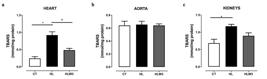
Lipid peroxidation levels in the heart (a) and kidney (c) increased in the HL dams when compared to the CT group, with reversal of the effect in the HLꞶ3 group only in the heart and no significant difference in the aorta (b). The vertical bars represent the mean and the upper bars represent the S.E.M and were analyzed using one-way ANOVA followed by Tukey’s post-test (*p<0.05, HL vs CT; #p < 0.05, HLꞶ3 vs HL).
Biochemical parameters evaluated
At the end of the lactation period, no significant differences were observed among the groups of dams related to the levels of triglycerides, total cholesterol and blood glucose (Table I).
Absolute mass of the heart and kidneys and cardiac hypertrophy index
There was no significant difference between the groups of dams in the absolute mass of the heart, right and left border, as well as in the values of the cardiac hypertrophy index (Table II).
Absolute mass of the heart and kidneys, and cardiac hypertrophy index values of the mother rats at the end of lactation.
Effect of high-fat diets on offspring
Changes in body composition and murinometric measurements
According to the data presented in Table III, on the first day of life of the offspring, no significant difference was observed between the groups in body mass, NL and AC as well as in BMI and Lee index.
After weaning, on the 21st day, the body mass of the HL and HLꞶ3 groups were greater compared to the CT group (HL = 57.62 ± 1.46 g; HLꞶ3 = 61.22 ± 1.26 g vs CT = 50.31 ± 0.47 g, n = 10) [F (2, 39) = 30.79, p < 0.05] (Table III).
A similar result was observed in the NL and AC parameters, there was an increase in the HL and HLꞶ3 groups compared to the CT group as demonstrated by the values (NL: HL = 18.76 ± 0.20 cm; HLꞶ3 = 19.34 ± 0.19 cm vs CT = 18.06 ± 0.11 cm, p < 0.05) (AC: HL = 9.90 ± 0.17 cm; HLꞶ3 = 9.96 ± 0.15 cm vs CT = 8.81 ± 0.08 cm, p < 0.05).
At 30 days of life, the body mass of the HL and HLꞶ3 groups sustained to increase compared to the CT group (HL = 99.31 ± 3.50 g; HLꞶ3 = 103.00 ± 0.98 g vs CT = 91.00 ± 0.99 g, n = 10) [F (2, 37) = 8.59, p < 0.05], as well as the nasocaudal length (HL = 24.95 ± 0.39 cm; HLꞶ3 = 25.32 ± 0.22 cm vs CT = 23.79 ± 0.13 cm, n = 10) (Table III).
At the beginning of the adult phase, at 60 days of life, the HL and HLꞶ3 groups presented greater body mass when compared to the CT group (HL = 309.50 ± 5.87 g; HLꞶ3 = 306.40 ± 4.66 g vs CT = 283.00 ± 5.03 g, n = 10) [F (2, 33) = 7.91, p < 0.05], and greater AC (HL = 16.22 ± 0.15 cm; HLꞶ3 = 16.46 ± 0.10 cm vs CT = 15.27 ± 0.17 cm, p < 0.05) (Table III).
Still in the adult phase, at 90 days of life, the offspring of the HL group continued to have greater body mass, as did the HLꞶ3 group when compared to the CT group (HL = 388.70 ± 9.30 g; HLꞶ3 = 380.40 ± 4.54 g vs CT = 347.00 ± 7.66 g, n = 10) [F (2, 43) = 9.43, p < 0.05] (Table III).
Regarding NL and AC, the HL and HLꞶ3 groups presented higher values when compared to the CT group (NL: HL = 40.23 ± 0.46 cm; HLꞶ3 = 42.72 ± 0.37 cm vs CT = 40.23 ± 0.46 cm, p < 0.05) (AC: HL = 17.05 ± 0.21 cm; HLꞶ3 = 16.77 ± 0.12 cm vs CT = 16.09 ± 0.21 cm, p < 0.05). The BMI and Lee index values did not differ between the groups in any of the periods evaluated (Table III).
Weekly monitoring of food and water
During the eight weeks of measurement, assessed from thirty to ninety days of life, the group of HL pups whose mothers were fed a high-fat diet, when compared to the CT group, presented greater body mass, as well as the HLꞶ3 pups, as demonstrated in Figure 4a.

Figure 4. The body mass (a) of the offspring of the HL () was greater than CT () group during the weeks of monitoring, except the week 3. There was no difference between the groups in terms of food (b) and water (c) consumption in any week of evaluation. Also, the and HLꞶ3 () group did not alter the parameters presented here. Values are expressed as mean ± standard error of the mean and were analyzed using two-way repeated measurement ANOVA followed by Tukey’s post-test (*p < 0.05, HL vs. CT).
Regarding the average food consumption, no significant difference was observed between the groups (Figure 4b), as well as water consumption (Figure 4c).
Systolic and diastolic blood pressure recordings
At 60 days of life, SBP significantly increased in the HL group compared to the CT group, (HL = 152.60 ± 1.32 mmHg vs CT = 126.90 ± 6.47, n = 9, p < 0.05), while, in relation to the HLꞶ3 group, no reduction in these levels was observed with the treatment (HL = 152.60 ± 1.32 vs HLꞶ3 = 135.70 ± 6.29 mmHg, n = 9) [F (5, 43) = 9.45, p < 0.05] (Figure 5a). Furthermore, no significant difference was found between the groups in DBP values at 60 days (CT = 87.02 ± 5.17, n = 9; HL = 89.75 ± 3.75 mmHg, n = 12; HLꞶ3 = 99.40 ± 4.16 mmHg, n = 9) (Figure 5b).

It shows the changes in (a) systolic and (b) diastolic blood pressure in the offspring at 60 and 90 days of life. Values are expressed as mean ± S.E.M and were analyzed using repeated measurement ANOVA followed by Tukey’s post-test (*p<0.05, HL vs. CT).
Similar result was found at 90 days of life with an increase in SBP in the HL group compared to the CT group (HL = 160.20 ± 3.72, n = 10 vs CT = 139.30 ± 2.93 mmHg, n = 9) and no changes in the omega-3-enriched group in relation to the HL group (HLꞶ3 = 150.40 ± 2.32 vs. HL = 160.20 ± 3.72 mmHg, n = 9) [F (6, 44) = 14.81, p < 0.05] (Figure 5c). Regarding DBP, no significant difference was observed between the groups (CT = 94.63 ± 2.17 mmHg, n = 8; HL = 100.40 ± 2.62 mmHg, n = 9; HLꞶ3 = 104.20 ± 3.14 mmHg, n = 7) (Figure 5d).
Analysis of biochemical parameters
At 90 days of age, no significant difference was observed between the groups of offspring in the levels of triglycerides, total cholesterol and glycemia (Table IV).
Biochemical parameters of offspring from groups CT, HL and HLꞶ3. Note: CT = control group (standard diet/AIN-93G); HL = high-fat diet group (rich in saturated fatty acids) and HLꞶ3 = high-fat diet group rich in saturated fatty acids and omega-3 (addition of flaxseed oil). Values were expressed as mean ± s.e.m., (n = 7-8).
Absolute weight of the heart, kidneys and cardiac hypertrophy index
The HL group presented increased mass of the heart and both kidneys, when compared to the control group, with the effect not attenuated by enrichment in the HLꞶ3 group (Table V). Also, the cardiac hypertrophy index was higher in the HL group, compared to the CT group and linseed oil was not able to reverse this effect.
Absolute mass of the heart and kidneys, and cardiac hypertrophy index values of offspring from the CT, HL and HLꞶ3 groups. Note: CT = control group (standard diet/AIN-93G); HL = high-fat diet group (rich in saturated fatty acids) and HLꞶ3 = high-fat diet group rich in saturated fatty acids and omega-3 (addition of flaxseed oil). Values were expressed as mean ± s.e.m., (n = 10–18).
Vascular reactivity in aortic artery rings
It was observed that the contractions induced by cumulative concentrations of Phe in the HL group were higher in comparison to the CT group (HL = 122.30 ± 14.60, n = 5 vs. CT = 83.62 ± 7.89%, n = 6), illustrated by the greater displacement of the curve in the graph (Figure 6a). The contraction curve in the HLꞶ3 group showed a reduction in the ME of Phe compared to the HL group as evidenced in Figure 6a (HLꞶ3 = 74.76 ± 13.32%, n = 5 vs HL = 122.30 ± 14.60%, n = 5) [F (2, 13) = 4.35, p < 0.05].

(a) Increased contraction induced by Phe in the HL () group when compared to the CT () group. HLꞶ3 (£) group induced a reduction in Phe-induced contraction compared to HL group. Vasorelaxation caused by ACh was reduced in the HL () compared to the CT () group without reversal of the effect in the HLꞶ3 group (b). Values are expressed as mean ± standard error of the mean and were analyzed using one-way ANOVA followed by Tukey’s post-test (*p<0.05, HL vs CT; #p < 0.05, HLꞶ3 vs HL).
In order to investigate if the vasorelaxant response was modified in the hyper lipidic rats or after the dams’ treatment with the flaxseed oil, we induced different vasorelaxant curves in the presence of ACh. The ME of the ACh was reduced in HL compared to te control group (HL = 69.40 ± 16.17 vs CT = 94.83 ± 8.88%, n = 5). The HLꞶ3 group did not alter the damage caused by the HL group [F (2, 11) = 2.87, p < 0.05]. The potency of ACh in causing vasorelaxation was reduced in the HL group when compared to the CT group (pD2: HL = 7.30 ± 0.31, n = 4 vs. CT = 8.54 ± 0.20, n = 6) and the introduction of flaxseed oil in the HLꞶ3 group was not able to change the vasorelaxant response in HL group (HLꞶ3 = 7.36 ± 0.37, n = 4 vs. HL = 7.30 ± 0.31, n = 5) [F (2, 11) = 7.02, p < 0.05] (Figure 6b).
DISCUSSION
In the present study the consumption of a diet rich in saturated fat contributed to an increase in body mass during pregnancy and lactation in the dams of the HL group, as well as reflected in the offspring’s adulthood. The increase in body mass and AC has a strong correlation with the amount of body adipose tissue (Macêdo et al. 2021, Gerbaix et al. 2010, Neto Angéloco et al. 2012). The hyper lipidic diet also increased systolic pressure in adult life. However, the enrichment with omega-3 by flaxseed oil delayed the increase in body mass of the dams of the HLꞶ3 group, which was shown to increase only during lactation.
The prior data reveal that metabolic changes observed in the offspring’s adult life may occur due to exposure to maternal overnutrition during the critical periods of development, such as a high-fat diet and/or obesity in the dams (Masuyama & Hiramatsu 2012, Yang et al. 2012, Blackmore et al. 2014, Umekawa et al. 2015). Regarding the weight of maternal organs, the heart and kidneys did not undergo changes due to the dietary protocol in the current study. Taylor et al. (2003) also observed similar result, however, they compared the use of a high-fat diet before and during pregnancy with a standard diet. The lack of increase in heart mass between the groups was reflected in the lack of significant differences in the cardiac hypertrophy index in dams.
Omega-3 has a significant potential to reduce oxidative stress in high-fat diet dams, since it exhibits antioxidant and anti-inflammatory properties, reducing the exacerbated inflammation that often coexists with increased reactive oxygen species (ROS) production versus reduced antioxidant defense, since oxidative stress is commonly associated with inflammation and also can stimulate the production of pro-inflammatory cytokines and in turn further increase the generation of ROS by target cells (Leghi & Muhlhausler 2016, Ilekis et al. 2016, Burton & Jauniaux 2011). In our study, the flaxseed oil, which is rich in omega-3, induced a decrease in lipid peroxidation levels in the heart compared to the HL group. This finding is due, probably, to the antioxidant action of omega-3 and the n-6/n-3 ratio of the diet. In contrast, MDA levels were elevated in the cardiac and renal tissue of the group of dams that consumed the high-fat diet without enrichment during pregnancy and lactation, indicating increased levels of lipid peroxidation which can be associate with an increase in oxidative damage. These findings corroborate the study by Yan et al. (2020), which dams of mice that consumed a high-fat diet during preconception, pregnancy and lactation had reduced superoxide dismutase (SOD) levels and increased MDA in the serum.
It is interesting to note that MDA levels increased in the heart of the hyperlipidemic group, but not in the aorta, despite both structures being closely interconnected within the cardiovascular system. Arnoso et al. (2022) had a finding in this sense, using a model of obesity induced by a high-fat diet, they observed that NOX4, an isoform of NADPH oxidase, an important source of ROS production and widely expressed in endothelial cells, cardiac myocytes and fibroblasts, was increased in the aorta, but not in the heart.
Regarding the offspring evaluated in this study, body mass did not significantly differ among the groups on the first day of life. However, from 21 days, at weaning, at 30 days and until adulthood (60 and 90 days), both the HL and HLꞶ3 groups presented greater body mass compared to the control group. Furthermore, the body mass of both groups was greater during the eight weeks of monitoring of food and water consumption, from the 30th to the 90th day of life. Therefore, dietary enrichment was not able to reverse this effect on the body mass of the offspring.
On the first day of life, we did not observe any difference in body mass between the groups studied, nor in other measures such as AC and NL, although adverse metabolic events appeared in adulthood, such as blood pressure, oxidative stress and endothelial dysfunction. A previous study showed a similar result, which the authors found no difference in the birth weight, length and CA between the groups of offspring whose dams were fed with an obese diet (Zambrano et al. 2016).
In the current study, the increase in body mass of the HL and HLꞶ3 groups occurred at weaning (21 days) and remained throughout the evaluated periods (30, 60, 90), including weekly monitoring from the 30th to the 90th day of the offspring’s life. Previous studies corroborate with this finding in which offspring exposed to a maternal diet rich in fat during pregnancy and/or lactation, which may also include preconception, presented greater weight at the end of breastfeeding (21 days) (Lin et al. 2021) or even in adulthood (Masuyama & Hiramatsu 2012, Samuelsson et al. 2008, Torrens et al. 2012) when compared to offspring whose dams consumed a standard diet. In addition, studies have evaluated the composition of milk from rats fed a high-fat diet during pregnancy and lactation and found that it had a higher caloric and fat content than milk from rats fed a standard diet, and that this could have contributed to greater weight gain in the offspring (Del Prado et al. 1997).
Here, the offspring of dams that consumed the HL and HLꞶ3 diets presented higher NL at weaning, at 30 and 90 days of life when compared to the control group, demonstrating greater longitudinal growth in these animals. It is important to remember that body mass also increased in these groups, which correlates with NL. It is in agreement with a study that demonstrated a positive correlation between body mass and NL in rodents. It shows that the offspring of these dams subjected to a high-fat diet during pregnancy and lactation are larger and heavier (Santiago et al. 2015).
Curiously, the Lee index and BMI of the HL and HLꞶ3 groups did not differ from the control group in any of the periods evaluated. Although these parameters are widely used and frequently cited as a consistent indicator of obesity in rats, some authors argue this association (Malafaia et al. 2013, Bernardis & Patterson 1928, Stephens 1980, Nery 2011, Novelli et al. 2007, Carvalho et al. 2013). Corroborating with our result, previous reports from collaborators used a maternal hyperlipidemic diet during pregnancy and lactation, with standard feed for the offspring at weaning, and also observed no significant difference in the Lee index and BMI in adulthood of the offspring when compared with the control group (Bloise et al. 2019, Carvalho et al. 2013).
It is important to note that no difference was observed in the groups of dams in relation to the biochemical parameters (glucose, triglycerides and total cholesterol). Likewise, no changes were reported in the offspring at 90 days of life, although the adverse events in adulthood occurred independently. Similar results were found in other studies that used a high-fat diet during critical periods of development and evaluated the long-term biochemical profile. Khan et al. (2003), for example, did not observe any effect of maternal diet among groups of offspring at 80 and 180 days of age on circulating cholesterol, triglycerides, and glucose levels. Moreover, Nascimento et al. (2020) did not find any effect on the glycemic and triglyceride profiles at 90 days. Also corroborating our results, Yan et al. (2012) did not identify any change in the biochemical profile (insulin, triglycerides, and cholesterol) of dams fed a high-fat diet during pregnancy and lactation, nor of their offspring.
We next assessed the systolic and diastolic blood pressure of the offspring. SBP was augmented in adulthood (60 and 90 days) in the offspring whose mother consumed a diet rich in saturated fat. This may have occurred as a direct consequence of the increase in fat mass, considering the increase in abdominal circumference and in body mass in these animals. It is known that systemic arterial hypertension is often associated with obesity due to the metabolic changes present related to excess fat and that substantially increase the risk of arterial hypertension (Bray 2004). Programmed hypertension by the consumption of a maternal high-fat diet has also been associated with sympathetic hyperactivity with the contribution of increased peripheral chemoreceptor sensitivity (Guimarães et al. 2017, Araújo et al. 2019), impaired baroreflex function (Araújo et al. 2019), renal oxidative stress linked to nitric oxide (NO) deficiency (Hsu et al. 2019) and the participation of the renin-angiotensin system (RAS) in offspring (Tain et al. 2017).
Here we observed an endothelial dysfunction in adult offspring (90 days) of HL group, which presented a reduction in the ACh-induced vasorelaxant response in aortic artery rings. The HLꞶ3 group did not alter the injury in the vasorelaxant response obtained in HL group. In contrast, the increase in SBP occurred only in HL offspring. This demonstrates that endothelial dysfunction and high blood pressure are not always linked (Khan et al. 2003), although the offspring with 300 days showed an increase in SBP in the HLꞶ3 group, as demonstrated a recent study performed by our colleagues that have used the direct measurement of blood pressure (Alves-de-Oliveira et al. 2022).
Our results corroborate with the study by Torrens et al. (2012), who observed a reduction in ACh-mediated vasorelaxation in 30-week-old mice exposed to a maternal high-fat diet and increased SBP, as well as Samuelsson et al. (2008), who found impaired ACh-induced relaxation and increased SBP in offspring at 90 days of age whose dams were fed with an obesogenic diet during critical periods of development.
Vasoconstriction is also associated with impaired vasorelaxation, which also may be influenced by the maternal high-fat diet and contribute to increased blood pressure in the offspring. This was observed in the current study, which showed an increase in Phe-induced contraction in the aortic rings of the HL group when compared with the control group, in addition to the presence of decreased endothelium-dependent vasorelaxation, suggesting endothelial dysfunction. Furthermore, Lin et al. (2021) similarly observed both findings, increased contraction and reduced relaxation functions in arteries of offspring of the adult rats exposed to a maternal high-fat diet, also associated with increased SBP.
Our data show that the HLꞶ3 group induced a decrease in the Phe-induced vasoconstriction compared to the HL group. A reduction in aorta contraction induced by omega-3 fatty acids (docosahexaenoic and eicosapentaenoic acids) was demonstrated in previous study using norepinephrine-induced contractions (Engler 1992). Curiously, a recent investigation showed that the linolenic acid increased the Phe-induced contraction in endothelium-intact aorta, while the incubation with the fatty acid decreased this contraction in endothelium-denuded aorta, and this last effect, similar to our study, could be explained by inhibiting intracellular calcium concentration induced the fatty acid (Lee et al. 2021). In spite of to highlight the direct effects of omega-3 in the Phe-induced contractions, from our knowledge, the current study provides the first demonstration of the vascular contraction effect in offspring of dams which were fed with an omega-3-enriched diet.
As expected, the heart weight and IHC from the HL group were increased compared to the CT group, and the HL group enriched with flaxseed oil did not alter this result. Although obesity concomitant with arterial hypertension plays an important role in the development of cardiac hypertrophy (Bray 2004, Balakumar et al. 2007), studies related to the developmental programming suggest that cardiac hypertrophy is a very early consequence induced by a maternal high-fat diet, which occurs independently of the body weight of the offspring and may be accompanied by cardiac dysfunction (Blackmore et al. 2014, Fernandez-Twinn et al. 2012).
Regarding the absolute weight of the kidneys, we also found an augment in the HL group when compared with the CT group and no change in HLꞶ3 group. Prior studies also reported similar result in rats from dams subjected to a high-fat diet before mating and during gestation and lactation and it was also associated with other findings such as increased lipid deposition, renal fibrosis, oxidative stress and reduced sodium excretion (Nguyen et al. 2017, Kasper et al. 2017). These data reinforce the influence of overnutrition through a high-fat diet on renal programming, contributing to associated diseases in adulthood.
In summary, our data suggest that maternal high-fat diet contributes to increased body mass in offspring, in addition to adverse cardiometabolic outcomes, and that the addition of flaxseed oil with the aim of increasing omega-3 concentrations, although capable of partially attenuating the effects observed in dams, it was unable to reverse the effects in offspring.
Acknowledgements
We would like to thank to the Fundação de Amparo à Ciência e Tecnologia do Estado de Pernambuco (FACEPE) grant number [APQ-0890-2.07/21]. This study was also financed in part by the Coordenação de Aperfeiçoamento de Pessoal de Nível Superior - Brazil (CAPES) - Finance Code 001. The funding sources had no involvement in this work that could have influenced its outcome
References
- ALBERT BB, VICKERS MH, GRAY C, REYNOLDS CM, SEGOVIA SA, DERRAIK JGB, GARG ML, CAMERON-SMITH D, HOFMAN PL & CUTFIELD WS. 2017. Fish oil supplementation to rats fed high-fat diet during pregnancy prevents development of impaired insulin sensitivity in male adult offspring. Sci Rep 7: 5595.
- ALVES-DE-OLIVEIRA DS ET AL. 2022. Maternal consumption of ɷ3 attenuates metabolic disruption elicited by saturated fatty acids-enriched diet in offspring rats. Nutr Metab Cardiovasc Dis 32: 279-289.
- ARAÚJO EV, GUIMARÃES KSL, MAGNANI M, CRUZ JC, VIDAL H, BRAGA VA & BRITO ALVES JL. 2019. Maternal dyslipidemia during pregnancy and lactation increases blood pressure and disrupts cardiorespiratory and glucose hemostasis in female rat offspring. Appl Physiol Nutr Metab 44: 925-936.
- ARNOSO BJM ET AL. 2022. Açaí seed extract (ASE) rich in proanthocyanidins improves cardiovascular remodeling by increasing antioxidant response in obese high-fat diet-fed mice. Chem Biol Interact 5: 109721.
- BAHIA L, COUTINHO ES, BARUFALDI LA, ABREU GDE A, MALHÃO TA, DE SOUZA CP & ARAUJO DV. 2012. The costs of overweight and obesity-related diseases in the Brazilian public health system: cross-sectional study. BMC Pub Health 12: 440.
- BALAKUMAR P, SINGH AP & SINGH M. 2007. Rodent models of heart failure. J Pharm Tox Methods 56: 1-10.
- BARBOSA CM, FIGUEIREDO VP, BARBOSA MA, CARDOSO LM & ALZAMORA AC. 2020. Maternal high-fat diet triggers metabolic syndrome disorders that are transferred to first and second offspring generations. Br J Nutr 123: 59-71.
- BERNARDIS LL & PATTERSON BD. 1968. Correlation between “lee index” and carcass fat content in weanling and adult female rats with hypothalamic lesions. J Endocrinol 40: 527-528.
- BLACKMORE HL, NIU Y, FERNANDEZ-TWINN DS, TARRY-ADKINS JL, GIUSSANI DA & OZANNE SE. 2014. Maternal diet-induced obesity programs cardiovascular dysfunction in adult male mouse offspring independent of current body weight. Endocrinology 155: 3970-3980.
- BLOISE AMNLG. 2019. Efeitos da ingestão de uma dieta com alto teor de ácidos graxos saturados enriquecida com ômega-3 durante a gestação e lactação sobre os parâmetros respiratórios e metabólicos de ratos jovens. Dissertação (Mestrado em Nutrição, Atividade física e Plasticidade fenotípica - PPGNAFPF) – Universidade Federal de Pernambuco, Centro Acadêmico de Vitória. (Unpublished).
- BRAY GA. 2004. Medical Consequences of Obesity. J Clin Endocrinol Metab 89: 2583-2589.
- BUEGE JA & AUST SD. 1978. Microsomal lipid peroxidation. Methods Enzymol 52: 302-310.
- BURTON GJ & JAUNIAUX E. 2011. Oxidative stress. Best Pract Res Clin Obstet Gynaecol 25: 287-299.
- CABRAL CA ET AL. 2013. Protein restriction does not impair adaptations induced in cardiomyocytes by exercise in rats. Int J Sports Med 11: 1015-1019.
- CARVALHO MF, COSTA MKME, MUNIZ GS, CASTRO RM DE & NASCIMENTO E. 2013. Experimental diet based on the foods listed in the Family Budget Survey is more detrimental to growth than to the reflex development of rats. Rev Nutr 26: 177-196.
- DEL PRADO M, DELGADO G & VILLALPANDO S. 1997. Maternal Lipid Intake During Pregnancy and Lactation Alters Milk Composition and Production and Litter Growth in Rats. J Nutr 127: 458-462.
- ELAHI MM, CAGAMPANG FR, MUKHTAR D, ANTHONY FW, OHRI SK & HANSON MA. 2009. Long-term maternal high-fat feeding from weaning through pregnancy and lactation predisposes offspring to hypertension, raised plasma lipids and fatty liver in mice. Br J Nutr 102: 514-519.
- ENGLER MB. 1992. Effect of omega3 fatty acids, docosahexaenoic and eicosapentaenoic, on norepinephrineinduced contractions. Can J Physiol Pharmacol 70: 675-679.
- FERNANDEZ-TWINN DS, BLACKMORE HL, SIGGENS L, GIUSSANI DA, CROSS CM, FOO R & OZANNE SE. 2012. The programming of cardiac hypertrophy in the offspring by maternal obesity is associated with hyperinsulinemia, AKT, ERK, and mTOR activation. Endocrinology 153: 5961-5971.
- GERBAIX M, METZ L, RINGOT E & COURTEIX D. 2010. Visceral fat mass determination in rodent: validation of dual-energy X-ray absorptiometry and anthropometric techniques in fat and lean rats. Lipids Health Dis 9: 140.
- GUIMARÃES KSL, ARAÚJO EV, AQUINO JS, GADELHA DA, BALARINI CM, COSTA-SILVA JH, MAGNANI M, VIDAL H, BRAGA VA & BRITO ALVES JL. 2017. Effect of maternal dyslipidemia on the cardiorespiratory physiology and biochemical parameters in male rat offspring. Br J Nutr 118: 930-941.
- HALL JE, DO CARMO JM, DA SILVA AA, WANG Z & HALL ME. 2015. Obesity induced hypertension: interaction of neurohumoral and renal mechanisms. Circ Res 116: 9911006.
- HOFFMAN DJ, POWELL TL, BARRETT ES & HARDY DB. 2021. Developmental origins of metabolic diseases. Physiol Rev 101: 739-795.
- HSU CN, LIN YJ & TAIN YL. 2019. Maternal Exposure to Bisphenol A Combined with High-Fat Diet-Induced Programmed Hypertension in Adult Male Rat Offspring: Effects of Resveratrol. Int J Mol Sci 20: 1-12.
- ILEKIS JV ET AL. 2016. Placental origins of adverse pregnancy outcomes: potential molecular targets: an Executive Workshop Summary of the Eunice Kennedy Shriver National Institute of Child Health and Human Development. Am J Obstet Gynecol 215: S1-S46.
- INCA – INSTITUTO NACIONAL DE CÂNCER. 2019. Posicionamento do Instituto Nacional de Câncer José Alencar Gomes da Silva acerca do Sobrepeso e Obesidade. RBC 63: 7-12.
- JIMÉNEZ-CHILLARÓN JC, DÍAZ R, MARTÍNEZ D, PENTINAT T, RAMÓN-KRAUEL M, RIBÓ S & PLÖSCH T. 2012. The role of nutrition on epigenetic modifications and their implications on health. Biochimie 94: 2242-2263.
- KAC G, & PÉREZ-ESCAMILLA R. 2003. A transição nutricional e a epidemiologia da obesidade na América Latina. Cad Saúde Púb 19: 4-5.
- KASPER P ET AL. 2017. Renal Metabolic Programming Is Linked to the Dynamic Regulation of a Leptin-Klf15 Axis and Akt/AMPKα Signaling in Male Offspring of Obese Dams. Endocrinology 158: 3399-3415.
- KHAN IY, DEKOU V, DOUGLAS G, JENSEN R, HANSON MA, POSTON L & TAYLOR PD. 2005. A high-fat diet during rat pregnancy or suckling induces cardiovascular dysfunction in adult offspring. Am J Physiol Regul Integr Comp Physiol 288: R127-133.
- KHAN IY, TAYLOR PD, DEKOU V, SEED PT, LAKASING L, GRAHAM D, DOMINICZAK AF, HANSON MA & POSTON L. 2003. Gender-linked hypertension in offspring of lard-fed pregnant rats. Hypertension 41: 168-175.
- LEE SH, KWON SC, OK SH, AHN SH, BAE SL, HWANG Y, PARK KE & SOHN JT. 2021. Linolenic acid enhances contraction induced by phenylephrine in isolated rat aorta. Eur J Pharmacol 890: 173662.
- LEGHI GE & MUHLHAUSLER BS. 2016. The effect of n-3 LCPUFA supplementation on oxidative stress and inflammation in the placenta and maternal plasma during pregnancy. PLEFA 113: 33-39.
- LIN XH ET AL. 2021. Maternal high-fat-diet exposure is associated with elevated blood pressure and sustained increased leptin levels through epigenetic memory in offspring. Sci Rep 11: 316.
- MACÊDO APA, CORDEIRO GS, SANTOS LS, SANTO DAE, PEREZ GS, COUTO RD, MACHADO MEPC & MEDEIROS JMB. 2021. Murinometric measurements and retroperitoneal adipose tissue in young rats exposed to the high-fat diet: Is there correlation? Braz J Biol 81: 246-250.
- MACHADO H, GUERRA MO & PETERS VM. 2010. Implementation and Standardization of Technique for Indirect Blood Pressure Measurement in Wistar Rats from the Colony of Vivarium of the Center for Reproductive Biology (CBR). Rev Interdiscip Estud Exp 2: 60-61.
- MALAFAIA AB, NASSIF PAN, RIBAS CAPM, ARIEDE BL, SUE KN & CRUZ MA. 2013. Indução de obesidade com sacarose em ratos. ABCD 26: 17-21.
- MALESZA IJ, MALESZA M, WALKOWIAK J, MUSSIN N, WALKOWIAK D, ARINGAZINA R, BARTKOWIAK-WIECZOREK J & MĄDRY E. 2021. High-Fat, Western-Style Diet, Systemic Inflammation, and Gut Microbiota: A Narrative Review. Cells 10: 3164.
- MARTINS MB, SUAIDEN AS, PIOTTO RF & BARBOSA M. 2008. Propriedades dos ácidos graxos poli-insaturados – Ômega 3 obtidos de óleo de peixe e óleo de linhaça. JHSI 26: 153-156.
- MASUYAMA H & HIRAMATSU Y. 2012. Effects of a High-Fat Diet Exposure in Utero on the Metabolic Syndrome-like Fennomen in Mouse Offspring through Epigenetic Changes in Adipocytokine Gene Expression. Endocrinology 153: 2823-2830.
- MATOS SL, RODRIGUES RWP, ABRAHIN OSA, LEÃO SC, WICHI SB & MARÇAL AC. 2017. Resistance Training Associated with the Use of Dexamethasone Does Not Promote Change in the Weight of Heart and Ventricles in Rats. JEP Online 20: 12-22.
- MAZZOCCANTE RF, MORAES JFVN & CAMPBELL CSG. 2013. Gastos públicos diretos com a obesidade e doenças associadas no Brasil. RICM 21: 25.
- MUSIAL B, VAUGHAN OR, FERNANDEZ-TWINN DS, VOSHOL P, OZANNE SE, FOWDEN AL & SFERRUZZI-PERRI AN. 2017. A Western-style obesogenic diet alters maternal metabolic physiology with consequences for fetal nutrient acquisition in mice. J Physiol 595: 4875-4892.
- NASCIMENTO LCP, NETO JPRC, DE ANDRADE BRAGA V, LAGRANHA CJ & BRITO ALVES JL. 2020. Maternal exposure to high-fat and high-cholesterol diet induces arterial hypertension and oxidative stress along the gut-kidney axis in rat offspring. Life Sci 261: 118367.
- NERY CS. 2011. Medidas Murinométricas e Eficiência Alimentar em Ratos Provenientes de Ninhadas Reduzidas na Lactação e Submetidos ou Não ao Exercício de Natação. RBME 17: 49-55.
- NEVES SMP. 2013. Manual de Cuidados e Procedimentos com Animais de Laboratório do Biotério de Produção e Experimentação da FCF-IQ/USP. São Paulo: FCF-IQ/USP.
- NETO ANGÉLOCO LR, DEMINICE R, LEME IA, LATARO RC & JORDÃO AA. 2012. Bioelectrical impedance analysis and anthropometry for the determination of body composition in rats: effects of high-fat and high-sucrose diets. Rev Nutr 25: 331-339.
- NGUYEN LT, CHEN H, POLLOCK C & SAAD S. 2017. SIRT1 reduction is associated with sex-specific dysregulation of renal lipid metabolism and stress responses in offspring by maternal high-fat diet. Sci Rep 7: 8982.
- NICULESCU MD, LUPU DS & CRACIUNESCU CN. 2012. Perinatal manipulation of α-linolenic acid intake induces epigenetic changes in maternal and offspring livers. FASEB J 27: 350-358.
- NOVELLI EL, DINIZ YS, GALHARDI CM, EBAID GM, RODRIGUES HG, MANI F, FERNANDES AA, CICOGNA AC & NOVELLI FILHO JL. 2007. Anthropometrical parameters and markers of obesity in rats. Lab Anim 41: 111-119.
- OLIVEIRA GMM ET AL. 2020. Estatística Cardiovascular – Brasil 2020. ABC Cardiol 115: 308-439.
- OTERO M, LAGO R, GOMEZ R, DIEGUEZ C, LAGO F, GÓMEZREINO JJ & GUALILLO O. 2006. Towards a proinflammatory and immunomodulatory emerging role of leptin. Rheumatology (Oxford) 45: 944-950.
- PRIOR LJ, DAVERN PJ, BURKE SL, LIM K, ARMITAGE JA & HEAD GA. 2014. Exposure to a high-fat diet during development alters leptin and ghrelin sensitivity and elevates renal sympathetic nerve activity and arterial pressure in rabbits. Hypertension 63: 338-345.
- RAKHRA V, GALAPPATHTHY SL, BULCHANDANI S & CABANDUGAMA PK. 2020. Obesity and the Western Diet: How We Got Here. Mo Med 117: 536-538.
- RAMALINGAM L, MENIKDIWELA KR, SPAINHOUR S, EBOH T & MOUSTAID-MOUSSA N. 2021. Sex Differences in Early Programming by Maternal High Fat Diet Induced-Obesity and Fish Oil Supplementation in Mice. Nutrients 21: 3703.
- SAFI-STIBLER S & GABORY A. 2020. Epigenetics and the Developmental Origins of Health and Disease: Parental environment signalling to the epigenome, critical time windows and sculpting the adult phenotype. Semin Cell Dev Biol 97: 172-180.
- SAMUELSSON AM ET AL. 2008. Diet-induced obesity in female mice leads to offspring hyperphagia, adiposity, hypertension, and insulin resistance: a novel murine model of developmental programming. Hypertension 51: 383-392.
- SANTIAGO HA, DE PIERRO LR, REIS RM, CALUZ AG, RIBEIRO VB & VOLPON JB. 2015. Allometric relationships among body mass, MUZZLE-tail length, and tibia length during the growth of Wistar rats. Acta Cir Bras 30: 743-748.
- SILVA LML, BLOISE AMNLG, FONTES DAF, ARAÚJO KS, BARBOSA MO & COSTA-SILVA JH. 2019. Composition of a maternal high fat diet rich in satured fats and omega 3 in gestation and lactation for studies with rodents. Rev Nutr 32: e180292.
- SOMMER RJ. 2020. Phenotypic Plasticity: From Theory and Genetics to Current and Future Challenges. Genetics 215: 1-13.
- STEPHENS DN. 1980. Does the Lee Obesity Index Measure General Obesity? Physiol Behav 25: 313-315.
- TAIN YL, LIN YJ, SHEEN JM, LIN IC, YU HR, HUANG LT & HSU CN. 2017. Resveratrol prevents the combined maternal plus postweaning high-fat-diets-induced hypertension in male offspring. J Nutr Biochem 48: 120-127.
- TAYLOR PD, KHAN IY, LAKASING L, DEKOU V, O’BRIEN-COKER I, MALLET AI, HANSON MA & POSTON L. 2003. Uterine artery function in pregnant rats fed a diet supplemented with animal lard. Exp Physiol 88: 389-398.
- TORRENS C, ETHIRAJAN P, BRUCE KD, CAGAMPANG FR, SIOW RC, HANSON MA, BYRNE CD, MANN GE & CLOUGH GF. 2012. Interaction between maternal and offspring diet to impair vascular function and oxidative balance in high fat fed male mice. PLoS One 7: e50671.
- UMEKAWA T, SUGIYAMA T, DU Q, MURABAYASHI N, ZHANG L, KAMIMOTO Y, YOSHIDA T, SAGAWA N & IKEDA T. 2015. A maternal mouse diet with moderately high-fat levels does not lead to maternal obesity but causes mesenteric adipose tissue dysfunction in male offspring. J Nutr Biochem 26: 259-266.
- VOLPATO AM, SCHULTZ A, MAGALHÃES-DA-COSTA E, CORREIA ML, ÁGUILA MB & MANDARIM-DE-LACERDA CA. 2012. Maternal high-fat diet programs for metabolic disturbances in offspring despite leptin sensitivity. Neuroendocrinology 96: 272-284.
- WITHROW D & ALTER DA. 2011. The economic burden of obesity worldwide: a systematic review of the direct costs of obesity. Obes Rev 12: 131-141.
-
WHO - WORLD HEALTH ORGANIZATION. 2018.Noncommunicable diseases country profiles 2018. Available at: https://iris.who.int/handle/10665/274512
» https://iris.who.int/handle/10665/274512 - YAN S, WANG F & SHI Q. 2020. The effect of maternal high-fat-diet mediated oxidative stress on ovarian function in mice offspring. Exp Ther Med 20: 135.
- YANG KF, CAI W, XU JL & SHI W. 2012. Maternal high-fat diet programs Wnt genes through histone modification in the liver of neonatal rats. J Mol Endocrinol 25: 107-114.
- YIN FC, SPURGEON HA, RAKUSAN K, WEISFELDT ML & LAKATTA EG. 1982. Use of tibial length to quantify cardiac hypertrophy: application in the aging rat. Am J Physiol 243: H941-7.
- ZAMBRANO E, IBÁÑEZ C, MARTÍNEZ-SAMAYOA PM, LOMAS-SORIA C, DURAND-CARBAJAL M & RODRÍGUEZ-GONZÁLEZ GL. 2016. Maternal Obesity: Lifelong Metabolic Outcomes for Offspring from Poor Developmental Trajectories During the Perinatal Period. Arch Med Res 47: 1-12.
- ZHANG YP, HUO YL, FANG ZQ, WANG XF, LI JD, WANG HP, PENG W, JOHNSON AK & XUE B. 2018. Maternal high-fat diet acts on the brain to induce baroreflex dysfunction and sensitization of angiotensin II-induced hypertension in adult offspring. Am J Physiol Heart Circ Physiol 314: H1061-1069.


