Tooth replacement of the filter-feeding pterosaur Forfexopterus and its implications for ecological adaptation
Abstract
A “comb-dentition”, characterized by long, needle-like, and closely-spaced teeth, is found in the ctenochasmatid pterosaurs as an adaptation for filter-feeding. However, little is known about their tooth replacement pattern, hindering our understanding of the development of the filter-feeding apparatus of the clade. Here, we describe the tooth replacement of the pterosaur Forfexopterus from the Jehol Biota based on high-resolution X-ray Computed Tomography (CT) reconstruction. As in the ornithocheirid Coloborhynchus, the tooth germs are relatively medially positioned along the middle line; the replacement teeth erupt at the posteromedial side of the functional tooth; no more than one replacement tooth present in each alveolus. The replacement teeth are less than half of the length of the full-grown tooth, and alternatively positioned along the tooth row. The alternatively-positioned young and mature functional teeth are dominant and abrased, maintaining the active tooth-tooth occlusion of the filter-feeding apparatus. Reconstruction of Zahnreihen shows an average Z-spacing of 2.02, comparable to that of the simple alternate replacement (Z-spacing = 2) between odd- and even-numbered tooth positions in reptiles. Based on comparisons with Balaenognathus, Ctenochasma, and Pterodaustro, the tooth replacement pattern appears to be varied in ctenochasmatids, and needs to be further studied in the future.
Key words
Ctenochasmatidae; filter-feeding; CT scan; tooth replacement; Jehol Biota
INTRODUCTION
Tooth replacement pattern is rarely known in pterosaurs (Edmund 1960, Fastnacht 2008). Fastnacht (2008) firstly examined the tooth replacement pattern in the ornithocheirid Coloborhynchus robustus by X-ray Computed Tomography (CT), revealing some tooth replacement patterns in pterosaurs, such as the replacement tooth positioned posteromedially against the functional tooth, the replacement process starting while the replacement tooth reaches about two-thirds the size of the functional tooth, the replacement process taking more than two thirds of the tooth life, enlarged anterior teeth have a higher replacement rate than posterior teeth, which are possibly adapted to the fish-grabbing feeding behavior (Fastnacht 2008).
The filter-feeding ctenochasmatids are characterized by long, needle-like, and closely-spaced teeth which form “comb-dentitions” (e.g. Wellnhofer 1991, Zhou et al. 2017, Bestwick et al. 2018). The “comb-dentitions” are highly diverse to filter varied food items from water with exceptionally weak bites (e.g. Henderson 2018), although a more active tooth-tooth occlusion is involved in Forfexopterus (Zhou et al. 2022). However, their tooth replacement pattern is still poorly known. Recently, Cerda & Codorniú (2023) examined the dental histology and the iconic ctenochasmatid Pterodaustro, revealing an absence of replacement teeth, which demonstrates a monophyodont or diphyodont pattern. Martill et al. (2023) hypothesized that an alternating replacement pattern in the ctenochasmatid Balaenognathus, based on the tooth size gradients identified in the tooth row, while the process of tooth replacement is unexplored.
In 2022, a mandibular fragment of the Jehol Biota ctenochasmatid Forfexopterus was reported by Zhou et al. (2022) using CT scan and three-dimensional visualization. This specimen preserves the rostral portion of the mandible and associated teeth. Nine pairs of alveoli are occupied by the functional and/or replacement teeth, providing an opportunity to investigate the tooth replacement pattern. Here, the functional and replacement teeth are reconstructed along with the functional teeth based on the CT scan data. Details of the tooth development, tooth replacement, Zahnreihen and Z-spacing are revealed to enrich our knowledge of the tooth replacement pattern in ctenochasmatids.
MATERIALS AND METHODS
SDUST-V1007 includes the rostral portion of the mandible of Forfexopterus from the Jiufotang Formation at the Dayaogou site, Jianchang, western Liaoning Province (Zhou et al. 2022). It is heavily damaged and compressed dorsoventrally, as common in other Jehol pterosaurs. Most of the teeth especially the roots are damaged (Figure 1).

Digital reconstruction of lower teeth in the ctenochasmatid Forfexopterus (SDUST-V1007). a) transparent reconstruction of the rostral fragment of the mandible in ventral view; b) lower teeth in ventral view. Colors marked: dark, mandibular body; green, functional teeth; purple, replacement teeth.
The specimen was CT scanned at China University of Geosciences (Beijing) with a High-Resolution X-ray CT scanner (Nikon XT H 225 ST; Nikon, Tokyo, Japan), 150 kV, 46 µA. 4284 slices were acquired with a resolution of 2000×2000 and a voxel size of 16.58 µm. The data was processed with VG Studio 3.0 (Volume Graphics, Heidelberg, Germany).
Zahnreihen and Z-spacing are constructed using the replacement index (Fastnacht 2008), which is the ratio between heights of replacement teeth and functional teeth. As mentioned by Zhou et al. (2022), many of the functional teeth of SDUST-V1007 are worn and some of them are broken off, which undermines their height estimation (Figure 1, Table I). In order to calculate the replacement index, all the functional teeth are assumed to have the same height of the left sixth functional tooth (lt6), which is the longest preserved teeth and is almost completely preserved, as the rostrally-positioned teeth appear subequal in height in most ctenochasmatids (e.g. Balaenognathus, Ctenochasma, Feilongus, Forfexopterus, Gnathosaurus, Pterofiltrus; Wellnhofer 1991, Wang et al. 2005, Jiang & Wang 2011, Jiang et al. 2016, Martill et al. 2023). The replacement index of the dentition of SDUST-V1007 is listed in Table II.
Measurement of the teeth of the ctenochasmatid Forfexopterus (SDUST-V1007) (mm). ?: unknown; N/A: not applicable.
Institutional abbreviations: SDUST, Shandong University of Science and Technology, Qingdao, Shandong Province, China; SMNS, Staatliches Museum für Naturkunde, Stuttgart, Germany.
RESULTS
Description of the dentition
Nine pairs of alveoli are preserved in the rostral portion of the mandible of SDUST-V1007. All alveoli except for the first right one are occupied by functional and/or replacement teeth. The first right functional tooth is preserved but is displaced from its alveolus, as a taphonomic artifact. A total of 18 functional teeth and 13 replacement teeth are identified using CT-scan (Figure 1). Six functional teeth (rt2, rt6, rt7, rt8, lt7, lt9) are damaged on their crowns, and only the roots remain. The rest of the functional teeth are abrased on their crowns, implying the presence of a tooth-tooth occlusion (Zhou et al. 2022). The functional teeth are comparable in the crown size, but the size of the roots are generally divergent (Figure 1, Table I). The basial surface of the root is closed in the mature functional teeth (rt2, rt4, rt6, rt8, lt1, lt3, lt5, lt7, lt9), but the posteromedial side of the root is secondary open due to resorption. The basial surface of the young functional teeth (rt1, rt3, rt5, rt7, rt9, lt2, lt4, lt6, lt8) is open. The open roots are thin-walled, heavily damaged, making them appear broader than the enclosed roots, as taphonomic artifacts. These two stages of the functional teeth are positioned alternatively along the tooth series and are asymmetrically on both sides (Figure 1).
The replacement teeth are present in thirteen alveoli, and are absent in the rest (rt1, rt3, rt5, rt7, lt8). As in the ornithocheirid Coloborhynchus, no more than one replacement tooth is present in each alveolus. The replacement teeth appear to be medially or posteromedially positioned in relative to the functional teeth, but much smaller in size (Figure 1, Table I). Nine replacement teeth are at a relatively developed stage and less than half size of the functional tooth. They erupt against the posteromedial side of the functional teeth. Their crowns are not completely formed, showing a slender and sharp profile in contrast to the relatively robust functional teeth. The nine replacement teeth are positioned in the alveoli along with the mature functional teeth. Other four replacement teeth (rt9, lt2, lt4, lt5) as tooth germs are tiny, cap-like, and deeply imbedded in the bony mandibles. The tooth germs appear more medially positioned in relative to the more developed replacement teeth. A similar condition is highlighted in Coloborhynchus, in which the tooth germs are developed even beyond the middle line (Fastnacht 2008).
Tooth development process
Based on the variation in morphology and size (e.g. Edmund 1960, Fastnacht 2008, Hanai & Tsuihiji 2019), the tooth development process can be represented by four stages: germ, replaced, young functional, and mature functional (Figure 2). At the first stage, the tooth germ rises medially to the functional tooth along the midline. Its crown apex is formed first, and then enlarged basally. At the replaced stage (stage II), the tooth migrates laterally against its predecessor. The tooth crown is further enlarged until the half of its full-grown length, and partially erupted at the posteromedial side of its predecessor. Subsequently, the tooth continues to develop, and replaces its predecessor completely. At the young functional stage (stage III), the tooth is well functional and evidenced by the occurrence of tooth abrasion, but its root is still unclosed, representing an immature condition. At this stage, the enclosed root is not observable even in the more mature condition when its successor arise. The full-sized root is present in the mature functional stage (Figure 2: stage IV), about one third the length of the entire tooth. Meanwhile, the tooth is resorbed on the posteromedial side, and its successor continuously grows and reaches the replaced stage of the second development cycle. At the end of the fourth stage, the root would be further eroded, resulting the functional tooth shed from the alveolus, but unobservable in SDUST-V1007. Only two mature functional teeth (lt3, rt4) are slightly displaced from their alveoli, as the taphonomic artifacts.

Tooth development process with four stages I-IV in the ctenochasmatid Forfexopterus (SDUST-V1007). Colors marked: green, functional teeth, and purple, replacement teeth, in the same developmental circle; light green, functional teeth of the previous developmental circle; light purple, replacement teeth of the next developmental circle.
Tooth replacement process
The replacement process matches well with the four tooth developmental stages. At the first two stages (stage I and II), the replacement tooth coexists with its functional predecessor in the alveolus. After the predecessor shed, the replacement tooth is functional at the last two stages, and preserved associated with its successor of the next circle. The replacement circle is identified along the tooth series, as Zahnreihe defined by Edmund (1960). Zahnreihen are revealed based on the replacement index in Figure 3. Five complete Zahnreihen are identified on the right side, while four complete Zahnreihen present on the left side. They include a range of 2-4 tooth positions. The first Zahnreihe involves 2 tooth positions on both sides. The following two Zahnreihen have a longer range of 4 tooth positions on the right side, and a range of 3 tooth positions on the left side. The last two Zahnreihen are shorter to involve 3 tooth positions on the right side. In contrast, the last Zahnreihe involves 4 tooth positions on the left side. Caudally, a partial Zahnreihe involves one or two tooth positions on the both sides respectively. The Z-spacing is calculated between each Zahnreihen, with a mean value of 1.97 on the left side and 2.06 on the right side. The mean value of Z-spacing is 2.02 in the whole dentition, comparable to the Z-spacing (2) of the simple alternative replacement between odd- and even-numbered tooth positions in reptiles (Edmund 1960).

Z-Spacing diagrams of the ctenochasmatid Forfexopterus (SDUST-V1007). a) the left and b) the right lower dentition. X-axis is the tooth position, Y-axis is the replacement index.
DISCUSSION
The ctenochasmatid pterosaurs are adapted to a filter-feeding behavior, showing a highly-diversified dentition, but little is known about their tooth replacement pattern. Recently, Cerda & Codorniú (2023) revealed that the iconic Pterodaustro lacks replacement teeth, showing a monophyodont or diphyodont pattern, which is unusual among reptiles and is possibly related to its numerous filamentous teeth. As a contrast, Forfexopterus exhibits a tooth replacement pattern similar to other reptiles.
Tooth replacement pattern
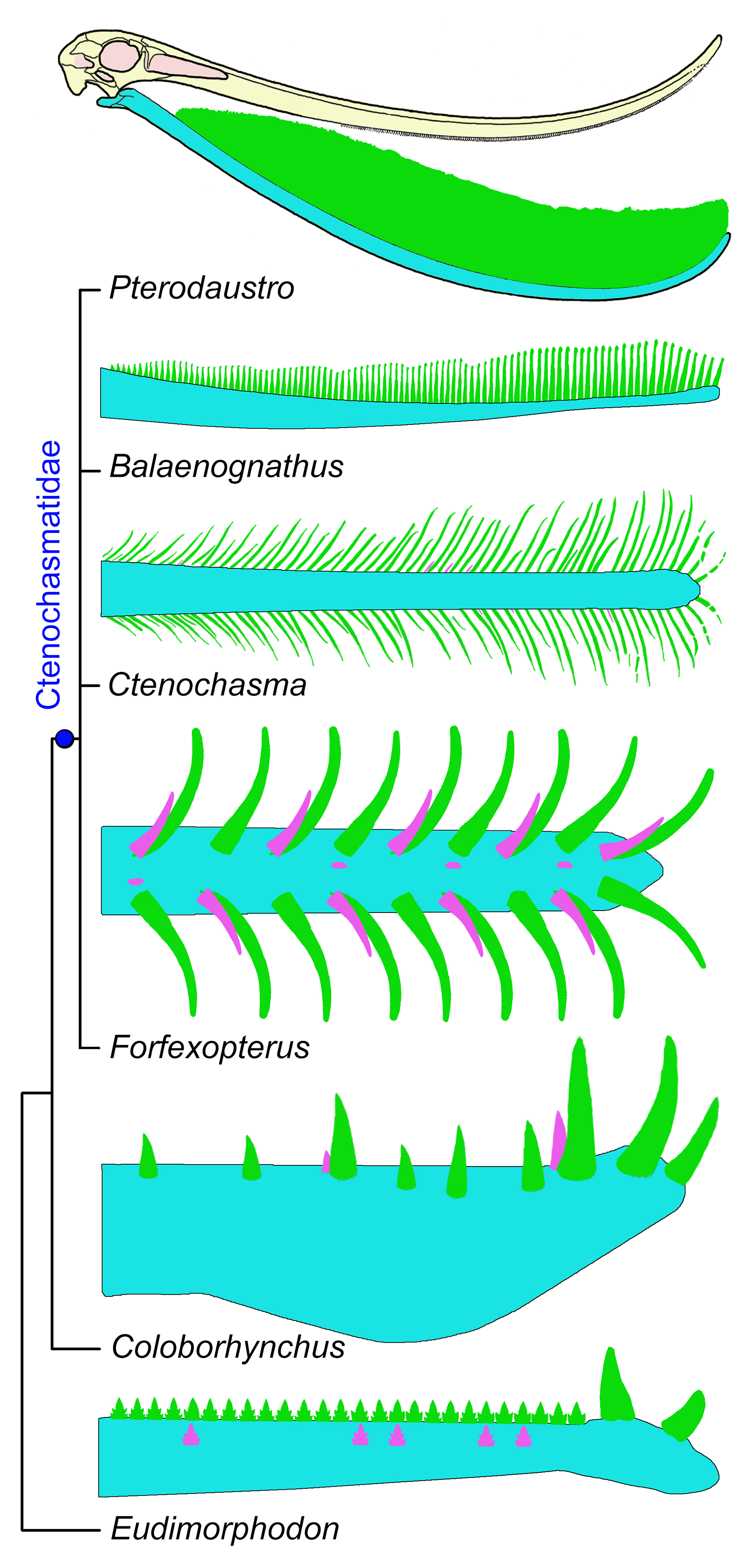
The tooth replacement of Forfexopterus (SDUST-V1007) is comparable to that of the ornithocheirid Coloborhynchus, in that the tooth germ is more medially positioned along the middle line, the replacement tooth erupting at the posteromedial side of the functional tooth, and no more than one replacement tooth present in each alveolus. The pathway of the replacement tooth appears to be in a lateral-anterior direction to replace its predecessor and occupy the alveolus, which is also known as “varanid method” (Edmund 1960, Bertin et al. 2018). It seems to be widely distributed in pterosaurs, evidenced by the replacement tooth erupted posteromedial to the functional tooth frequently known in the Jurassic-Cretaceous pterosaurs (e.g. Anhanguera, Fastnacht 2008; Ctenochasma, Pterodactylus, Rhamphorhynchus, Scaphognathus, Edmund 1960; Jianchangnathus, Zhou 2014). However, a lateral-vertical replacement path, as an alternative pattern, is reported in the basal pterosaur Eudimorphodon (Wild 1978; Figure 4), which is common in other archosaurs (e.g. crocodiles, dinosaurs, toothed birds; Edmund 1960, Dumont et al. 2016, Bertin et al. 2018). One or less replacement tooth in each alveolus is not frequently known in archosaurs (e.g. Edmund 1960, 1962, Dumont et al. 2016). In contrast, multiple generations of the replacement tooth are well known in some ornithischian dinosaurs (e.g. ceratopsids, hadrosaurians), associated with a high replacement ratio (e.g. Erickson 1996a). The tooth replacement rate can be measured based on the dentinal growth line counts between functional and replacement teeth (e.g. Erickson 1996a, b, D’Emic et al. 2019, Kosch & Zanno 2020, Maho et al. 2022). The tooth replacement rate is not yet known in pterosaurs, due to the lack of histological data of the teeth. A preliminary histological study in Hamipterus only reveals a tooth formation time of about 80 days, without any information on the tooth replacement (Chen et al. 2023). Furthermore, Cerda & Codorniú (2023) reported that the dentinal growth lines could not be identified in the teeth of Pterodaustro, which could either be a taphonomic artifact or an adaption related to its filamentous teeth. Based on values of Z-spacing, the relative tooth replacement ratio has been estimated in Coloborhynchus by Fastnacht (2008) that the enlarged anterior teeth are replaced faster with a lower value of Z-spacing than the posterior teeth along the tooth row, to maintain the fish-grabbing feeding behavior. In Forfexopterus (SDUST-V1007), however, the values of Z-spacing are comparable, implying the replacement ratio is relatively stable along the preserved tooth row to adapt to the filter-feeding strategy.

Comparisons of the tooth replacement of Forfexopterus with selected pterosaurs in a simple cladogram. Colors marked: blue, mandibular body; green, functional teeth; purple, replacement teeth. Line drawings of the lower dentitions of Ctenochasma is based on SMNS 81803; Balaenognathus, Coloborhynchus, Eudimorphodon, and Pterodaustro are modified or reconstructed from the literatures (Wild 1978, Fastnacht 2001, 2008, Zhou et al. 2017, Martill et al. 2023).
The tooth replacement of Forfexopterus (SDUST-V1007) is alternative between odd- and even-numbered tooth positions on both sides, and showing an asymmetrical pattern on both sides of the lower jaw. In contrast, it is more complex in Coloborhynchus (Fastnacht 2008). However, the replacement pattern is varied in ontogeny: a simple alternative pattern is frequently reported in young individuals, and becomes more irregular in adults (e.g. Edmund 1960, 1962, Hanai & Tsuihiji 2019). Unfortunately, this variation is untestable here, lacking the information of the mature specimen of Forfexopterus and the young specimen of Coloborhynchus. Asymmetry of the tooth replacement between both sides in Forfexopterus (SDUST-V1007) is similar with that of Coloborhynchus, possibly in order to maintain the functional occlusion on at least one side of the lower jaws. Actually, all young and mature functional teeth are worn somewhat on their crown, fully functioning during the replacement circle in each tooth position in Forfexopterus (SDUST-V1007; Figures 2 and 4). In addition, as hypothesized by Lawson et al. (1971), the tooth count in each stage may reflect the relative duration of the stage in the replacement cycle. The relative duration of teeth in function of Forfexopterus can be estimated by the percentage of the tooth count in stages III and IV as about 58%, which is longer than the relative duration of the tooth formation.
Ecological adaptation
Generally, there is a functional gap between the shedding of the former tooth and the full growing of the replacement tooth in gnathostomes (e.g. Berkovitz & Shellis 2017). To increase the duration of the functional tooth and the growth time of the replacement tooth, it would be appropriate to reduce the functional gap between tooth generations. As documented by Fastnacht (2008), the replacement tooth is positioned posteromedial to the functional tooth, commonly in pterosaurs, as functional for increasing the duration of the functional tooth and further improving the growth of the replacement tooth. This strategy may be highlighted in Forfexopterus by the strong ligamentous anchoring in the alveolus, evidenced by the full functionality of the young functional tooth with incompletely-developed root. In contrast to other ctenochasmatids, Forfexopterus feeds more actively, having a tooth-tooth occlusion evidenced by the tooth abrasion, and with a larger interdental space of about 6 mm (Zhou et al. 2022). Larger prey are expected to be more challenging for filter feeding. Therefore, the fully functioning dentition may play a significant role in maintaining the active feeding of Forfexopterus.
The tooth replacement is poorly known in other ctenochasmatids. Based on new fossil observation, the tooth replacement may be more complex than we prospected previously. Recently, a study of the dental histology of Pterodaustro has been performed by Cerda & Codorniú (2023), revealing that the replacement tooth is absent in Pterodaustro evidenced as a monophyodont or diphyodont pattern, possibly representing its extremity in feeding adaption. Another case is present in the bizarre Balaenognathus (Martill et al. 2023), in which an alternative replacement pattern is hypothesized based on the tooth size gradients identified along the exposed tooth row on the mandible. The tooth size gradients are unusual, in having a long period of 10-13 teeth (Martill et al. 2023, Figure 4). The small teeth in the size gradients were interpreted as “replacement teeth” in the original literature. However, the tooth replacement process is unknown in Balaenognathus. In contrast, more information of the tooth replacement is revealed in Ctenochasma (Bennett 2007, Figure 4). As in Balaenognathus, the dentitions of Ctenochasma is exposed on the jaws. Along the dentition, the tooth size gradients can be identified, implying an alternative pattern of replacement. Each size gradient involves two or three succeeding teeth. The anterior teeth are larger, and the posteriormost tooth is smallest, varied from one third to more than half the size of the anteriormost tooth. Some replacement teeth are tiny and just erupted posterior to the associated functional teeth (SMNS 81803; Figure 4). In contrast to Forfexopterus, the young functional teeth that are varied from one third to more than half-size of the mature functional teeth of Ctenochasma are not fully functional, evidenced as a functional gap during the tooth replacement process, which hinder more or less the functional occlusion of the filter feeding apparatus. These variations in the tooth replacement of the ctenochasmatids need to be further studied in the future.
ACKNOWLEDGMENTS
The authors would like to thank Dr. Rui Pei (Institute of Vertebrate Paleontology and Paleoanthropology (IVPP), Chinese Academy of Sciences) for his corrections to an early version of this manuscript, Qin-Fang Fang (China University of Geosciences) for help in CT scanning of the specimen, and Jiahao Wang (Shandong University of Science and Technology) for help in 3D reconstruction of the specimen. We thank Dr. Shunxing Jiang (IVPP, Chinese Academy of Sciences) for his kind invitation for this special issue, and thank Dr. Taissa Rodrigues (Universidade Federal do Espírito Santo) and two anonymous reviewers for their helpful comments and suggestions on our manuscript. This work was supported by the National Natural Science Foundation of China (42161134003); Taishan Scholar Program of Shandong Province (tsqn201812070, tstp20240514).
REFERENCES
- BENNETT SC. 2007. A review of the pterosaur Ctenochasma: taxonomy and ontogeny. N Jb Geol Paläont Abh 92: 254-271.
- BERKOVITZ B & SHELLIS P. 2017. The Teeth of Non-Mammalian Vertebrates. Amsterdam: Elsevier, 342 p.
- BERTIN TJC, THIVICHON-PRINCE B, LEBLANC ARH, CLADWELL MW & VIRIOT L. 2018. Current perspectives on tooth implantation, attachment, and replacement in Amniota. Front Physiol 9: 1630. doi:10.3389/fphys.2018.01630.
- BESTWICK J, UNWIN DM, BUTLER RJ, HENDERSON DM & PURNELL MA. 2018. Pterosaur dietary hypotheses: a review of ideas and approaches. Biol Rev 93: 2021-2048.
- CERDA IA & CODORNIÚ L. 2023. Palaeohistology reveals an unusual periodontium and tooth implantation in a filter-feeding pterodactyloid pterosaur, Pterodaustro guinazui, from the Lower Cretaceous of Argentina. J Anat 243: 579-589.
- CHEN H, LI ZH, JIANG SX, WU Q, GONG YX, ZHU XF & WANG XL. 2023. A preliminary analysis of dental microstructure in Hamipterus (Pterosauria, Pterodactyloidea). The Anatomical Record, p. 1-15. doi:10.1002/ar.25289.
- D’EMIC MD, O’CONNOR PM, PASCUCCI TR, GAVRAS JN, MARDAKHAYAVA E & LUND EK. 2019. Evolution of high tooth replacement rates in theropod dinosaurs. PLoS ONE 14: e0224734.
- DUMONT M, TAFFOREAU P, BERTIN T, BHULLAR B-A, FIELD D, SCHULP A, STRILISKY B, THIVICHON-PRINCE B, VIRIOT L & LOUCHART A. 2016. Synchrotron imaging of dentition provides insights into the biology of Hesperornis and Ichthyornis, the “last” toothed birds. BMC Evol Biol 16: 178.
- EDMUND AG. 1960. Tooth replacement phenomena in the lower vertebrates. R Ontario Mus Contrib Life Sci Div 52: 1-190.
- EDMUND AG. 1962. Sequence and rate of tooth replacement in the Crocodilia. R Ontario Mus Contrib Life Sci Div 56: 1-42.
- ERICKSON GM. 1996a. Incremental lines of von Ebner in dinosaurs and the assessment of tooth replacement rates using growth line counts. Proc Natl Acad Sci USA 93: 14623-14627.
- ERICKSON GM. 1996b. Daily deposition of dentine in juvenile Alligator and assessment of tooth replacement rates using incremental line counts. J Morphol 228: 189-194.
- FASTNACHT M. 2001. First record of Coloborhynchus (Pterosauria) from the Santana Formation (Lower Cretaceous) of the Chapada do Araripe, Brazil. PalZ 75: 23-36.
- FASTNACHT M. 2008. Tooth replacement pattern of Coloborhynchus robustus (Pterosauria) from the Lower Cretaceous of Brazil. J Morphol 269: 332-348.
- HANAI T & TSUIHIJI T. 2019. Description of tooth ontogeny and replacement patterns in a juvenile Tarbosaurus bataar (Dinosauria: Theropoda) using CT-Scan data. Anat Rec 302: 1210-1225.
- HENDERSON DM. 2018. Using three-dimensional, digital models of pterosaur skulls for the investigation of their relative bite forces and feeding styles. Geol Soc London Special Publications 455: 25-44.
- JIANG SX, CHENG X, MA YX & WANG XL. 2016. A new archaeopterodactyloid pterosaur from the Jiufotang Formation of western Liaoning, China, with a comparison of sterna in Pterodactylomorpha. J Vertebr Paleontol 36: e1212058.
- JIANG SX & WANG XL. 2011. A new ctenochasmatid pterosaur from the Lower Cretaceous, western Liaoning, China. An An Acad Bras Cienc 83: 1243-1249. DOI 10.1590/S0001-37652011000400011.
- KOSCH JCD & ZANNO LE. 2020. Sampling impacts the assessment of tooth growth and replacement rates in archosaurs: implications for paleontological studies. PeerJ 8: e9918.
- LAWSON R, WAKE DB & BECK NT. 1971. Tooth replacement in the redbacked salamander, Plethodon cinereus. J Morphol 134: 259-269.
- MAHO T, MAHO S, SCOTT D & REISZ RR. 2022. Permian hypercarnivore suggests dental complexity among early amniotes. Nat Commun 13: 4882.
- MARTILL DM, FREY E, TISCHLINGER H, MÄUSER M, RIVERA-SYLVA HE & VIDOVIC SU. 2023. A new pterodactyloid pterosaur with a unique filterfeeding apparatus from the Late Jurassic of Germany. PalZ 97: 383-424.
- WANG XL, KELLNER AWA, ZHOU ZH & CAMPOS DA. 2005. Pterosaur diversity and faunal turnover in Cretaceous terrestrial ecosystems in China. Nature 437: 875-879.
- WELLNHOFER P. 1991. The Illustrated Encyclopedia of Pterosaurs. London: Salamander, 192 p.
- WILD R. 1978. Die Flugsaurier (Reptilia, Pterosauria) aus der Oberen Trias von Cene bei Bergamo, Italien. Boll Soc Paleontol Ital 17: 176-256.
- ZHOU C-F. 2014. Cranial morphology of a Scaphognathus-like pterosaur, Jianchangnathus robustus, based on a new fossil from the Tiaojishan Formation of western Liaoning, China. J Vertebr Paleontol 34: 597-605.
- ZHOU C-F, GAO K-Q, YI HY, XUE JZ, LI QG & FOX RC. 2017. Earliest filter-feeding pterosaur from the Jurassic of China and ecological evolution of Pterodactyloidea. R Soc Open Sci 4: 160672.
- ZHOU C-F, WANG XY & WANG JH. 2022. First evidence for tooth-tooth occlusion in a ctenochasmatid pterosaur from the Early Cretaceous Jehol Biota. Geol Soc London Special Publications 521: 9-17.