
 Assessment of Renal Function and Bilirubin Measurement in Urine as Prognostic Value in Immune-Mediated Hemolytic Anemia in Dogs
Abstract
Immune-mediated hemolytic anemia (IMHA) is the leading cause of hemolytic anemia in dogs. Although renal injury has been reported in IMHA patients, laboratory findings defining the type and extent of renal damage remain inconsistent. The aim of this study was to evaluate key laboratory findings in the assessment of renal function in dogs with a suspected diagnosis of IMHA and to correlate these findings with disease prognosis. A prospective study was conducted on 21 dogs treated at a veterinary hospital with a suspected diagnosis of IMHA. Blood samples were collected for complete blood count and biochemical analyses, and urinalysis was performed. Significant alterations in renal function were observed in IMHA patients. The urinary protein-to-creatinine ratio (UPC) was elevated compared to controls (P < 0.0002) and correlated with the degree of anemia (R = -0.638) and the inflammatory status, assessed by white blood cell count (R = 0.550). In addition, urinary bilirubin levels were increased in IMHA patients (P < 0.0001), suggesting that bilirubin is an important prognostic marker of the disease. Renal function is impaired in IMHA patients, and urinary bilirubin levels serve as a valuable prognostic indicator.
Key words
Biomarkers; Canine; Hemolysis; Proteinuria; Veterinary clinical pathology
INTRODUCTION
Immune-mediated hemolytic anemia (IMHA) is one of the main causes of hemolytic anemia in dogs (Balch & Mackin 2007). The disease may occur either idiopathically or secondarily, with the latter triggered by underlying conditions such as hemoparasitosis, drugs, or vaccination (Gianesini et al. 2023, Morrow & White 2020, Neelawala et al. 2021). Hemolysis can occur intravascularly or extravascularly and is mediated either by the direct destruction of erythrocytes or by the phagocytosis of red blood cells opsonized with IgM or IgG. Additionally, this process may involve complement activation (Balch & Mackin 2007). In this immune-mediated hemolytic state, dogs may present clinical signs such as lethargy, weakness, pale and/or icteric mucous membranes, hemoglobinuria, and bilirubinuria. Laboratory evaluation is essential for disease characterization and commonly reveals regenerative anemia, bilirubinemia and/or bilirubinuria, spherocytosis, autoagglutination, and a positive Coombs test (Garden et al. 2019, Piek 2011).
IMHA is associated with a poor prognosis, with studies reporting mortality rates between 50% and 70% (McAlees 2010). High morbidity and mortality are associated with intense hemolysis, which results in severe anemia and consequent tissue hypoxia. Furthermore, a systemic inflammatory state is triggered, primarily through the release of heme and free hemoglobin, which activate cells of the innate immune system and promote hemostatic activation in response to inflammation. Free hemoglobin also generates large amounts of reactive oxygen species (ROS), leading to endothelial injury and damage to organs such as the liver and kidneys, further aggravating the clinical condition (Hamzianpour & Chan 2016, Kidd & Mackman 2013).
Several studies have characterized the hematological and biochemical parameters of IMHA canine patients, emphasizing regenerative anemia, alterations in liver enzymes such as alanine aminotransferase (ALT) and alkaline phosphatase (ALP), and increased bilirubin levels (Balch & Mackin 2007, Elwood & Polton 2008, Piek et al. 2008). Other studies have also investigated hemostatic findings, reporting prolonged prothrombin time (PT) and activated partial thromboplastin time (aPTT), as well as hypercoagulable states detected by thromboelastography (Hamzianpour & Chan 2016, Sinnott & Otto 2009). In addition, these laboratory findings are correlated with patient prognosis. For example, elevated serum levels of urea, ALP, and bilirubin have been associated with poorer outcomes (Carr et al. 2002, Swann & Skelly 2015).
In this context, the laboratory characterization of dogs with IMHA is of great clinical relevance, particularly regarding the assessment of renal function. This is because intense hemolysis, hypoxia, and systemic inflammation can compromise the kidneys, however, the literature still lacks detailed studies on this topic. Furthermore, the presence of urinary bilirubin, frequently observed in IMHA, may reflect both the degree of hemolysis and renal involvement. Indeed, when identified in the urine, especially in the form of indirect bilirubin, this finding may be associated with renal injury, as indirect bilirubin binds to serum proteins and is normally not excreted by an intact glomerulus. Therefore, measuring total bilirubin and its fractions can serve as a marker of renal impairment and have prognostic value. Accordingly, this study aimed to comprehensively evaluate renal function and urinary bilirubin levels in dogs with IMHA, investigating their associations with disease severity and prognosis.
MATERIALS AND METHODS
Overall Study Design
This was a prospective study conducted with canine patients treated at the Veterinary Hospital of Unesp, Botucatu, SP, between April and October 2017. The inclusion criteria were animals diagnosed with hemolytic anemia based on clinical signs, such as lethargy, pale and/or icteric mucous membranes, and bilirubinuria (dark and/or orange-colored urine), along with laboratory tests, such as complete blood count (CBC) showing macrocytic hypochromic anemia, increased RDW (red cell distribution width), anisocytosis, and polychromasia (varying degrees), positive saline agglutination test and/or spherocytosis, accompanied by bilirubinemia (icteric plasma) and bilirubinuria (assessed using chemical reagent dipsticks). The exclusion criteria included patients with a provisional diagnosis of leptospirosis, hepatopathies and/or renal diseases, and a provisional diagnosis of neoplasia. The study included 21 patients with a suspected diagnosis of primary and/or secondary IMHA, and the data were compared with those of 10 healthy dogs. The study adhered to ethical principles approved by the Animal Use Ethics Committee (protocol no. 194/2016).
Sample Collection and Processing
Samples were collected in EDTA tubes (ethylene diamine tetra-acetic acid - BD Vacutainer) and tubes without anticoagulant (BD Vacutainer), along with urine collected via urethral catheterization. These samples were collected immediately upon admission, prior to any medication interference. Serum was obtained after centrifugation at 1800 × g for 10 minutes. Importantly, the serum and urine samples were protected from light until processing.
Clinical Findings, Laboratory Results, and Prognosis
Clinical data were obtained from medical records in the veterinary hospital system. Hematological and biochemical parameters were performed in accordance with American Society for Veterinary Clinical Pathology (ASVCP) guidelines (Gunn-Christie et al. 2012). Prognosis was defined as survival or death within 30 days after admission, and in cases where the patient’s outcome was not recorded in the electronic file, our team contacted the owner to confirm the outcome.
Renal Function Evaluation
Renal function assessment was also performed in accordance with the ASVCP guidelines. Serum urea (Bioclin®) and creatinine (Bioclin®) levels were measured in both patients and controls via a biochemical analyzer (Cobas Mira Plus®). For urinalysis, approximately 10 mL of urine was collected via catheterization, protected from light, and processed within 1 hour after collection. The samples were subjected to physical, chemical, and sediment examinations. Reagent strips Combur Test M (Roche®) were used for chemical evaluation, and the urinary sediment was examined by microscopy (400×). In addition, from the urine supernatant, the levels of microprotein (Bioclin®), creatinine (Bioclin®), total bilirubin (Bioclin®), and direct and indirect bilirubin (Bioclin®) were measured.
Statistical analyses
Quantitative data were expressed as medians and percentiles. For comparisons between groups and experiments, variables were treated as nonparametric, and comparisons were made via the Mann-Whitney test (independent variables), whereas Fisher’s exact test was used for categorical variables. Correlations were assessed using the Spearman test. Statistical significance was considered when P < 0.05. All the statistical analyses were performed via SPSS version 26 (IBM) or GraphPad Prism 7.0 Software (GraphPad, Inc.).
RESULTS
In this study, 21 canine patients diagnosed with IMHA and 10 healthy dogs were included. Among the affected dogs, most were mixed-breed (62%, 13/21), followed by Poodles (14%, 3/21), Pitbull, Brazilian Terrier, Shih Tzu, Yorkshire Terrier and Labrador Retriever breeds (24%, 5/21). The mean age was 4.5 ± 4 years, with a male-to-female ratio of 10:11. All animals were diagnosed based on clinical signs and laboratory findings. Among these patients, 10% exhibited spherocytosis and 19% tested positive in the saline agglutination test. Regarding the healthy controls, most were mixed-breed dogs (40%, 4/10), followed by Pitbulls (20%, 2/10), Golden Retrievers (20%, 2/10), and Labradors and Shih Tzus (20%, 2/10). The mean age was 4 ± 1.5 years, with a male-to-female ratio of 3:7. The detailed hematological and biochemical characteristics of the study population are presented in Table SI – Supplementary Material.
Regarding the evaluation of renal parameters, the main relevant findings from urinalysis are presented in Table I.
We observed that the urine protein-to-creatinine ratio (UPC) was greater in dogs with immune-mediated hemolytic anemia (IMHA) than in to control dogs (Table I and Figure 1a). A trend toward reduced urine specific gravity (USG) was also observed in IMHA patients (Table I and Figure 1b).
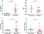
Evaluation of renal function parameters. (a) The urine protein-to-creatinine ratio (UPC) was greater in IMHA patients (median, IQR: 1.22, 0.76–2.12) than in control dogs (median, IQR: 0.06, 0.03–0.09) (P < 0.0002). (b) Assessment of the USG in control dogs and IMHA patients (dashed line represents a value of 1.030). (c-d) Measurement of serum urea and creatinine levels in control dogs and IMHA patients (dashed lines represent values of 21–59 mg/dL for urea; 0.5–1.5 mg/dL for creatinine). The medians (horizontal lines) represent nonGaussian data; similarly, the P value was obtained from the Mann-Whitney test according to the data distribution.
IMHA patients presented increased microscopic cellularity, indicating an active acute renal process. Renal or pelvic epithelial cells were present in the urinary sediment in 52% of the IMHA patients, along with granular casts in 38% and bilirubin crystals in 33% of the cases. In the assessment of serum biomarkers of renal function, IMHA patients tended to have higher urea levels than controls did (Figure 1c). Conversely, creatinine levels were lower in IMHA patients (Figure 1d).
For the assessment of prognostic biomarkers, the data were analyzed by dividing IMHA patients into survivors and nonsurvivors. There was a trend toward higher levels of total bilirubin (TB), ALP, and gamma-glutamyltransferase (GGT) in nonsurvivors. Serum direct bilirubin (DB) levels were also higher in nonsurvivors than in survivors (Figure S1).
The measurement of TB, DB and indirect bilirubin (IB) were also measured in the urine samples immediately after collection. Both urinary total bilirubin (UTB) and urinary direct bilirubin (UDB) were identified as severity biomarkers in IMHA patients, as evidenced by higher levels in nonsurvivors (Figure 2a, b). Using the ROC curve, optimal cutoff values were determined for the UTB (9.7 mg/dL – 70% sensitivity and 88.9% specificity) and UDB (5.1 mg/dL – 60% sensitivity and 88.9% specificity) (Figure 2c, d). Urinary indirect bilirubin (UIB) levels did not demonstrate significant prognostic value, although a trend toward higher values was observed in patients who died (median 5.03 mg/dL) than in survivors (median 2.33 mg/dL) (P = 0.16).

Evaluation of urinary bilirubins as a prognostic marker in IMHA. Urinary total bilirubin (UTB) levels (a) and urinary direct bilirubin (UDB) levels (b) were greater in patients who died. (c-d) ROC curve based on the UTB and UDB values. The medians (horizontal lines) represent non-Gaussian data; similarly, the P value was obtained from the Mann-Whitney test according to the data distribution.
Finally, a correlation analysis was performed between the UPC and other relevant laboratory parameters in the IMHA. In terms of hematological parameters, UPC was inversely correlated with hematocrit and positively correlated with total leukocytes and the NLR. For the serum biomarkers, the UPC was inversely correlated with the serum albumin concentration and positively correlated with the ALP, TB, DB and IB levels. Additionally, UPC was inversely correlated with USG and positively correlated with UTB and UDB (Table II).
DISCUSSION
The main contribution of our study was to demonstrate that renal injury is an evident alteration in patients with IMHA, primarily indicated by an increase in the UPC and its association with the degree of anemia. In addition, we observed that the UTB and UDB behaved as prognostic markers of the disease, with higher values in IMHA patients who died.
Regarding demographic data, we found that most patients were mixed-breed, differing from other studies that describe a predominance in purebred animals (McAlees 2010, Piek et al. 2008). However, it is important to note that mixed-breed patients are frequently treated at veterinary hospitals, which explains their predominance in our sample. The mean age of the patients was 4.5 years, while the literature reports an average age of approximately 6 years, although it also highlights that the disease can affect animals of any age (Balch & Mackin 2007).
When classical markers of renal function, such as urea and creatinine (Braun et al. 2003, Yadav et al. 2020), are evaluated, no significant increases in urea levels are observed, although a tendency toward elevation is observed in dogs with IMHA. On the other hand, median creatinine values were reduced in IMHA patients. This finding was probably influenced by the presence of serum icterus (Bowers & Wong 1980, Ji & Meng 2011), since icterus was one of the inclusion criteria for this study. The reduction in creatinine levels does not appear to be physiologically consistent in these patients, as other renal function parameters, such as the UPC and urinary sediment findings, were altered.
Urinalysis revealed that USG, which is considered an earlier marker than increases in serum urea and creatinine (Reine & Langston 2005), tended to be lower in dogs with IMHA than in healthy controls. Although the difference did not reach statistical significance, 72% of the dogs with IMHA were dehydrated (dehydration equal to or greater than 5%), and most of them had a USG lower than 1.030. This finding may differ from what is expected in dehydrated animals, as most patients presented with hypernatremic dehydration (due to reduced water intake) or isotonic dehydration (associated with vomiting), conditions in which urine is normally concentrated when renal function is preserved (Reine & Langston 2005, Yadav et al. 2020). These results suggest that IMHA patients presented renal dysfunction, especially due to a reduced ability to concentrate urine.
Other urinary findings reinforce this hypothesis, including increased cellularity in the urinary sediment, with the presence of renal or pelvic epithelial cells (52% of cases), granular casts (38%), and bilirubin crystals (33%). These findings are consistent with an acute renal injury process, possibly resulting from immune complex deposition in the glomeruli and tubular precipitation of compounds such as hemoglobin, bilirubin, and cellular debris, leading to the formation of granular casts. Altogether, these alterations indicate renal injury involving both tubular and glomerular components (Hokamp & Nabity 2016, Reine & Langston 2005, Yadav et al. 2020).
In the biochemical analysis of urine, the UPC was greater in IMHA patients than in healthy individuals. It is well established that UPC is an important marker for assessing glomerular injury (Lees et al. 2005, Moraes et al. 2017). Therefore, UPC data were explored and correlated with other laboratory parameters (Table II). Among these, we observed an inverse correlation between UPC and hematocrit (R = -0.638), and a positive correlation with serum bilirubins (R = 0.739), suggesting that patients with greater red blood cell destruction also have increased UPC values. Positive correlations were also found between UPCs and WBCs, as well as between UPCs and the NLR, indicating an association with a stronger inflammatory response. The UPC was also inversely correlated with the USG, suggesting that the lower USG observed in IMHA patients may be associated with concurrent tubular injury. Finally, a positive correlation was observed between UPC, TBU and DBU, which are discussed in more detail below.
The measurement of total bilirubin and its urinary fractions was exploratory, aiming to quantify these fractions more accurately. Serum fractions were positively correlated with their respective urinary fractions, including TB and UTB (R = 0.775; p = 0.001), IB and UIB (R = 0.591; p = 0.001), and BD and UBD (R = 0.660; p = 0.001). It is known that IB, which is bound to plasma proteins, when found at high urinary levels may indicate glomerular injury (Kalakonda et al. 2022). Moreover, urinary bilirubin levels were analyzed, which revealed that higher TBU and DBU values were associated with a worse prognosis, as evidenced by patient death. Regarding IBU, a tendency toward elevation was observed in nonsurviving patients (p = 0.16).
To complement the analysis of prognostic markers, the serum levels of DB, TB, ALP and GGT were also associated with a worse prognosis (Figure S1), as previously reported by other studies (Elwood & Polton 2008, Piek et al. 2008, Swann & Skelly 2015).
This study has some limitations that should be acknowledged, such as the small sample size and the absence of postmortem renal histopathological evaluation, which is often limited by the lack of owner authorization. In addition, the outcome of one patient could not be determined due to incomplete medical records and the inability to contact the owner, and urinary bilirubin analysis was not performed for one dog. Although these limitations exist, our results are consistent, and this is the first study to explore urinary bilirubin fractions in dogs with IMHA.
In conclusion, this study clearly indicates that renal function is impaired in patients diagnosed with IMHA. Additionally, the urinary UTB and UDB proved to be important prognostic markers. Therefore, we emphasize the importance of monitoring renal function parameters in IMHA patients, since a broader evaluation beyond hematology may be beneficial in selecting more specific and targeted therapeutic strategies for these individuals.
Acknowledgements
This study was funded by the Veterinary Hospital of the Faculty of Veterinary Medicine and Animal Science at São Paulo State University – Botucatu Campus.
References
- BALCH A & MACKIN A. 2007. Canine immune-mediated hemolytic anemia: pathophysiology, clinical signs, and diagnosis. Compendium 29: 217-225.
- BOWERS LD & WONG ET. 1980. Kinetic serum creatinine assays. II. A critical evaluation and review. Clin Chem 26: 555-561.
- BRAUN JP, LEFEBVRE HP & WATSON ADJ. 2003. Creatinine in the dog: a review. Vet Clin Pathol 32: 162-179.
- CARR AP, PANCIERA DL & KIDD L. 2002. Prognostic factors for mortality and thromboembolism in canine immune-mediated hemolytic anemia: a retrospective study of 72 dogs. J Vet Intern Med 16: 504-509.
- ELWOOD C & POLTON G. 2008. Idiopathic immune-mediated hemolytic anemia: treatment outcome and prognostic factors in 149 dogs. J Vet Intern Med 22: 1075.
- GARDEN OA ET AL. 2019. ACVIM consensus statement on the diagnosis of immune-mediated hemolytic anemia in dogs and cats. J Vet Intern Med 33: 313-334.
- GIANESINI G, DRIGO M & ZOIA A. 2023. Immune-mediated hemolytic anemia and clinically suspected acute pancreatitis in dogs, a pilot study. Top Companion Anim Med 56-57: 100821.
- GUNN-CHRISTIE RG, FLATLAND B, FRIEDRICHS KR, SZLADOVITS B, HARR KE, RUOTSALO K, KNOLL JS, WAMSLEY HL & FREEMAN KP. 2012. ASVCP quality assurance guidelines: control of preanalytical, analytical, and postanalytical factors for urinalysis, cytology, and clinical chemistry in veterinary laboratories. Vet Clin Pathol 41: 18-26.
- HAMZIANPOUR N & CHAN DL. 2016. Thromboelastographic assessment of the contribution of platelets and clotting proteases to the hypercoagulable state of dogs with immune-mediated hemolytic anemia. J Vet Emerg Crit Care 26: 295-299.
- HOKAMP JA & NABITY MB. 2016. Renal biomarkers in domestic species. Vet Clin Pathol 45: 28-56.
- JI JZ & MENG QH. 2011. Evaluation of the interference of hemoglobin, bilirubin, and lipids on Roche Cobas 6000 assays. Clin Chim Acta 412: 1550-1553.
- KALAKONDA A, JENKINS BA & JOHN S. 2025. Physiology, Bilirubin. StatPearls [Internet]. Treasure Island (FL): StatPearls Publishing.
- KIDD L & MACKMAN N. 2013. Prothrombotic mechanisms and anticoagulant therapy in dogs with immune-mediated hemolytic anemia. J Vet Emerg Crit Care 23: 3-13.
- LEES GE, BROWN SA, ELLIOTT J, GRAUER GE, VADEN SL & AMERICAN COLLEGE OF VETERINARY INTERNAL MEDICINE. 2005. Assessment and management of proteinuria in dogs and cats: 2004 ACVIM Forum consensus statement (small animal). J Vet Intern Med 19: 377-385.
- MCALEES TJ. 2010. Immune-mediated haemolytic anaemia in 110 dogs in Victoria, Australia. Aust Vet J 88: 25-28.
- MORAES LF, TAKAHIRA RK & GOLIM MA. 2017. Avaliação hematológica e da função renal em cães com AHIM. Acta Sci Vet 45: 1510.
- MORROW L & WHITE C. 2020. Does vaccination increase the risk of immune-mediated haemolytic anaemia in dogs? Vet Rec 187: 274-275.
- NEELAWALA D, DISSANAYAKE DRA, PRASADA DVP & SILVA ID. 2021. Analysis of risk factors associated with recurrence of canine babesiosis caused by Babesia gibsoni. Comp Immunol Microbiol Infect Dis 74: 101572.
- PIEK CJ. 2011. Canine idiopathic immune-mediated haemolytic anaemia: a review with recommendations for future research. Vet Q 31: 129-141.
- PIEK CJ, JUNIUS G, DEKKER A, SCHRAUWEN E, SLAPPENDEL RJ & TESKE E. 2008. Idiopathic immune-mediated hemolytic anemia: treatment outcome and prognostic factors in 149 dogs. J Vet Intern Med 22: 366-373.
- REINE NJ & LANGSTON CE. 2005. Urinalysis interpretation: how to squeeze out the maximum information from a small sample. Clin Tech Small Anim Pract 20: 2-10.
- SINNOTT VB & OTTO CM. 2009. Use of thromboelastography in dogs with immune-mediated hemolytic anemia: 39 cases (2000-2008). J Vet Emerg Crit Care 19: 484-488.
- SWANN JW & SKELLY BJ. 2015. Systematic review of prognostic factors for mortality in dogs with immune-mediated hemolytic anemia. J Vet Intern Med 29: 7-13.
- YADAV SN, AHMED N, NATH AJ, MAHANTA D & KALITA MK. 2020. Urinalysis in dog and cat: a review. Vet World 13: 2133-2141.
Edited by
-
Handling editor
Alexander Kellner
All data generated or analyzed during this study are included in this published article and its supplementary material.


