Immunophenotypic analysis and clinical significance in pediatric hemophagocytic lymphohistiocytosis
Abstract
This study characterizes changes in peripheral blood lymphocyte subsets among pediatric hemophagocytic lymphohistiocytosis (HLH) patients. Compared to healthy controls, HLH patients exhibited significantly elevated levels of CD8⁺ T cells, with concomitant reductions in B cells, CD4⁺ T cells, DPT cells, DNT cells, and the CD4⁺/CD8⁺ T cell ratio as determined by multiparametric flow cytometry. Organ dysfunction was further correlated with distinct immunophenotypic alterations. Specifically, respiratory dysfunction was associated with decreased CD4⁺ CM T cells, naïve B cells, plasmablasts, and transitional B cells; hepatic dysfunction with increased CD4⁺ Temra T cells; circulatory failure with reduced transitional B cells; and neurological dysfunction with elevated CD4⁺ Temra T cells and CD8⁺ EM T cells. Moreover, HLH patients exhibiting three or more organ dysfunctions demonstrated significantly lower levels of DPT cells, memory B cells, transitional B cells, and plasmablasts. Additionally, CD8⁺ T cells and CD8⁺ CM T cells showed positive correlations with pro-inflammatory cytokines (IL-6, TNF-α). These results suggest that depletion of CD4⁺ T cells and B cells, along with aberrant CD8⁺ T cell differentiation, may contribute to the pathogenesis of cytokine storms. These findings support dynamic monitoring of lymphocyte subsets, demonstrating potential utility for early HLH diagnosis and organ damage assessment.
Key words
diagnosis; hemophagocytic lymphohistiocytosis; immunophenotyping; lymphocyte subsets; organ dysfunction
INTRODUCTION
Hemophagocytic lymphohistiocytosis (HLH) is a life-threatening inflammatory syndrome characterized by persistent fever, cytopenias, hepatomegaly, splenomegaly, and lymphadenopathy (Canna & Marsh 2020). This disease progresses rapidly and exhibits a high mortality rate (Li et al. 2020). Primary HLH is caused by gene mutations, which lead to a deficiency in cytotoxic activity in lymphocytes (Steen et al. 2021). In contrast, secondary HLH is often associated with infections, tumors, and autoimmune diseases. Epstein-Barr virus (EBV) is a prevalent cause of secondary HLH (Xu et al. 2022). Although the HLH-1994/HLH-2004 treatment protocol has reduced mortality (Trottestam et al. 2011, Bergsten et al. 2017), the fatality rate among patients with multiple organ dysfunction syndrome (MODS) remains above 70% (Eichenauer et al. 2021, Parajuli et al. 2021). The systemic inflammatory storm in HLH promptly leads to complications in multiple systems, such as coagulation disorders, acute respiratory distress syndrome, and central nervous system impairment, which pose significant risks to patient survival (Canna & Marsh 2020). Exploring markers for early diagnosis, disease progression, and prognosis of HLH is clinically valuable. However, the nonspecific clinical features in the early stages and the time required for specific diagnostic criteria render early diagnosis challenging.
Moreover, the pathophysiology of HLH is initiated by a disruption in immune homeostasis, which causes immune effector cells to become overactivated and proliferate excessively. Consequently, a cytokine storm driven by proinflammatory factors such as IL-6, IFN-γ, and TNF-α ultimately results in MODS (Keenan et al. 2021). Peripheral blood lymphocyte counts serve as crucial biological indicators that reflect the body’s immune status. Considering the substantial heterogeneity of lymphocyte subsets in terms of functional phenotype and immune regulation, this study utilized multiparametric flow cytometry to evaluate the immune status of HLH patients more accurately and comprehensively.
This study aimed to investigate the quantitative variations in lymphocyte subsets in HLH and their correlations with organ function impairments, providing experimental data for the early diagnosis, disease severity evaluation, and prognosis assessment of HLH.
MATERIALS AND METHODS
Research object and inclusion criteria
This study included 20 patients with HLH admitted to the Pediatric Hematology and Pediatric Intensive Care Units at the Affiliated Hospital of Zunyi Medical University. A healthy control group (HC group) consisting of 20 healthy individuals, matched for age and gender, was also selected. The inclusion criteria for HLH patients were as follows: age ranging from 1 month to 14 years; diagnosis conforming to the HLH-2004 guidelines of the Histiocyte Society; treatment with the standardized HLH-1994/2004 protocol, or the HLH-1994/2004 protocol combined with blood purification therapy (Huang et al. 2020). Data collected encompassed gender, age, underlying causes, clinical manifestations, organ dysfunction status, laboratory test results, treatment approaches, and outcomes. Diagnoses of organ dysfunction were based on the criteria for Pediatric MODS (Table I) (Goldstein et al. 2005). Patients were classified into two groups based on the number of dysfunctional organs: organ dysfunction ≥ 3 and organ dysfunction ≤ 2. This study was approved by the Ethics Committee of the Affiliated Hospital of Zunyi Medical University (Approval number: KLLY-2022-141).
Sample processing
Blood samples were collected from healthy children undergoing health check-ups and patients with HLH. The analysis of peripheral blood leukocytes (WBC), lymphocyte counts, and cytokine levels was conducted in the Clinical Laboratory of the Affiliated Hospital of Zunyi Medical University. The remaining blood samples were analyzed using multiparametric flow cytometry analysis. Prior to analysis, samples were diluted at a ratio of 1:1 in RPMI-P/S medium (10% FBS, 100 U/mL penicillin, and 100 μg/mL streptomycin). Subsequently, the samples were layered on Ficoll and centrifuged at 300×g for 25 minutes with a gradually increasing and then decreasing rotation speed. The peripheral blood mononuclear cells were aspirated, washed with RPMI-P/S solution, and adjusted to a cell concentration of 2 × 10⁶ cells/mL for subsequent experiments.
Lymphocyte classification and multiparametric flow cytometry methods
The staining protocol and antibody panel for multiparametric flow cytometry are presented in Table II. Two flow cytometry detection panels were used: T cell Panel: Anti-human CD3 (Percp, 130113693, BD), Anti-human CD4 (FITC, 561005, BD), Anti-human CD8 (BV510, 563256), Anti-human CD45RA (PE-cy7,560675, BD), Anti-human TCRαβ (PE,561674, BD), Anti-human TCRγδ (BV421,562560, BD), Anti-human CD27 (APC,561786, BD). B cell Panel: Anti-human CD19 (APC, 555415, BD), Anti-human CD38 (Percp, 561106, BD), Anti-human CD24 (PE, 560991, BD), Anti-human CD27 (BV450, 560448, BD), Anti-human IgD (FITC, 555778, BD). The samples were mixed with the antibodies and incubated at room temperature in the dark for 20 minutes. Each test tube was washed once with 1 mL of PBS buffer, and flow cytometry analysis was performed.
Table III Laboratory examination of patients with HLH.| Laboratory examination | [N (%)] |
|---|---|
| NEUT < 1.0×10⁹ /L | 12/20 (60.00) |
| HB < 90 g/L | 11/20 (55.00) |
| PLT < 100×109 /L | 17/20 (85.00) |
| FIB ≤ 1.5 g/L | 12/20 (60.00) |
| ALT > 50 U/L | 12/20 (60.00) |
| AST > 40 U/L | 15/20 (75.00) |
| LDH > 300 U/L | 17/20 (85.00) |
| TG ≥ 3 mmol/L | 15/20 (75.00) |
| IL-6 > 5.31 pg/mL | 18/20 (90.00) |
| IL-10 > 4.91 pg/mL | 16/20 (80.00) |
| TNF -α > 2.31 pg/mL | 13/20 (65.00) |
| IFN -γ > 7.42 pg/mL | 18/20 (90.00) |
| SF > 500 μg/L | 19/20 (95.00) |
| Soluble CD25 ≥ 2400 U/mL | 16/20 (80.00) |
| Low or absent NK-cell cytotoxicity | 10/16 (62.50) |
| Hemophage was found in bone marrow | 11/16 (68.75) |
Statistical Analysis
The experimental data were analyzed using SPSS 29.0. The data are presented as the mean ± standard deviation (SD). The normality of the data distribution was assessed using the Shapiro-Wilk test. For inter-group comparisons, paired t-tests were used on data that met assumptions of normality and homogeneity of variances; otherwise, the Mann-Whitney U test was applied. The correlation between continuous variables was calculated using Pearson correlation. P < 0.05 was considered statistically significant.
RESULTS
General Conditions and Etiological Factors of the Patients
A total of twenty patients were diagnosed with HLH, including 12 male and 8 female. Ages ranged from 2 months to 14 years. Genetic screening for HLH-associated mutations was negative in all 20 patients. All patients were associated with EBV infection.
Clinical Manifestations and Organ Dysfunction
The clinical manifestations observed in the 20 patients included fever (100%), hepatomegaly in 12 patients (60.0%), lymphadenopathy in 16 patients (80.0%), splenomegaly in 15 patients (75.0%), pleural and peritoneal effusion in 9 patients (45.0%), purpura in 7 patients (35.0%), gastrointestinal hemorrhage in 4 patients (20.0%), and pulmonary hemorrhage in 2 patients (10.0%). Hepatic dysfunction (HD) was noted in 12 patients (60.0%), respiratory system dysfunction (RSD) in 12 patients (60.0%), central nervous system dysfunction (CNSD) in 6 patients (30.0%), and circulatory system dysfunction (CSD) in 7 patients (35.0%). Furthermore, 9 patients (45.0%) presented with organ dysfunction ≤ 2 organs, while 11 patients (55.00%) had organ dysfunction in ≥ 3 organs.
Laboratory examination
Among the 20 HLH patients, thrombocytopenia (PLT < 100 × 10⁹/L) was observed in 17 patients (85.0%). Elevated levels of ALT were detected in 12 patients (60.0%), AST in 15 patients (75.0%), LDH in 17 patients (85.0%), and TG in 15 patients (75.0%). Cytokine abnormalities included elevated levels of IL-6 and IFN-γ in 18 patients (90.0%), increased IL-10 in 16 patients (80.0%), and elevated TNF-α in 13 patients (65.0%). Additionally, hyperferritinemia (SF > 500 μg/L) was observed in 19 patients (95.0%), and soluble CD25 was elevated in 16 patients (80.0%). Among the 16 patients who underwent functional immunologic assessments, impaired or absent NK cell activity was documented in 10 patients (62.5%), and hemophagocytosis was observed in the bone marrow samples from 11 patients (68.75%).
Treatment and Outcomes
In this study, 7 patients were treated solely with the HLH-1994/2004 regimen, while 13 patients received the HLH-1994/2004 regimen combined with blood purification. Blood purification methods included continuous renal replacement therapy (CRRT) in 2 patients, plasma exchange (PE) in 2 patients, and a combination of CRRT and PE in 9 patients. Clinical outcomes were as follows: 15 patients survived (75.00%), 3 patients discontinued treatment (15.00%), and 2 patients died (10.00%).
Comparison of the Counts of Peripheral Blood WBCs and Lymphocytes
HLH patients exhibited significantly lower absolute counts of WBC (P < 0.001) and lymphocytes (P = 0.001) in peripheral blood compared to the HC group (Figure 1a). Moreover, the absolute count of T lymphocytes (P = 0.003) and B lymphocytes (P < 0.001), as well as the relative count of B lymphocytes (P < 0.001), were significantly lower in HLH patients than in the HC group (Figures 1b-e).

Altered Peripheral Blood Lymphocyte Profiles in HLH. Comparison of the relative and absolute quantities of WBCs (a), T lymphocytes (b), and B lymphocytes (c) in the peripheral blood between the HC group and the HLH group; Flow cytometry analysis of T and B lymphocytes (d, e). Data are presented as mean ± SD with n = 20; *P < 0.05, **P < 0.01, and ***P < 0.001 compared with the HC group; ns, not statistically significant.
Immunotyping of T and B cells by multiparametric flow cytometry
The relative counts of CD4+ T cells (P = 0.024), CD8+ naïve T cells (P = 0.002), DNT cells (P < 0.001), and TCRγδ+ T cells (P < 0.001), along with the CD4+/CD8+ T cell ratio (P < 0.001), were significantly decreased in HLH patients compared to the HC group. Conversely, the relative counts of CD8+ T cells (P < 0.001) and CD8+ CM T cells (P = 0.003) were significantly increased (Figure 2a, b, d; Figure 2f, i). The absolute counts of CD4+ T cells (P < 0.001), CD4+ CM T cells (P = 0.001), CD4+ EM T cells (P = 0.003), CD4+ naïve T cells (P < 0.001), CD8+ naïve T cells (P < 0.001), DPT cells (P = 0.028), DNT cells (P < 0.001), TCRγδ+ T cells (P < 0.001), and TCRαβ+ DNT cells (P < 0.001) were all significantly lower in HLH patients compared to the HC group (Figure 2c, e; Figure 2h, j).

Comparison of lymphocyte subsets between HC and HLH groups. Flow cytometry analysis of DPT cells, DNT cells, TCRγδ+ T cells, TCRαβ+ DNT cells, CD4+ T cells, and CD8+ T cells (a). Comparison of the relative and absolute quantities of DPT cells, DNT cells, TCRγδ+ T cells, TCRαβ+ DNT cells, CD4+ T cells, CD8+ T cells, and the CD4/CD8 ratio between the HC group and the HLH group (b-e). Flow cytometry analysis of CD4+ T cell subsets and CD8+ T cell subsets (f). Comparison of the relative and absolute quantities of CD4+ T cell subsets and CD8+ T cell subsets between the HC group and the HLH group (g-j). Flow cytometry analysis of B lymphocyte subsets (k, l). Comparison of the relative and absolute quantities of B lymphocyte subsets between the HC and HLH groups (m, n). Data are presented as mean ± SD with n = 20; *P < 0.05, **P < 0.01, and ***P < 0.001 compared with the HC group; ns, not statistically significant.
The relative count of transitional B cells in HLH patients was significantly decreased (P = 0.001) compared to the HC group. The absolute counts of naïve B cells (P < 0.001), memory B cells (P < 0.001), transitional B cells (P < 0.001), and plasmablasts (P = 0.005) were all significantly lower compared to the HC group (Figure 2k-n).
Analysis of lymphocyte subsets in patients with HLH complicated by organ dysfunction
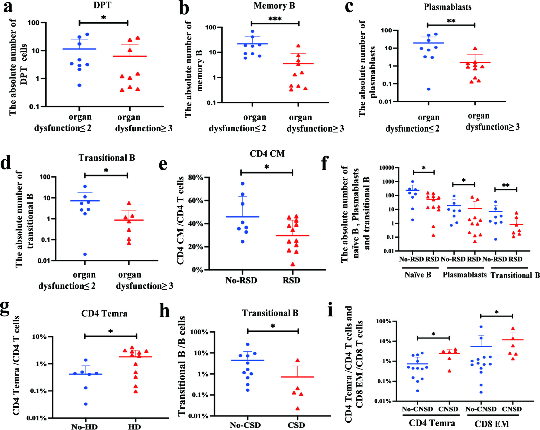
The absolute counts of DPT cells (P = 0.044), memory B cells (P = 0.001), plasmablasts (P = 0.007), and transitional B cells (P = 0.024) were significantly lower in the HLH group with ≥3 dysfunctional organs compared to those with ≤2 dysfunctional organs (Figure 3a-d). The relative counts of CD4+ CM T cells (P = 0.030), the absolute counts of naïve B cells (P = 0.017), plasmablasts (P = 0.025), and transitional B cells (P = 0.008) were significantly decreased in the group with RSD compared to those without (Figure 3e, f). The relative counts of CD4+ Temra T cells (P = 0.025) were significantly increased in the HD group compared to those without (Figure 3g). Additionally, the relative counts of transitional B cells (P = 0.043) were significantly decreased in the group with CSD compared to those without (Figure 3h). The relative counts of CD4+ Temra T cells (P = 0.013) and CD8+ EM T cells (P = 0.017) were significantly increased in the CNSD group compared to those without (Figure 3i).

Analysis of lymphocyte subsets in patients with HLH complicated by organ dysfunction. Comparison between the organ dysfunction ≤ 2 group and organ dysfunction ≥ 3 group (a, b, c, d). Comparison between those without respiratory system dysfunction (RSD) and those with it (e, f). Comparison between those without hepatic dysfunction (HD) and those with it (g). Comparison between those without circulatory system dysfunction (CSD) and those with it (h). Comparison between those without central nervous system dysfunction (CNSD) and those with it (i). Data are presented as mean ± SD with n = 20. *P < 0.05, **P < 0.01, and ***P < 0.001 compared with the organ dysfunction ≤ 2 group or no organ dysfunction group.
Correlation analysis between lymphocyte subsets and Laboratory examination
DNT cells (R = 0.701, P = 0.001) and TCRγδ+ T cells (R = 0.697, P = 0.001) showed a positive correlation with fibrinogen levels (Figure 4a, b). CD8+ CM T cells (R = -0.597, P = 0.005) exhibited a negative correlation with hemoglobin levels (Figure 4c). Transitional B cells (R = 0.455, P = 0.044) exhibited a positive correlation with the hemoglobin levels (Figure 4d). Memory B cells (R = 0.632, P = 0.003) and plasmablasts (R = 0.528, P = 0.017) showed a positive correlation with platelets counts (Figure 4e, f). CD8+ T cells were positively associated with AST (R = 0.677, P = 0.001), LDH (R = 0.464, P = 0.040), IL-6 (R = 0.615, P = 0.004), and TNF-α (R = 0.515, P = 0.020) (Figure 4g, i, k, m). CD8+ CM T cells were positively associated with AST (R = 0.703, P = 0.001), LDH (R = 0.617, P = 0.004), IL-6 (R = 0.781, P < 0.001), and TNF-α (R = 0.670, P = 0.001) (Figure 4h, j, l, n).

Correlation analysis of lymphocyte subsets and laboratory examination results. DNT cells (a) and TCRγδ+ T cells (b) positively correlate with fibrinogen levels. CD8+ CM T cells (c) demonstrate a negative correlation with hemoglobin levels. Transitional B cells positively correlate with hemoglobin levels (d); Memory B cells (e) and plasmablast (f) positively correlate with platelet counts; CD8+ T cells and CD8+ CM T cells are positively associated with AST, LDH, IL-6, and TNF-α (g-n).
DISCUSSION
Our comprehensive immunophenotypic analysis of pediatric HLH patients reveals profound and characteristic alterations in peripheral blood lymphocyte subsets, demonstrating both quantitative and compositional imbalances with significant clinical implications. Exposure to infections, malignant tumors, or autoimmune diseases disrupts immune homeostasis, resulting in an imbalance in lymphocyte subsets (Wang et al. 2024). This imbalance may severely impair pathogen clearance, potentially contributing to the uncontrolled immune activation characteristic of HLH.
The discovery of novel cell surface markers has enabled the identification of a growing number of lymphocyte subsets involved in the progression of HLH. For instance, the T lymphocyte subset CD4dimCD8+ T cells showed a substantial increase in secondary HLH and macrophage activation syndrome, and this increase demonstrated a strong correlation with the severity of the disease (De Matteis et al. 2022). These findings imply that performing a more comprehensive phenotypic analysis of lymphocyte subsets in HLH patients and dynamically monitoring these immune cells can contribute to a more precise assessment of disease progression and prognosis.
CD4⁺ T cells are functionally heterogeneous and can be categorized into subsets (including naïve, CM, EM, and Temra T cells) based on their differentiation state, function, and surface markers. CD4+ naïve cells are antigen-inexperienced and poised to differentiate, while CD4⁺ CM T cells mediate rapid systemic recall responses (Raphael et al. 2020). The depletion of CD4⁺ naïve and CD4⁺ CM T cells observed in this study indicates an impairment in cellular immunity. Crucially, CD8⁺ T cell dysfunction, characterized by the induction of an inflammatory cytokine storm, is a well-established driver of HLH pathogenesis (Griffin et al. 2020). During the immune paralysis phase of sepsis, CD8⁺ T cells undergo significant expansion, particularly the CD8⁺ CM T cells subset endowed with self-renewal and proliferative capacity (Sung et al. 2023). CD8⁺ CM T cells exhibit cytotoxic activity and directly kill target cells by releasing cytotoxic effector molecules such as perforin and granzyme B (Knorck et al. 2022). However, these expanded CD8⁺ CM T cells in such contexts often exhibit functional impairment or exhaustion, as evidenced by their strong correlation with key pro-inflammatory cytokines (IL-6, TNF-α) and tissue damage markers (AST, LDH) (Sung et al. 2023). This paradoxical expansion of potentially dysfunctional CD8⁺ CM T cells may contribute to, rather than control, the cytokine storm in conditions like HLH (Jensen et al. 2021).
Unconventional T cell populations also play crucial roles in immune regulation and defense. DPT cells perform effector and regulatory functions, which assist in immune defense and modulate disease progression. For example, they secrete IFN-γ and TNF-α to enhance the cytotoxicity of macrophages and NK cells (Hagen et al. 2023). DNT cells, especially the TCRαβ⁺ subset, can suppress inflammatory responses and alleviate tissue damage by regulating the activity of macrophages and B cells and exerting cytotoxic effects (Wu et al. 2022; Velikkakam et al. 2022). TCRγδ⁺ T cells rapidly initiate innate immune responses and eliminate infected cells through the secretion of cytokines (IFN-γ, IL-17) and chemokines (Chien et al. 2014). Research indicates that the development and maturation of B cells in HLH patients are significantly inhibited (Shim et al. 2023). Notably, this study found substantial decreases not only in CD4⁺ T cell subsets (naïve, CM, EM) and CD8⁺ naïve T cells but also in DPT cells, DNT cells (including both TCRαβ⁺ and TCRγδ⁺ subsets), and B cells (including naïve, memory, transitional, and plasmablasts subsets) in HLH patients. This result implies that this extensive impairment in adaptive immune function may be a key factor contributing to the dysregulated, compensatory systemic hyperinflammation characteristic of HLH. The underlying mechanisms of this lymphocyte depletion and its causal relationship with the hyperinflammatory state require further investigation. Therefore, for children with EBV infection, actively conducting multiparametric flow cytometry immune cell typing analysis to evaluate immune status and changes in lymphocyte subsets can provide a vital basis for ultra-early intervention in HLH. Furthermore, MODS is the leading cause of death in HLH patients (Goldman et al. 2018). Our study elucidates distinct immunophenotypic signatures associated with specific organ dysfunctions, providing potential mechanistic insights and diagnostic clues.
Although Temra T cells are at the terminal stage of differentiation, they retain the capacity for an immune response (Milner et al. 2020). This study revealed that the proportions of CD4+ Temra T cells were significantly increased in HLH patients with hepatic and central nervous system dysfunction. Moreover, in HLH patients with organ dysfunction ≥ 3 and respiratory dysfunction, the levels of naïve B cells, memory B cells, transitional B cells, plasmablasts, CD4+ CM T cells, and DPT cells in their peripheral blood were significantly decreased. These findings suggest that abnormalities in lymphocyte subsets may be associated with the development of organ dysfunction. Dynamic monitoring of alterations in these lymphocyte subsets in the peripheral blood of HLH patients may provide a reliable theoretical basis for the early detection of organ dysfunction.
Among the classic clinical symptoms of HLH-2004, decreased blood cell counts, reduced fibrinogen levels, and impaired organ function have been noted (Canna 2020). This study demonstrated that DNT and TCRγδ+ T cells were positively correlated with fibrinogen expression. CD8+ CM T cells were negatively correlated with hemoglobin levels. Memory B cells, plasmablasts, and transitional B cells were positively correlated with platelet and hemoglobin levels. CD8+ T cells and CD8+ CM T cells were positively associated with AST and LDH. In this study, DNT, TCRγδ+ T cells, memory B cells, plasmablasts, and transitional B cells were significantly down-regulated in HLH patients, while CD8+ T cells and CD8+ CM T cells were substantially up-regulated. These findings imply that these changes may be associated with the reduced fibrinogen and hemoglobin levels and with organ function damage observed in HLH. IL-6 and TNF-α are key factors contributing to the cytokine storm observed in HLH (Canna et al. 2020). CD8+ T cells and CD8+ CM T cells show a positive correlation with IL-6 and TNF-α levels, further highlighting the crucial role of CD8+ T cells in the pathogenesis of HLH. The consistency between the quantitative changes in specific lymphocyte subsets and the classic HLH indicators reflects the severity of systemic inflammation (Shim et al. 2023).
In conclusion, this study delineates a characteristic immunophenotypic landscape in pediatric HLH, marked by combined immunodeficiency and pathogenic expansion of CD8+ T cells (particularly the CM subset), which drives inflammation. The association between specific lymphocyte subset alterations and organ dysfunction, as well as classic HLH laboratory markers, highlights their potential as valuable biomarkers. Incorporating dynamic immunophenotypic profiling via multiparametric flow cytometry into clinical management offers a promising approach for enhancing early diagnosis, risk stratification for organ damage, and ultimately improving outcomes for children with this life-threatening condition. While the limited sample size precludes definitive conclusions on the impact of specific triggers and calls for larger validation studies, our findings provide a crucial experimental foundation for utilizing lymphocyte subset analysis in pediatric HLH management.
Acknowledgements
This research was supported by grants from the Zunyi City Science and Technology Plan Project (Grant Number: HZ [2023] 214).
References
- BERGSTEN E ET AL. 2017. Confirmed efficacy of etoposide and dexamethasone in HLH treatment: long-term results of the cooperative HLH-2004 study. Blood 130(25): 2728-2738.
- CANNA SW & MARSH RA. 2020. Pediatric hemophagocytic lymphohistiocytosis. Blood 135(16): 1332-1343.
- CHIEN Y, MEYER C & BONNEVILLE M. 2014. γδ T cells: first line of defense and beyond. Annu Rev Immunol 32: 121-55.
- DE MATTEIS A ET AL. 2022. Expansion of CD4dimCD8+ T cells characterizes macrophage activation syndrome and other secondary HLH. Blood 140(3): 262-273.
- EICHENAUER DA, LACHMANN G & LA ROSéE P. 2021. Hemophagocytic lymphohistiocytosis in critically ill patients. Med Klin Intensivmed Notfmed 116(2): 129-134.
- GOLDMAN J ET AL. 2018. Hepatobiliary Dysfunction and Disseminated Intravascular Coagulation Increase Risk of Mortality in Pediatric Hemophagocytic Lymphohistiocytosis. Pediatr Crit Care Med 19(10): e522-e530.
- GOLDSTEIN B, GIROIR B & RANDOLPH A. 2005. International pediatric sepsis consensus conference: definitions for sepsis and organ dysfunction in pediatrics. Pediatr Crit Care Med 6(1): 2-8.
- GRIFFIN G, SHENIO S & HUGHE GC. 2020. Hemophagocytic lymphohistiocytosis: An update on pathogenesis, diagnosis, and therapy. Best Pract Res Clin Rheumatol 34(4):101515.
- HAGEN M, PANGRAZZI L, ROCAMORA-REVERTE & WEINBERGER B. 2023. Legend or Truth: Mature CD4+CD8+ Double-Positive T Cells in the Periphery in Health and Disease. Biomedicines 11(10): 2702.
- HUANG P, HUANG C, XU H, LU J, TIAN R, WANG Z & CHEN Y. 2020. Early Use of Blood Purification in Severe Epstein-Barr Virus-Associated Hemophagocytic Syndrome. Pediatrics 145(6): e20193197.
- JENSEN IJ ET AL. 2021. Sepsis leads to lasting changes in phenotype and function of memory CD8 T cells. Elife 10: e70989.
- KEENAN C, NICHOLS KE & ALBEITUNI S. 2021. Use of the JAK Inhibitor Ruxolitinib in the Treatment of Hemophagocytic Lymphohistiocytosis. Front Immunol 12: 614704.
- KNORCK A, SCHAFER G, ALANSARY D, RICHTER J, THURNER L, HOTH M & SCHWARZ EC. 2022. Cytotoxic Efficiency of Human CD8+ T Cell Memory Subtypes. Front Immunol 13: 838484.
- LI X, YAN H, ZHANG X, HUANG J, XIANG ST, YAO Z, ZANG P, ZHU D, XIAO Z & LU X. 2020. Clinical profiles and risk factors of 7-day and 30-day mortality among 160 pediatric patients with hemophagocytic lymphohistiocytosis. Orphanet J Rare Dis 15(1): 229.
- MILNER JJ ET AL. 2020. Delineation of a molecularly distinct terminally differentiated memory CD8 T cell population. Proc Natl Acad Sci U S A 117(41): 25667-25678.
- PARAJULI B ET AL. 2021. Hemophagocytic Lymphohistiocytosis in a PICU of a Developing Economy: Clinical Profile, Intensive Care Needs, Outcome, and Predictors of Mortality. Pediatr Crit Care Med 22(1): e44-e57.
- RAPHAEL I, JOERN R R & FORSTHUBER TG. 2020. Memory CD4+ T Cells in Immunity and Autoimmune Diseases. Cells 9(3): 531.
- SHIM J ET AL. 2023. Early B-cell development and B-cell maturation are impaired in patients with active hemophagocytic lymphohistiocytosis. Blood 142(23): 1972-1984.
- STEEN EA, HERMISTON ML, NICHOLS KE & MEYER LK. 2021. Digenic Inheritance: Evidence and Gaps in Hemophagocytic Lymphohistiocytosis. Front Immunol 12: 777851.
- SUNG M, KIM JH, MIN H, JANG S, HONG J, CHOI BK, SHIN J, CHUNG KS & PARK YR. 2023. Three-dimensional label-free morphology of CD8+ T cells as a sepsis biomarker. Light Sci Appl 12(1):265.
- TROTTESTAM H ET AL. 2011. Chemoimmunotherapy for hemophagocytic lymphohistiocytosis: long-term results of the HLH-94 treatment protocol. Blood 118(17): 4577-4584.
- VELIKKAKAM T, GOLLOB KJ & DUTRA WO. 2022. Double-negative T cells: Setting the stage for disease control or progression. Immunology 165(4): 371-385.
- WANG SY, YANG NS & ZHANG H. 2024. Metabolic dysregulation of lymphocytes in autoimmune diseases. Trends Endocrinol Metab 35(7): 624-637.
- WU Z, ZHENG Y, SHENG J, HAN Y, YANG Y, PAN H & YAO J. 2022. CD3+CD4-CD8- (Double-Negative) T Cells in Inflammation, Immune Disorders and Cancer. Front Immunol 13: 816005.
- XU L, GUO X & GUAN H. 2022. Serious consequences of Epstein-Barr virus infection: Hemophagocytic lymphohistocytosis. Int J Lab Hematol 44(1): 74-81.
The data sets used and/or analyzed during the current study are available from the corresponding author on reasonable request.