Effect of vitamin K and calcium in bone loss in ovariectomized rats
Abstract
Osteoporosis compromises bone mineral composition and microarchitecture. Given the limited ability of current treatments to restore lost bone mass, prevention remains crucial in clinical practice. In the present study, we investigated the effect of vitamin K2, administered alone or in combination with calcium and vitamin D3, on ovariectomy-induced osteoporosis in rats. We assessed serum parameters, bone mineral content, bone strength, histomorphometry, radiography, and microtomography to evaluate trabecular volume and bone microarchitecture. Serum calcium levels were similar between ovariectomized rats supplemented with either vitamin K2 or calcium. Vitamin K2 protected against the loss of trabecular bone volume in ovariectomized rats. Supplementation with calcium and/or vitamin D3 showed inconsistent effects on bone mass. Histologic, microtomographic, and radiographic analyses provided similar results; however, microtomography was more effective in detecting differences between groups. In our study vitamin K2 helped in the treatment of bone loss, while calcium and vitamin D3 continued to demonstrate inconsistent effects.
Key words
Cholecalciferol; Menaquinone; Osteoporosis; Ovariectomy; Treatment
INTRODUCTION
Osteoporosis is a systemic skeletal disease characterized by decreased bone mass and deterioration of bone microarchitecture, leading to an increased risk of fractures (Camacho et al. 2020, Kulczyński et al. 2023, Feng et al. 2024). Given the crucial role of bone calcium stores in the pathogenesis of osteoporosis, calcium supplementation - alone or in combination with vitamin D - has become a common recommendation for postmenopausal women (Camacho et al. 2020). However, the low estrogen levels typically observed after menopause inhibit mineral fixation in the skeleton, even with increased calcium intake from dietary supplements (McClung et al. 2021, Gao et al. 2021, Ukon et al. 2019). In addition, estrogen has been shown to enhance the expression and functionality of the plasma membrane calcium ATPase 1b (PMCA1b) in duodenal mucosal cells via estrogen receptor beta (ERβ), thereby promoting duodenal calcium absorption and contributing to the improvement of postmenopausal osteoporosis (Wu et al. 2025).
Vitamin K2, also known as menaquinone (MK), has been shown to provide unique benefits in the prevention and treatment of osteoporosis (Khéde et al. 2017, Aaseth et al. 2024). Iwamoto et al. (2021) evaluated the effect of MK-4 and MK-7 administration on bone loss and the microstructural mechanical properties of bone in an ovariectomy-induced osteoporosis model in female Wistar rats. They found that MK-7 preserved bone microstructural properties in this model, potentially resulting in greater resistance to fractures. Ünal et al. (2023) examined the effects of MK-7 on fracture healing in male rats and found that vitamin K2 positively influences bone metabolism during the healing and promotes the formation of more durable callus tissue. Vitamin K2 plays a role in bone remodeling and bone health by activating proteins that promote bone mineralization, as well proteins containing gamma-carboxyglutamic acid-rich (GLA) domain (Cancela et al. 2012, Katsuyama et al. 2015, Aaseth et al. 2024). Furthermore, vitamin K2 may increase both the number and activity of osteoblasts (Urayama et al. 2000, Kim et al. 2013).
In animal models, ovariectomy decreases bone formation and trabecular thickness while increasing bone resorption and trabecular separation, mirroring the impact of low estrogen levels on bone loss observed in postmenopausal women. This confirms that ovariectomy is a suitable model for simulating postmenopausal bone loss (Kalu 1991, Thompson et al. 1995, Mishima et al. 2020). In this study, we aimed to investigate the effect of vitamin K2 supplementation, alone or associated with vitamin D3 and calcium, on bone mass in a rat model of osteoporosis.
Abbreviations
AI: Aluminium; AIN-93M: Standard diet for adult rodents’ maintenance; ANOVA: One-way analysis of variance; BV/TV: Trabecular volume; DRIs: Dietary reference intakes; DTO: Osteoporosis Therapeutic Guidelines; GLA: Gamma-carboxyglutamic acid-rich; HCl: Hydrochloric acid; IU: International unit; MK: Menaquinone; PBS: Phosphate buffered saline; Tb.N: Trabecular number; Tb.Sp: Trabecular separation; Tb/Th: Trabecular thickness.
MATERIALS AND METHODS
Animals
Sixty healthy 30-day-old female rats (Rattus norvegicus, Wistar strain, albinus variation), weighing 250–350 g, were obtained from the Central Animal House of the Federal University of Viçosa, Minas Gerais, Brazil. The animals were acclimated for three days under standard room conditions prior to the start of the experiment. Rats were fed a commercial diet and had ad libitum access to water and food. They were housed individually (one animal per cage) in an air-conditioned environment maintained at 22 ± 2°C with a 12 h light/12 h dark photoperiod. Based on sample size and allowable loss margin calculations (Mera et al. 1998), ten animals were allocated to each group. All experimental procedures were conducted in accordance with the ethical principles of animal experimentation. This study was approved by the Animal Research Ethics Committee of the Federal University of Viçosa (protocol number 068/2016).
Ovariectomy-induced osteoporosis
At 12 weeks of age, rats underwent ovariectomy (abdominal incision and removal of the ovaries). Thirty minutes prior to the procedure, the animals received subcutaneous doses of the anti-inflammatory flunixin meglumine (0.68 mg/kg) and the antibiotic enrofloxacin (10 mg/kg). The animals were then anesthetized with isoflurane diluted in 100% oxygen, administered via inhalation through a calibrated vaporizer. The isoflurane concentration was as needed to maintain adequate anesthesia. After anesthetic induction and stabilization, the animals were positioned in the supine position on a heated surgical pad. The operative field was prepared with povidone-iodine, and a 10% solution of morphine (5 mg/kg) was administered subcutaneously for analgesia. Following surgery, the animals were kept in a heated recovery chamber to maintain body temperature and were subsequently returned to individual cages. All anesthetic, surgical and post-surgical procedures were performed in the University Veterinary Hospital under the responsibility of a veterinarian.
The post-ovariectomy osteoporosis induction period lasted six weeks before the start of the intervention. During this period, the rats were housed individually and received a commercial pellet diet and water ad libitum.
Standard diet composition
The standard diet offered to the animals alongside the dietary treatments was formulated according to the recommendations described in AIN-93M (Reeves et al. 1993). Recommendations for the micronutrients analyzed in this study are as follows: vitamin K, 750 IU/kg diet; vitamin D3, 1.000 IU/kg diet; and calcium, 5.000 mg/kg diet. It is noteworthy that dietary recommendations for vitamin K refer to the use of vitamin K1. The AIN-93 diet is free of vitamin K2.
Supplement dosages
The supplements were added to the diet in powder and homogenized. The dosages used for calcium and vitamin D3 supplementation were proportional to the recommendations of the Osteoporosis Therapeutic Guidelines (DTO) (Brasil 2014) for humans. The DTO recommends supplementing calcium and vitamin D3 at 1× and 1.67× of their dietary reference intakes (DRIs), respectively (Ross et al. 2011). The recommended DRIs considered were for women aged 51 to 70 years, which includes the menopausal period, and the additional supplementation suggested by the DTO was calculated accordingly. The same calcium and vitamin D3 supplementation rations (1x and 1.67x, respectively) were applied to the AIN-93M diet to determine the supplementation levels for the experimental groups. There are no official nutritional recommendations for vitamin K2 intake in humans or animals. Therefore, the dosage of K2 supplementation was based on previous toxicity studies, which used doses ranging from 141 µg/kg of diet (Yamaguchi et al. 2000) to 201 mg/kg of diet (Fu et al. 2012). The dosage used in this study was the median of the lowest and highest dosages reported in those studies, corresponding to 12.5 mg/kg of diet. The experimental groups, the type and composition of diet received by each group, and the supplementation doses added in each experimental diet are detailed in Table I.
Experimental groups and euthanasia procedures
At 18 weeks of age, 6 weeks post-surgery, the rats were distributed by body weight into six groups of 10 animals each: (1) Control: AIN-93M - untreated control, (2) Ca: AIN-93M + calcium, (3) K2: AIN-93M + K2, (4) Ca+K2: AIN-93M + calcium + K2, (5) Ca + D3 / Ca + K2: AIN-93M + calcium + D3, and (6) Ca + D3 + K2: AIN-93M + calcium + K2 + D3. At the end of 4 weeks of treatment, five animals per group were euthanized, and the remaining five were euthanized at the end of 8 weeks, to evaluate the effects of supplementation duration on bone health. All groups received their respective diets throughout the experimental period, except for Group 5, which was switched from a calcium + vitamin D3 diet to a calcium + vitamin K2 diet at the 4-week mark. This procedure was designed to assess the effects of a treatment change on the outcome variables.
Animal weight and food intake were monitored weekly. Euthanasia was performed after a 12 h fasting period by exsanguination via cardiac puncture of animals anesthetized with isoflurane (Isoforine, Cristália®). Approximately 6 mL of blood was collected from the left ventricle using a needle. Blood samples were centrifuged at 3000 rpm and 4°C for 10 min. The right and left femurs were removed and washed in PBS buffer. Portions of each tissue were either stored in 10% formalin for histological examination or frozen at −80°C for further analysis.
Blood analyses
Serum levels of alkaline phosphatase, calcium, and magnesium were determined by colorimetry using a BS200 analyzer and specific kits supplied by Bioclin®.
Determination of bone mineral content
Right femur specimens were dried in a forced-air oven at 68–72°C for 72 h. The samples were then weighed and ground for bone mineral content determination. All glassware was cleaned and decontaminated with 2% HCl solution for 5 min and subsequently rinsed with deionized water. For acid digestion, a 4:1 ratio of nitric to perchloric acid solution was added to the material in digestion tubes, which were placed on a preheated plate at 80°C. The temperature was gradually increased to 200°C until the material became crystalline. Calcium and magnesium levels were determined by atomic absorption spectrophotometry, potassium by flame photometry, and phosphorus by the ascorbic acid colorimetric method (Altunay et al. 2019).
Bone strength
Bone strength was measured using a three-point bending test on a universal mechanical testing machine (Instron) equipped with Bluehill software. Each femur was positioned on the device with its ends resting on supports adapted to the size of the bone. Force was applied to the center of the diaphysis at a speed of 2 mm/min. The maximum load at failure, expressed in newton (N), was recorded by a computer system connected to the testing machine.
Bone histomorphometry
Right femur specimens were fixed in 10% formalin for 72 h and then stored in 70% ethanol until use. Samples were treated with a solution of sodium citrate and formic acid, which was replaced every 4 days for 12 days to ensure sufficient decalcification. Subsequently, the samples were rinsed under running water for 24 h and immersed in 5% sodium sulfate solution for an additional 24 h. Longitudinal sections of the epiphysis were prepared, dehydrated in a graded ethanol series (70%, 80%, 90%, and 100%%), treated with xylol, and embedded in paraffin using a Leica TP1020 apparatus. Then, 5 µm thick slices (6 per sample) were cut using a rotary microtome (Leica RM2245). Histological sections were oven-dried for 2 h, mounted on slides, deparaffinized in xylol, rehydrated in water and decreasing ethanol concentrations, and stained with hematoxylin and eosin. Histological images were captured using a light microscope (Zeiss, Scope A1) at 40× magnification with an image capture system (Zeiss AxioCam 105 Color) in randomly selected fields. Images were analyzed using ImageJ software by applying a standardized 266-point grid. Trabecular volume was measured in the region located just below the growth line.
Radiography
The left femurs of animals were radiographed. The size of analyzed areas (femur head) was consistent between samples. The equipment was operated using the following parameters: 44 kV, 100 mA, an exposure time of 0.07 s, and a distance of 1 m between the X-ray source and the radiographic film. An aluminum penetrometer (25 mm wide and 60 mm long) with five 3 mm-high steps was positioned next to the femurs to determine radiographic density. Radiographs were scanned and analyzed using ImageJ software. The images were converted to grayscale, and the gray levels of each step of the penetrometer were quantified to construct a curve for linear regression analysis. Density values expressed in millimeters of aluminum (mm AI), were generated for each femoral radiographs based on the corresponding penetrometer gray levels.
Microtomography
Microstructural quantification of the femur was performed using a high-resolution X-ray microtomograph (SkyScan 1174v2, Bruker) along with software provided by the manufacturer (NRecon, DataViewer, and CT-Analyzer). Images were acquired using a voltage of 50 kV, an electric current of 670 µA, a 0.5 mm aluminum filter, 360º rotation, and a resolution of 18.11 µm. Fifty slices of trabecular bone, located at 10 slices below the growth plate, were analyzed. The following parameters were measured: trabecular volume (BV/TV), trabecular thickness (Tb/Th), trabecular number (Tb.N), trabecular separation (Tb.Sp) and connectivity density (Conn.D).
Statistical analysis
The data were first subjected to a Kolmogorov–Smirnov normality test. Subsequently, results were analyzed using one-way analysis of variance (ANOVA) at a 5% significance level, followed by the Newman-Keuls post hoc test to assess differences between experimental groups. Within-group comparisons between weeks 4 and 8 were performed using Student’s t-test. The level of significance was set at p < 0.05. Data are presented as mean ± standard deviation. Statistical analysis was conducted using the GraphPad Prism software, version 9.0.
RESULTS
Body weight and food intake
Final body weight did not differ among the experimental groups at the end of week 4 or at the end of week 8. Regarding supplementation duration, only rats supplemented with Ca + K2 showed an increase in body weight over time (Table II).
Body weight and food intake of rats after 4 and 8 weeks under different supplementation conditions.
At the end of week 4, animals supplemented with Ca + D3 (prior to the treatment change) showed lower food intake compared to the groups supplemented with Ca, K2 and Ca+K2+D3. By the end of week 8, there were no differences in food intake among the groups. However, regarding supplementation time, the group that received Ca + D3/Ca + K2 (after treatment change) showed higher food intake at the end of week 8 (Table II).
Blood analysis
Serum calcium analysis (Table III) at week 4 showed that the only difference between groups was that the group supplemented with Ca+K2+D3 had lower serum calcium levels than the group supplemented with K2. At week 8, the groups supplemented with calcium or K2 had higher serum calcium levels compared to the other groups. Supplementation duration did not affect serum calcium levels. At week 4, the group supplemented with Ca+D3 (before the treatment change) showed the highest serum magnesium levels, while the group supplemented with Ca+K2+D3 showed the lowest. By week 8, magnesium levels did not differ among the groups (Table III). Regarding the supplementation time, the group supplemented with Ca+D3/Ca+K2 had lower serum magnesium levels at week 8 compared to week 4. Alkaline phosphatase levels did not differ between groups at either week 4 or week 8 (Table III). However, in terms of the supplementation time, animals without supplementation (control) and those supplemented with Ca+D3/Ca+K2 showed higher alkaline phosphatase levels at week 8 than at week 4.
Serum levels of calcium, magnesium, and alkaline phosphatase in ovariectomized rats after 4 and 8 weeks under different supplementation conditions.
Bone mineral content
There were no differences in bone phosphorus, potassium, calcium or magnesium levels among the experimental groups at the end of week 8 (Table IV).
Bone mineral content in the femur of rats after 8 weeks and bone strength of femur after 4 and 8 weeks under different supplementation conditions.
Bone strength
There was no difference among the groups in maximum bone compression strength at the end of week 4 or at the end of group 8. Additionally, no differences were observed with respect to supplementation time (Table IV).
Bone histomorphometry
There were no differences among the groups in bone histomorphometry at the end of week 4 or at the end of group 8. Trabecular volume decreased over time in Ca and Ca + K2 groups (Figure 1 and Table V).
Femoral histological analysis of the percentage of trabecular bone and bone density based on radiographs of the femurs, in rats after 4 and 8 weeks under different supplementation conditions
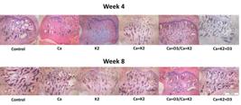
Hematoxylin and eosin-stained histological slides of the femur of rats after 4 and 8 weeks under different dietary conditions. Ca: calcium supplementation; K2: vitamin K2 supplementation; D3: vitamin D3 supplementation.
Radiography
At 4 weeks of treatment, Ca + K2 and Ca + K2 + D3 supplementation increased bone density, while Ca + D3 treatment reduced bone density. After 8 weeks of treatment, radiological analysis did not detect any differences between groups (Table V).
Microtomography
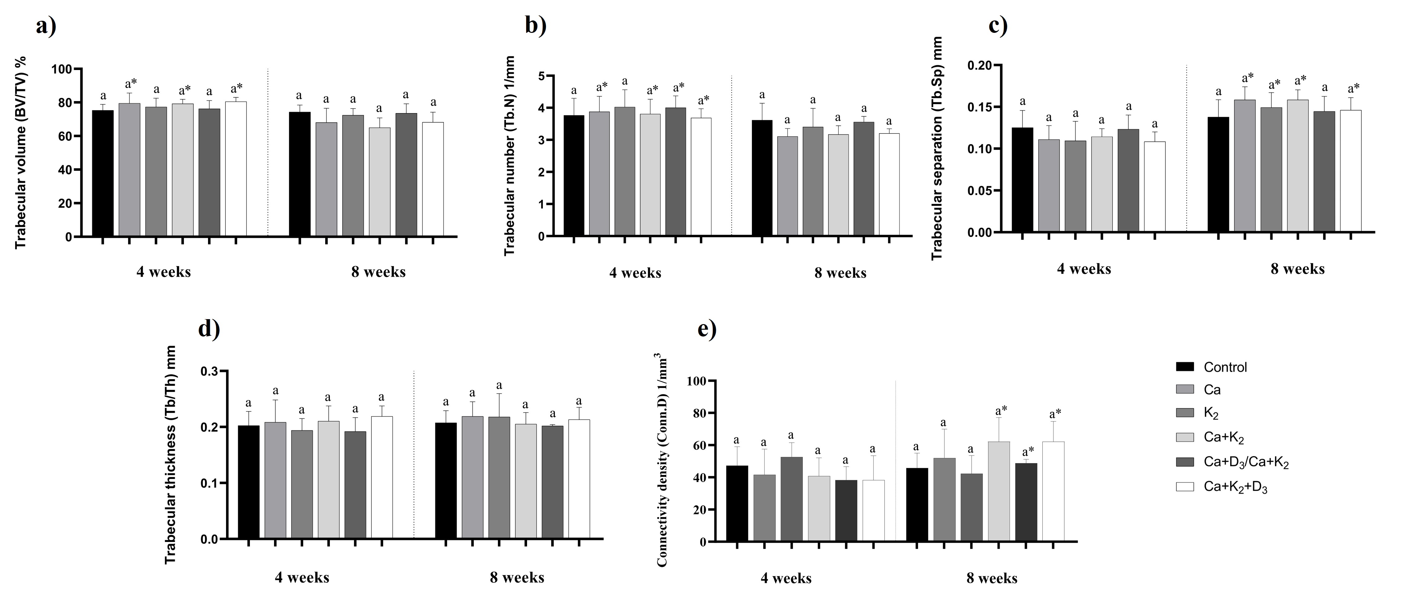
There were no differences in trabecular volume, trabecular number, connectivity density, trabecular thickness, or trabecular separation among the groups at either week 4 or week 8 (Figures 2 and 3). Regarding supplementation time, in the groups that received Ca, Ca+K2 or Ca+K2+D3, trabecular volume and trabecular number were higher at week 4 than at week 8, while trabecular separation was higher in week 8 than at week 4. The group that received Ca+D3/Ca+K2 also showed a higher trabecular number at week 4 (when the animals received only Ca+D3 supplementation) compared to week 8 (after switching to Ca+K2). In the group that received K2, trabecular separation was higher at week 8 than in week 4. Trabecular thickness didn’t change over time. Regarding connectivity density, the groups that received Ca and vitamin K2 (Ca+K2, Ca+D3/Ca+K2 and Ca+K2+D3) showed higher connectivity density at week 8 than at week 4 (Figures 2 and 3).
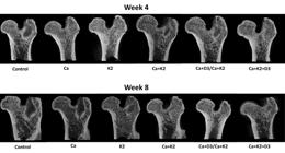
Microtomography analysis of standardized portion of the femur of rats from different experimental groups. Ca: calcium supplementation; K2: vitamin K2 supplementation; D3: vitamin D3 supplementation.

Microtomography analysis of femur. Ca: calcium supplementation; K2: vitamin K2 supplementation; D3: vitamin D3 supplementation. a) Trabecular volume (BV/TV); b) Trabecular number (Tb.N); c) Trabecular separation (Tb.Sp); d) Trabecular thickness (Tb/Th) and e) Connectivity density(Conn.D). Differences between groups were analyzed using ANOVA followed by Newman-Keuls post hoc test and the same small letters (a) means no significant differences at 5% threshold of probability. t-test was conducted to analyze differences within groups at weeks 4 and 8 and * means significant differences (p < 0.05). Data expressed as mean ± standard deviation.
DISCUSSION
Vitamin K2 supplementation is still not widely used in clinical practice for the treatment of osteoporosis, but it has shown promising results, as demonstrated in the present study. Calcium and vitamin D3 are commonly used in osteoporosis treatment, and in our study, we used dosages proportionally similar to those recommended for the treating the disease in Brazil. This study evaluated, in an animal model, the effect of vitamin K2 supplementation - alone or associated with calcium and vitamin D3 - on bone mass in a rat model of osteoporosis. Ovariectomy is well established as a reliable model for simulating postmenopausal bone loss in animals, as it induces typical osteoporotic changes such as reduced bone formation and trabecular thickness, along with increased bone resorption and trabecular separation (Kalu 1991, Thompson et al. 1995, Wu et al. 2014, Mishima et al. 2020).
In our study, the animals’ final body weight and food intake were similar across all groups. At week 4, it was observed that the group supplemented with the combination of Ca+K2+D3 had lower serum calcium levels than the group supplemented with vitamin K2 alone, and lower serum magnesium levels than the groups that received combined vitamin K2 (Ca+K2 or Ca+D3/Ca+K2). In the group where supplementation was changed, replacing vitamin D3 with K2 let to a reduction in serum magnesium levels and an increase in alkaline phosphatase over time. At week 8, the groups supplemented with vitamin K2 alone or Ca alone showed higher serum calcium levels compared to the other groups. The literature indicated that the increase in serum calcium levels resulting from supplementation is not associated with increased bone mineral density, does not provide relevant protection against fractures and is associated with an increased risk of coronary artery disease (Cerani et al. 2019, Myung et al. 2021, Migliorini et al. 2025).
At week 4, the groups did not receive vitamin K2 had lower bone density. Furthermore, the group that received only Ca+D3 had the lowest bone density among all groups. However, when vitamin D3 was replaced by vitamin K2 at week 8, this difference was no longer observed. An important finding of this study was the reduction in bone mass over time in animals treated with calcium. This aligns with concerns raised in the literature suggesting that the protective effects of calcium supplement on bone health in the general population are not entirely clear and may not, in fact, provide benefits for bone health outcomes (Cerani et al. 2019). We emphasize that, overall, supplementation with vitamin K2 – either alone or in combination with calcium and/or vitamin D3 (throughout the entire period or for only 4 weeks) - was effective in preventing bone loss (Iwamoto et al. 2021, Aaseth et al. 2024). This is consistent with the findings of AlHajri et al. (Alhajri et al. 2021), who found that the administration of vitamin K2 alongside vitamin D and calcium, rather than each one alone, is likely to be more beneficial.
The isolated administration of calcium led to a reduction in trabecular volume and number, along with an increase in the spacing between trabeculae over time. In contrast, the group that received vitamin K2 alone did not show a decrease in trabecular volume or number. Similarly, in the group in which supplementation was switched from D3 to K2 (Ca+D3/Ca+K2), no reduction in trabecular volume or increase in spacing was observed. However, when vitamin K was supplemented with calcium (Ca+K2) or with both calcium and vitamin D3 (Ca+D3+K2), it was not sufficient to prevent the decrease in trabecular volume and number or the increase in spacing between trabeculae. Importantly, animals whose supplementation was changed at week 4 (from Ca + D3 to Ca + K2) did not exhibit the same bone mass reduction pattern observed in the groups not treated with K2. Thus, the change in supplementation helped prevent the reduction in bone volume and the increase in trabecular spacing, suggesting that switching from vitamin D3 to K2 may have had a beneficial effect. Additionally, for connectivity density - a computational measure of trabecular interconnectivity and a potential indicator of the mechanical strength of the trabecular architecture (Ohs et al. 2020) - the groups that received calcium and vitamin K2 (Ca+K2, Ca+D3/Ca+K2, and Ca+K2+D3) presented higher values at week 8 than at week 4. This finding indicates improved structural integrity of the trabecular bone over time. The increase in connectivity density reflects enhanced interconnection among trabeculae, which strengthens the bone microarchitecture. This suggests that supplementation with calcium and vitamin K2—either alone or in combination with vitamin D3—had a positive and progressive effect on trabecular bone quality. In osteoporosis models, such a response may indicate structural recovery of the bone or attenuation of bone loss associated with estrogen deficiency.
The observed bone loss in calcium-supplemented groups over time may be associated with competition among calcium, magnesium, and phosphorus for intestinal absorption, as these minerals are crucial for bone mineralization (Mott et al. 2022). Excess calcium may act as an antagonist to phosphorus and magnesium absorption by forming insoluble chelates, thereby reducing their bioavailability and potentially causing secondary deficiencies (Vellasco et al. 2016, McClung et al. 2021).
Most studies use the MK-4 form vitamin K2 at doses ranging from 30 to 45 mg/day. Although MK-7 is usually administered at much lower doses, its effects are generally comparable to those of MK-4. In this study, we used MK-7 at a dose of 12.5 mg/kg diet equivalent to 180 µg/ animal/ day. Wu et al. (2014) treated ovariectomized rats with 2, 4, or 8 µg/day of MK-7 or cheonggukjang - a fermented soybean paste used in Korean cuisine containing the same amounts of MK-7 - and observed reductions in alkaline phosphatase levels and prevention of changes in trabecular volume. In our study, although vitamin K2 did not reduce alkaline phosphatase, it did prevent changes in trabecular volume. A review supports the role of vitamin K2 in modulating bone metabolism by enhancing the expression and/or synthesis of key bone biomarkers. Vitamin K2 promotes bone formation by increasing osteoblast differentiation and reducing their apoptosis, while also decreasing bone resorption by inhibiting osteoclast differentiation (Khéde et al. 2017).
Our results did not support the effectiveness of calcium supplementation alone for osteoporosis treatment, as commonly recommended in clinical practice and by the Brazilian Osteoporosis Therapeutic Guidelines (DTO), given that calcium supplementation actually exacerbated bone loss. The Auckland Calcium Study found no relationship between dietary calcium intake (ranging from 400 to 1,500 mg/day) and the rate of bone loss over five years in healthy elderly women. Within this intake range, the authors concluded that calcium supplements were unnecessary to compensate for a possible dietary deficiency. As in our study, they found no antifracture efficacy of calcium supplements (except in individuals with severe vitamin D deficiency). Moreover, calcium supplementation was associated with a 17% increase in kidney stones and a 20–40% increase in the risk of myocardial infarction, which the authors argue outweighs any potential benefit in fracture prevention (Reid et al. 2015). Other researchers have suggested that the benefits of calcium supplementation may only occur when dietary calcium intake is deficient, noting that 1,000–1,200 mg/day of dietary calcium appears sufficient to prevent general fractures (Ströhle et al. 2015, Zhang & Feng 2017). The systematic review by Migliorini et al. (2025) investigated whether different doses of vitamin D and calcium supplementation in postmenopausal women with osteoporosis undergoing antiresorptive therapy are associated with changes in bone mineral density, serum markers of osteoporosis, fracture rate, adverse events, and mortality. The study found that calcium supplementation showed no association with any of the outcomes of interest.
The main contribution of this article was that we used the Brazilian guidelines for the treatment of osteoporosis (DTO) as a basis for calculations of calcium and vitamin D3 supplementation for the groups of animals, and made the adaptations, adding vitamin K2 to test whether the results would be better or worse than just following current guidelines. We conclude that, in an ovariectomized rat model, there are benefits to adding vitamin K2 to the current recommendations by guidelines (calcium and vitamin D3 supplementation), and these benefits are evident even with isolated vitamin K2 supplementation. It is important to note that, although the ovariectomy procedure was performed following established protocols and has been widely validated in the literature as a reliable model for simulating postmenopausal estrogen deficiency (Kalu 1991, Thompson et al. 1995), we did not include direct physiological confirmation of estrogen depletion, such as estrous cycle monitoring, uterine weight assessment, or serum hormone measurements. While the primary aim of this study was to evaluate the effects of vitamin K2, alone or in combination with vitamin D3 and calcium, on bone mass under conditions of estrogen deficiency, we recognize this as a limitation. This was a study conducted with animals and does not allow us to extrapolate the results to application in humans, but it can give us direction for future efforts.
CONCLUSIONS
In conclusion, this study suggests that administration of vitamin K2 (MK-7) for 8 weeks, either alone or in combination with calcium and vitamin D3 supplements, protects against the loss of trabecular bone volume and bone calcium content in rats with ovariectomy-induced osteoporosis. Our findings also indicate that calcium supplementation, when not accompanied by vitamin K2, may worsen bone microarchitecture. The effects of vitamin D3 supplementation on bone mass were inconsistent.
Acknowledgements
The study was supported by the Conselho Nacional de Desenvolvimento Científico e Tecnológico (CNPq), the Coordenação de Aperfeiçoamento de Pessoal de Nível Superior (CAPES, Finance Code 001) and Fundação de Amparo à Pesquisa do Estado de Minas Gerais (FAPEMIG).
References
- AASETH JO, FINNES TE, ASKIM M & ALEXANDER J. 2024. The Importance of Vitamin K and the Combination of Vitamins K and D for Calcium Metabolism and Bone Health: A Review. Nutrients 16: 1-15.
- ALHAJRI L, AYOUB A, AHMED H & ALMULLA M. 2021. Effect of Vitamin K 2 Alone or in Combination on Various Bone Turnover Markers Amongst Postmenopausal Females. J Taibah Univ Med Sci 28: 11-26.
- ALTUNAY N, ELIK A & GÜRKAN R. 2019. Monitoring of some trace metals in honeys by flame atomic absorption spectrometry after ultrasound assisted-dispersive liquid liquid microextraction using natural deep eutectic solvent. Microchem Journal 147: 49-59.
- BRASIL. 2014. Ministério da Saúde. Secretaria de Atenção à Saúde. Portaria nº 224, de 26 de março de 2014. Aprova o Protocolo Clínico e Diretrizes Terapêuticas da Osteoporose. Brasília: Ministério da Saúde.
- CAMACHO PM ET AL. 2020. American association of clinical endocrinologists/American college of endocrinology clinical practice guidelines for the diagnosis and treatment of postmenopausal osteoporosis-2020 update. Endocr Pract 26: 1-46.
- CANCELA ML, CONCEIÇÃO N & LAIZÉ V. 2012. Gla-rich protein, a new player in tissue calcification? Adv Nutr 3: 174-181.
- CERANI A, ZHOU S, FORGETTA V, MORRIS JA, TRAJANOSKA K, RIVADENEIRA F, LARSSON SC, MICHAËLSSON K & RICHARDS JB. 2019. Genetic predisposition to increased serum calcium, bone mineral density, and fracture risk in individuals with normal calcium levels: mendelian randomisation study. BMJ 366: l4410.
- FENG B, LU J, HAN Y, HAN Y, QIU X & ZENG Z. 2024. The role of short-chain fatty acids in the regulation of osteoporosis: new perspectives from gut microbiota to bone health: Medicine (Baltimore) 103: e39471.
- FU X, MOREINES J & BOOTH SL. 2012. Vitamin K supplementation does not prevent bone loss in ovariectomized Norway rats. Nutr Metab (Lond) 9: 1-5.
- GAO Y, PATIL S & JIA J. 2021. The development of molecular biology of osteoporosis. Int J Mol Sci 22: 1-23.
- IWAMOTO D, MASAKI C, SHIBATA Y, WATANABE C, NODAI T, MUNEMASA T, MUKAIBO T, KONDO Y & HOSOKAWA R. 2021. Microstructural and mechanical recovery of bone in ovariectomized rats: The effects of menaquinone-7. J Mech Behav Biomed Mater 120: 104570.
- KALU DN. 1991. The ovariectomized rat model of postmenopausal bone loss. Bone Miner 15: 175-191.
- KATSUYAMA H, FUSHIMI S, YAMANE K, WATANABE Y, SHIMOYA K, OKUYAMA T, KATSUYAMA M, SAIJOH K & TOMITA M. 2015. Effect of vitamin K2 on the development of stress-induced osteopenia in a growing senescence-accelerated mouse prone 6 strain. Exp Ther Med 10: 843-850.
- KHÉDE J, VILLA D, ALVES M, DIAZ N, RAMOS V, STAMPINI H, MARTINO D & KH J. 2017. Effect of vitamin K in bone metabolism and vascular calcification: A review of mechanisms of action and evidences mechanisms of action and evidences. Crit Rev Food Sci Nutr 57: 3959-3970.
- KIM M, NA W & SOHN C. 2013. Vitamin K1 (phylloquinone) and K2 (menaquinonee4) supplementation improves bone formation in a high-fat diet induced obese mice. J Clin Biochem Nutr 53: 108-113.
- KULCZYŃSKI B, SIDOR A, BRZOZOWSKA A & GRAMZA-MICHAŁOWSKA A. 2023. The role of carotenoids in bone health – a narrative review. Nutr 119: 112306.
- MCCLUNG MR, PINKERTON JV, BLAKE J, COSMAN FA, LEWIECKI EM & SHAPIRO M. 2021. Management of osteoporosis in postmenopausal women: the 2021 position statement of The North American Menopause Society. Menopause 28: 973-997.
- MERA R, THOMPSON H & PRASAD C. 1998. How to Calculate Sample Size for an Experiment: A Case-Based Description. Nutr Neurosci 1: 87-91.
- MIGLIORINI F, MAFFULLI N, COLAROSSI G, FILIPPELLI A, MEMMINGER M & CONTI V. 2025. Vitamin D and calcium supplementation in women undergoing pharmacological management for postmenopausal osteoporosis: a level I of evidence systematic review. Eur J Med Res 30: 170.
- MISHIMA MDV, PEREIRA B, LOPES RC, BRUNORO NMC & MARTINO HSD. 2020. Bioavailability of Calcium from Chia (Salvia hispanica L.) in Ovariectomized Rats Fed a High Fat Diet. J Am Coll Nutr 39: 1-11.
- MOTT A, BRADLEY T, WRIGHT K, COCKAYNE ES, SHEARER MJ, ADAMSON J, LANHAM-NEW SA & TORGERSON DJ. 2022. Effect of Vitamin K on Bone Mineral Density and Fracture Risk in Adults: Systematic Review and Meta-Analysis. Biomedicines 10: 1543-1559.
- MYUNG S-K, KIM H-B, LEE Y-J, CHOI Y-J & OH S-W. 2021. Calcium Supplements and Risk of Cardiovascular Disease: A Meta-Analysis of Clinical Trials. Nutr 13: 3682.
- OHS N, COLLINS CJ & ATKINS PR. 2020. Validation of HR-pQCT against micro-CT for morphometric and biomechanical analyses: A review. Bone Rep 13: 3682.
- REEVES PG, NIELSEN FH & FAHEY GC. 1993. AIN-93 Purified Diets for Laboratory Rodents: Final Report of the American Institute of Nutrition Ad Hoc Writing Committee on the Reformulation of the AIN-76A Rodent Diet. J Nutr 123: 1939-1951.
- REID IR, BRISTOW SM & BOLLAND MJ. 2015. Calcium supplements: Benefits and risks. J Intern Med 278: 354-368.
- ROSS AC ET AL. 2011. The 2011 report on dietary reference intakes for calcium and vitamin D from the Institute of Medicine: What clinicians need to know. J Clin Endocrinol Metab 96: 53-58.
- STRÖHLE A, HADJI P & HAHN A. 2015. Calcium and bone health - Goodbye, calcium supplements? Climacteric 18: 702-714.
- THOMPSON DD, SIMMONS HA, PIRIE CM & KE HZ. 1995. FDA guidelines and animal models for osteoporosis. Bone 17: 125-133.
- UKON Y, MAKINO T, KODAMA J, TSUKAZAKI H, TATEIWA D, YOSHIKAWA H & KAITO T. 2019. Molecular-Based Treatment Strategies for Osteoporosis: A Literature Review. Int J Mol Sci 20: 2557.
- ÜNAL ÖK, DAĞTAŞ MZ, İPEK BÖ, SITAR ME & UĞUTMEN E. 2023. Morphological and biomechanical effects of vitamin K2 on fracture healing: An animal study on the rat tibia fracture model. Acta Orthop Traumatol Turc 57: 17-22.
- URAYAMA S ET AL. 2000. Effect of vitamin K2 on osteoblast apoptosis: Vitamin K2 inhibits apoptotic cell death of human osteoblasts induced by Fas, proteasome inhibitor, etoposide, and staurosporine. J Lab Clin Med 136: 181-193.
- VELLASCO CR, GOMES PC, DONZELE JL, ROSTAGNO HS, CALDERANO AA, DE CARVALHO MELLO HH & PASTORE SM. 2016. Níveis de cálcio e relação cálcio: Fósforo em rações para poedeiras leves de 24 A 40 semanas de idade. Cienc Anim Bras 17: 206-216.
- WU WJ, LEE HY, LEE GH, CHAE HJ & AHN BY. 2014. The antiosteoporotic effects of cheonggukjang containing vitamin K2 (Menaquinone-7) in ovariectomized rats. J Med Food 17: 1298-1305.
- WU Y, GUO X, JIANG A, BAI J & NIE X. 2025. Estrogen regulates duodenal calcium absorption and improves postmenopausal osteoporosis by the effect of ERβ on PMCA1b. Sci Rep 15: 32761.
- YAMAGUCHI M, KAKUDA H, GAO YH & TSUKAMOTO Y. 2000. Prolonged intake of fermented soybean (natto) diets containing vitamin K2 (menaquinone-7) prevents bone loss in ovariectomized rats. J Bone Miner Metab 18: 71-76.
- ZHANG Y & FENG B. 2017. Systematic review and meta-analysis for the association of bone mineral density and osteoporosis/osteopenia with vascular calcification in women. Int J Rheum Dis 20: 154-160.
Edited by
-
Handling editor
Denise Brentan
Data will be made available upon reasonable request.