Developmental defects in planarian hatchlings (Platyhelminthes: Rhabditophora)
ABSTRACT
Developmental plasticity of the planarians is well-illustrated by whole-body regeneration from any tissue fragments and body size scaling regulated by food intake. Here we report another form of developmental plasticity that appears to be shared by multiple planarian species. We have observed developmental defects at rates up to ~10% in batches of Schmidtea mediterranea (Benazzi, 1975) and Schmidtea polychroa (Schmidt, 1862) hatchlings. The most frequent defect is duplicated body parts which occurs in both two species, which can be corrected after amputation and regeneration, supporting that these developmental defects were not caused by genetic mutations. Taken together, our data described a new form of developmental plasticity and an interesting comparison between development and regeneration in the hatchlings of freshwater planarians, that the two processes can have different outcomes.
KEY WORDS:
Embryonic development; hatchlings; mutants; planarians; regeneration
INTRODUCTION
Embryonic developmental defects occur throughout the animal kingdom. In humans, declining oocyte quality over age leads to chromosome segregation errors. Surprisingly, the first mitotic division of the zygotes also has a high error rate leading to aneuploidy and embryonic development defects (Delhanty 2005, Mantikou et al. 2012, Schneider and Ellenberg 2019). In animals that perform spawning before fertilization, the embryonic development defect rate seems even higher (Gowland et al. 2002). While eggs almost always hatch in the copepod, Pseudocalanus newmani (Frost, 1989), embryonic abnormality can be as high as 20~40% of the hatchlings (Ban et al. 2000). Why the development process is prone to errors is unclear.
Planarians are great models for the study of adult stem cells and whole-body regeneration (Newmark and Alvarado 2002, Reddien and Alvarado 2004, Saló and Agata 2012). In the past few decades, lots of progress was made on the characterization of adult stem cells (Tanaka and Reddien 2011) and signals guiding the patterning of regenerating tissues (Reddien 2022). While it is well established that regeneration uses pathways important for embryonic development to build tissues in an adult body, comprehensive comparisons remain elusive in planarians. One reason for the slow progress is due to our poor understanding of planarian embryonic development.
Limited work on planarian embryonic development extended from morphological characterizations in 1880s (Metchnikoff 1883, Ijima 1884, Hallez 1887, Sakurai 1991, Alvarado 2003, Martín-Durán et al. 2012a) to molecular studies in 2010s (Alvarado 2003, Martín-Durán et al. 2010, 2012a, 2012b, Davies et al. 2017, Monjo and Romero 2015, Solana and Romero 2009). These studies revealed that planarian embryonic development deviates from standard spiral cleavage. At the early stage of development, planarian zygotes are surrounded by yolk cells, which will form a syncytium at 4-cell stage (Sakurai 1991). Such an embryonic primordium will then proliferate and differentiate into a “cryptic larva”, possessing an embryonic pharynx, which pumps external yolk cells into its interior, and is resorbed at mid-stage of development as the definitive pharynx is formed (Cardona et al. 2006). Interestingly, molecular studies implicated shared programs regulating development of pharynx (Koinuma et al. 2000, Martín-Durán and Romero 2011, Adler et al. 2014), eyes (Martín-Durán et al. 2012b), and nervous system (Cebrià et al. 2002, Cowles et al. 2013, Monjo and Romero 2015, Roberts-Galbraith et al. 2016,) in the embryos, and in the regeneration of the adults. In addition, the planarian homologue of piwi-1 gene marks both proliferating pluripotent blastomeres in the embryos and the pluripotent stem cells in the adult (Reddien et al. 2005, Davies et al. 2017), providing additional support for the value to compare development and regeneration in planarians.
Herein, we reported developmental defects in two species of sexual planarians, Schmidtea mediterranea (Benazzi, 1975) and Schmidtea polychroa (Schmidt, 1862), and asked if regeneration of these defective hatchlings will reproduce the errors caused by development or correct such errors leading to a properly patterned hatchling. In all defective hatchlings that are big enough to regenerate lost tissues, we observed that the newly regenerated tissues were free from developmental errors.
MATERIAL AND METHODS
Planarian culture
All planarians were maintained in 1x Montjuïc salts, in plastic containers, at 18 °C, with once a week feeding of organic cow liver as previously described (Cebrià and Newmark 2005). Egg capsules were collected weekly and maintained in petri dishes (60 x 15 mm, Fisherbrand) with 1x Montjuïc salts. Hatchlings were examined two weeks after egg capsule collection. Line LAF (Los Angeles Fertile) was an inbred line established from a progeny produced by crossing D2E and S2F8b, two lines with high fecundity. Animals reported in this study that produce abnormal hatchlings were not amputated routinely. No gentamicin was used in culture unless noted specifically in text. The ages of these parental animals range from four months old to 15 months old. Animals were maintained in incubators without light cycles. Animals were exposed to light only during feeding and water changes. A medium Tupperware (10.80 cm W x 10.80 cm L x 7.77 cm H, 473 mL, Ziploc) houses 5~10 animals, and a large Tupperware (14.91 cm W x 22.86 cm L x 8.25 cm H 2130 mL Ziploc) houses 20~50 animals.
Abnormal hatchling identification
The ratio is calculated by dividing number of abnormal hatchlings to number of total hatchlings. The frequency varies between culturing conditions (e.g., with or without gentamycin) and between genetic backgrounds (e.g., inbreeding, no inbreeding, or diverse mating schemes). For example, the inbred strain S2F8b (Guo et al. 2016) or its more inbred progeny S2F12 (Dai et al. 2023) has very low frequency of abnormal hatchlings (e.g., 1 out of 64 in a most recent batch). A newly established inbred line, LAF, had 2 abnormal hatchlings out of 84 in a recent collection. The mixed populations have much higher frequency of abnormal hatchling, especially when maintained with gentamycin (e.g., eight out of 81 in a documented note). In a mixed population of hatchlings from the S2 lineage, one observation noted four out of 79.
Another important note for the observation is that the numbers are recorded based on newly hatched individuals. The abnormal hatchlings require special attention to identify as most of them are around 1.0 mm in sizes and are nearly transparent. Some of them die within a week. If observations are made one to two weeks after hatching, the frequency of abnormal hatchlings will be much lower or zero.
In Schmidtea polychroa, the abnormal hatchlings can be relatively bigger (~2 mm2 or bigger) and are much easier to identify.
Species identification
Planarian species were identified by morphology, chromosome karyotyping (Guo et al. 2018), genome sequencing, and compared to known features of S. mediterranea and S. polychroa.
Imaging
Images were taken with Leica S9i and processed by Fiji (Schindelin et al. 2012) and Adobe PhotoshopTM. Animal sizes were measured as total area of the animal in Fiji.
RESULTS
Hatchling sizes vary in the freshwater planarian
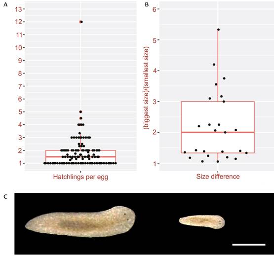
Within the gravid egg capsules, 1 to 10 embryos may be found in S. mediterranea (Guo et al. 2016). The number of embryos per egg capsule varies among different inbred lines. In line LAF, out of 110 hatched egg capsules, we found on average 1.8 embryos per egg capsule (Fig. 1A, Supplementary Table S1).
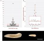
Hatchlings of the planarian, Schmidtea mediterranea: (A) number of hatchlings per egg capsule, total egg capsules quantified = 110; (B) sizes of the biggest hatchling divided by sizes of the smallest hatchling from the same egg capsule; (C) an example of two normal hatchlings from the same egg capsules that are of different sizes. Scale bar: 1 mm.
Hatchlings coming out of the same egg capsule can vary significantly in sizes, suggesting embryos within one egg capsule can develop at strikingly different rate. We examined the size of hatchlings that came out of the same egg capsules. Out of a total of 61 hatchlings that came out of a total of 25 egg capsules, the biggest hatchling can be 5.3 times the sizes of the smallest hatchling from the same egg capsule. Only two out of the 25 egg capsules gave rise to hatchlings of the same sizes. On average, the biggest hatchling was 2.2 times the sizes of the smallest hatchling from the same egg capsule (Fig. 1B, C, Supplementary Table S1). The size of hatchlings ranged from 0.5 to 4.8 mm2.
Developmental defects in Schmidtea mediterranea
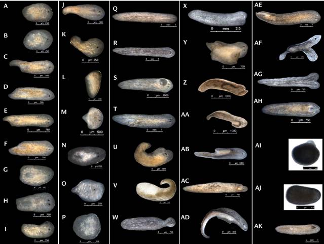
We found several categories of developmental defects that repeatedly occurred in hatchlings of S. mediterranea. In general, total developmental defects we examined were estimated to occur at a frequency lower than 5%. Among these defective hatchlings, some failed to develop a complete body plan, missing the tails (Fig. 2A, B, G, H), half of the tails (Fig. 2C-F), or half of the body (Fig. 2I). Some completely failed at patterning the body (Fig. 2J-P, AI, AJ). A majority of these defective hatchlings failed to survive long-term in laboratory culturing (Fig. 2A, B, G-P, AI, AJ), except those missing half of the tails, which could properly heal the missing tails (Fig. 2C-F).
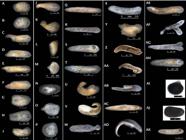
Some hatchlings were born with eye defects. Among these, missing one eye was the most common error (Fig. 2A-D, G, I, K, O, Q, U). Hatchlings with near complete body plans (Fig. 2Q) properly developed and developed standard two eyes with consistent feeding. Occasionally, hatchlings with three eyes were observed (Fig. 2R). Such hatchlings can make it into adulthood with three eyes. Hatchlings with ectopic eyes were reported in earlier literature in different species (Harangphy and Balázs 1964).
Another repeatedly observed defect was duplicated body parts. These hatchlings can appear as one fused head with three eyes (Fig. 2X), extra body parts (Fig. 2Z-AD), double tails (Fig. 2AE), double heads (Fig. 2AF-AH), or two complete bodies fused together (Fig. 2Y). This category of developmental errors can usually make it into adulthood with the defects sustained.
Some other rare defects can occur. For example, hatchlings can be born with blisters (Fig. 2S) or bloated bodies (Fig. 2V), suggesting defects in protonephridia (Rink et al. 2011, Thi-Kim Vu et al. 2015). All hatchlings (Fig. 2V) with bloated bodies died. Tumor like outgrowth could also occur (Fig. 2T, AK).
Developmental defects in Schmidtea polychroa
Generally, eggs produced per week per worm by S. polychroa is higher than S. mediterranea, likely due to their asexual mode of reproduction through parthenogenesis (D’Souza et al. 2004, D’Souza and Michiels 2006, 2010, Sánchez et al. 2013). Such an asexual mode of reproduction can reduce the negative impact from inbreeding in a short-term (Guo et al. 2016, 2022) and increase fecundity without spending energy in finding a partner or mating. The long-term benefit of parthenogenesis remains unclear. Occasional genetic information exchange and homologous recombination were reported in S. polychroa populations, which can potentially rescue the deleterious effect of parthenogenesis in long-term (D’Souza et al. 2004, Sánchez et al. 2013).
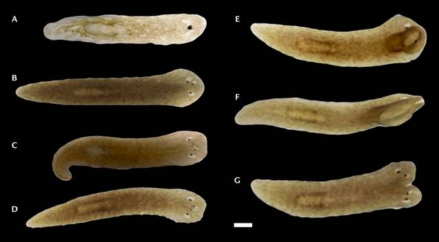
Larger numbers of eggs produced by S. polychroa allowed us to quantitatively score developmental defects in the hatchlings. We observed developmental defects in S. polychroa more frequently than in S. mediterranea, but this may be due to abnormal hatchlings being relatively bigger (~2 mm2 or bigger) in S. polychroa and much easier to identify. We uncovered 52 defected hatchlings in two weeks, out of a total of 480 hatchlings. The error rate is about ~10%.
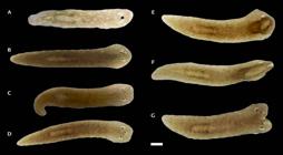
Even though the error rate is much higher in S. polychroa, we did not observe the full spectrum of developmental defects in S. polychroa (Fig. 3) as we observed in S. mediterranea (Fig. 2). Instead, defective hatchlings of S. polychroa most frequently showed eye defects (Fig. 3A-D) or fused bodies (Fig. 3E-G), which were observed in S. mediterranea as well. All such defective hatchlings in S. polychroa made into sexual adulthood.
Regeneration corrected developmental errors
Frequent occurrence of developmental errors in planarians is interesting as they allow the comparison of patterning regulation during regeneration with patterning in development. Clearly, the patterning signals during embryonic development did not execute properly in hatchlings with defects. This could be caused by genetic mutations segregating in the culturing populations or caused by specific developmental errors in the confined space of egg capsules.
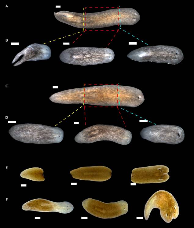
We raised the hatchlings to 5~8 mm in sizes and amputated them into three pieces from anterior to posterior to examine regeneration (Fig. 4). In all cases we examined, we found regenerated worms did not recapitulate the erroneous phenotypes from development. The head and trunk fragments of the double tailed hatchling regenerated one single tail (Fig. 4A, B). The trunk and tail fragments of the single eyed hatchling regenerated two eyes (Fig. 4C, D). The trunk and tail fragments of the double headed hatchling regenerated single heads (Fig. 4E, F). This is consistent in both S. mediterranea (Fig. 4A-D) and S. polychroa (Fig. 4E, F). In addition, we cultured these hatchlings corrected by regeneration to sexually maturity to collect their progeny in clonal crosses (Guo et al. 2016). Frequency of the parental defects are comparable in hatchlings between the regenerated progeny and their parents. This suggests the defects were likely due to erroneous developmental regulation, instead of inheritable genetic mutations.
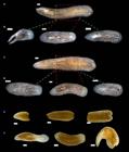
Regeneration leads to corrected body patterning. (A-D) Schmidtea mediterranea: one double tailed animal (A) and one one-eyed animal (C) was amputated into three fragments (B, D), 12 days post regeneration. (E-F) Schmidtea polychroa: one double headed animal (E) was amputated into three fragments (F), 12 days post regeneration. Scale bars: 0.2 mm.
DISCUSSION
Laboratory culture of sexual S. mediterranea and S. polychroa produced such defected hatchlings at various rates, which allowed us to ask if regeneration of these hatchlings could reproduce the developmental errors. We found that regeneration robustly produced corrected body plans in hatchlings even with striking developmental errors (e.g., double heads or double tails). Our work illustrated an interesting form of developmental plasticity.
Developmental errors may arise from fusion of multiple embryos within the confined space of the egg capsules. The egg capsules can contain multiple embryos, each of which forms its own syncytium space in their early stage of development. Before the embryos become individualized with embryonic epithelium, fusion of these early embryos seems likely. In addition, throughout the development course within the egg capsule, competition of space is unavoidable. They also share the same source of yolk cells which can function as both nutrition and mechanical scaffolds within the space of the capsule. These features make it possible for the interference on each other’s development program and rate, leading to defects and size differences of the hatchlings. It is possible that adelphophagy, a phenomenon of embryos cannibalizing their siblings (Harrath et al. 2009), also play a role in variable sizes and defects of the hatchlings.
How does regeneration know the right tissue architecture to build? While comparative analysis of the regulatory programs between development and regeneration has been of strong interest to the research community, our data here does not imply that embryonic development of planarians uses different molecular programs from regeneration. Instead, our work here showed that even though development built an erroneous body plan, the regeneration program remains robustly capable of building the correct tissue architecture. Considering the recent finding that age-associated changes in tissue architecture can be restored to youthful state (Dai et al. 2023), it seems truly remarkable that the regeneration program has its own tissue memory.
ACKNOWLEDGEMENTS
This work was supported by grants from Global Consortium for Reproductive Longevity & Equality (GCRLE Grant-0423), the Biological Sciences Scholar Program, the Endowment for the Basic Sciences, and Geriatrics Center Richard A. Miller, MD, PhD Emerging Scholar in Aging Research from University of Michigan, and from the startup fund from the Department of Molecular and Integrative Physiology, University of Michigan (LG).
LITERATURE CITED
-
Adler CE, Seidel CW, McKinney SA, Alvarado AS (2014) Selective amputation of the pharynx identifies a FoxA-dependent regeneration program in planaria. eLife 3: e02238. https://doi.org/10.7554/eLife.02238
» https://doi.org/10.7554/eLife.02238 -
Alvarado AS (2003) The freshwater planarian Schmidtea mediterranea: Embryogenesis, stem cells and regeneration. Current Opinion in Genetics & Development 13: 438-444. https://doi.org/10.1016/S0959-437X(03)00082-0
» https://doi.org/10.1016/S0959-437X(03)00082-0 -
Ban S, Lee H-W, Shinada A, Toda T (2000) In situ egg production and hatching success of the marine copepod Pseudocalanus newmani in Funka Bay and adjacent waters off southwestern Hokkaido, Japan: Associated to diatom bloom. Journal of Plankton Research 22: 907-922. https://doi.org/10.1093/plankt/22.5.907
» https://doi.org/10.1093/plankt/22.5.907 -
Cardona A, Hartenstein V, Romero R (2006) Early embryogenesis of planaria: A cryptic larva feeding on maternal resources. Development Genes and Evolution 216: 667-681. https://doi.org/10.1007/s00427-006-0094-3
» https://doi.org/10.1007/s00427-006-0094-3 -
Cebrià F, Nakazawa M, Mineta K, Ikeo K, Gojobori T, Agata K (2002) Dissecting planarian central nervous system regeneration by the expression of neural-specific genes. Development, Growth & Differentiation 44: 135-146. https://doi.org/10.1046/j.1440-169x.2002.00629.x
» https://doi.org/10.1046/j.1440-169x.2002.00629.x -
Cebrià F, Newmark PA (2005) Planarian homologs of netrin and netrin receptor are required for proper regeneration of the central nervous system and the maintenance of nervous system architecture. Development 132: 3691-3703. https://doi.org/10.1242/dev.01941
» https://doi.org/10.1242/dev.01941 -
Cowles MW, Brown DDR, Nisperos SV, Stanley BN, Pearson BJ, Zayas RM (2013) Genome-wide analysis of the bHLH gene family in planarians identifies factors required for adult neurogenesis and neuronal regeneration. Development 140: 4691-4702. https://doi.org/10.1242/dev.098616
» https://doi.org/10.1242/dev.098616 -
Dai X, Li X, Pletcher S, Paris D, Kruglyak L, Sobota J, Guo L (2023) Regeneration leads to global tissue rejuvenation in aging sexual planarians. bioRxiv Preprint. https://doi.org/10.1101/2023.06.24.546358
» https://doi.org/10.1101/2023.06.24.546358 -
Davies EL, Lei K, Seidel CW, Kroesen AE, McKinney SA, et al. (2017) Embryonic origin of adult stem cells required for tissue homeostasis and regeneration. eLife 6: e21052. https://doi.org/10.7554/eLife.21052
» https://doi.org/10.7554/eLife.21052 -
Delhanty JD (2005) Mechanisms of aneuploidy induction in human oogenesis and early embryogenesis. Cytogenetic and Genome Research 111: 237-244. https://doi.org/10.1159/000086894
» https://doi.org/10.1159/000086894 -
D’Souza TG, Michiels NK (2006) Genetic signatures of occasional sex in parthenogenetic subpopulations of the freshwater planarian Schmidtea polychroa Freshwater Biology 51: 1890-1900. https://doi.org/10.1111/j.1365-2427.2006.01622.x
» https://doi.org/10.1111/j.1365-2427.2006.01622.x -
D’Souza TG, Michiels NK (2010) The costs and benefits of occasional sex: theoretical predictions and a case study. Journal of Heredity 101: S34-S41. https://doi.org/10.1093/jhered/esq005
» https://doi.org/10.1093/jhered/esq005 -
D’Souza TG, Storhas M, Schulenburg H, Beukeboom LW, Michiels NK (2004) Occasional sex in an “asexual” polyploid hermaphrodite. Proceedings of the Royal Society B: Biological Sciences 271: 1001-1007. https://doi.org/10.1098/rspb.2004.2675
» https://doi.org/10.1098/rspb.2004.2675 -
Gowland FC, Moltschaniwskyj NA, Steer MA (2002) Description and quantification of developmental abnormalities in a natural Sepioteuthis australis spawning population (Mollusca: Cephalopoda). Marine Ecology Progress Series 243: 133-141. https://doi.org/10.3354/meps243133
» https://doi.org/10.3354/meps243133 -
Guo L, Accorsi A, He S, Guerrero-Hernández C, Sivagnanam S, McKinney S, et al. (2018) An adaptable chromosome preparation methodology for use in invertebrate research organisms. BMC Biology 16: 25. https://doi.org/10.1186/s12915-018-0497-4
» https://doi.org/10.1186/s12915-018-0497-4 -
Guo L, Bloom JS, Dols-Serrate D, Boocock J, Ben-David E, Schubert OT, et al. (2022) Island-specific evolution of a sex-primed autosome in a sexual planarian. Nature 606: 329-334. https://doi.org/10.1038/s41586-022-04757-3
» https://doi.org/10.1038/s41586-022-04757-3 -
Guo L, Zhang S, Rubinstein B, Ross E, Alvarado AS (2016) Widespread maintenance of genome heterozygosity in Schmidtea mediterranea Nature Ecology & Evolution 1: 19. https://doi.org/10.1038/s41559-016-0019
» https://doi.org/10.1038/s41559-016-0019 - Hallez P (1887) Embryogénie des Dendrocoeles d’eau douce. O. Doin, Paris.
-
Haranghy L, Balázs A (1964) Ageing and rejuvenation in planarians. Experimental Gerontology 1: 77-91. https://doi.org/10.1016/0531-5565(64)90009-9
» https://doi.org/10.1016/0531-5565(64)90009-9 -
Harrath AH, Sluys R, Zghal F, Tekaya S (2009) First report of adelphophagy in flatworms during the embryonic development of the planarian Schmidtea mediterranea (Benazzi, Baguñà, Ballester, Puccinelli & Del Papa, 1975) (Platyhelminthes, Tricladida). Invertebrate Reproduction & Development 53: 117-124. https://doi.org/10.1080/07924259.2009.9652297
» https://doi.org/10.1080/07924259.2009.9652297 - Ijima I (1884) Untersuchungeniiber den Bau und die Entwicklungsgeschichte der stisswasser-Dendrocoelen (Tri-claden).
-
Koinuma S, Umesono Y, Watanabe K, Agata K (2000) Planaria FoxA (HNF3) homologue is specifically expressed in the pharynx-forming cells. Gene 259: 171-176. https://doi.org/10.1016/s0378-1119(00)00426-1
» https://doi.org/10.1016/s0378-1119(00)00426-1 -
Mantikou E, Wong KM, Repping S, Mastenbroek S (2012) Molecular origin of mitotic aneuploidies in preimplantation embryos. Biochimica et Biophysica Acta - Molecular Basis of Disease 1822: 1921-1930. https://doi.org/10.1016/j.bbadis.2012.06.013
» https://doi.org/10.1016/j.bbadis.2012.06.013 -
Martín-Durán JM, Amaya E, Romero R (2010) Germ layer specification and axial patterning in the embryonic development of the freshwater planarian Schmidtea polychroa Developmental Biology 340: 145-158. https://doi.org/10.1016/j.ydbio.2010.01.018
» https://doi.org/10.1016/j.ydbio.2010.01.018 -
Martín-Durán JM, Monjo F, Romero R (2012a) Morphological and molecular development of the eyes during embryogenesis of the freshwater planarian Schmidtea polychroa Development Genes and Evolution 222: 45-54. https://doi.org/10.1007/s00427-012-0389-5
» https://doi.org/10.1007/s00427-012-0389-5 -
Martín-Durán, JM, Monjo F, Romero R (2012b) Planarian embryology in the era of comparative developmental biology. The International Journal of Developmental Biology 56: 39-48. https://doi.org/10.1387/ijdb.113442jm
» https://doi.org/10.1387/ijdb.113442jm -
Martín-Durán JM, Romero R (2011) Evolutionary implications of morphogenesis and molecular patterning of the blind gut in the planarian Schmidtea polychroa Developmental Biology 352: 164-176. https://doi.org/10.1016/j.ydbio.2011.01.032
» https://doi.org/10.1016/j.ydbio.2011.01.032 - Metchnikoff E (1883) Die Embryologie von Planaria polychroa Zeitschrift für wissenschaftliche Zoologie 38: 331-354.
-
Monjo F, Romero R (2015) Embryonic development of the nervous system in the planarian Schmidtea polychroa Developmental Biology 397: 305-319. https://doi.org/10.1016/j.ydbio.2014.10.021
» https://doi.org/10.1016/j.ydbio.2014.10.021 -
Newmark PA, Alvarado AS (2002) Not your father’s planarian: A classic model enters the era of functional genomics. Nature Reviews Genetics 3: 210-219. https://doi.org/10.1038/nrg759
» https://doi.org/10.1038/nrg759 -
Reddien PW (2022) Positional information and stem cells combine to result in planarian regeneration. Cold Spring Harbor Perspectives in Biology 14: a040717. https://doi.org/10.1101/cshperspect.a040717
» https://doi.org/10.1101/cshperspect.a040717 -
Reddien PW, Alvarado AS (2004) Fundamentals of planarian regeneration. Annual Review of Cell and Developmental Biology 20: 725-757. https://doi.org/10.1146/annurev.cellbio.20.010403.095114
» https://doi.org/10.1146/annurev.cellbio.20.010403.095114 -
Reddien PW, Oviedo NJ, Jennings JR, Jenkin JC, Alvarado AS (2005) SMEDWI-2 is a PIWI-like protein that regulates planarian stem cells. Science 310: 1327-1330. https://doi.org/10.1126/science.1116110
» https://doi.org/10.1126/science.1116110 -
Rink JC, Vu HTK, Alvarado AS (2011) The maintenance and regeneration of the planarian excretory system are regulated by EGFR signaling. Development 138: 3769-3780. https://doi.org/10.1242/dev.066852
» https://doi.org/10.1242/dev.066852 -
Roberts-Galbraith RH, Brubacher JL, Newmark PA (2016) A functional genomics screen in planarians reveals regulators of whole-brain regeneration. eLife 5: e17002. https://doi.org/10.7554/eLife.17002
» https://doi.org/10.7554/eLife.17002 -
Sakurai T (1991) An electron-microscopic study of syncytium formation during early embryonic development of the freshwater planarian Bdellocephala brunnea Hydrobiologia 227: 113-118. https://doi.org/10.1007/BF00027590
» https://doi.org/10.1007/BF00027590 -
Saló E, Agata K (2012) Planarian regeneration: a classic topic claiming new attention. International Journal of Developmental Biology 56: 3-4. https://doi.org/10.1387/ijdb.123495es
» https://doi.org/10.1387/ijdb.123495es -
Sánchez Navarro B, Jokela J, Michiels NK, D’Souza TG (2013) Population genetic structure of parthenogenetic flatworm populations with occasional sex. Freshwater Biology 58: 416-429. https://doi.org/10.1111/fwb.12070
» https://doi.org/10.1111/fwb.12070 -
Schindelin J, Arganda-Carreras I, Frise E, Kaynig V, Longair M, Pietzsch T, et al. (2012) Fiji: An open-source platform for biological-image analysis. Nature Methods 9: 676-682. https://doi.org/10.1038/nmeth.2019
» https://doi.org/10.1038/nmeth.2019 -
Schneider I, Ellenberg J (2019) Mysteries in embryonic development: How can errors arise so frequently at the beginning of mammalian life? Plos Biology 17: e3000173. https://doi.org/10.1371/journal.pbio.3000173
» https://doi.org/10.1371/journal.pbio.3000173 -
Solana J, Romero R (2009) SpolvlgA is a DDX3/PL10-related DEAD-box RNA helicase expressed in blastomeres and embryonic cells in planarian embryonic development. International Journal of Biological Sciences 5: 64-73. https://doi.org/10.7150/ijbs.5.64
» https://doi.org/10.7150/ijbs.5.64 -
Tanaka EM, Reddien PW (2011) The cellular basis for animal regeneration. Developmental Cell 21: 172-185. https://doi.org/10.1016/j.devcel.2011.06.016
» https://doi.org/10.1016/j.devcel.2011.06.016 -
Thi-Kim Vu H, Rink JC, McKinney SA, McClain M, Lakshmanaperumal N, Alexander R, Alvarado AS (2015) Stem cells and fluid flow drive cyst formation in an invertebrate excretory organ. eLife 4: e07405. https://doi.org/10.7554/eLife.07405
» https://doi.org/10.7554/eLife.07405
ADDITIONAL NOTES
- ZooBank register
-
How to cite this article
Dai X, Sobota J, Li X, Guo L (2024) Developmental defects in planarian hatchlings (Platyhelminthes: Rhabditophora). Zoologia 41: e23087. https://doi.org/10.1590/S1984-4689.v41.e23087
-
Published by
Sociedade Brasileira de Zoologia at Scientific Electronic Library Online - https://www.scielo.br/zool
-
Global Consortium for Reproductive Longevity & Equality (GCRLE Grant-0423) This work was supported by grants from Global Consortium for Reproductive Longevity & Equality (GCRLE Grant-0423), the Biological Sciences Scholar Program, the Endowment for the Basic Sciences, and Geriatrics Center Richard A. Miller, MD, PhD Emerging Scholar in Aging Research from University of Michigan, and from the startup fund from the Department of Molecular and Integrative Physiology, University of Michigan (LG).
Supplementary material 1
Authors: Dai X, Sobota J, Li X, Guo L.
Data type: Experiment source data
Copyright notice: This dataset is made available under the Open Database License (http://opendatacommons.org/licenses/odbl/1.0/). The Open Database License (ODbL) is a license agreement intended to allow users to freely share, modify, and use this Dataset while maintaining this same freedom for others, provided that the original source and author(s) are credited.