Sexual dimorphism and morphological characterization of antennal sensilla in the pine processionary moth Thaumetopoea pityocampa (Lepidoptera: Notodontidae)
ABSTRACT
The pine processionary moth Thaumetopoea pityocampa (Denis & Schiffermüller, 1755) is widely distributed in Mediterranean areas where its larval phases colonize different Pinus species, often inducing a complete defoliation. According to the most common pattern in Insects, T. pityocampa shows a size dimorphism, with males being smaller than females. Despite its great socioeconomic relevance, the morphological characteristics of this species are still not well-documented. Available studies mainly focus on general morphology, biological cycle and ecological traits, thus leaving a knowledge gap on this topic. Here, we provide for the first time a detailed morphological description of T. pityocampa adults with particular emphasis on sexual dimorphism through an in-depth analysis under both stereomicroscope and scanning electron microscope. The shape and distribution of scales in the wings of males and females have been evaluated to reveal putative differences in their morphology and/or location and the hindwings of males have a much greater number of hairy scales. Since insects rely on their antennal sensilla to accomplish vital cues, including mate selection and host plant recognition, we investigated the antennal arrangement and the distribution of sensilla in both sexes. A notable dimorphism in the length of the lateral branches of the antennae was highlighted but no differences were observed in the general distribution of the sensilla. Males possess exclusively one type of trichoid sensilla (type 1), which are longer than those observed in females.
KEYWORDS:
Morphology; scanning electron microscopy; sexual dimorphism; sensorial pattern; wing scales
INTRODUCTION
Thaumetopoeinae includes about 100 species of moths in 20 genera widely distributed across the Palearctic, Asia, Australasia, New Caledonia, and Australia (Schintlmeister 2013, Battisti et al. 2017, St. Laurent et al. 2025). The pine processionary moth, Thaumetopoea pityocampa (Denis & Schiffermüller, 1755), is a polyphagous Thaumetopoeinae species that feeds on Pinus, Cedrus and Larix. It has recently been distinguished from its sister species T. mediterraneaTrematerra & Scalercio, 2017 and T. hellenica Trematerra & Scalercio, 2017 (Trematerra et al. 2017) and, in Mediterranean black pine forests, represents the most abundant species among nocturnal macromoth communities (Scalercio and Greco 2018, Azcárate et al. 2023).
As with most Thaumetopoeinae, it is considered an univoltine species, and its caterpillars, which possess urticating setae, colonise the leaves of host plants (Battisti et al. 2017, Pimentel et al. 2006).
The females lay their eggs (70-300) on the ends of the pine branches, it is building a sticky cocoon and covering it with the scales from the last segments of their abdomen. Hatching occurs after 35-40 days. During the development period, which includes five instars, larvae are gregarious and can move around the tree while building silky nests where they overwinter. Once development is complete, they move in their characteristic procession, searching for a suitable ground as a pupation site. Pupae rest in a cocoon below the ground until the adult flies at night during summer, but some individuals enter prolonged diapause, which may endure more than a year (Trematerra and Colacci 2019, Salman et al. 2016), resulting in a semivoltine life cycle.
Thaumetopoea pityocampa is recognized as one of the most damaging pests in Mediterranean pine forests because larval foraging can severely affect the diameter and volume of the tree, often causing complete defoliation (Bonamonte et al. 2013, Ferracini et al. 2023, Mantzoukas et al. 2024). As a thermophilous species, T. pityocampa is widely distributed in Mediterranean Europe, but its range is expanding rapidly northward and upward. This phenomenon is largely driven by improved winter survival rates due to global warming, as higher winter temperatures significantly influence larval development (Battisti et al. 2006, Salman et al. 2016). The observed increase in outbreak areas and the predicted impact on forests in the coming decades raise great concern about maintaining tree growth potential and related forest productivity (Salman et al. 2016, Ferracini et al. 2023, Pimentel et al. 2006). Caterpillars are also recognized as a serious public health issue because their urticating setae are known to induce rashes and severe allergic responses in humans and domesticated animals (Azcárate et al. 2023, Battisti et al. 2017).
Despite its great economic importance and being the most studied species of the Thaumetopoea genus, the morphological characteristics of this species are surprisingly not well-documented. Previous studies have primarily focused on general morphology, biological cycles, and ecological traits, leaving a knowledge gap in detailed morphological characterization (Agenjo 1941, Basso et al. 2016, 2017, 2023, Trematerra and Colacci 2018).
On this basis, we provide a detailed morphological description for the first time of T. pityocampa, with particular emphasis on sexual dimorphism.
One of the most characteristic features of butterflies and moths (Lepidoptera) are wings covered with scales. These chitin extensions may confer different colour patterns to wings depending on pigmentation and light interaction (Wang et al. 2022, Zeng et al. 2011, Gomez et al. 2021). They are not limited to the wings and typically cover the entire body, being involved in several functions, including thermoregulation, antipredator strategies, signaling during mating behavior, communication, and water repellency (Zeng et al. 2011, Gomez et al. 2021). Research on the scales of T. pityocampa is currently limited, with only three studies focusing on the color and shape of scales covering egg batches as diagnostic characters for species recognition (Tsankov et al. 1991, Doģanlar et al. 2005, İpekdal et al. 2016).
To fill this gap, here we evaluated the shape and distribution of scales in the wings of males and females to reveal potential differences in their morphology and/or location.
The antennae of insects act as sensory organs that allow them to explore and perceive their environment. They are equipped with a series of sensilla, each performing distinct sensory functions: olfactory, tactile, gustatory, and thermo-/hygro-reception (Amat et al. 2022, Varga et al. 2023). Sensilla are classified according to their morphology which, along with their distribution in the body region, allows their functional determination (Schneider 1957, Steinbrecht 1970, Zacharuk 1985, Taszakowski et al. 2023). The general structure of the antenna and sensilla varies among taxa, and an outstanding variety has been reported in Lepidoptera, within and between diurnal and nocturnal species (Li et al. 2024). Moreover, previous investigations revealed a sex-related difference in the types and functions of antennal sensilla (Ma and Du 2000, Ndomo-Moualeu et al. 2014). In moths, the morphology of antennae has been investigated in several species (Pophof et al. 2005, Sun et al. 2011); however, as far as we know, no previous study has investigated the antennal sensilla of T. pityocampa.
Given that antennal morphology, as well as the number and types of sensilla, vary among many Noctuidae moths, we performed a qualitative morphological assessment to define the sensilla types and their distribution in males and females. Moreover, a morphometric analysis of antennae and sensilla in both sexes has been carried out to allow for a more objective evaluation.
The present study aimed to gather significant morphological data on Thaumetopoea pityocampa, emphasising the sexual dimorphism, to address a gap in the existing literature. To the best of our knowledge, this is the first study documenting the morphological, morphometric, and functional features of wings and antennae in T. pityocampa by Ultra High-Resolution SEM. Given the ecological and economic significance of T. pityocampa, one of the most destructive pests affecting Mediterranean black pine forests, the findings of this study provide essential knowledge to support the development of novel control strategies.
MATERIAL AND METHODS
Studied specimens (n = 16, 8 males and 8 females) were collected in Calabrian black pine (Pinus nigra J.F. Arnold subsp. laricio Palib. ex Maire) dominated forests of the Sila Massif, Calabria, southern Italy. Samplings were performed by using UV-LED light traps (Infusino et al. 2017) during comprehensive monitoring of forest macro-moth communities (Scalercio and Greco 2018). Both males and females were collected on 5th of July 2016 in the locality Mangiatoie (coordinates: 39.2380°N; 16.6647°E), San Giovanni in Fiore municipality, Cosenza province, at an altitude of 1,270 meters. Specimens were preserved pinned, then, they were spread on setting boards for Lepidoptera in accordance with standard protocols (Gullan and Cranston 2014). Forewings were positioned on the setting board the morning after their collection, with the posterior margin forming about 90° with the body axis. Only the anterior margin of the hindwings was settled below the forewings, with most of the hindwing surface visible. The material was deposited in the Lepidoptera collection of the Wildlife Management and Forest Biodiversity Lab of the Research Centre for Forestry and Wood, Rende, Italy.
For morphological observations, the dried specimens were photographed using a Nikon Research Stereo Microscope SMZ25. For scanning electron microscope (SEM), the dried samples were attached to a holder using electrically conductive adhesive tape, sputter-coated with graphite in a Sputter-Carbon Coater (QUORUM Q150T-ES) and examined under Ultra High-Resolution UHR-SEM ZEISS CrossBeam 350, operating at an accelerating voltage of 15 kV. Observations were performed at the Microscopy and Microanalysis Centre (CM2), University of Calabria, Italy. For sensilla terminology were referred to Schneider (1964) and Varga et al. (2023).
Statistical analyses were run in GraphPad Prism 8.00 (GraphPad Software Inc., San Diego, CA, USA) at a significance level of 0.05. The data, presented as mean ± standard error of the mean, were analyzed using the “unpaired t test”, the normality assumption was verified using the Shapiro-Wilk test and equality of variances was evaluated with the F-test.
RESULTS
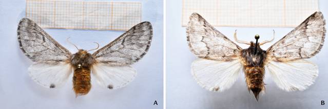
Sexual dimorphism in T. pityocampa is evident. Males are smaller and equipped with bipectinate antennae with elongated rami, while females are larger and have antennae with shorter branches (Fig. 1A, B).
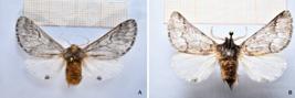
Adults Thaumetopoea pityocampa: (A) female and (B) male. The coloration pattern of both forewing and hindwing did not differ between the sexes. The thorax and abdomen of the male show longer and more numerous hairs. Note the bipectinate antennae of the male holding long branches.
Females
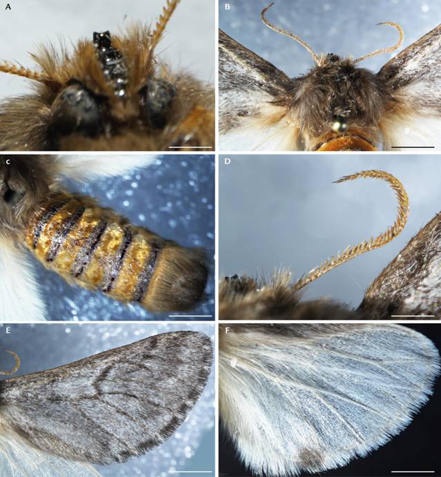
The head of the female is light brown and has bipectinate antennae. In the central middle region of the head, a chitinous brown and shiny frontal process (canthus) is well distinguishable, showing five teeth, the first of which is longer than the remaining four (Fig. 2A). The eyes are large and located in a ventrolateral position. Females show a thorax covered by light-brown hairs (Fig. 2B) and a prominent cylindrical abdomen. The abdomen is ochraceous with dark bands, and its last segments hold a tuft of long hairs that partially covers the large anal papillae scales (Fig. 2C). The bipectinate antennae, from which arise short lateral branches, are ochraceous (Fig. 2D).
The coloration pattern of the forewing and hindwing is different (Fig. 1A). The forewings have a light brown background with three thin, blackish transverse bands and are crossed by dark and evident brown veins (Fig. 2E). The margins of the costa are darkly marked, while the fringed termen alternating brown and white. Hindwings (Fig. 2F) are whitish-cream with hairy-fringed margins and a defined dark analspot. The distal margin is light-brown-cream, and honey-colored veins cross the wing.
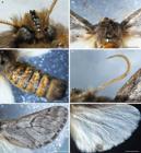
Thaumetopoea pityocampa female. (A) canthus in ventral view, a chitinous brown and shiny frontal process showing five teeth. (B) thorax in dorsal view, covered by light-brown hairs -like setae; (C) abdomen in dorsal view, patterned with ochraceous abdomen and dark bands, note the tuft of long hairs that covers the large anal scales; (D) bipectinate antennae bearing short lateral branches; (E) forewings in dorsal view showing an obscure pattern, note the fringed termen alternatively, colored in brown and white; (F) hindwing in dorsal view exhibiting a disruptive coloration, note the round black spot. Scale bar = 1mm.
Males
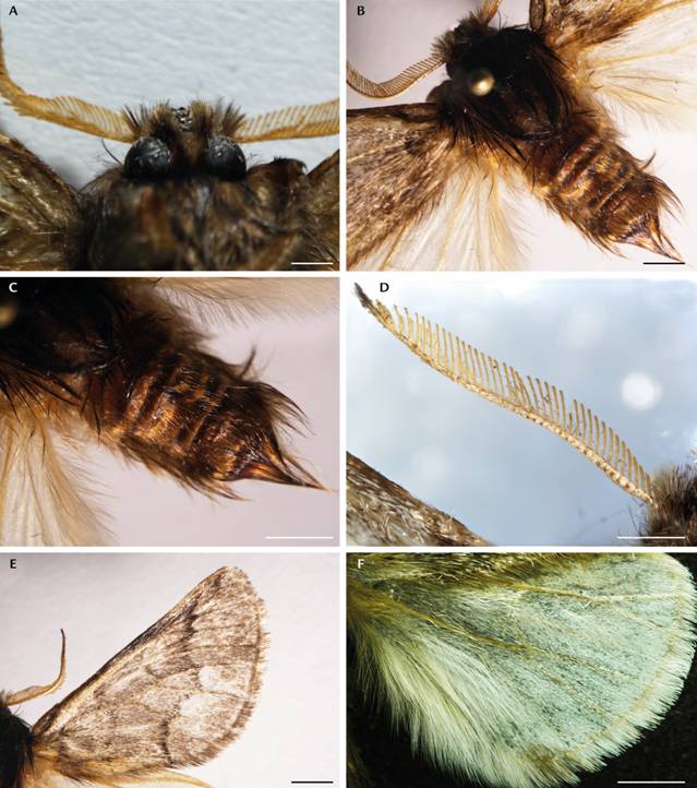
Head morphology of males is similar to described for females except for the dimorphic antennae (see below, Fig. 3A) and the thorax is covered by numerous long dark brown hairs (Fig. 3B). The abdomen is less developed than that of females, and it is equipped with a greater amount of hair. It has a characteristic conic shape and is dark ochre, with smaller and less evident brown bands than those observed in females (Fig. 3C). Antennae, bipectinate, ochraceous colored, lateral branches much more developed than those observed in females, giving them a plumose appearance (Fig. 3D). As in females, the forewing and hindwing exhibit different coloration patterns (Fig. 1B). Forewings (Fig. 3E) light brown with a darker costal margin. The margins of the costa are darkly marked, and the termen shows brown and white fringe. Yet, the males’ forewing surface is crossed by darker veins that branch across and by three vertical dark brown to black lines (Fig. 3E). Hindwing (Fig. 3F) coloration pattern is similar to that of females with a whitish cream base color, suffused with brown along the distal margin and with an evident circular dark analspot.
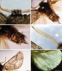
Thaumetopoea pityocampa male. (A) head in dorsal view, canthus could be observed along with the eyes located in a ventrolateral position;.(B) thorax in dorsal view, covered by long dark brown hairs-like setae; (C) abdomen in dorsal view, less developed than in females, equipped with a greater amount of hair; note the characteristic conic shape; (D) bipectinate antenna bearing well-developed lateral branches;(E) forewings in dorsal view; (F) hindwing. Scale bar = 1 mm.
Wings
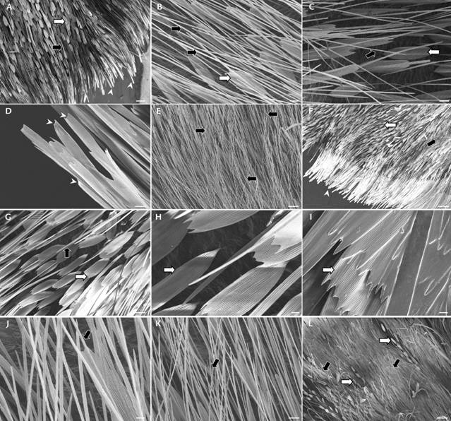
Three types of scales can be distinguished: type a) piliform scales, monofid, thin and elongated, similar to bristles; type b) lamellar scales, showing a notched apex; type c) border scales, deeply elongated with two or more projections on an apical border (Fig. 4A-C).
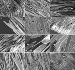
Scanning Electron Microscopy (SEM) images of wing scales of females (A-E) and males of Thaumetopoea pityocampa (F- L). (A) differential distribution of piliform, lamellar, and border scales, scale bar = 200 µm; (B) further enlargement showing the elongated monofid piliform scales and the lamellar scales, scale bar = 30 µm; (C) detail of lamellar scales showing a notch at their apical fin, scale bar = 30 µm; (D) detail of elongated border scales, note the apical border with two or more projections, scale bar = 30 µm; (E) numerous piliform scales that cover border scales along the termen, scale bar = 100 µm; (F) differential distribution of piliform, lamellar, and border scales, scale bar = 300 µm; (G) lamellar scales and sparse piliform scales in the central area of forewings, scale bar = 40 µm; (H) further enlargement showing three or more projections at their apical border, scale bar = 10 µm; (I) lamellar scales positioned like roof tiles near the upper edge of the wings, scale bar = 10 µm; (J) lamellar and piliform scales on the hindwings in the central area, scale bar = 10 µm; (K) numerous piliform scales that densely cover the other scales, scale bar = 30 µm; (L) numerous piliform scales cover border scales along the termen, scale bar = 200 µm. arrow black: piliform scales; arrow white: lamellar scales; white arrowhead: border scales.
The overall morphology of each type of scale is the same in both sexes, but they are arranged differently in males and females.
In female forewings, lamellar scales cover the central area, while piliform scales are less numerous and scattered throughout the entire wing surface (Fig. 4B, C). Lamellar scales mainly show a single notch at their apical fin (Fig. 4C). Border elongated marginal scales are found only along the wing’s outer edge (Fig. 4A, D). The piliform and lamellar scales are distributed in the central area on the hindwings. Along the termen, in addition to the long border scales, numerous piliform scales could be seen, forming a dense covering (Fig. 4E).
In male forewings (Fig. 4F), the central area is covered by lamellar scales and sparse piliform scales (Fig. 4G). Lamellar scales often have three or more apical projections (Fig. 4H). Near the upper edge of the wing, lamellar scales are positioned like roof tiles (Fig. 4I). The border scales are distributed only along the wing margin. Both lamellar and piliform scales on the hindwings in the central area (Fig. 4J), but the latter are much more numerous and densely cover the other scales (Fig. 4K). As in females, along the termen, we found border scales and numerous piliform scales that form a hairy covering of the wings (Fig. 4L).
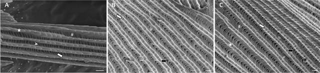
Chitinous wing scales have a highly structured upper lamina, which consists of a series of parallel ridges/ribs made of slightly overlapping lamellae running along their entire length. From the lateral wall of each rib, short micro-ribs depart perpendicularly. Due to their different lengths, not all the micro ribs extend to the adjacent ridge. They are called cross-ribs when they reach the adjacent ridge, connecting them to each other. Parallel cross-ribs originate in a structure with more or less large windows that open onto the scale lumen. This general arrangement is similar across the three scale types, but some differences become evident upon further magnification.
In the piliform scales (type a), the ribs are distant from each other by 1±0.05 µm. Cross ribs that join adjacent ribs leave a large window (Fig. 5A). In the lamellar and border elongated scales (types b and c), the ribs are distant from each other by 1.3±0.07 µm. Cross-ribs appear like a reverse C-shaped structure; no large window is found at the center of this membranous structure, but there are some small perforations (Fig. 5B, C).

Scanning Electron Microscopy (SEM) images of scales of Thaumetopoea pityocampa in high magnification showing the highly structured upper lamina. (A) piliform scales (type a), scale bar = 2 µm; (B) lamellar scales (type b), scale bar = 1 µm; (C) border scales (type c), scale bar = 1 µm. star: ribs; #: crossribs; white arrow: lamellae; white head arrow: windows; black arrow: perforations.
Antennae
Antenna consists of a scape, a pedicel, and a bipectinate flagellum. The length of the antennae is significantly greater in females, and this results from a significantly longer flagellomeres, while the number does not differ significantly between males and females (Fig. 1A, B; Table 1). Two lateral branches arise from flagellomeres, which differ significantly between sexes, with males having longer branches (Table 1). The length of the branches is not uniform along the flagellomeres and increases progressively from the proximal to the median region and then decreases towards the distal region. At tip of antenna some elongated scales are visible. The surface of the scape and peduncle, as well as the dorsal surface of the flagellum, is covered with overlapping lamellar scales.
Morphometric measurements of antennae of T. pityocampa (significance level of 0.05). SEM: standard error of the mean; df: degree of freedom; F: F-test; p: p-value). Asterisk indicates significance.
Females
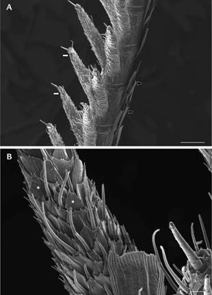
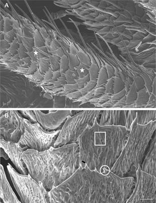
Antennae total length measure 8.9 ± 0.4 mm, and each flagellomere measures 208.99 ± 8 µm. The length of lateral branches is 248.5 ± 35 µm (Table 1). The dorsal surface of the antenna is widely covered by scales, whereas the sensilla are only distributed on the lateral branches (Fig. 6A). Coeloconic sensilla are the most abundant and almost uniformly cover the branch surface and exhibit a typical leaf-like shape (Fig. 6B). Each sensillum shows a flattened rough surface with numerous pores (Fig. 7A, B). They exhibit a certain degree of morphological variability, and it is possible to recognize narrow sensilla with a single tip and larger sensilla with two or more apical tips (Fig. 7A). Each tip shows a pore at its apical end, and in females, they are significantly more developed than in males (Table 1).
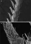
Scanning Electron Microscopy (SEM) images of antennae of females of Thaumetopoea pityocampa. (A) dorsal surface, scale bar = 100 µm; (B) antennal branch (pectin), scale bar = 10 µm. black arrow: scales; white arrow: branches; star: coeloconic sensilla. The dorsal surface is covered by scales; note the short lateral branches equipped with sensilla (A). Coeloconic sensilla, representing the most abundant type of sensilla, uniformly cover the branches’ surface. Scattered through the branches, trichoid sensilla of type 2 could also be seen (B).
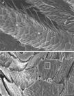
Scanning Electron Microscopy (SEM) images of antennal branch of Thaumetopoea pityocampa female. (A) equipped with coeloconic sensilla, scale bar = 10 μm. It is possible to recognize narrow sensilla that end with a single tip and larger sensilla with two or more tips at their apex; (B) higher magnification showing a flattened rough surface of coeloconic sensilla, scale bar = 3 μm. Note the pore at the apical end of tips. star: coeloconic sensilla; white circle: tips; white square: rough surface of tips; black arrow = pore.
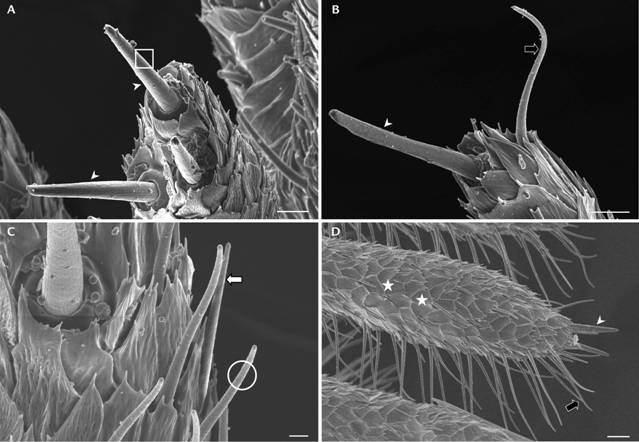
There are three sensilla chaetica of similar length (45.7 ± 15 μm) at the apical region of each branch which are significantly longer than in males (Table 1). Each sensillum arises from a socket and has a robust appearance, with a single pore at its end. The diameter of their setal shaft gradually decreases from the proximal to the distal portion. The external surface is thick and shows longitudinal grooves (Fig. 8A).
In the same area, it is possible to observe a few long, flagellate trichoid sensilla (type 1) (Fig. 8B). Numerous shorter trichoid sensilla (type 2) of 26.668 ± 1.1 µm in length (Tab.1) are distributed along the axis of the branches scattered through the coeloconic sensilla. The surface of trichoid sensilla, both type 1 and 2, shows a spiral annulations pattern (Figs 6B and 8B, C).
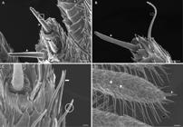
Scanning Electron Microscopy (SEM) images of sensilla chaetica of Thaumetopoea pityocampa (A-C) female and (D) male located at the apical region of each antennal branch; (A-B) note the socket at the base of each sensillum and longitudinal grooves on their external surface scale bar = 10 µm. (B) trichoid sensilla of type 1, note the elongated shape and the annulation in their external surface, scale bar = 10 µm. (C) shorter trichoid sensilla of type 2 in female antennae, scale bar = 3 µm. (D) shorter trichoid sensilla of type 2 along the ventral side of the branches in the male antenna, scale bar = 20 µm. white head arrow: sensilla chaetica; white square: longitudinal grooves; black arrow: trichoid sensilla type 1; white arrow: trichoid sensilla type 2; white circle: spiral annulation; star: coeloconic sensilla.
Males
Antennae of males (Fig. 8D) measure 7.75 ± 0.3 mm in length, and each flagellomere is 169 ± 5 μm long. The length of lateral branches is 417.3 ± 20.8 µm (see Table 1). As in females, the dorsal surface of the antenna is densely covered by scales, whereas sensilla cover the lateral branches. Coeloconic sensilla densely covered the surface of branches. As in females, they are characterized by one or more tips that show a length of about 1.4±0.4 µm. It is also possible to recognize three long sensilla chaetica with a length of 27.6±7.3 µm in the apical portion of each branch.
In males, only one type of sensillum trichodeum (type 1) was observed (Fig. 8D). They are significantly longer than those observed in females (86.7 ± 6.2 µm) and are mainly distributed on the ventral side of the branches (Fig. 8D). Their surface shows the typical spiral annulation pattern.
DISCUSSION
In this study, we provide a description of Thaumetopoea pityocampa, with particular emphasis on sexual dimorphism, thus enhancing the limited information available on this subject. Regarding general morphology, we have confirmed that males are smaller than females but are characterized by antennae equipped with significantly more developed lateral branches. Additionally, the male’s thorax and abdomen are covered with a dense layer of hair. Although wing coloration is not a reliable characteristic for distinguishing males from females, microscopic examinations show that males have a number abundance of hairy structures on their hindwings.
Wings
Lepidoptera wings are characterized by the presence of scales, which are colored by both structural and pigmentary phenomena, resulting in opaque color patterns (Stavenga 2014, Gomez et al. 2021). These patterns could be involved in different functions, including mate attraction, antipredator defences (camouflage, deflection, and aposematism), thermoregulation, sound production, or sound resonation (Mallet and Singer 1987, Nakano et al. 2008). As reported in many moths, in T. pityocampa the forewings and hindwings display distinct patterns. Forewings show an obscure pattern in the dorsal surface, while hindwings are whitish-cream colored, with hairy-fringed margins and an evident rounded dark spot. No differences in coloration patterns could be seen in males and females.
The coloration of the forewings of T. pityocampa is cryptic, which is not surprising, as it is well-established that moths adapt their color patterns to their surroundings to evade predators. A significant body of literature indicates that such mimetic phenotypes reduce the likelihood of detection and enable the moths to hide during daytime rest (Nokelainen et al. 2024 and references therein, Kang et al. 2012, Mishra et al. 2017). In contrast, it has been hypothesized that disruptive hindwing coloration may help avoid attacks when forewing camouflage fails (Mishra et al. 2017, Lindstedt et al. 2011, Schaefer and Stobbe 2006). In addition, the hind wings of T. pityocampa are characterized by the distinct black spot that contrasts with the color of the surrounding area. Similar circular markings, known as eye spots, are common among Lepidoptera and are generally found in both sexes. Several hypotheses have been proposed regarding the role of eyespots, and they are often conflicting. According to literature, eyespots are thought to serve several putative functions, including mate choice, intrasexual competition, and predator avoidance (Stevens 2005 and reference therein, Schaefer and Stobbe 2006, Kodandaramaiah 2011, Mishra et al. 2017). However, only a few studies have explored the role of eyespots in mate choice or intrasexual competition among lepidopterans, and no studies are available on this topic in T. pityocampa. A great research effort is needed to define whether the presence of these signals serves an antipredatory function or not.
Based on their morphology, three types of scales can be identified on the wings of T. pityocampa. The general organization of these scales is similar in both males and females; however, their distribution varies between the sexes. In males, lamellar scales near the upper edge of the forewings are arranged like roof tiles. Additionally, the hindwings of males have a much greater number of piliform scales, which densely cover the other scales. It has been suggested that body scales play a role in thermoregulation and insulation, but in males, they may also be involved in hormone production (Ghosh and Mishra 2018).
Antenna
Since insects rely on their antennal sensilla to accomplish vital cues, including mate selection and host plant recognition, we investigated the antennal arrangement and the distribution of sensilla in both sexes. The morphological features of the antennae in T. pityocampa are similar to those described for other notodontid moths, and each antenna consists of a scape, a pedicel, and a bipectinate flagellum from which two branches arise laterally (Mark et al. 2018). On the surface of the noctuids’ antennae, scales are usually found alongside the sensilla. This arrangement likely helps the insect in detecting the direction of odor stimuli and may also serve as a mechanism for capturing and concentrating odour molecules (Wall 1978, Van der Pers et al. 1980). Our ultrastructural analysis revealed that in both male and female T. pityocampa, the dorsal surface of the antenna is extensively covered with scales, as previously reported in many species of noctuids (Mark et al. 2018), whereas sensilla only occur on the lateral branches. Although there are no differences in general arrangement between males and females, a notable dimorphism is observed in the length of the lateral branches. In males, these are significantly longer than in females, resulting in a more plumose appearance. The difference in the length of later branches is species-specific in bombycoid and notodontid, even if males with longer branches represent the most common pattern (Mark et al. 2018).
Li and colleagues (2024) noted that the type, function, quantity, and distribution of antennal sensilla tend to be stable within taxonomic groups, highlighting a consistent pattern across Lepidoptera. This stability suggests that these sensilla have evolved to fulfil specific ecological roles, particularly in olfactory and tactile perception, which are critical for behaviours such as foraging and mate selection.
We identified four types of sensilla in T. pityocampa: trichoid type 1, trichoid type 2, coeloconica and chaetica.
Trichoid sensilla of insects are typically involved in mechano- or chemoreception (Amornsak et al. 1998, Onagbola and Fadamiro 2008, Roux et al. 2005, Yang et al. 2009). In Lepidoptera, it has been shown that trichoid sensilla are responsible for identifying the sexual pheromones of the females (Faucheux et al. 2006, Sun et al. 2011). Accordingly, in different species of day-flying moths (Heliozelidae), it has been demonstrated that sex pheromones contact the receptors through the pores of such sensilla (Wang et al. 2018). The presence of long trichoid sensilla in males has been previously reported in several noctuid species, suggesting their role in the reception of female sex pheromones ( Jefferson et al. 1970, Zhang et al. 2001, Seada 2015).
In this study, we demonstrated that the well-developed bipectinate antennae of male specimens not only provide a greater surface area but are also equipped with significantly longer type 1 trichoid sensilla compared to females (Mark et al. 2018), thus supporting their role in the detection of pheromones. In contrast, the shorter type 2 trichoid sensilla are found exclusively on the ventral side of the antennal branches in females, where they may serve a different function, such as detecting the volatile compounds of plants.
Wang and colleagues (2018) showed that variations in the spatial arrangement of sensilla in moths indicate their receptor functions, suggesting that long trichoid sensilla are devoted to sex pheromones detection, whereas short coeloconic sensilla may be able to detect airborne signals with a high diffusion coefficient, such as plant-associated odours (Rani et al. 2021 and references therein). Coeloconic sensilla, while showing some morphological variability across different species, are generally considered olfactory receptors (Rani et al. 2021, Seada 2015) but also receptors of humidity and temperature (Yan et al. 2014, Romani et al. 2009, Sun et al. 2011) due to the presence of numerous pores on the cuticular walls. In agreement, our observations revealed the presence of pores on both the surface and at the apical ends of each tip in the coeloconic sensilla surface. The distribution and general morphology of coeloconic sensilla did not differ in males and females, suggesting that some receptorial cues provided by these sensilla are probably related to shared ecological needs between the sexes, such as chemo- or hydro-reception. However, we found that in females, the tips of the coeloconic sensilla are significantly longer than in males. This suggests that the role of coeloconic sensilla in the reception of plant-associated odors assumes a greater relevance in females of T. pityocampa, probably in relation to the choice of the deposition sites.
The sensilla chaetica exhibit a consistent morphology across moth species and, more broadly, in Lepidoptera. They emerge from a basal socket and appear as robust hair-like structures with a terminal pore. Sensilla chaetica belong to a broader group of uniporous sensilla, which share morphological and functional features and are typically associated with contact chemoreception (Amat et al. 2022). These taste sensilla contain two to four gustatory receptor neurons, with their dendrites exposed to the environment through a single apical pore (Popescu et al. 2013, Amat et al. 2022) and have often been observed on antennae of various species of noctuids and pyralids. Additionally, the presence of a basal socket is linked to tactile function, indicating a bimodal role of these sensilla.
The distribution pattern of sensilla chaetica observed here is similar to that reported in bipectinate antennae of other noctuid species (Varga et al. 2023). However, it is worth noting that in other investigated species, females displayed a higher number of sensilla or longer ones than males suggesting a specific role for sensilla chaetica in female host plant selection for oviposition (Seada, 2015, Rani et al. 2021). In contrast, in T. pityocampa we did not reveal any sex-related differences in the morphology or number of such sensilla. One may suppose that plant recognition is also crucial for males since mating takes place on the host plant. Additionally, it should be noted that sensilla chaetica have a dual role, as they are also involved in the reception of contact and short-range contact cues are essential for partner recognition in both sexes.
LITERATURE CITED
- Agenjo R (1941) Monografia de la familia Thaumetopoeidae (Lep.). Revista Espanola de Entomologia 17: 69-130.
-
Amat C, Marion-Poll F, Navarro-Roldán MA, Gemeno C (2022) Gustatory function of sensilla chaetica on the labial palps and antennae of three tortricid moths (Lepidoptera: Tortricidae). Scientific Reports 12(1): 18882. https://doi.org/10.1038/s41598-022-21825-w
» https://doi.org/10.1038/s41598-022-21825-w - Amornsak W, Cribb B, Gordh G (1998) External morphology of antennal sensilla of Trichogramma australicum Girault (Hymenoptera: Trichogrammatidae). International Journal of Insect Morphology and Embryology 27: 67-82.
-
Azcárate FM, Seoane J, Silvestre M (2023) Factors affecting pine processionary moth (Thaumetopoea pityocampa) incidence in Mediterranean pine stands: a multiscale approach. Ecological Management 529: 120728. https://doi.org/10.1016/j.foreco.2022.120728
» https://doi.org/10.1016/j.foreco.2022.120728 -
Basso A, Simonato M, Cerretti P, Paolucci P, Battisti A (2016) A review of the “summer” Thaumetopoea spp. (Lepidoptera: Notodontidae, Thaumetopoeinae) associated with Cedrus and Pinus Turkish Journal of Agriculture and Forestry 17: 31-39. https://doi.org/10.18182/tjf.23128
» https://doi.org/10.18182/tjf.23128 -
Basso A, Negrisolo E, Zilli A, Battisti A, Cerretti P (2017) A total evidence phylogeny for the processionary moths of the genus Thaumetopoea (Lepidoptera: Notodontidae: Thaumetopoeinae). Cladistics 33: 557-573. https://doi.org/10.1111/cla.12181
» https://doi.org/10.1111/cla.12181 -
Basso A, Avtzis D, Burban C, Kerdelhué C, İpekdal K, et al. (2023) The pine processionary moth Thaumetopoea pityocampa (Notodontidae) species complex: a phylogeny-based revision. Arthropod Systematics & Phylogeny 81: 1031-1050. https://doi.org/10.3897/asp.81.e102928
» https://doi.org/10.3897/asp.81.e102928 - Battisti A, Stastny M, Buffo E, Larsson S (2006) A rapid altitudinal range expansion in the pine processionary moth produced by the 2003 climatic anomaly. Global Change Biology 12: 662-671.
- Battisti A, Larsson S, Roques A (2017) Processionary moths and associated urtication risk: Global change-driven effects. Annual Review of Entomology 62: 323-342.
-
Bonamonte D, Foti C, Vestita M, Angelini G (2013) Skin reactions to pine processionary caterpillar Thaumetopoea pityocampa Schiff. The Scientific World Journal 2013: 867431. https://doi.org/10.1155/2013/867431
» https://doi.org/10.1155/2013/867431 - Doğanlar M, Doğanlar O, Doğanlar F (2005) Morphology and systematics of European species of Traumatocampa Wallengren, 1871 with descriptions of two new species from the Mediterranean region of Turkey (Lepidoptera, Thaumetopoeidae). Entomofauna 13: 229-240.
- Faucheux MJ (2006) External ultrastructure of sensilla on the male and female antennal flagellum of Noctua pronuba L. (Lepidoptera: Noctuidae). Annales de la Société Entomologique de France 26(2): 173-184.
- Ferracini C, Saitta V, Rondoni G, Rollet I (2023) Variables Affecting the Pine Processionary Moth Flight: A Survey in the North-Western Italian Alps. Forests 14: 31.
- Ghosh S, Mishra M (2018) Fine nanostructural variation in the wing pattern of a moth Chiasmia eleonora Cramer (1780). Journal of Biosciences 43(4): 673-684.
-
Gomez D, Pinna C, Pairraire J, Arias M, Barbut J, et al. (2021) Wing transparency in butterflies and moths: structural diversity, optical properties, and ecological relevance. Ecological Monographs 91(4): e01475. https://doi.org/10.17605/OSF.IO/Q8TWE
» https://doi.org/10.17605/OSF.IO/Q8TWE - Gullan PJ, Cranston PS (2014) The insects: an outline of entomology. John Wiley & Sons, Oxford.
- Infusino M, Luzzi G, Scalercio S (2017) I macrolepidotteri notturni dell’Arboreto Sbanditi, Area MAB-UNESCO, Parco Nazionale della Sila (Calabria, Italia). Memorie della Società Entomologica Italiana 94(1-2): 137-153.
- İpekdal K, Helvacı Z, Çağlar S (2016) Preliminary results of the shape analysis of pine processionary moth scales in Turkey. Turkish Journal of Agriculture and Forestry 17: 40-43.
- Jefferson RN, Rubin RE, Mcfarland SU, Shorey HH (1970) Sex pheromones of noctuid moths, XXII. The external morphology of the antennae of Trichoplusiani, Heliothiszea, Prodenia ornithogalli and Spodoptera exigua Annals of the Entomological Society of America 63: 1227-1238.
- Kang CK, Moon JY, Lee SI, Jablonski P (2012) Camouflage through an active choice of a resting spot and body orientation in moths. Journal of Evolutionary Biology 25: 1695-1702.
-
Kodandaramaiah U (2011) The evolutionary significance of butterfly eyespots. Behavioral Ecology 22(6): 1264-1271. https://doi.org/10.1093/beheco/arr123
» https://doi.org/10.1093/beheco/arr123 -
Li YP, Zhou HY, Yang Y, Ye H, Haack RA, Cao J (2024) Morphological characterization and distribution of antennal sensilla of Spodoptera frugiperda (Lepidoptera: Noctuidae) using scanning electron microscopy. Microscopy Research and Technique: 1-14. https://10.21203/rs.3.rs-5285978/v1
» https://10.21203/rs.3.rs-5285978/v1 - Lindstedt C, Eager H, Ihalainen E, Kahilainen A, Stevens M, Mappes J (2011) Direction and strength of selection by predators for the color of the aposematic wood tiger moth. Behavioral Ecology 22: 580-587.
- Ma RY, Du JW (2000) Insect antennal sensilla. Entomological Knowledge 37(3): 179-183.
- Mallet J, Singer MC (1987) Individual selection, kin selection, and the shifting balance in the evolution of warning colours: the evidence from butterflies. Biological Journal of the Linnean Society 32: 337-350.
-
Mantzoukas S, Lagogiannis I, Zarmakoupi C, Kitsiou F, Eliopoulos PA, Patakioutas G (2024) Evaluation of commercial virus biopesticides for the control of moth pests in laboratory conditions: the cases of Thaumetopoea pityocampa and Helicoverpa armigera Applied Sciences 14: 506. https://doi.org/10.3390/app14020506
» https://doi.org/10.3390/app14020506 - Mark CJ, Parsons S, Holwell G (2018) Antennal morphology and microsensory architecture of the New Zealand magpie moth, Nyctemera annulata (Lepidoptera: Erebidae): diversity, distribution and dimorphism. Austral Entomology 57: 303-323.
- Mishra M, Nayak N, Sahoo H (2017) Ultrastructural variation tune wing coloration of a moth Asota caricae Fabricius, 1775. Tissue and Cell 49: 648-656.
- Nakano R, Skals N, Takanashi T, Surlykke A, Koike T, et al. (2008) Moths produce extremely quiet ultrasonic courtship songs by rubbing specialized scales. Proceedings of the National Academy of Sciences of the United States of America 105: 11812-11817.
- NdomoMoualeu AF, Ulrichs C, Radek C, Adler C (2014) Structure and distribution of antennal sensilla in the Indian meal moth, Plodia interpunctella (Hübner, 1813) (Lepidoptera: Pyralidae). Journal of Stored Products Research 59: 66-75.
-
Nokelainen O, Silvasti SA, Strauss SY, Wahlberg N, Mappes J (2024) Predator selection on phenotypic variability of cryptic and aposematic moths. Nature Communications 15(1): 1678. https://doi.org/10.1038/s41467-024-45329-5
» https://doi.org/10.1038/s41467-024-45329-5 - Onagbola EO, Fadamiro HY (2008) Scanning electron microscopy studies of antennal sensilla of Pteromalus cerealellae (Hymenoptera: Pteromalidae). Micron 39: 526-535.
- Pimentel C, Calvão T, Santos M, Ferreira C, Neves M, Nilsson JÅ (2006) Establishment and expansion of a Thaumetopoea pityocampa (Den. & Schiff.) (Lep. Notodontidae) population with a shifted life cycle in a production pine forest, CentralCoastal Portugal. Forest Ecology and Management 233: 108-115.
- Popescu A, Couton L, Almaas TJ, Rospars JP, Wright GA, MarionPoll F, Anton S (2013) Function and central projections of gustatory receptor neurons on the antenna of the noctuid moth Spodoptera littoralis Journal of Comparative Physiology 199: 403-416.
- Pophof B, Stange G, Abrell L (2005) Volatile organic compounds as signals in a plantherbivore system: electrophysiological responses in olfactory sensilla of the moth Cactoblastis cactorum Chemical Senses 30: 51-68.
-
Rani AT, Shashank PR, Meshram NM, Sagar D, Srivastava C, et al. (2021) Morphological characterization of antennal sensilla of Earias vittella (Fabricius) (Lepidoptera: Nolidae). Micron 140: 102957. https://doi.org/10.1016/j.micron.2020.102957
» https://doi.org/10.1016/j.micron.2020.102957 - Romani R, Stacconi MV, Riolo P, Isidoro N (2009) The sensory structures of the antennal flagellum in Hyalesthes obsoletus (Hemiptera: Fulgoromorpha: Cixiidae): a functional reduction? Arthropod Structure and Development 38: 473-483.
- Roux O, van Baaren J, Gers C, Arvanitakis L, Legal L (2005) Antennal structure and oviposition behavior of the Plutella xylostella specialist parasitoid: Cotesia plutellae Microscopy Research and Technique 68: 36-44.
- Salman MHR, Hellrigl K, Minerbi S, Battisti A (2016) Prolonged pupal diapause drives population dynamics of the pine processionary moth (Thaumetopoea pityocampa) in an outbreak expansion area. Forest Ecology and Management 361: 375-381.
- Scalercio S, Greco S (2018) Heterocera fauna of the Calabrian black pine forest, Sila Massif (Italy) (Insecta: Lepidoptera). SHILAP Revista de Lepidopterologia 46(183): 455-472.
- Schaefer HM, Stobbe N (2006) Disruptive coloration provides camouflage independent of background matching. Proceedings of the Royal Society of London, Series B 273: 2427-2432.
- Schneider D (1957) Electrophysiological investigation on the antennal receptors of the silk moth during chemical and mechanical stimulation. Experientia 13: 89-91.
- Schneider D (1964) Insect antennae. Annual Review of Entomology 9: 103-122.
- Schintlmeister A (2013) Notodontidae & Oenosandridae (Lepidoptera). World Catalogue of Insects. Brill, Leiden, vol. 11, 608 pp.
- Seada MA (2015) Antennal morphology and sensillum distribution of female cotton leaf worm Spodoptera littoralis (Lepidoptera: Noctuidae). The Journal of Basic & Applied Zoology 68: 10-18.
-
Stavenga DG (2014) Thin film and multilayer optics cause structural colors of many insects and birds. Materials Today: Proceedings 1S: 109-121. https://doi.org/10.1016/j.matpr.2014.09.007
» https://doi.org/10.1016/j.matpr.2014.09.007 - Steinbrecht RA (1970) Zur Morphometrie der Antenne des Seidenspinners, Bombyx mori L.: Zahl und Verteilung der Riechsinneszellen (Insecta Lepidoptera). Zeitschrift für Morphologie und Ökologie der Tiere 68: 93-126.
- Stevens M (2005) The role of eyespots as anti-predator mechanisms, principally demonstrated in the Lepidoptera. Biological Reviews 80: 573-588.
-
St Laurent RA, Goldstein PZ, Prada-Lara L, Schintlmeister A, Miller SE, et al. (2025) Phylogenomics of prominent moths (Lepidoptera: Notodontidae). A subfamily-level reclassification. Smithsonian Institution Scholarly Press, Washington, DC, https://doi.org/10.5479/si.28912337
» https://doi.org/10.5479/si.28912337 - Sun X, Wang MQ, Zhang G (2011) Ultrastructural observations on antennal sensilla of Cnaphalocrocis medinalis (Lepidoptera: Pyralidae). Microscopy Research and Technique 74: 113-121.
- Taszakowski A, Masłowski A, Daane KM, Brożek J (2023) Closer view of antennal sensory organs of two Leptoglossus species (Insecta, Hemiptera, Coreidae). Scientific Reports 13(1): 617.
- Trematerra P, Scalercio S, Colacci M (2017) Thaumetopoea hellenica sp. n. and Thaumetopoea mediterranea sp. n., new taxa from Southern Europe (Lepidoptera Notodontidae Thaumetopoeinae). Redia 100: 3-10.
- Trematerra P, Colacci M (2018) Morphology and ethology of Thaumetopoea hellenica and Thaumetopoea mediterranea (Lepidoptera, Notodontidae, Thaumetopoeinae). Redia 101: 13-22.
- Trematerra P, Colacci M (2019) Recent advances in management by pheromones of Thaumetopoea moths in urban parks and woodland recreational areas. Insects 10: 395.
- Tsankov G, Breuer M, Schmidt GH, Maslov A (1991) Scale shape and colour of egg batches of some Thaumetopoea species (Insecta, Lepidoptera, Thaumetopoeidae). Bollettino di Zoologia Agraria e di Bachicoltura Ser II 23: 45-60.
- Van der Pears JNC, Cuperus PL, Den Otter CJ (1980) Distribution of sense organs on male antennae of small ermine moths, Yponomeuta spp. (Lepidoptera: Yponomeutidae). International Journal of Insect Morphology and Embryology 9: 15-23.
- Varga Z, Ronkay L, Rákosy L (2023) Differentiation in the ultrastructure of pectiniform antennae in species groups of the genus Ctenoceratoda Varga, 1992 (Lepidoptera, Noctuidae). Contributions to Entomology 73(1): 95-107.
- Wall C (1978) Morphology and histology of the antenna of Cydia nigricana (F.) (Lepidoptera: Tortricidae). International Journal of Insect Morphology and Embryology 7: 237-250.
- Wang Q, Shang Y, Hilton DS, Inthavong K, Zhang D, Elgar MA (2018) Antennal scales improve signal detection efficiency in moths. Royal Society Open Science 285: 1-9.
- Wang J, Zhang W, Engel MS, Sheng X, Shih C, Ren D (2022) Early evolution of wing scales prior to the rise of moths and butterflies. Current Biology 32(17): 3808-3814.
- Yan ZX, Deng CP, Sun XJ, Hao C (2014) Effects of various degrees of antennal ablation on mating and oviposition preferences of the diamondback moth, Plutella xylostella L. Journal of Integrative Agriculture 13(6): 1311-1319.
- Yang H, Yan SC, Liu D (2009) Ultrastructural observations on antennal sensilla of Coleophora obducta (Meyrick) (Lepidoptera: Coleophoridae). Micron 40: 231-238.
- Zacharuk RY (1985) Antennae and sensilla. In: Kerkut GA, Gilbert LI (Eds) Comparative Insect Physiology, Biochemistry and Pharmacology. Pergamon Press, Oxford, vol. 6, 1-70.
- Zhang S, Maida R, Steinbrecht A (2001) Immunolocalization of odorantbinding proteins in noctuid moth (Insecta: Lepidoptera). Chemical Senses 26: 885-896.
- Zeng J, Xiang N, Jiang L, Jones G, Zheng Y, et al. (2011) Moth wing scales slightly increase the absorbance of bat echolocation calls. Plos One 6(11): e27190
ADDITIONAL NOTES
-
ZooBank register
https://zoobank.org/FD97F771-CE50-461A-9011-736252D74D24
-
Data Availability Statement
All data generated and/or analyzed are included in this article.
-
Funding
This research received no external funding.
-
Ethical Statement
This study did not involve live vertebrate animals and therefore did not require approval by an ethics committee.
-
AI Statement
No artificial intelligence tools were used in the preparation of this manuscript.
-
How to cite this article
Talarico F, Macirella R, Scalercio S, Mezzasalma M, Brunelli E (2026) Sexual dimorphism and morphological characterization of antennal sensilla in the pine processionary moth Thaumetopoea pityocampa (Lepidoptera: Notodontidae). Zoologia 43: e25055. https://doi.org/10.1590/S1984-4689.v43.e25055
-
Published by
Sociedade Brasileira de Zoologia at Scientific Electronic Library Online - https://www.scielo.br/zool
All data generated and/or analyzed are included in this article.