 An update on cactus viruses (2025)
ABSTRACT
The Cactaceae family is known for its adaptation to dry habitats and peculiar morphology of different forms and sizes, composed of perennial succulent stem-bearing spine plants, lacking broad green leaves. Cactaceous plants have been cultivated for food production, medicinal and cosmetic products, animal fodder, construction, and ornamental applications. Among them, the widely cultivated dragon fruit (genus Hylocereus and Selenicereus) is gaining prominence. Physiologically, they have reduced their photorespiration and achieved higher water-use efficiency rates than the other metabolic types. For this reason, cacti have the potential to become a stable food source in the future scenario of global climate change. Many pests and diseases have been described affecting cactaceous plants, including viruses. Viral infection of cacti is mostly asymptomatic though examples of mottling, chlorotic, or necrotic ringspots have been described, and their economic impact is unknown as yet. The cytopathic effect, such as spindle-shaped inclusions in epidermal and parenchymal cells, was first observed in the late 19th century. Subsequently, it was discovered that they were indeed aggregate particles of potexvirus. Until the turn of the 21st century, few viruses among cacti were known (genera Potexvirus, Tobamovirus, Carlavirus, and Alphacarmovirus); however, since the advent of advances in molecular tools, several new cactus viruses (families Tospoviridae, Solemoviridae, Caulimoviridae, Geminiviridae and Genomoviridae) have been described. Currently, 31 distinct virus species, belonging to 13 genera of nine families, as well as three viroids of two genera, have been documented to naturally infect cacti. This review updates the number of known cactus viruses, providing a brief description of each of them.
Keywords:
Hylocereus; Selenicereus; Opuntia; Cactaceae; virome
Introduction
The Cactaceae family is recognized for its distinctive morphology, diverse forms, and varied sizes, as well as its adaptation to dry environments (Nobel, 2002). Generally, a cactus lacks typical broad green leaves. Instead, the leaves have become the signature spines produced on highly specialized axillary buds, known as areoles. Cacti belong to the Caryophyllales order, with five subfamilies: Pereskeoideae, Leuenbergioideae, Maihuenioideae, Opuntioideae, and Cactoideae (Stevens, 2017; Kamikawachi, 2019; Singh, 2019; Anderson, 2001; Nobel, 2002). These subfamilies are subdivided based on structural criteria, such as the presence or absence of leaves, aspects of the seeds, and the presence or absence of glochidia (Nobel, 2002). The family Cactaceae consists of 177 genera, from which 2,360 different species are grouped (Lodé, 2015).
These perennial succulent stem plants are native to the New World, ranging from Canada to the Southern tip of South America (Anderson, 2001). It has a wide ecological and climatic range, comprising deserts with limited rain such as western Atacama, Chile, at one extreme to the tropical rainforest at the other (Taylor, 1997); their endemism and center of diversity are located in Mexico, the southern United States, the central region of the Andes Mountains, and eastern Brazil (Taylor and Zappi, 2004). Due to their Crassulacean Acid Metabolism (CAM) pathway, Cactaceae plants can reduce their photorespiration and achieve higher water use efficiency rates than other metabolism types. Thus, cacti demonstrate a remarkable ability to thrive in water-constrained environments, emerging as innovative food and water resources, playing a crucial role in the future scenario of minimum demand for irrigation and water-stress tolerance (Mizrahi et al., 2002).
Archaeological records indicate the presence of cacti plants around 12,000 and 9,000 years ago associated with human activities. One such record was found in a cave at Serra da Capivara, in the state of Piauí, Brazil, where the painting depicted what appeared to be Tacinga inamoena (K. Schum.) N. P. Taylor & Stuppy. The production of fishhooks from the spines of Neoraimondia arequipensis (Meyen) Backeb. is another example from the pre-Ceramic period. Numerous other data support cacti becoming a significant element in human subsistence following the human migration from North and South America, nearly 40,000 years ago (Anderson, 2001).
Cacti plants have had significance as cultural, religious, and food sources. Lophophora williamsii (Lem. ex J. F. Cels) J. M. Coult. (peyote) and Echinopsis pachanoi (Britton & Rose) H. Friedrich & G. D. Rowley (San Pedro cactus), which contains the mind-affecting alkaloid mescaline, were used in the religious-medicinal practices of Native Americans. Saguaro (Carnegiea gigantea Britton & Rose) and Indian fig cactus [Opuntia ficus-indica (L.) Mill.] have had a significant role in ancient cultures such as provision of wine and food, respectively (Anderson, 2001). Plants of the genus Opuntia, commonly known as prickly pears and nopales, are used to cultivate female cochineal insects (Dactylopius coccus Costa), which produce the dye carminic acid. This dye remains widely in use in the food, textile, cosmetic, and pharmaceutical industries (Greenfield, 2009; Galappaththi and Patabendige, 2021; Renita et al., 2023).
In the modern context, cacti have a diverse range of uses: human and animal food, medicine, ornamental, living fence, ceremonial and religious cults, sources of dye, horticulture, and other common usages, especially by indigenous and rural populations, in areas where cacti are abundant (Anderson, 2001; Lucena et al., 2013). New applications have emerged, such as employing ‘leather’ made from O. ficus-indica by craftsmen for the production of shoes and bags, registered as Desserto® (Desserto, 2023). In cosmetics, extracts from Cereus jamacaru Hort. Vindob. ex Salm-Dyck, known as Mandacaru, are also utilized (L'occitane au Brésil, 2016). Another tendency is the inclusion of some cacti, such as Cereus hildmannianus Schumann, Opuntia cochenillifera (L.) Mill., O. ficus-indica, Pereskia aculeata (L.) Karsten among "Non-Conventional Food Plants" (Kinupp and Lorenzi, 2014; Sartori et al., 2020).
Within this family, dragon fruit (species of Hylocereus and Selenicereus- see below) has gained significant economic relevance internationally. Originally from Central and South America, its production is distributed among most of these countries, and the fruit has now been introduced in the Bahamas, Bermuda, the United States, Australia, Israel, and the Asian continent. Recently, dragon fruit has become a significant crop in Southeast Asia, having been the world's largest producer since its introduction in the 16th century. The high aggregated value of the fruit, combined with its growing demand, makes the cultivation of this fruit tree especially attractive (Ortiz-Hernández and Carrillo-Salazar, 2012; Faleiro, 2022).
Authors have cited dragon fruit as belonging to the genera Hylocereus and Selenicereus, with a preference for Hylocereus. Although Korotkova et al. (2017) used various comparisons to conclude that both genera are similar and suggested merging them under the name Selenicereus, a preference favors Hylocereus. Among the 14 species of this genus, four are the most cultivated worldwide: Hylocereus undatus (Haw.) Britton & Rose; H. monacanthus (Lem.) Britton & Rose (Syn. H. polyrhizus); H. costaricensis Britton & Rose; and H. megalanthus (K. Schum. ex Vaupel) Ralf Bauer (Syn. Selenicereus megalanthus). However, it has been challenging to identify dragon fruit species and cultivars due to intense intra- and interspecific hybridization (Abirami et al., 2021).
The global dragon fruit market is estimated to be worth almost US$ 14 billion in 2024, and is expected to reach US$ 18 billion by 2029 (Mordorintelligence, 2024). Vietnam is one of the world's largest dragon fruit producers, with a planted area of more than 55,000 ha and an average productivity of 22-35 ton ha–1. Together with Indonesia and China, they represent over 90 % of the world's dragon fruit production (Dragonfruit.net, 2024). The leading dragon fruit importers are the United States, Germany, France, the Netherlands, Russia, the United Kingdom and Canada (Faleiro, 2022; Dragonfruit.net, 2024).
Several pests and diseases have been reported affecting Cactaceae, including viruses (Zimmermann and Granata, 2002). Initial studies identified mainly potexviruses, tobamoviruses, and carlaviruses (Chessin, 2002). Until 2019, the comprehensive list of plant viruses compiled by Sastry et al. (2019) included a several species from the genera Orthotospovirus and Polerovirus. Because of the rapidly expanding culture of dragon fruit worldwide, more attention has been focused on viral diseases. Moreover, the intensive use of genome sequencing techniques allowed for the detection and identification of numerous new viruses infecting cacti, mostly in asymptomatic plants. In this review, we update and provide brief comment on viruses identified as naturally infecting species of the Cactaceae family.
Viruses in Cactaceae
Observation of spindle-shaped inclusion in epidermal cells of Epiphyllum sp. by Molisch (1885), now known to be the aggregate of the potexvirus cactus virus X (CVX) particles (Amelunxen, 1958; Brandes and Wetter, 1959), is the oldest report of cactus viruses. This initial description of CVX in cacti was followed by the finding of the tobamovirus Sammon's Opuntia virus (SOV) (Sammons and Chessin, 1961) and the alphacarmovirus saguaro cactus virus (SgCV) (Milbrath, 1971; Milbrath et al., 1973). After these pioneering works, new potexvirus and tobamovirus species as well as viruses of the genera Carlavirus, Orthotospovirus and Polerovirus infecting cacti have been described (Brunt et al., 1990; Chessin, 2002; Sastry et al., 2019).
In the last decade, use of genome sequencing has revealed new viruses, with DNA genome (families Caulimoviridae and Geminiviridae), in the genera Badnavirus, Cavemovirus, Becurtovirus, Opunvirus, Gemykolovirus, Gemyduguivirus, Gemygorvirus, Gemykroznavirus, Gemycircularvirus, and Gemykibivirus, mostly in asymptomatic cacti (Lan et al., 2019; Zheng et al., 2020; Fontenele et al., 2020a, b; 2021a). Furthermore, three viroids were detected by High-Throughput Sequencing (HTS) in O. ficus-indica in Mexico (Ortega-Acosta et al., 2023; 2024).
Presently, 31 distinct virus species, belonging to 13 genera and nine families, and three viroids, from two genera, were found to be naturally infecting Cactaceae plants (Table 1). Table 2 presents a list of species of Cactaceae found naturally infected by one or more viruses, as well as the three viroids identified in Mexico. Most of them seem to be specific to cacti (genera Potexvirus, Tobamovirus, Carlavirus, Alphacarmovirus, families Caulimoviridae, and Geminiviridae). However, just a few of them (genera Orthotospovirus and Polerovirus) must have been transmitted from non-cacti plants. Although virus-infected cacti have a large geographic distribution, cacti-specific viruses likely originate from the Americas, the center of the origin of most cacti species, where they were spread by human activities such as cloning, grafting, and plant manipulation, especially among commercially exploitable species.
Cactus species found naturally infected by viruses and viroids, descriptor, geographic localization, virus and viroids acronyms.
The lines below provide a brief description of these virus along with comments, presented in chronological order. Nomenclature and classification of the viruses follow the most recent ICTV Master Species List "ICTV_Master_Species_List_2023_MSL39.v2.xlsx", of the International Committee for Virus Taxonomy (ICTV, 2024). Additionally, comments are also made on both internal and external symptoms caused by cacti viruses in infected plants, and there have been several previous brief reviews on cacti viruses (Uschdraweit, 1965; Chessin and Lesemann, 1972; Chessin, 2002), with an emphasis on dragon fruits (Li et al., 2015; Mao et al., 2018; Balendres and Bengoa, 2019; Evallo et al., 2021).
Realm Riboviria, Kingdom Orthornavirae, Phylum Kitrinoviricota, Class Alsuviricetes, Order Tymovirales, Family Alphaflexiviridae
Genus Potexvirus (7 species)
Cactus virus X (CVX) - Potexvirus ecscacti
The presence of spindle-shaped inclusions in epidermal cells of several cacti species such as Epiphyllum sp., Schlumbergera sp., and Zygocactus sp. (Molisch, 1885) was one of the first reports of the cytopathic effect of viral infection in plants. Mikosch (1908) and Rosenzopf (1951) demonstrated that such cytopathology was graft-transmissible. Detailed research by Amelunxen (1958) in Germany revealed that such spindle-shaped inclusions from Opuntia monacantha (Wild.) Haw represented aggregates of elongated (ca. 500 nm long) particles, which were included in the potato virus X (PVX) group and named CVX (Brandes and Wetter, 1959).
Several authors also described cell inclusions in cactus species as Rhipsalis cereuscula Haw. ex Phil., Epiphyllum truncatum Haw., Opuntia subulata Engelm in Austria (Weber and Kenda, 1952a, b), in Opuntia brasiliensis Haw., in Yugoslavia (Miličić, 1954; Miličić and Plavšić, 1956), in Epiphyllum sp., Cereus sp., Echinocereus sp., and Echinopsis sp. in Russia [Goldin and Fedotina, 1956, quoted in the review by Miličić (1959/1960)], which may have been associated with CVX. In the USA, Sammons and Chessin (1961) detected tobamovirus (Sammon's Opuntia virus- SOV) and potexvirus-like (possibly CVX) particles in samples of O. monacantha and Opuntia sp., from Montana and California, associated with the presence of spindle-shaped inclusions in the hypodermal cells.
Surveys made in southern Arizona by Milbrath et al. (1973) detected CVX in Platypuntia sp. CVX was also found infecting barrel cactus [Ferocactus acanthodes (Lem.) Britton & Rose] showing mottling, necrosis and distorted areoles, collected in the Clark Mountains (southeast California, USA) (Attathom et al., 1978). Fudl-Allah et al. (1983) described CVX infecting the ornamental H. undatus exhibiting stunting and deformation in a nursery in California, USA. In Japan, Nakamura and Mukoo (1973) sampled a large number (112) of cacti species, varieties, and hybrids, mostly symptomatic, in which they detected spindle-shaped inclusions, potex- and tobamovirus-like particles, but did not identify these possible viruses. These authors had previously (Mukoo et al., 1967) similarly sampled 40 symptomatic cacti species, detecting cell inclusions and potexvirus-like particles, and suggested cases of infection with CVX.
Morris (1977) analyzed the presence of cacti viruses in 137 Opuntia spp. samples from several countries [Europe (Germany, the Netherlands, Austria, Scotland, UK, Sweden, Russia, Portugal) and the Americas (USA and Brazil)], and, based on the morphology of particles present in extracts, observed by electron microscopy, suggested that these plants were infected by CVX. An extensive survey made on several cacti species in Brazil resulted in the detection of CVX in Cactus bahiensis Rose & Russel, Cereus triangularis Haw., C. hildmannianus, C. hexagonus (L.) Miller, Echinocereus sp., Lobivia sp., Mamillaria sp., Myrtilocactus sp., Nopallea cochenillifera (L.) Salm-Dick, Opuntia vulgaris Mill., O. leucotricha DC., P. aculeata, Pereskia bleo (HBK) De Candolle (Aragão et al., 1993).
In Taiwan, CVX was found to induce stunting and chlorosis in O. ficus-indica (Chen and Tzeng, 1996), as well as in in dragon fruit (H. undatus) with systemic mottling (Hung et al., 1999; Liou et al., 2001; 2004; Liao et al., 2003). CVX was also detected in dragon fruit with mosaic symptoms in Japan (Natsuaki and Shinkai, 2001). In Ukraine, Marliarenko and Mudrak (2013) reported infection of Echinocereus, Echinopsis, and Mammilaria by CVX. A case of simultaneous infection of Notocactus leninghausii f. cristatus P. V. Heath. by six different viruses: the potexviruses CVX, Schlumbergera virus X (SchVX), pitaya virus X (PiVX) and Zygocactus virus X (ZyVX), the tobamoviruses cactus mild mottle virus (CMMoV) and rattail cactus necrosis-associated virus (RCNaV) was reported by Park et al. (2018) in South Korea.
Lee (2020), in Taiwan, studied the transcriptome of healthy and CVX- and PiVX-infected dragon fruit plants and observed differences in the expression of several proteins. In Venezuela, Izaguirre-Mayoral and Marys (1996) evaluated the effect of radiation levels on CAM and infection by CVX in N. cochenillifera and Acanthocereus tetragonus (L.) Hummelinck. They concluded that there was a 1.3-fold increase in virus concentration under lower illumination, but there was no effect on CAM. In South Korea, transgenic Hylocereus trigonus Saff., used as rootstock, expressed CVX capsid protein up to the 5th generation. Such a transgenic plant was immune to CVX upon experimental inoculation (Kim et al. 2011; 2012a).
Dragon fruit (Hylocereus spp., Selenicereus spp.) crop is in a period of rapid expansion around the world. Following the initial description of potexvirus infection in Taiwan, Japan, and Brazil, several cases of infection of dragon fruit with CVX have been reported worldwide: Malaysia (Masanto et al., 2018), USA (De Soto et al., 2014; Gazis et al., 2018), China (Peng et al., 2016; Zeng et al., 2017), Philippines (Evallo et al., 2022), India (Parameswari et al., 2022), and South Korea (Kim et al., 2016). Santos et al. (2022) and Medeiros et al. (2023) mentioned the infection of dragon fruit (Selenicereus sp.) by CVX in Brazil. A recent and extensive virus survey in Brazilian dragon fruit orchards, covering the northern, midwestern, southeastern, and southern regions was conducted using RT-PCR. The survey detected not only CVX but also PiVX, SchVX, and ZyVX, either alone or in combination, associated with external symptoms (Silveira et al., 2024).
Opuntia virus X (OpVX) - Potexvirus ecsopuntiae, Schlumbergera virus X (SchVX) - Potexvirus ecschlumbergerae, and Zygocactus virus X (ZyVX) - Potexvirus ecszygocacti
Further works with cacti potexviruses revealed that what was considered CVX was indeed a complex of distinct potexvirus species. Brandes and Bercks (1963) described a potex-like virus in Zygocactus sp. and regarded it as possibly distinct from CVX. The virus had a distant serological relationship with other potexviruses, and had distinct symptomatology in assay plants. Later, Casper and Brandes (1969) described a potexvirus infecting an asymptomatic hybrid of Zygocactus × Schlumbergera and considered it distinct from CVX due to significent serological differences, tentatively naming this virus Zygocactus virus. Miličić et al. (1966) and Pleše and Miličić (1966) had also noticed differences between CVX isolates. Giri and Chessin (1972; 1975a) described in the USA, a Zygocactus-infecting potexvirus, with host range and serological relationship distinct from CVX and Zygocactus virus, and named it Zygocactus virus X (ZyVX).
This diversity of potexviruses infecting cacti was suggested by Miličić et al. (1966) when they noticed remarkable serological differences among isolates of CVX. Such diversity was confirmed by Koenig et al. (2004) when comparing genome sequences of several isolates of cacti potexviruses obtained from Opuntia sp., Schlumbergera sp., and Zygocactus sp. They concluded that what had been referred to as CVX was indeed several different potexviruses, named SchVX, ZyVX, and Opuntia virus X (OpVX).
Schlumbergera virus X and another potexvirus (CVX or ZyVX) were detected infecting Opuntia cochellinifera (L.) Mill. [=N. cochenillifera (L.) Saim.] in the state of Pernambuco, Brazil (Lamas et al., 2014); the genome of this SchVX isolate was entirely sequenced (Sanches et al., 2015). In Mexico, Alonso-Barrera et al. (2015) cited an unidentified potexvirus inducing chlorotic spots in O. ficus-indica, possibly the same situation in which De la Torre-Almaráz et al. (2016b) detected SchVX in this host plant. SchVX as well as ZyVX and PiVX were identified infecting dragon fruit (Hylocereus spp.) in Taiwan [Mao (2008), quoted in Mao et al., 2018]. In Brazil, studies of samples of certain cacti species [(Opuntia tuna Mill., Schlumbergera truncata (Haw.) Moran, H. undatus] collected in the state of São Paulo, demonstrated infection of O. tuna by ZyVX, mixed infection by SchVX and ZyVX in H. undatus, and triple infection of S. truncata by OpVX, SchVX, and ZyVX, by RT-PCR assays (Duarte et al., 2008).
Zygocactus virus X was detected infecting dragon fruit (Hylocereus spp.) in India (Parameswari et al., 2023). Bae and Park (2022) reported ZyVX co-infecting dragon fruit with PiVX in South Korea. SchVX was detected on dragon fruit in China (Zeng et al., 2017), Spain (Janssen et al., 2022), and Ecuador (Espinoza-Lozano et al., 2024). SchVX and ZyVX, together with CVX and PiVX were found in samples of dragon fruit collected in several parts of Brazil (Silveira et al., 2024). Koenig et al. (2006) generated a ZyVX-based expression vector for capsid proteins of soil-borne cereal mosaic virus and beet necrotic yellow vein virus. Up till now OpVX has not been reported in dragon fruit.
Pitaya virus X (PiVX) - Potexvirus ecspitayae
Pitaya virus X was first described and characterized in Taiwan, infecting dragon fruit (Hylocereus sp.) (Mao, 2008; Li, 2010), and in South Korea (Kim et al., 2023; Bae and Park, 2022), being the fourth potexvirus described in this fruit cacti, as well as CVX, SchVX, and ZyVX. RT-PCR assays are the most commonly used for the detection and identification of viruses infecting dragon fruit. However, methods such as loop-mediated isothermal amplification have been proposed to detect CVX (Zhang et al., 2016) and PiVX (Zhao et al., 2023). Furthermore, a multiplex assay, which simultaneously detects the five potexvirus that cause cacti infections, was developed by Park et al. (2021). Zhao et al. (2023) suggested that PiVX capsid protein suppresses the RNA silencing mechanism. PiVX was part of the complex of potexviruses found in field dragon fruit plants from different regions in Brazil (Silveira et al., 2024).
Opuntia potexvirus A (OpV-A)
HTS analysis was carried out on samples of O. ficus-indica, collected from fruits and "nopalitos" (edible pads) in the state of Mexico, Mexico, showing chlorotic spots and rings. The complete genome sequence of a new potexvirus, designated Opuntia potexvirus A (OpV-A) was found, together with genomes of the tobamovirus Opuntia virus 2 (OV-2), the carlavirus cactus carlavirus 1 (CCV-1), as well as of the apscaviroid Opuntia viroid 1 (OVd-1), Opuntia viroid 2 (OVd-2), and the pospiviroid mexican Opuntia viroid (MOVd) (Ortega-Acosta et al., 2023; 2024).
Alternanthera mosaic virus (AltMV) - Potexvirus alternantherae
Alternanthera mosaic virus (AltMV), initially described in Australia infecting Alternanthera pungens Kunth, was detected in a hybrid of Epiphyllum (‘Dragon heart’) showing irregular chlorotic patches, in the USA. The sequence of AltMV was obtained by metagenomics, and the virus was demonstrated to be graft-transmissible to the same cacti species. Further surveys by RT-PCR detected AltMV also in an asymptomatic Epiphylllum hybrid (Wu et al., 2019). AltMV was found naturally infecting Pereskia aculeata in Brazil, inducing mosaic and ringspot symptoms (Eiras et al., 2025).
Realm Riboviria, Kingdom Orthornavirae, Phylum Kitrinovicola, Class Alsuviricetes, Order Martellivirales, Family Virgaviridae
Genus Tobamovirus (5 species)
Sammon's Opuntia Virus (SOV) - Tobamovirus opuntiae
A tobamovirus was the second virus recorded among cacti plants, following CVX. In 1961, during the initial works on cacti viruses in the USA, the presence of spindle-shaped inclusions in the cells of O. monacantha (Wild.) Haw. f. variegata and Opuntia lindheimeri Engelm was observed by Sammons and Chesin (1961). This condition was mechanically transmitted by sap injection, and transmission electron microscopy (TEM) revealed the presence of tobamovirus and potexvirus-like particles in the extracts. Potexvirus was not identified; however, the tobamovirus was later considered distinct from other known species and was named SOV (Brandes and Chessin, 1965). Serological assays confirmed that SOV was distinct from other tobamoviruses (Wetter and Paul, 1967). A survey made on Opuntia spp. by Morris (1977) detected SOV in samples from Germany and the UK.
Tobacco mosaic virus (TMV) - Tobamovirus tabaci
Giri and Chessin (1975b) reported the infection of Opuntia basilaris Engelm. & J. M. Bigelow, collected in Kingman, AZ, USA by a tobamovirus, identified as an isolate of tobacco mosaic virus (TMV) based on mechanical transmission assays, virion morphology, and immunoassays.
Cactus mild mottle virus (CMMoV) - Tobamovirus cacti
A tobamovirus was detected in Gymnocalycium mihanovichii (Frič & Gürke) Britton & Rose, grafted onto the top of H. trigonus, in Suwon, South Korea, associated with ringspot mottling symptoms. Immunoassays demonstrated that this tobamovirus was unrelated to SOV, and subsequent sequence analysis of the 3’- terminal of the viral genome revealed that it was a distinct species, and was named CMMoV (Min et al., 2006a, b). The complete genome sequence of CMMoV was reported by Min et al. (2009). Park et al. (2018) described a case of mixed infection of the tobamoviruses CMMoV and RCNaV, with four other potexviruses (CVX, PiVX, SchVX, and ZyVX) in the cactus N. leninghausii f. cristatus, collected in the province of Gyeonggi, South Korea.
Rattail cactus necrosis-associated virus (RCNaV) - Tobamovirus muricaudae
Infection of the Cactaceae Aporocactus flagelliformis Lem., characterized by necrotic lesions, with a tobamovirus was observed in a sample obtained from the National Institute of Horticultural & Herbal Science Institute, Suwon, South Korea. This tobamovirus was identified as a new species and named RCNaV and its genome entirely sequenced (Kim et al., 2012b). Opuntia albicarpa Scheinvar plants were found on the Central High Plateau of Mexico exhibiting small yellow ringspots. Tobamovirus-like particles were detected in these plants and mechanical transmission assays resulted in the infection of several test plants. Molecular assays identified the causal virus as RCNaV (De La Torre-Almaráz et al., 2016a).
Rattail cactus necrosis-associated virus was found infecting native black-spined prickly pear (Opuntia macrocentra Engelmann) in Phoenix, AZ, USA, showing concentric ringspots and chlorotic spots. Molecular assays made the detection and identification of the virus. This virus was graft-transmitted to Opuntia sp. and mechanically to Phaseolus vulgaris L. and Medicago sativa L. (Murcia-Bermudez et al., 2024).
Park et al. (2018) described a case of co-infection of N. leninghausii f. cristatus with several potexviruses (CVX, PiVX, SchVX, and ZyVX) and the tobamoviruses CMMoV and RCNaV, by molecular assays, during a survey of cactus viruses in samplings carried out in Gyeonggi province, South Korea. RCNaV was detected in Brazil in a case of symptomless infection of O. leucotricha from a cactus collection in the Instituto Plantarum. Identification was made based on electron microscopy, mechanical transmission, and molecular assays (Guimarães et al., 2025).
Opuntia virus 2 (OpV2)*
HTS of samples from prickly pears (O. albicarpa, O. ficus-indica) with chlorotic annular spots, collected in Otumba, Mexico, detected a new species of tobamovirus, tentatively named Opuntia virus 2 (OpV2)* (Salgado-Ortíz et al., 2020).
*Obs.: The tobamovirus OpV2 has not been included in the ICTV species list yet.
Realm Riboviria, Kingdom Orthornaviae, Phylum Kitrinoviricota, Class Alsuviricetes, Order Tymovirales, Family Betaflexiviridae, Subfamily Quinviridae
Genus Carlavirus (3 species)
Cactus virus 2 (CV2) - Carlavirus cacti
Brandes and Wetter (1959) were responsible for the first attempt to classify elongated plant viruses based on their true intrinsic characteristics (particle morphology and immuno-properties), and not by symptoms of the diseases they caused. Two cacti viruses were part of their classification- CVX among potexviruses (flexible particles ca. 500 nm long), and another from Opuntia sp., collected in Germany, in the group which today is referred to as the Carlavirus genus (semi-rigid particles, ca. 600 nm long), and was designated as Cactus virus 2 (CV2).
Based on particle morphology, Morris (1977) described the presence of CV2 in Opuntia spp. samples from several European countries (Germany, the Netherlands, Austria, Georgia, Portugal, Spain, UK) as well as from the Americas (USA and Brazil). Mudrak et al. (2008) identified CV2 infecting Opuntia spp., based on serological assays, after screening cactus viruses in a collection from the botanical garden of the Kyiv at Krakov National University in Ukraine. Richert-Pöggeler et al. (2015) also found CV2 infecting Opuntia sp. and Peniocereus maculatus (Weing.) Cutak, by biological, morphological, and molecular assays, in Germany, during a survey of ornamental viruses.
Cactus carlavirus 1 (CCV-1) and cactus carlavirus 2 (CCV-2)
Molecular analysis by HTS carried out in the USA, on samples of Epiphyllum sp., with mottling and ringspot symptoms, detected two new carlavirus species, designated respectively cactus carlavirus 1 (CCV-1) and cactus carlavirus 2 (CCV-2). These viruses could not be mechanically transmitted to certain test plants, but were graft-transmitted onto the Epiphyllum hybrid ‘prof. Ebert’ (Peng et al., 2019).
Realm Riboviria, Kindgom Orthornavira, Phylum Kitrinoviricota, Class Tolucaviricetes, Order Tolivirales, Family Tombusviridae, Subfamily Procedovirinae
Genus Alphacarmovirus (1 species)
Saguaro cactus virus (SgCV) - Alphacarmovirus cacti
An isometric virus was found infecting the saguaro cactus (Carnegiae gigantea) asymptomatically in Saguaro National Monument, near Tucson, AZ, USA. The virus was purified, demonstrating a content of 20 % RNA. Mechanically inoculated Chenopodium quinoa Willd. reacted with chlorotic local lesions, while C. capitatum (L.) Asch became systemically infected. This virus was named SgCV (Milbrath, 1971; Milbrath and Nelson, 1972). Specific additional physicochemical properties of SgCV were determined, as well as the absence of a serological relationship with several other isometric viruses (Nelson and Tremaine, 1975).
Saguaro cactus virus was found infecting asymptomatic Chamacereus sylvestrii Britton & Rose ‘aureus’, grown in Ghent, Belgium (Samyn and Welvaert, 1978), and more recently in Gyeonggi Province, South Korea, infecting asymptomatically G. mihanovichii (Lim et al., 2022). The complete genome sequence of SgCV was obtained, comprising 3,879 nucleotides and covering five open reading frames, and was confirmed as as a distinct species of the genus Alphacarmovirus genus (Weng and Xiong, 1997). An isometric virus, co-infecting Opuntia sp. with a phytoplasma, found in Europe, was considered a possible isolate of SgCV (Casper et al., 1970).
Realm Riboviria, Kingdom Orthornavirae, Phlum Pisuviricota, Class Pisoviricetes, Order Sobolivirales, Family Solemoviridae
Genus Polerovirus (1 species)
Beet western yellows virus (BWYV) - Polerovirus BWYV
Eicholtz et al. (2018) found hybrids of Epiphyllum with mottling and chlorotic ringspots at a nursery in New York, USA. Molecular tools detected several previously described cacti viruses, and in one asymptomatic hybrid (‘prof. Ebert’), the HTS assay detected an isolate of beet western yellows virus (BWYV). This virus could be graft-transmitted to virus-free Epiphyllum plants.
Realm Riboviria. Kingdom Orthornavirae, Phylum Negarnaviricola, subphylum Polyploviricotina, Class Elioviricetes, Order Bunyavirales, Family Tospoviridae
Genus Orthotospovirus (3 species)
Impatiens necrotic spot virus (INSV) – Orthotospovirus impatiensnecromaculae
Impatiens necrotic spot virus (INSV) was detected infecting Opuntia microdasys (Lehm.) Pfeiff. var. ‘albata’, in the UK, causing necrotic ringspots. Identification was made by serological and molecular assays (Blockley and Mumford, 2001).
Tomato chlorotic spot virus (TCSV) - Orthotospovirus tomatoflavi
Schlumbebergera truncata plants showing chlorotic blotches and stripes were found at a nursery in Lake County, FL, USA. RT-PCR assays detected tomato chlorotic spot virus (TCSV) in these plants (Baker and Adkins, 2015).
Tomato spotted wilt virus (TSWV) - Orthotospovirus tomatomaculae
Incidence of tomato spotted wilt virus (TSWV) in Schlumbergera bridgesii (Lem.) Loefgr., in Canada, was quoted by Tehrani et al. (1990). During a survey of tospoviruses in ornamentals in commercial nurseries in the state of Pennsylvania, USA, an isolate of TSWV/Impatiens was detected in potted Opuntia sp. exhibiting mosaic and necrosis (Hausbeck and Gildow, 1991; Hausbeck et al., 1992).
Realm Riboviria Kingdom Pararnavirae Phylum Artverviricota Class Revtraviricetes Order Ortervirales Family Caulimoviridae
Genus Badnavirus (1 species)
Epiphyllum mottle-associated virus (EpMoaV) - Badnavirus maculaepiphylli
A hybrid of Epiphyllum, exhibiting mottling, necrotic and chlorotic spots, was discovered in a garden in California, USA. Molecular assays to detect viruses reported in Cactaceae were negative. Semi-purified preparations revealed the presence of badnavirus-like particles, and molecular assays directed to this virus group allowed the detection and the obtention of the complete sequence of a new badnavirus, named Epiphyllum mottle-associated virus (EpMoaV). This virus was demonstrated to be graft-transmissible (Lan et al., 2019). EpMoaV could have been the uncharacterized badnavirus described in Epiphyllum sp., which exhibits ringspot symptoms in Japan (Yamashita et al., 1991).
Genus Cavemovirus (1 species)
Epiphyllum virus 4 (EpV-4) - Cavemovirus deltaepiphyllii
A plant of the Epiphyllum hybrid, ‘Prof.Ebert’, with stripes in flowers, from a commercial nursery in the USA, was found to have a mixed infection with CVX, ZyVX, and EpMoaV. Additional molecular assays permitted the detection of a new cavemovirus, whose genome was entirely sequenced, and the virus was named Epiphyllum virus 4 (EpV-4) (Zheng et al., 2020).
Realm Monodnaviria, Kingdom Shotokuvirae, Phylum Cressdnaviricola, Class Repensivircetes, Order Geplafuvirales
Family Geminiviridae
Genus Opunvirus
Opuntia virus 1 (OpV1) - Opunvirus opuntiae
An extensive survey was conducted by HTS on 527 samples of cactaceae (Cactoideae and Opuntioideae clades), collected worldwide (Americas, Africa, Asia, Europe). A single divergent virus species of the family Geminiviridae was identified and named Opuntia virus 1 (OpV1) (Fontenele et al., 2020b; 2021a). A new genus, Opunvirus, was subsequently created for this species (Roumagnac et al., 2022).
This virus was detected only in samples collected in Arizona, USA, and Baja California, Mexico, mostly belonging to the Opuntia species [O. arbuscula Engelm., O. aureispina (S. Brack & K .D. Heil) Pinkava & B. D. Parfitt, O. basilaris, O. cespitosa Raf., O. echios T. Howell, O. engelmannii Salm-Dick, O. mackensenii Rose, O. martiniana, (L. D. Benson) B. D. Parfitt, O. phaeacantha Engelm., O. polyacantha Haw., O. robusta H. L. Wend. ex Pfeiff., O. rooneyi M. P. Griff., O. rufida Engelm., O. santa-rita (Griffiths & Hare) Rose, O. spinosibacca M. S. Anthony, O. stenopetala Engelm., O. tapona Engelm., and also in Laphocereus schotii (Engelm.) Britton & Rose, Cylindropuntia arbuscula (Engelm.) F. M. Knuth., Cylindropuntia echinocarpa (Engelm. & J. M. Bigelow) F. M. Knuth., Cylindropuntia fulgida (Engelm.) F. M. Knuth., Cylindropuntia spinosior (Engelm.) F. M. Knuth. OpV1 was also detected in the cochineal insect Dactylopius sp. Nicotiana benthamiana Domin., and O. microdasys plants were successfully infected by an infectious clone but remained symptomless (Fontenele, 2020b; 2021a).
Genus Becurtovirus
Spinach curly top Arizona virus (SCTAV) - Becurtovirus spinaciae
In continuing the survey of cactus geminiviruses by HTS to evaluate their diversity, genomes of an Opuntia-infecting becurtovirus were identified and considered as a strain of the spinach curly top Arizona virus (SCTAV). This virus was detected only in samples obtained in the USA, mainly from Arizona, and one from Utah, in several Opuntia species and cochineal insects (Fontenele et al., 2021a).
Opuntia virus 2 (OpV2) (Putative Genus Becurtovirus)
In the same study, these authors identified another geminivirus, considered closely related to becurtovirus, which was designated OpV2*. It was also present only in Opuntia samples from the USA (Arizona and Utah). These viruses occurred mostly in mixed infection, together with the Opuntia virus OpV1 (Fontenele et al., 2021a).
Note: The name OpV2 was also given to a recently described cactus tobamovirus from Mexico (Salgado-Ortíz et al., 2020). Since none of these viruses are mentioned in the last ICTV Master virus species list (2024), the question is likely under discussion in the pertinent committee.
Unclassified putative Geminiviridae
Utkilio virus
A bipartite, circular single-stranded DNA virus, made up of two 1.8 kb circular DNA components, possibly of bipartite nature, was found in wild prickly pear Opuntia discolor Britton & Rose from the Chaco ecoregion (Bolivia) of South America, and was named the utkilio virus (Fontenele et al., 2021b).
Family Genomoviridae
In an extensive study, screening 88 plant species from 19 families collected in various parts of the world, Fontenele et al. (2020a) detected 98 genomoviruses, circular single-stranded DNA viruses, representing genus Gemykolovirus, Gemyduguivirus, Gemygorvirus, Gemykroznavirus, Gemycircularvirus, and Gemykibivirus, using molecular means. The authors suggest that these Genomoviridae viruses were likely present in fungi associated with the cacti; however, they do not rule out the possibility that these viruses could also be infecting the cactus plants. Five Genomoviridae viruses were detected in Cactaceae plants as follows.
Genus Gemycircularvirus
Plant-associated genomoviridae 25 (PaGmV 25) Gemycircularvirus opunt1
Opuntia sp. (France).
Genus Gemygorvirus
Plant-associated genomoviridae 26 (PaGmV 26) Gemygorvirus opunt1
Opuntia sp. (France).
Genus Gemykibivirus
Plant-associated genomoviridae 27 (PaGmV 27)*
Cylindropuntia ramosissima (Engelm.) F. M. Knuth (USA).
Plant-associated genomoviridae 28 (PaGmV 28)* and plant-associated genomoviridae 29 (PaGmV 29) - Gemikibivirus planta2
Carnegiea gigantea (USA).
(*) not included in the list of Genomoviridae of Varsani and Krupovic (2021) and the last ICTV Master species list (2024).
Viroids
Family Pospiviroidae
Genus Apscaviroid
Opuntia viroid 1 (OVd-1)
Opuntia viroid 2 (OVd-2)
Genus Pospiviroid
Mexican Opuntia viroid (MOVd)
A survey using HTS was conducted to detect viruses in O. ficus-indica, which exhibited chlorotic spots and rings in the pads from commercially producing areas in the state of Mexico, Mexico. In addition to viruses such as the tobamovirus OpV2, CCV-1, potexviruses OpV-A, sequences of three viroids, respectively from the genus Apscaviroid, Opuntia viroid 1 and 2 (OVd-1 and OVd-2), and genus Pospiviroid, MOVd were also described (Ortega-Acosta et al., 2023; 2024).
Viruses found in Cactaceae plants pending identification
Chessin and Lesemann (1972) reported the presence of tobamovirus and potexvirus in cacti samples collected in the southeastern USA, but did not identify them. Studies conducted on O. ficus-indica cladodes exhibiting mosaic, chlorosis, fruit deformation, and thickening symptoms, collected in Nopaltepec, Mexico, detected dsRNA and particles 950-1,700 nm long. This presumptive virus was mechanically transmitted to several test plants. RT-PCR assays were negative for tobamovirus, potexvirus or potyviruses (Suaste-Dzul et al., 2012).
Symptoms of viral infection in cacti
External symptoms
External symptoms caused by cacti viruses have been described as mottling or mosaic, chlorotic or necrotic streaks and ringspots, depression, reddening, distortion of the cladode, spine malformation, which are mainly caused by potexviruses (Chessin, 2002) (Figure 1). However, many of them cause asymptomatic infection, and these viruses have been detected by cytology (detection of inclusions), electron microscopy (detection of particles in extracts), serology, and, more recently, by molecular assays.

Examples of symptoms associated with viral infection in Cactaceae plants. A) Cereus jamacaru infected by an unidentified potexvirus, showing yellow blotches in cladodes; B and C) symptoms associated with Zygocactus virus X infection (B = mottling; C = chlorotic rings) in dragon fruit (Hylocereus sp.); D) Symptoms of rings and mosaic on a leaf of Pereskia aculeata infected with Alternanthera mosaic virus (Eiras et al., 2025).
Weingart (1920) seems to have been the first to associate the presence of cellular inclusions in cacti with external symptoms. He detected inclusions in C. triangularis, Cereus pictus DC and Opuntia monachanta f. variegata Anon. with white/yellowish spots. Soon after, Blattny and Vukolov (1932) observed sap-transmitted mosaic symptoms in E. truncatum. Mosaic/mottling in several cacti species was described by Pape (1932), though no association with cytological changes was mentioned.
Studies by the Austrian group of Weber at the Universität Ganz (Kenda, 1954; Weber, 1953a, b, c; Weber, 1954; Weber and Kenda, 1952a, b; Weber and Kenda, 1953; Weber et al., 1952a, b; Weber et al., 1953), and the Yugoslavian group of Miličić, at the University of Zagreb (Miličić, 1954; Miličić, 1956; Miličić, 1959/1960; Miličić and Plavšić, 1956) reported cases of cell inclusions in different cacti species, some of which were associated with symptoms (mostly mottling and chlorotic flecking), and others, with asymptomatic plants.
These examples were related to CVX-infection. Potexviruses (CVX, OpVX, PiVX, SchVX, ZyVX, AltMV) - infection of cacti has been mostly associated with symptom expression (Asatani and Inoue, 1969; Chessin and Lesemann, 1972; Milbrath et al., 1973; Nakamura and Mukoo, 1973; Giri and Chessin, 1972; Giri and Chessin, 1975a; Attathom et al. 1978; Liou et al., 2001; Natsuaki and Shinkai, 2001; Liao et al., 2003; De Soto et al., 2014; Duarte et al., 2008; Park et al., 2018; Alonso-Barrera et al., 2015; De La Torre-Almaráz et al., 2016a; Peng et al., 2016; Gazis et al., 2018; Masanto et al., 2018; Parameswari et al., 2022; Parameswari et al., 2023; Bae and Park, 2022; Janssen et al., 2022; Kim et al., 2023). However, there are reports of symptomless infection with cacti potexviruses (Lamas et al., 2014; Sanches et al., 2015; Kim et al., 2016). Figure 1 A-C illustrates cases of external symptoms associated with infection in some cacti, associated with infection by potexviruses. Recently, P. aculeata plants were found to be infected with AltMV, exhibiting conspicuous rings and mosaic symptoms on their leaves (Eiras et al., 2025) (Figure 1D).
The tobamovirus SOV causes diagnostic chlorotic rings and arcs in the cladodes (Chessin et al., 1963; Milbrath et al., 1973). The description of TMV infecting O. basilaris (Giri and Chessin, 1975b) was based on an asymptomatic plant. CMMoV (Min et al., 2006b), RCNaV (Kim et al., 2012; De La Torre-Almaráz et al., 2016a), and OpV2 (Salgado-Ortíz et al., 2020) were found in symptomatic (mottling, chlorotic and/or necrotic spots) cacti species.
The CV2, like most carlaviruses, causes asymptomatic infection and was initially identified in co-infection with CVX (Brandes and Wetter, 1959). CV2, CCV-1 and 2 were detected in co-infection with CVX and SOV (Richert-Pogler et al., 2015) but probably cause asymptomatic infection as in the case of Epiphyllum (Peng et al., 2019).
Alphacarmovirus SgCV was originally found in an asymptomatic saguaro cactus in the USA (Milbrath, 1971; Milbrath and Nelson, 1972), and was described later in Belgium, infecting asymptomatically C. sylvestrii imported from Brazil (Samyn and Welvaert, 1978), and G. mihanovichii in South Korea (Lim et al., 2022).
Eicholtz et al. (2018) detected BWYV in the USA, in co-infection with several other previously described cacti viruses by HTS in Epiphyllum hybrids with mottling and chlorotic spots; however, it remains unclear whether BWYV infection alone causes symptoms.
Orthotospoviruses TCSV and TSWV were found in symptomatic (chlorotic/necrotic spots, sunken lesions, distortion of cladodes) in Opuntia and Epiphyllum in nurseries, in the USA and UK (Hausbeck and Gildow, 1991; Hausbeck et al., 1992; Blockley and Mumford, 2001; Baker and Adkins, 2015).
The EpMoaV, a badnavirius, was found in an Epiphyllum hybrid with mottling and chlorotic/necrotic ringspots in the USA. RT-PCR assays for known cactus viruses were negative (Lan et al., 2019). Yamashita et al. (1991) described an uncharacterized badnavirus in Epiphyllum, which showed ringspots, co-infected with CVX and SOV.
The cavemovirus, Epiphyllum virus 4, was detected in an Epiphyllum hybrid with flower stripes and co-infected with CVX, ZyVX and EpMoaV (Zheng et al., 2020).
Species of genera Opunvirus, Becurtovirus (family Geminiviridae), Gemycircularvirus, Gemygorvirus, and Gemykibivirus (family Genomoviridae) were found by metagenomics in a large number of asymptomatic cacti, collected randomly from several parts of the world (Fontenele et al., 2020a, b; Fontenele et al., 2021a, b).
Subcellular
As already mentioned, the first evidence of viral infection in cacti was subcellular, through the detection of spindle-shaped inclusions ("proteinkörper") in Epiphyllum by Molisch (1885). These inclusions were initially considered reservoir substances (Molisch, 1885), or products of a secretion process (Chmielewsky, 1887). There were subsequently several similar descriptions involving numerous cacti species (Mikosch, 1890; Mikosch, 1908; Gicklhorn, 1913; Küster, 1934). Demonstration that these inclusions were graft or sap-transmissible (Weingart, 1920; Blattny and Vukolov, 1932; Rosenzopf, 1951; Weber and Kenda, 1952b; Miličić and Plavšić, 1956) led to the suggestion of their viral nature (Rosenzopf, 1951; Weber et al., 1952b; Weber and Kenda, 1952a, b).
The viral nature of the inclusions was unequivocally demonstrated by Amelunxen (1958), who combined physicochemical techniques with electron microscopy. The virus, with filamentous particles ca. 500 nm long, was named CVX and placed in the group of PVX (Brandes and Wetter, 1959). These spindle-shaped inclusions have been observed in a large number of cacti species (Table 3), in symptomatic or asymptomatic plants. Later Štefanac et al. (1967) confirmed by immunofluorescence that the spindle-shaped inclusions in E. bridgesii are formed by CVX particles. Spindle-shaped inclusions were also detected in assay plants experimentally infected by CVX (Miličić, 1962a, b; Miličić and Udjbinac, 1961; Pleše and Miličić, 1966).
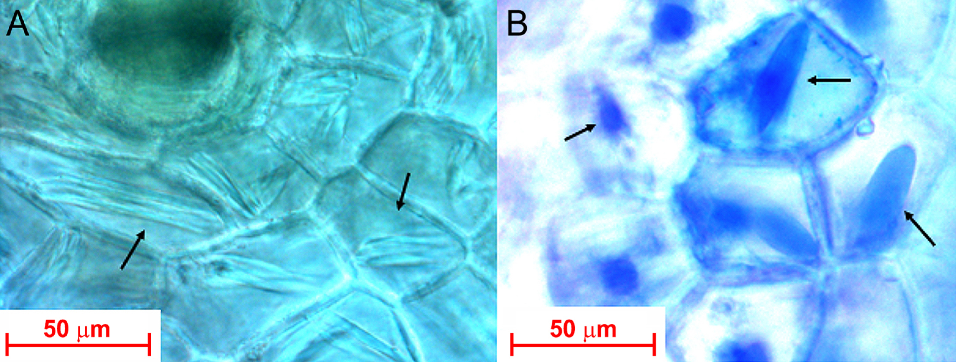
Before morphological, immunological, or molecular techniques for virus detection were available, these inclusions were the sole diagnostic evidence for possible viral infection in cacti. Stomatal abnormalities, such as deformation and fusion of guard cells with accessory cells, were also associated with viral infection (Weber and Kenda, 1953). After these pioneer works on viral inclusions of cacti made by European researchers, similar works were carried out in the USA (Sammons and Chessin, 1961; Chessin et al., 1963; Milbrath et al., 1973; Giri and Chessin, 1975b; Attathom et al., 1978; Fudl-Allah et al., 1983), in Japan (Mukoo et al., 1967; Asatani and Inoue, 1969; Nakamura and Mukoo, 1973), and in Brazil (Aragão et al., 1993) as part of investigations to detect and characterize cacti viruses, mostly potexviruses. Descriptions of cell inclusions, however, have been mostly neglected after the introduction of molecular assays for their detection. An example of a spindle-shaped inclusion in dragon fruit cortical cells, infected by the potexvirus ZyVX, as seen under a light microscope is presented in Figure 2.

Light micrographs of spindle-shaped fibrous inclusions (arrows) in cortical cell of cladode from an asymptomatic moonlight cactus [Selenicereus inermis (Otto) Britton & Rose] infected by Zygocactus virus X. A) Image of a fresh unstained, handmade section showing spindle-shaped inclusions in most cortical cells; B) Same sample, after being stained with Azure A. Inclusions were deeply stained in blue (Del Corona, unpublished data).
Ultrastructural
There are not too many in situ observations of cactus viruses. Virions of cactus viruses have been detected by TEM in extracts from potexviruses CVX-, ZyVX-, CV2-, SOV-, SgCV-infected plants (Amelunxen, 1958; Brandes and Wetter, 1959; Sammons and Chessin, 1961; Mukoo et al., 1967; Casper and Brandes, 1969; Casper et al., 1970; Milbrath and Nelson, 1972; Chessin and Lesemann, 1972; Nakamura and Mukoo, 1973; Milbrath et al., 1973; Giri and Chessin, 1975a, b), and in most of the recent works with cacti potexvirus, tobamovirus, carlavirus, alphacarmovirus, and badnavirus. On the contrary, images of polerovirus, orthotospovirus, cavemovirus, as well as of geminivirus and genomoviridae particles from cacti are not available.
A witches’ broom type of symptoms in O. tuna, previously considered to be of viral etiology (Uschdraweit, 1965), was demonstrated to be of phytoplasma etiology, after the detection of pleomorphic, wall-less bodies in the lumen of sieve tubes (Casper et al., 1970). In this study, the authors also detected particles of CVX and isometric particles ca. 30 nm in extracts and tissue sections. The latter was not identified but may have represented the alphacarmovirus SgCV. Maramorosch et al. (1972) confirmed the phytoplasma etiology of this witches’ broom type of symptoms in O. tuna Mill. "monstruosa", demonstrating the disappearance of pleomorphic bodies from the sieve tube, after tetracycline treatment. These authors also found the aggregate of unidentified tobamovirus-like particles in the cytoplasm. Attathom et al. (1978) reported masses of elongated particles in the cytoplasm of C. quinoa and Amaranthus caudatus L. after experimental inoculation withan isolate of CVX from barrel cactus (F. acanthodes). Similar observations were made with Brazilian isolates of CVX in naturally infected O. vulgaris and C. triangularis, as well as in experimentally infected C. quinoa and Gomphrena globosa L. (Aragão et al., 1993). Duarte et al. (2008) described aggregates of filamentous particles of potexvirus in the cytoplasm of parenchymal cells in Hylocereus undata Haw.) Britton & Rose co-infected withy ZyVX and SchVX, S. truncata co-infected with ZyX, SchVX, OpVX, and O. tuna infected with ZyVX. Kim et al. (2016) in South Korea, detected CVX in symptomless Hylocereus sp., and fibrous inclusion bodies in the cells. Cytoplasmic inclusions formed by aggregation of different potexvirus and tobamoviruses do not present peculiarities; thus, the detection of such inclusions does not have diagnostic value at the species level.
Detection and identification of cactus viruses
Although external symptoms, as mentioned above, may indicate a possible viral infection, virus infections of cacti are usuallyasymptomatic. Indeed, early detection of a cactus virus was a consequence of a cytological study that detected spindle-shaped inclusions (Molisch, 1885), later demonstrated to be formed by CVX particles (Amelunxen, 1958). Although not widely exploited in recent research on cactus viruses, searching for cell inclusions remains a powerful tool for detecting viral infections in cacti (Figure 2). In economically important crops, such as dragon fruit (Selenicereus spp.) and prickly pear (O. ficus-indica), potexviruses and tobamoviruses have been associated with external symptoms as well as in asymptomatic plants. However, there are no reports of economically significant outbreaks of diseases being caused by these viruses.
Identification of viruses in Cactaceae may pose several difficulties due to the frequent occurrence of mixed infection with two or more viruses. Additionally, handling extracts of these plants poses difficulty due to their sticky nature, which is caused by excess mucilage. This problem can be minimized by adding calcium chloride (0.3-0.5 M), which fluidizes the solution (Aragão et al., 1993). Metagenomics of bulk extracts from the sample cactus under study appears to be the best and the most effective method for detecting both known and novel viruses. However, though these methods are still costly and should be used selectively. Most recently described cacti viruses have been identified by metagenomics (Peng et al., 2019; Wu et al., 2019; Salgado-Ortíz et al., 2020; Zheng et al., 2020; Fontenele et al., 2020a, b; Fontenele et al., 2021a, b; Bae and Park, 2022; Ortega-Acosta et al., 2023; Ortega-Acosta et al., 2024; Guimarães et al., 2025).
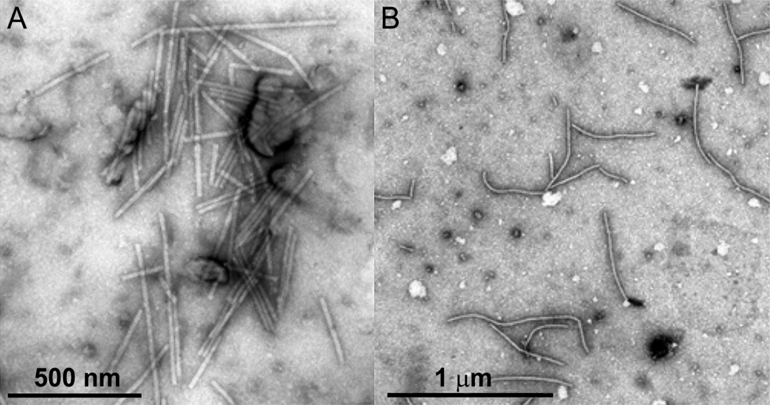
For routine surveys, examination of negatively stained tissue extracts (Figure 3) and tissue sections (Figure 4) by transmission electron microscopy may provide initial hints of viral infection through the detection of presumptive viral particles, especially those occurring in high concentrations as tobamoviruses, potexviruses, carlaviruses, and alphacarmoviruses. Moreover, trials to detect cell inclusion bodies in fresh sections of cacti tissues (Figure 2) may provide clues for viral infection. Mechanical and graft transmission assays may provide additional evidence for viral detection. Definitive identification can be achieved by serology, if specific antibodies are available, or by molecular RT-PCR or RT-qPCR assays, using specific primers and nucleotide sequencing of the amplicons, or by the metagenomic approach. It is always desirable, after their detection, to add some of the biological properties of the detected viruses.
Transmission electron micrographs of virions present in extract from cladode, negatively stained with uranyl acetate. A) Rod-shaped particles of rattail cactus necrosis-associated virus, a tobamovirus, in cladode extract from asymptomatic Opuntia leucotricha, sampled at Instituto Plantarum, Nova Odessa, SP, Brazil; B) Elongated, flexible particles of the potexvirus Zygocactus virus X present in cladode extract of an asymptomatic dragon fruit (Selenicereus sp.) collected in an experimental field at Piracicaba, SP, Brazil.

Transmission electron micrographs of tissue sections exhibiting aggregates of virions. A) A stacked mass of rod-like particles (V) of the tobamovirus rattail cactus necrosis associated virus in a cortical cell of a cladode from an asymptomatic Opuntia leucotricha; B) Fibrous bundle of elongated particles of the potexvirus Zygocactus virus in a cortical cell of an asymptomatic dragon fruit (Selenicereus sp.) plant. CW = cell wall; Vc = vacuole.
Dissemination of cactus viruses
Cacti tobamoviruses and potexviruses have been described in a large number of cultivated cacti (ornamentals, forage, fruit-producing) species. In most of these cases, the transmission must have occurred using infected rootstocks (such as H. trigonus, H. undatus, Cereus peruvianus (L.) Mill., Trichocereus pachanoi Britton & Rose, etc.) for grafting or by handling of plants during cultivation (such as pruning and harvesting).
The situation worsens since frequent virus infections in cacti are often symptomless, leading to the use of infected mother plants for vegetative propagation through cloning, for example, in dragon fruit cultivation. This fact may explain the widespreaddistribution of potexviruses infecting this crop globally. In the wild, one possibility means of dissemination for viruses, although likely with low efficiency, could involve flower-visiting organisms (insects, birds, bats), potentially through contaminated pollen, as suggested for SgCV (Milbrath et al., 1973); however, this has yet to be demonstrated. Therefore, the widespread distribution of some cactus viruses is the result of human activities, including the large-scale relocation of cactus species from their center of origin and the transmission of viruses through mechanical handling and grafting.
In the case of cacti infection by poleroviruses and orthotospoviruses, this was certainly mediated by infected aphids and thrips, originating from a non-cactus source. No information is available about the dissemination of newly described and widely spread Geminiviridae and Genomoviridae species detected by mass sequencing.
Final Remarks
To date, 31 distinct viruses, belonging to 13 genera and nine families, and three viroids of two genera, have been described as naturally infecting approximately 70 Cactaceae species, mostly belonging to the genus Opuntia, worldwide. Until recently, all of them belonged to the Riboviria realm, Orthornavirae kingdom (genera Potexvirus, Tobamovirus, Carlavirus, Alphacarmovirus, Polerovirus and Orthotospovirus), but in the last two decades, after the use of genome deep sequencing, several novel species belonging to the Pararnavirae kingdom, (family Caulimoviridae, genera Badnavirus and Cavemovirus) and Monodnaviria realm, Shotokuvirae kingdom, Geminiviridae family, (genera Opunvirus, Becurtovirus) and Genomoviridae family, (genera Gemykolovirus, Gemyduguivirus, Gemygorvirus, Gemykroznavirus, Gemycircularvirus and Gemykibivirus), as well as viroids, were found.
In general, these cacti viruses cause asymptomatic infections; however, cases of ringspots, necrosis, mottling, and deformations of cladodes have been reported, especially in dragon fruit and prickly pear. A number of these viruses infect economically important cacti such as dragon fruit and prickly pear, but there is no information available on possible losses due to the infection. Certain viruses found infecting cacti, such as AltMV, TMV, BWYV, and orthotospoviruses probably originate from non-Cactaceae plants, representing cases of accidental infections. However, cactus-specific potexviruses (CVX, OpVX, PiVX, SchVX, ZyVX), tobamoviruses (SOV, CMMoV, RCNaV, OpV2), carlaviruses (CV2, CCV-1 and 2), alphacarmovirus (SgCV), and the viruses discovered by metagenomics (Badnavirus, Cavemovirus and Geminiviridae, and Genomoviridae) are probably cacti viruses, present in the wild growing cacti species.
Several of them have been found in cultivated ornamental or economically exploited cacti, likely originating from wild plants and disseminated via nurseries through handling and grafting procedures. Interestingly, although these viruses can experimentally infect non-cactus plants, no naturally infecting plants outside the Cactaceae family have been identified yet; thus, their host range seems limited to cacti. More viruses in cactus plants, both wild and cultivated, will likely be found, especially with the increasing use of metagenomics.
Acknowledgments
This work received financial support from Fundação de Amparo à Pesquisa do Estado de São Paulo (FAPESP, 2021/02179-4; 2024/06440-7). The authors, FP Del Corona (88887.824702/2023-00), and LL. Pires (88887.702325/2022-00), are recipients of Doctoral scholarships from Coordenação de Aperfeiçoamento do Ensino Superior (CAPES). JJV Guimarães (88887.952284/2024-00) receives a master's scholarship, also from CAPES. The author, EW Kitajima, is a recipient of a Conselho Nacional de Desenvolvimento Científico e Tecnológico (CNPq) research dedication scholarship 1A (30.3715/2021-9).
References
-
Abirami K, Swain S, Baskaran V, Venkatesan K, Sakthivel K, Bommayasamy N. 2021. Distinguishing three Dragon fruit (Hylocereus spp.) species grown in Andaman and Nicobar Islands of India using morphological, biochemical and molecular traits. Scientific Reports 11: 2894. https://doi.org/10.1038/s41598-021-81682-x
» https://doi.org/10.1038/s41598-021-81682-x - Alonso-Barrera B, Mora-Aguilera G, Valdovinos-Ponce G, Ochoa-Martinez DL, Rodriguez-Leyva E, Tlapal-Bolaños B, et al. 2015. Association of Potexvirus as a causal agent of chlorotic spots on Opuntia ficus-indica Revista Mexicana de Fitopatología 33: 75-86.
-
Amelunxen F. 1958. Die Virus-Eiwei\spindeln der Kakteen. Darstellung, elektronenmikroskopische und biochemische Analyse des Virus. Protoplasma 49: 140-178 (in German). https://doi.org/10.1007/BF01248121
» https://doi.org/10.1007/BF01248121 - Anderson EF. 2001. The Cactus Family. Timber Press, Portland, OR, USA.
- Aragão FJL, Marinho VLA, Kitajima EW. 1993. Cactus virus X in Cactaceae in Brazil and a novel method to purify it directly from cactus tissues. Fitopatologia Brasileira 18: 112-117.
- Asatani M, Inoue T. 1969. Cactus virus X isolated from cactus. Nogaku Kenkyu 53: 35-48 (in Japanese).
- Attathom S, Weathers LG, Gumpf DJ. 1978. Identification and characterization of a Potexvirus from California barrel cactus. Phytopathology 68: 1401-1406.
-
Bae M, Park M-R. 2022. First report of pitaya virus X and zygocactus virus X infection of dragon fruit (Selenicereus undatus) in Korea. Journal of Plant Pathology 104: 1581. https://doi.org/10.1007/s42161-022-01206-y
» https://doi.org/10.1007/s42161-022-01206-y -
Baker CA, Adkins S. 2015. First report of Tomato chlorotic spot virus in Hoya wayetii and Schlumbergera truncata. Plant Health Progress 16: 29-30. https://doi.org/10.1094/PHP-BR-14-0043
» https://doi.org/10.1094/PHP-BR-14-0043 -
Balendres MA, Bengoa JC. 2019. Diseases of dragon fruit (Hylocereus species): Etiology and current management options. Crop Protection 126: 104920. https://doi.org/10.1016/j.cropro.2019.104920
» https://doi.org/10.1016/j.cropro.2019.104920 - Blattny G, Vukolov V. 1932. Mosaik bei Epiphyllum truncatum Gartenbauwissenschaft 6: 425 (in German).
-
Blockley AL, Mumford RA. 2001. Identification and isolation of Impatiens necrotic spot virus from prickly pear cactus (Opuntia microdasys). Plant Pathology 50: 805. https://doi.org/10.1046/j.1365-3059.2001.00628.x
» https://doi.org/10.1046/j.1365-3059.2001.00628.x -
Brandes J, Bercks R. 1963. Untersuchungen zur Identifizierung und Klassifizierung des Kakteen-X-Virus (cactus virus X). Phytopathogische Zeitschrift 46: 291-300 (in German). https://doi.org/10.1111/j.1439-0434.1963.tb02072.x
» https://doi.org/10.1111/j.1439-0434.1963.tb02072.x -
Brandes J, Chessin M. 1965. An electron microscope study on the size of Sammon's Opuntia virus. Virology 25: 674-674. https://doi.org/10.1016/0042-6822(65)90100-5
» https://doi.org/10.1016/0042-6822(65)90100-5 -
Brandes J, Wetter C. 1959. Classification of elongated plant viruses on the basis of particle morphology. Virology 8: 99-115. https://doi.org/10.1016/0042-6822(59)90022-4
» https://doi.org/10.1016/0042-6822(59)90022-4 - Brunt AA, Crabtree K, Gibbs A. 1990. Viruses of Tropical Plants. Descriptions and lists from VIDE database. CAB International, Wallingford, UK.
-
Casper R, Brandes J. 1969. A new cactus virus. Journal of General Virology 5: 155-156. https://doi.org/10.1099/0022-1317-5-1-155
» https://doi.org/10.1099/0022-1317-5-1-155 - Casper R, Lesemann D, Bartels R. 1970. Mycoplasma-like bodies and viruses in Opuntia tuna with witches’-broom disease. Plant Disease Reporter 54: 851-853.
- Chen MJ, Tzeng DDS. 1996. Identification of cactus X Potexvirus in Taiwan and localization of its existence and multiplication in infected cells. Plant Pathology Bulletin 5: 63-67 (in Chinese, with abstract in English).
-
Chessin M. 2002. Symptoms of virus infection in cactus. Acta Horticulturae 568: 73-77. https://doi.org/10.17660/ActaHortic.2002.568.9
» https://doi.org/10.17660/ActaHortic.2002.568.9 - Chessin M, Lesemann D. 1972. Distribution of cactus viruses in wild plants. Phytopathology 62: 97-99.
- Chessin M, Solberg RA, Fischer PC. 1963. External symptoms and giemsa-stainable cell inclusions associated with virus infection in cacti. Phytopathology 53: 988-989.
- Chmielewsky V. 1887. Eine Bemerkung über die von Molisch beschriebenen Proteïnkörper in den Zweigen von Epiphyllum. Botanisches Zentralblatt 31: 117-119 (in German).
-
De La Torre-Almaráz R, Salgado-Ortíz H, Salazar-Segura M, Pallas V, Sánchez-Navarro JA, Valverde RA. 2016a. First report of Rattail cactus necrosis-associated virus in Prickly pear fruit (Opuntia albicarpa Scheinvar) in Mexico. Plant Disease 100: 2339. https://doi.org/10.1094/PDIS-04-16-0554-PDN
» https://doi.org/10.1094/PDIS-04-16-0554-PDN -
De la Torre-Almaráz R, Salgado-Ortíz H, Salazar-Segura M, Pallas V, Sánchez-Navarro JA, Valverde RA. 2016b. First report of Schlumbergera virus X in Prickly pear (Opuntia ficus-indica). Plant Disease 100: 1799. https://doi.org/10.1094/PDIS-11-15-1326-PDN
» https://doi.org/10.1094/PDIS-11-15-1326-PDN - De Soto JF, Lobo R, Aguiar JL, Mathews DM, Tanizaki G. 2014. Cactus virus X (CVX) a new threat to pitahaya/dragon fruit (Hylocereus spp.) production in California. ASHS Annual Conference, Orlando, FL, USA.
-
Desserto®. 2023. Available at: https://desserto.com.mx/home [Accessed 10 Sept, 2023]
» https://desserto.com.mx/home -
Dragonfruit.net. 2024. Available at: https://dragonfruit.net.vn/news/185-world-s-dragon-fruit-suppliers-and-demand [Accessed Feb 20, 2024]
» https://dragonfruit.net.vn/news/185-world-s-dragon-fruit-suppliers-and-demand - Duarte LML, Alexandre MAV, Rivas EB, Harakava R, Galleti SR, Barradas MM. 2008. Potexvirus diversity in Cactaceae from São Paulo state in Brazil. Journal of Plant Pathology 90: 545-551.
-
Eicholtz M, Grinstead S, Wu L-P, Kinard G, Li R. 2018. First report of Beet western yellows virus infecting Epiphyllum spp. Plant Disease 102: 464. https://doi.org/10.1094/PDIS-08-17-1238-PDN
» https://doi.org/10.1094/PDIS-08-17-1238-PDN -
Eiras M, Chaves ALR, Del Corona FP, Guimarães JJV, Bello VH, Kitajima EW, et al. 2025. First report of Alternanthera mosaic virus infecting Pereskia aculeate in Brazil. Plant Disease 109: 1187. https://doi.org/10.1094/PDIS-01-25-0057-PDN
» https://doi.org/10.1094/PDIS-01-25-0057-PDN -
Espinoza-Lozano L, Sumba M, Cañada-Bautista MG, Quito-Ávila DF. 2024. Occurrence, distribution, and population structure of Schlumbergera virus X in dragon fruit in Ecuador. Plant Disease 108: 587-591. https://doi.org/10.1094/PDIS-03-23-0445-SC
» https://doi.org/10.1094/PDIS-03-23-0445-SC -
Evallo E, Taguiam JD, Balendres MA. 2021. A brief review of plant diseases caused by Cactus virus X Crop Protection 143: 105566. https://doi.org/10.1016/j.cropro.2021.105566
» https://doi.org/10.1016/j.cropro.2021.105566 -
Evallo E, Taguiam JD, Tumolva JA, Bengoa J, Maghirang R, Balendres MA. 2022. First report of Cactus virus X in Selenicereus monacanthus the Philippines. Indian Phytopathology 75: 1237-1239. https://doi.org/10.1007/s42360-022-00555-z
» https://doi.org/10.1007/s42360-022-00555-z - Faleiro FG. 2022. Pitaia: a fruta que está conquistando o Brasil. Anuário Campo e Negócios Hortifruti 11: 97-99 (in Portuguese).
-
Fontenele RS, Roumagnac R, Richet C, Kraberger S, Stainton D, Aleamotu'a M, et al. 2020a. Diverse genomoviruses representing twenty-nine species identified associated with plants. Archives of Virology 165: 2891-2901. https://doi.org/10.1007/s00705-020-04801-5
» https://doi.org/10.1007/s00705-020-04801-5 -
Fontenele RS, Salywon AM, Majure LC, Cobb IN, Bhaskara A, Avalos-Calleros JA, et al. 2020b. A novel divergent Geminivirus identified in asymptomatic New World Cactaceae plants. Viruses 12: 398. https://doi.org/10.3390/v12040398
» https://doi.org/10.3390/v12040398 -
Fontenele RS, Salywon AM, Majure LC, Cobb IN, Bhaskara A, Avalos-Calleros JA, et al. 2021a. New World Cactaceae plants harbor diverse Geminiviruses. Viruses 13: 694. https://doi.org/10.3390/v13040694
» https://doi.org/10.3390/v13040694 -
Fontenele RS, Köhler M, Majure LC, Avalos-Calleros JA, Argüello-Astorga GR, Font F, et al. 2021b. Novel circular DNA virus identified in Opuntia discolor (Cactaceae) that codes for proteins with similarity to those of geminiviruses. Journal of General Virology 102: 11. https://doi.org/10.1099/jgv.0.001671
» https://doi.org/10.1099/jgv.0.001671 -
Fudl-Allah AE-SA, Weathers LG, Greer Jr FC. 1983. Characterization of a Potexvirus isolated from night-blooming Cactus. Plant Disease 67: 438-440. https://doi.org/10.1094/PD-67-438
» https://doi.org/10.1094/PD-67-438 -
Galappaththi M, Patabendige N. 2021. Cochineal chemistry, related applications and problems: a mini review. Academia Letters 1792: 1-12. https://doi.org/10.20935/AL1792
» https://doi.org/10.20935/AL1792 -
Gazis R, Poudel B, Dey KK, Zhang S, Palmateer A, Campoverde EV, et al. 2018. First report of Cactus virus X in Hylocereus undatus (dragon fruit) in Florida. Plant Disease 102: 2666. https://doi.org/10.1094/PDIS-05-18-0725-PDN
» https://doi.org/10.1094/PDIS-05-18-0725-PDN - Giri L, Chessin M. 1972. Characterization of a virus isolated from a cultivated cactus, Zygocactus sp. Phytopathology 62: 1103.
-
Giri L, Chessin M. 1975a. Zygocactus Virus X. Phytopathogische Zeitschrift 83: 40-48. https://doi.org/10.1111/j.1439-0434.1975.tb03511.x
» https://doi.org/10.1111/j.1439-0434.1975.tb03511.x -
Giri L, Chessin M. 1975b. A severe strain of Tobacco mosaic virus from cactus. Phytopathology 65: 824–825. https://doi.org/10.1094/Phyto-65-824
» https://doi.org/10.1094/Phyto-65-824 - Gicklhorn J. 1913. Über das Vorkommen spindelförmiger Eiweiβkörper bei Opuntia Oesterreichische botanische Zeitschrift 63: 8-15 (in German).
- Goldin MI, Fedontina VL. 1956. Die Verbreitung der Eiweiss- (Virus-) Einschlusskörper in verschiedenen Kakteen. Byulleten’ Glavnogo Botanitscheskogo Sada (Moskow) 26: 80-84 (in Russian).
- Greenfield AB. 2009. A Perfect Red: Empire, Espionage, and the Quest for the Color of Desire. Harper Collins, New York, NY, USA.
-
Guimarães JJV, Del Corona FP, Bello V, Ramos-González PL, Lorenzi H, Kitajima EW. 2025. First report of rattail cactus necrosis-associated virus (Tobamovirus muricaudae) in symptomless Opuntia leucotricha in Brazil. Journal of Phytopathology 173: e70063. https://doi.org/10.1111/jph.70063.2025
» https://doi.org/10.1111/jph.70063.2025 -
Hausbeck MK, Gildow FE. 1991. Report of tomato spotted wilt virus on thanksgiving cactus. Plant Disease 75: 215. https://doi.org/10.1094/PD-75-0215C
» https://doi.org/10.1094/PD-75-0215C -
Hausbeck MK, Welliver RA, Derr MA, Gildow FE. 1992. Tomato spotted wilt virus survey among greenhouse ornamentals in Pennsylvania. Plant Disease 76: 795-800. https://doi.org/10.1094/PD-76-0795
» https://doi.org/10.1094/PD-76-0795 - Hung CL, Liou MR, Liou RF. 1999. Identification and characterization of a potexvirus on pitaya (Hylocereus undatus). Plant Pathology Bulletin 8: 177.
-
ICTV. 2024. ICTV_Master_Species_List_2023_MSL39.v2.xlsx. International Committee for Virus Taxonomy. Available at: https://ictv.global/msl [Accessed Jun 15, 2024]
» https://ictv.global/msl -
Izaguirre-Mayoral ML, Marys E. 1996. Interactions between irradiance levels and cactus X virus infection on the crassulacean acid metabolism in Nopalea cochenillifera and Acanthocereus tetragonus plants. Journal of Plant Physiology 149: 35-42. https://doi.org/10.1016/S0176-1617(96)80170-2
» https://doi.org/10.1016/S0176-1617(96)80170-2 -
Janssen D, García C, Ruiz L. 2022. First report of Schlumbergera virus X in dragon fruit (Hylocereus spp.) in Spain. Plant Disease 106: 1773-2006. https://doi.org/10.1094/PDIS-09-21-1879-PDN
» https://doi.org/10.1094/PDIS-09-21-1879-PDN - Kamikawachi RC. 2019. Estudo fitoquímico e potencial biológico de cactos da Mata Atlântica do gênero Rhipsalis Gärtner (Cactaceae). UNESP, São Vicente, SP, Brazil (in Portuguese, with abstract in English).
-
Kenda G. 1954. Eiweißspindeln in Opuntia monacantha f. variegata. Protoplasma 44: 192-193 (in German). https://doi.org/10.1007/BF01247960
» https://doi.org/10.1007/BF01247960 - Kim JB, Roh KH, Kim HU, Lee KR, Kim SH, Lee SH, et al. 2011. Transgenic resistance of trigonal cactus (Hylocereus trigonus) to cactus virus X (CVX). Korean Society for Plant Biotechnology 2011: 86 (in Korean).
- Kim JB, Roh KH, Kim HU, Lee KR, Kim SH, Lee HS, et al. 2012a. Reverification of transgenic resistance of trigonal cactus (Hylocereus trigonus) to cactus virus X (CVX). Journal of the Korean Plant Biotechnology Society 2012: 94 (in Korean).
-
Kim JS, Park CY, Nam M, Lee JS, Kim HG, Lee SH. 2016. First report of Cactus virus X infecting Hylocereus undatus in Korea. Plant Disease 100: 2544. https://doi.org/10.1094/PDIS-06-16-0889-PDN
» https://doi.org/10.1094/PDIS-06-16-0889-PDN -
Kim NR, Hong JS, Song YS, Chung BN, Park JW, Ryu KH. 2012b. The complete genome sequence of a member of a new species of tobamovirus (rattail cactus necrosis-associated virus) isolated from Aporcactus flagelliformis Archives of Virology 157: 185-187. https://doi.org/10.1007/s00705-011-1142-5
» https://doi.org/10.1007/s00705-011-1142-5 -
Kim N-Y, Jo YH, Bae YM, Lee K-P, Jeong R-D, Han YS, et al. 2023. First report of pitaya virus X infecting dragon fruit (Selenicereus undatus) in Korea. Journal of Plant Pathology 105: 1149. https://doi.org/10.1007/s42161-023-01335-y
» https://doi.org/10.1007/s42161-023-01335-y - Kinupp VF, Lorenzi H. eds. 2014. Plantas Alimentícias Não Convencionais (PANC) no Brasil. Plantarum, Nova Odessa, SP, Brazil (in Portuguese).
-
Koenig R, Pleij CWA, Loss S, Burgermeister W, Aust H, Schiemann J. 2004. Molecular characterisation of potexviruses isolated from three different genera in the family Cactaceae Archives of Virology 149: 903-914. https://doi.org/10.1007/s00705-003-0268-5
» https://doi.org/10.1007/s00705-003-0268-5 -
Koenig R, Lesemann D-E, Loss S, Engelmann J, Commandeur U, Demi G, et al. 2006. Zygocactus virus X-based expression vectors and formation of rod-shaped virus-like particles in plants by the expressed coat proteins of Beet necrotic yellow vein virus and Soil-borne cereal mosaic virus Journal of General Virology 87: 439-443. https://doi.org/10.1099/vir.0.81477-0
» https://doi.org/10.1099/vir.0.81477-0 -
Korotkova N, Borsch T, Arias S. 2017. A phylogenetic framework for the Hylocereus (Cactaceae) and implications for the circumscription of the genera. Phytotaxa 327: 1-46. https://doi.org/10.11646/phytotaxa.327.1.1
» https://doi.org/10.11646/phytotaxa.327.1.1 -
Küster E. 1934. Anisotrope fibrillenbündel in pflanzenzellen. Berichte der Deutschen Botanischen Gesellschaft 52: 564-573 (in German). https://doi.org/10.1111/j.1438-8677.1934.tb01715.x
» https://doi.org/10.1111/j.1438-8677.1934.tb01715.x - Lamas N, Sanches MM, Alves-Freitas DMT, Reis MBA, Sousa JGA, Romano E, et al. 2014. Genome sequencing of two potexvirus infecting prickly pear (Opuntia cochenillifera). Virus Reviews and Research 19: 217.
-
Lan P, Tian T, Pu L, Rao W, Li F, Li R. 2019. Characterization and detection of a new badnavirus infecting Epiphyllum spp. Archives of Virology 164: 1837-1841. https://doi.org/10.1007/s00705-019-04237-6
» https://doi.org/10.1007/s00705-019-04237-6 -
Lee K. 2020. Investigation of host genes affected by cactus virus X and Pitaya virus X infection. National Taiwan University, Taipei, Taiwan (in Chinese, with abstract in English). https://doi.org/10.6342/NTU202003267
» https://doi.org/10.6342/NTU202003267 -
Li Y-S. 2010. Characterization, infectious clone construction and antiserum preparation of Pitaya virus X. National Taiwan University, Taipei, Taiwan (in Chinese, with abstract in English). https://doi.org/10.6342/NTU.2010.02361
» https://doi.org/10.6342/NTU.2010.02361 - Li Y-S, Mao C, Kuo T-Y, Chang Y-C. 2015. Viral diseases of pitaya and other Cactaceae plants. p. 119-127. In: Jiang Y-L, Liu P-C, Huang P-H. eds. Improving Pitaya production and marketing: International Workshop Proceedings. Food and Fertilizer Technology Center, Taipei, Taiwan.
- Liao JY, Chang CA, Yan CR, Chen YC, Deng TC. 2003. Detection and incidence of Cactus virus X on pitaya in Taiwan. Plant Pathology Bulletin 12: 225-234 (in Chinese, with abstract in English).
-
Lim MS, Min BE, Choi SH. 2022. First report of Saguaro cactus virus infecting Gymnocalycium mihanovichii in South Korea. Plant Disease 106: 1310. https://doi.org/10.1094/PDIS-04-21-0770-PDN
» https://doi.org/10.1094/PDIS-04-21-0770-PDN -
Liou MR, Hung CL, Liou RF. 2001. First report of Cactus virus X on Hylocereus undatus (Cactaceae) in Taiwan. Plant Disease 85: 229. https://doi.org/10.1094/PDIS.2001.85.2.229A
» https://doi.org/10.1094/PDIS.2001.85.2.229A -
Liou MR, Chen YR, Liou RF. 2004. Complete nucleotide sequence and genome organization of a Cactus virus X strain from Hylocereus undatus (Cactaceae). Archives of Virology 149: 1037-1043. https://doi.org/10.1007/s00705-003-0251-1
» https://doi.org/10.1007/s00705-003-0251-1 -
L’Occitane au Brésil. 2016. Available at: https://br.loccitaneaubresil.com[Accessed Sept 10, 2023.
» https://br.loccitaneaubresil.com - Lodé J. 2015. Taxonomía de las cactáceas. La nueva clasificación de los cactus basada en la investigación molecular y explicada. 1-2ed. Ediciones Cactus-Aventuras, Almería, Spain (in Spanish).
-
Lucena CM, Lucena RFP, Costa GM, Carvalho TKN, Costa GGS, Alves RRN, et al. 2013. Use and knowledge of Cactaceae in Northeastern Brazil. Journal of Ethnobiology and Ethnomedicine 9: 62. https://doi.org/10.1186/1746-4269-9-62
» https://doi.org/10.1186/1746-4269-9-62 - Mao C-H. 2008. Molecular characterization and detection of new Zygocactus virus X and Pitaya virus X from pitaya. National Taiwan University, Taipei, Taiwan (in Chinese, with abstract in English).
-
Mao C-H, Kuo T-Y, Lu Y, Li Y-S, Chang Y-C. 2018. Pitaya viral diseases and their detection methods in Taiwan. Dragon Fruit Regional Network Initiation Workshop, Taipei, Taiwan. https://doi.org/10.56669/WQEJ8648
» https://doi.org/10.56669/WQEJ8648 -
Maramorosch K, Klein M, Wolanski BS. 1972. Beitrag zur Ätioloogie der Hexenbesenkrankheit der Kaktee Opuntia tuna (= tuna monstrosa). Experientia 28: 362-363 (in German). https://doi.org/10.1007/BF01928742
» https://doi.org/10.1007/BF01928742 -
Marliarenko VM, Mudrak TP. 2013. Cactus viruses in fasciated plants. Biologija 59: 213-218. https://doi.org/10.6001/biologija.v59i2.2754
» https://doi.org/10.6001/biologija.v59i2.2754 -
Masanto M, Sijam K, Awang Y, Satar MGM. 2018. First report of necrotic spot disease caused by Cactus virus X on dragon fruit (Hylocereus spp.) in Peninsular Malaysia. Jurnal Perlindungan Tanaman Indonesia 22: 1-12. https://doi.org/10.22146/jpti.23763
» https://doi.org/10.22146/jpti.23763 - Medeiros ML, Figueira AR, Silveira ATL, Silva RMS, Carvalho CM, Holanda GC. 2023. Análise filogenética de dois isolados de Cactus virus X. UFLA, Lavras, MG, Brazil. (in Portuguese).
- Mikosch C. 1890. Ueber ein neues Vorkommen geformten Eiweisses. Berichte der Deutschen Botanischen Gesellschaft 8: 33-38 (in German).
-
Mikosch C. 1908. Über den Einfluß des Reises auf die Unterlage. p. 280. In: Wiesner J, Linsbauer K. eds. Wiesner-Festschrift. Konegen, Wien, German (in German). https://doi.org/10.5962/bhl.title.10912
» https://doi.org/10.5962/bhl.title.10912 - Milbrath GM. 1971. Etiology and epidemiology of viruses of native cactus species in Arizona. The University of Arizona. Tucson, AZ, USA.
-
Milbrath GM, Nelson MR. 1972. Isolation and characterization of a virus from Saguaro Cactus. Phytopathology 62: 739-742. https://doi.org/10.1094/Phyto-62-739
» https://doi.org/10.1094/Phyto-62-739 - Milbrath GM, Nelson MR, Wheeler RE. 1973. The distribution and electron microscopy of viruses of cacti in Southern Arizona. Phytopathology 63: 1133-1139.
-
Miličić D. 1954. Viruskörper und Zellteilungsanomalien in Opuntia brasiliensis. Protoplasma 43: 228-236 (in German). https://doi.org/10.1007/BF01250988
» https://doi.org/10.1007/BF01250988 - Miličić D. 1956. Eiweißkristalloide in Opuntia inermis. Österreichische Botanische Zeitschrift 103: 365-375 (in German).
- Miličić D. 1959/1960. Sind verschiedene Eiweisskristalle der Kakteen Viruskörper? Acta Botanica Croatica 18/19: 37-60 (in German).
-
Miličić D. 1962a. Neue Wirtspflanzen des Kakteenvirus. Berichte der Deutschen Botanischen Gesellschaft 75: 172-178 (in German). https://doi.org/10.1111/j.1438-8677.1962.tb02522.x
» https://doi.org/10.1111/j.1438-8677.1962.tb02522.x -
Miličić D. 1962b. Virus-Einschlußkörper in Lokalläsionen. Journal of Phytopathology 44: 282-294 (in German). https://doi.org/10.1111/j.1439-0434.1962.tb02018.x
» https://doi.org/10.1111/j.1439-0434.1962.tb02018.x -
Miličić D, Plavšić B. 1956. Eiweißkristalloide em Kakteen-Virusträgern. Protoplasma 46: 547-555 (in German). https://doi.org/10.1007/BF01248899
» https://doi.org/10.1007/BF01248899 -
Miličić D, Udjbinac Z. 1961. Virus-Eiweißspindeln der Kakteen in Lokalläsionen von Chenopodium Protoplasma 53: 584-596 (in German). https://doi.org/10.1007/BF01248233
» https://doi.org/10.1007/BF01248233 -
Miličić D, Pleše N, Bercks R, Brandes J, Caspar R, Chessin M. 1966. Vergleichende serologische und elektronenmikroskopische Untersuchungen an Isolaten des Kakteen-X-Virus. Journal of Phytopathology 55: 211-217 (in German). https://doi.org/10.1111/j.1439-0434.1966.tb02227.x
» https://doi.org/10.1111/j.1439-0434.1966.tb02227.x -
Min BE, Ryu KH, Chung BN. 2006a. Molecular evidence that cactus mild mottle virus is a new tobamovirus. Acta Horticulturae 722: 181-184. https://doi.org/10.17660/ActaHortic.2006.722.22
» https://doi.org/10.17660/ActaHortic.2006.722.22 -
Min BE, Chung BN, Kim MJ, Ha JH, Lee BY, Ryu KH. 2006b. Cactus mild mottle virus is a new cactus-infecting tobamovirus. Archives of Virology 151: 13-21. https://doi.org/10.1007/s00705-005-0617-7
» https://doi.org/10.1007/s00705-005-0617-7 -
Min BE, Song YS, Ryu KH. 2009. Complete sequence and genome structure of cactus mild mottle virus. Archives of Virology 154: 1371-1374. https://doi.org/10.1007/s00705-009-0435-4
» https://doi.org/10.1007/s00705-009-0435-4 - Mizrahi Y, Nerd A, Sitrit Y. 2002. New fruits for arid climates. p. 378-384. In: Janick J, Whipkey A. eds. Trends in new crops and new uses. American Society for Horticultural Science Press, Alexandria, VA, USA.
-
Mole-Bajer J. 1953. Experimental studies on protein spindles. Acta Societatis Botanicorum Poloniae 22: 811-828. https://doi.org/10.5586/asbp.1953.050
» https://doi.org/10.5586/asbp.1953.050 -
Molisch H. 1885. Ueber merkwürdig geformte Proteinkörper in den Zweigen von Epiphyllum. Berichte der Deutschen Botanischen Gesellschaft 3: 195-202 (in German). https://doi.org/10.1111/j.1438-8677.1885.tb04251.x
» https://doi.org/10.1111/j.1438-8677.1885.tb04251.x -
Mordorintelligence. 2024. Dragon fruit market size & share analysis – Growth trends & forecasts (2025-2030). Available at: https://www.mordorintelligence.com/industry-reports/dragon-fruit-market[Accessed Feb 20, 2024]
» https://www.mordorintelligence.com/industry-reports/dragon-fruit-market - Morris SJ. 1977. Viruses of Opuntias. Zeitschrift für Pflanzenkrankheiten und Pflanzenschutz 84: 468-472. (in German)
- Mudrak TP, Bysov AS, Kompanets TA, Korotyeyeva GV. 2008. Incidence of Cactus virus 2 in collection of Ukrainian botanical gardens. Біологія 51: 29-31.
- Mukoo H, Nakamura S, Kondo N. 1967. On cacti viral disease. Annals of the Phytopathological Society of Japan 33: 345 (in Japanese).
-
Murcia-Bermudez JM, Singh N, Chamberland M, Poudel-Ward B. 2024. First report of rattail cactus necrosis-associated virus infecting prickly pear (Opuntia macrocentra) in the United States. Plant Disease 108: 550-828. https://doi.org/10.1094/PDIS-07-23-1344-PDN
» https://doi.org/10.1094/PDIS-07-23-1344-PDN - Nakamura S, Mukoo H. 1973. A research on viral disease of cacti. Report from Department of Agricultural Sciences. Tokyo Agricultural University 18: 91-101 (in Japanese, with abstract in English)
- Natsuaki KT, Shinkai M. 2001. Characterization of Cactus virus X from Hylocereus undatus and Selenicereus megalanthus showing mosaic symptoms. Journal of Agricultural Science - Tokyo Nogyo Daigaku 45: 325-330 (in Japanese, with abstract in English).
-
Nelson MR, Tremaine JH. 1975. Physiochemical and serological properties of a virus from saguaro cactus. Virology 65: 309-319. https://doi.org/10.1016/0042-6822(75)90037-9
» https://doi.org/10.1016/0042-6822(75)90037-9 - Nobel PS. 2002. Cacti: Biology and Uses. University of California Press, Berkeley, CA, USA.
-
Ortega-Acosta C, Ochoa-Martínez DL, Rojas-Martínez RI, Nava-Díaz C, Valverde RA. 2023. Virome of the vegetable prickly pear cactus in the central zone of Mexico. Revista Mexicana de Fitopatologia 41: 3. https://doi.org/10.18781/R.MEX.FIT.2023-2
» https://doi.org/10.18781/R.MEX.FIT.2023-2 -
Ortega-Acosta C, Ochoa-Martínez DL, Rodríguez-Leyva E. 2024. High-Throughput Sequencing Reveals New Viroid Species in Opuntia in Mexico. Viruses 16: 1177. https://doi.org/10.3390/v16081177
» https://doi.org/10.3390/v16081177 -
Ortiz-Hernández YD, Carrillo-Salazar JA. 2012. Pitahaya (Hylocereus spp.): a short review. Comunicata Scientiae 3: 220-237. https://doi.org/10.14295/cs.v3i4.334
» https://doi.org/10.14295/cs.v3i4.334 - Pape H. 1932. Mosaikkrankheit an Glieder-, Blatt und Rutenkakteen. Gartenwelt 36: 731-732 (in German).
-
Parameswari B, Bajaru B, Karthikaiselvi L, Sivaraj N, Mangrauthia SK, Nagalakshimi S, et al. 2023. First report of the association of Zygocactus virus X with dragon fruit (Hylocereus sp.) plants from Telangana, India. Plant Disease 107: 1249. https://doi.org/10.1094/PDIS-05-22-1242-PDN
» https://doi.org/10.1094/PDIS-05-22-1242-PDN -
Parameswari B, Bajaru B, Sivaraj N, Mangrauthia SK, Nagalakshimi S, Baranwal VK, et al. 2022. First record of Cactus virus X in dragon fruit (Hylocereus spp.) in India. Indian Phytopathology 75: 297-299. https://doi.org/10.1007/s42360-021-00421-4
» https://doi.org/10.1007/s42360-021-00421-4 -
Park CH, Song EG, Ryu KH. 2018. Detection of co-infection of Notocactus leninglhausii f. cristatus with six virus species in South Korea. The Plant Pathology Journal 34: 65-70. https://doi.org/10.5423/PPJ.NT.08.2017.0187
» https://doi.org/10.5423/PPJ.NT.08.2017.0187 -
Park CH, Song EG, Ryu KH. 2021. A multiplex PCR assay for the simultaneous detection of five potexviruses infecting cactus plants using dual-priming oligonucleotides (DPO) primers. Journal of Virological Methods 298: 114280. https://doi.org/10.1016/j.jviromet.2021.114280
» https://doi.org/10.1016/j.jviromet.2021.114280 -
Peng C, Yu NT, Luo ZW, Fan HY, He F, Li XH, et al. 2016. Molecular identification of Cactus virus X infecting Hylocereus polyrhizus (Cactaceae) in Hainan Island, China. Plant Disease 100: 1956. https://doi.org/10.1094/PDIS-01-16-0048-PDN
» https://doi.org/10.1094/PDIS-01-16-0048-PDN -
Peng L, Grinstead S, Kinard G, Wu L-P, Li R. 2019. Molecular characterization and detection of two carlaviruses infecting cactus. Archives of Virology 164: 1873-1876. https://doi.org/10.1007/s00705-019-04279-w
» https://doi.org/10.1007/s00705-019-04279-w - Pleše N, Miličić D. 1966. Vergleichende untersuchungen an isolaten des kakteen-x-virus mit testplflanzen. Phytopathologische Zeitschrift 55: 197-210 (in German).
-
Reiter L. 1956. Zerfall homogener Epiphyllum-Erweißspindelen in Fibrillen. Protoplasma 45: 615-617. https://doi.org/10.1007/BF01252679
» https://doi.org/10.1007/BF01252679 -
Renita AA, Gajaria TK, Sathish S, Kumar JA, Lakshmi DS, Kujawa J, et al. 2023. Progress and prospective of the industrial development and applications of eco-friendly colorants: An insight into environmental impact and sustainability issues. Foods 12: 1521. https://doi.org/10.3390/foods12071521
» https://doi.org/10.3390/foods12071521 -
Richert-Pöggeler KR, Turhal A-K, Schuhmann S, Maaß C, Blockus S, Zimmermann E, et al. 2015. Carlavirus biodiversity in horticultural host plants: efficient virus detection and identification combining electron microscopy and molecular biology tools. Acta Horticulturae 1072: 37-45. https://doi.org/10.17660/ActaHortic.2015.1072.3
» https://doi.org/10.17660/ActaHortic.2015.1072.3 - Rosenzopf L. 1951. Sind Eiweißspindeln Virus-Einschlußkörper? Phyton 3: 95-101 (in German).
-
Roumagnac P, Lett J-M, Fiallo-Olivé E, Navas-Castillo J, Zerbini FM, Martin DP, et al. 2022. Establishment of five new genera in the Family Geminiviridae: Citlodavirus, Maldovirus, Mulcrilevirus, Opunvirus, and Topilevirus Archives of Virology 167: 695-710. https://doi.org/10.1007/s00705-021-05309-2
» https://doi.org/10.1007/s00705-021-05309-2 -
Salgado-Ortíz H, De La Torre-Almaraz R, Sánchez-Navarro JA, Pallás V. 2020. Identification and genomic characterization of a novel tobamovirus from prickly pear cactus. Archives of Virology 165: 781-784. https://doi.org/10.1007/s00705-020-04528-3
» https://doi.org/10.1007/s00705-020-04528-3 -
Sammons IM, Chessin M. 1961. Cactus virus in the United States. Nature 191: 517-518. https://doi.org/10.1038/191517b0
» https://doi.org/10.1038/191517b0 -
Samyn G, Welvaert W. 1978. About an isometric virus on a cultivated cactus: Chamaecereus sylvestrii ‘aureus’. Phytopathologische Zeitschrift 91: 276-279. https://doi.org/10.1111/j.1439-0434.1978.tb03268.x
» https://doi.org/10.1111/j.1439-0434.1978.tb03268.x -
Sanches MM, Lamas NS, Reis MBA, Arieta-Sosa JG, Romano E, Melo FL, et al. 2015. Genome assembly of Schlumbergera virus X infecting prickly pear (Opuntia cochenillifera) in Brazil. Genome Announcements 3: e00133-15. https://doi.org/10.1128/genomeA.00133-15
» https://doi.org/10.1128/genomeA.00133-15 - Santos DN, Müller DH, Procópio W, Silva WM. 2022. Sistema de produção da pitaya. p. 17-47. In: Santos DN, Pio LAS, Faleiro FG. eds. Pitaya: uma alternativa frutífera. Proimpress, Brasília, DF, Brazil (in Portuguese).
- Sartori VC, Theodoro H, Minello LV, Pansera MR, Basso A, Scur L. 2020. Plantas Alimentícias Não Convencionais - PANC: Resgatando a Soberania Alimentar e Nutricional. Educs, Caxias do Sul, RS, Brazil (in Portuguese).
-
Sastry KS, Mandal B, Hammond J, Scott SW, Bridon RW. 2019. Encyclopedia of Plant Virus and Viroids. Springer Nature, New Delhi, India. https://doi.org/10.1007/978-81-322-3912-3
» https://doi.org/10.1007/978-81-322-3912-3 -
Silveira ATL, Figueira AR, Fernandes SB, Medeiros ML, Barros SCIM, Carvalho CM, et al. 2024. Detection, identification and molecular analysis of viruses present in pitaya plants in Brazil. Crop Science 182: 106733. https://doi.org/10.1016/j.cropro.2024.106733
» https://doi.org/10.1016/j.cropro.2024.106733 - Singh G. 2019. Plant Systematics: An Integrated Approach. 4ed. CRC Press, Boca Raton, FL, USA.
-
Štefanac Z, Pende B, Miličić D. 1967. Untersuchung der Eiweißspindeln des Kakteen-X-Virus mit fluoreszierenden Antikörpern. Biologia Plantarum 9: 109-115 (in German). https://doi.org/10.1007/BF02929727
» https://doi.org/10.1007/BF02929727 -
Stevens PF. 2017. Angiosperm phylogeny website, version 14. Available at: http://www.mobot.org/MOBOT/research/APweb [Accessed Oct 20, 2023]
» http://www.mobot.org/MOBOT/research/APweb -
Suaste-Dzul A, Rojas-Martínez RI, Ochoa-Martínez D, Zavaleta-Mejía E, Pérez-Brito D, Hernández-Juárez C, et al. 2012. Virus associated with thickening of the cladodes of prickly pear (Opuntia fícus-indica Mill.). Journal of Biotechnology and Biodiversity 3: 100-107. https://doi.org/10.20873/jbb.uft.cemaf.v2n4.dzul
» https://doi.org/10.20873/jbb.uft.cemaf.v2n4.dzul - Suhov KS, Nikiforova GS. 1955. Virus-like particles in the juice of Epiphyllum which carry in the cells proteins crystals. Doklady Akadémii Nauk SSSR 103: 721 (in Russian).
- Taylor NP. 1997. Cactaceae. p. 17-20. In: Oldfield S. ed. Cactus and succulent plants: status survey and conservation action plan. IUCN/SSC Cactus and Succulent Specialist Group, Gland, Switzerland.
- Taylor NP, Zappi D. 2004. Cacti of Eastern Brazil. The Royal Botanic Gardens, Kew, UK.
- Tehrani B, Allen WR, Matteoni JA. 1990. Update on the incidence of tomato spotted wilt virus in greenhouse. Canadian Plant Disease Survey 70: 102-103.
- Uschdraweit HA. 1965. Viruskrankheiten bei Kakteen. Kakteen und andere Sukkulenten 16: 91-95 (in German).
-
Varsani A, Krupovic M. 2021. Family Genomoviridae: 2021 taxonomy update. Archives of Virology 166: 2911-2926. https://doi.org/10.1007/s00705-021-05183-y
» https://doi.org/10.1007/s00705-021-05183-y - Weber F. 1953a. Eiweißspindeln (Viruskörper) in vergilbenden Pereskia-Blättern. Osterreichicheses Botanische Zeitschrift 100: 319-321 (in German).
-
Weber F. 1953b. Eiweißpolyeder in Pereskiopsis-Virusträgern. Protoplasma 42: 283-286 (in German). https://doi.org/10.1007/BF01262996
» https://doi.org/10.1007/BF01262996 -
Weber F. 1953c. Viruskrankes Epiphyllum Österreichische Botanische Zeitschrift 100: 548-551 (in German). https://doi.org/10.1007/BF01815061
» https://doi.org/10.1007/BF01815061 -
Weber F. 1954. Kakteen-Virus-Übertragung durch Pfropfung. Protoplasma 43: 382-384 (in German). https://doi.org/10.1007/BF01248276
» https://doi.org/10.1007/BF01248276 -
Weber F, Kenda G. 1952a. Cactaceen-Virus-Eiweißspindeln. Protoplasma 41: 111-120 (in German). https://doi.org/10.1007/BF01250692
» https://doi.org/10.1007/BF01250692 -
Weber F, Kenda G. 1952b. Die Viruskörper von Opuntia subulata Protoplasma 41: 378-381 (in German). https://doi.org/10.1007/BF01254750
» https://doi.org/10.1007/BF01254750 - Weber F, Kenda G. 1953. Stomata-Anomalie von Opuntia Virusträgern. Österreichische Botanische Zeitschrift 100: 153-159 (in German).
-
Weber F, Kenda G, Thaler I. 1952a. Viruskörper in Kakteen-Zellen. Protoplasma 41: 277-286 (in German). https://doi.org/10.1007/BF01246609
» https://doi.org/10.1007/BF01246609 - Weber F, Kenda G, Thaler I. 1952b. Stachelkugeln in Opuntia Phyton 4: 98-100 (in German).
-
Weber F, Kenda G, Thaler I. 1953. Eiweißspindeln und cytoplasmatische Einschulßkörper in Pereskiopsis Protoplasma 42: 239-245 (in German). https://doi.org/10.1007/BF01252912
» https://doi.org/10.1007/BF01252912 - Weingart W. 1920. Buntgefleckte Kakteen. Monatschr. Kakteenkunde 30: 145-147 (in German).
-
Weng Z, Xiong Z. 1997. Genome organization and gene expression of Saguaro cactus carmovirus. Journal of General Virology 78: 525-534. https://doi.org/10.1099/0022-1317-78-3-525
» https://doi.org/10.1099/0022-1317-78-3-525 -
Wetter C, Paul HL. 1967. Serologische und physikalische Eigenschaften von Sammons' Opuntia virus. Journal of Phytopathology 60: 92-95 (in German). https://doi.org/10.1111/j.1439-0434.1967.tb02979.x
» https://doi.org/10.1111/j.1439-0434.1967.tb02979.x -
Wu L, Zheng L, Li R. 2019. First report of Alternanthera mosaic virus infecting Epiphyllum spp. Plant Disease 103: 780. https://doi.org/10.1094/PDIS-09-18-1529-PDN
» https://doi.org/10.1094/PDIS-09-18-1529-PDN - Yamashita M, Takeuchi T, Tsuchizaki T. 1991. A virus isolated from Epiphyllum sp. Annals of the Phytopathological Society of Japan 57: 73 (in Japanese).
-
Zeng Q-M, Wang Z, Peng Z-J, Ma Y-H, Cai Y-Q. 2017. High-throughput sequencing analysis of viruses in pitaya from Guizhou province, China. Acta Phytopathologica Sinica 47: 364-369 (in Chinese). https://doi.org/10.13926/j.cnki.apps.000101
» https://doi.org/10.13926/j.cnki.apps.000101 - Zhang Y, Liu Z, Huang Q, Luo Y, Penneman KK, Lai D, et al. 2016. Rapid detection of cactus virus X in pitaya by efficient reverse transcription loop-mediated isothermal amplification. Austin Journal of Biotechnology & Bioengineering 3: 1055.
-
Zhao Y, Yang S, Jiang L, Yang Q, Luo L, Jiang J, et al. 2023. Pitaya virus X coat protein acts as an RNA silencing suppressor and can be used as a specific target for detection using RT-LAMP. Plant Disease 107: 3378-3382. https://doi.org/10.1094/PDIS-11-22-2570-SC
» https://doi.org/10.1094/PDIS-11-22-2570-SC -
Zheng L, Cao M, Wu L, Liu H, Chen M, Li R. 2020. First identification and molecular characterization of a novel cavemovirus infecting Epiphyllum spp. Archives of Virology 165: 2083-2086. https://doi.org/10.1007/s00705-020-04688-2
» https://doi.org/10.1007/s00705-020-04688-2 -
Zimmermann HG, Granata G. 2002. Insect pests and diseases. p. 235-254. In: Nobel PS. ed. Cacti: biology and uses. University of California Press, Berkeley, CA, USA. https://doi.org/10.1525/california/9780520231573.003.0014
» https://doi.org/10.1525/california/9780520231573.003.0014
Edited by
-
Edited by:
Alice Kazuko Inoue-Nagata
All information used in this review is available upon request to the corresponding author.