A case of tuberculosis in the cavitary lesion suggestive of neoplasm
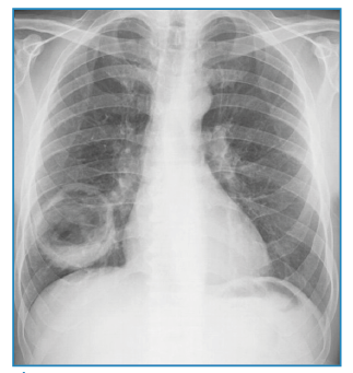
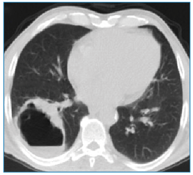
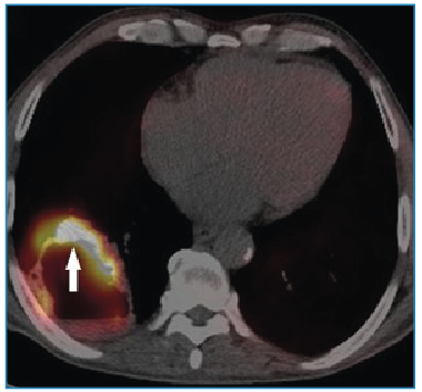
A 60-year-old man was admitted to the chest disease outpatient clinic with shortness of breath, hemoptysis, and cough. Chest radiography revealed a radiopaque, round, and cavitary lesion in the lower lobe of the right lung (Figure 1). The patient underwent non-contrast thorax imaging and positron emission tomography/computed tomography (PET-CT) imaging. PET-CT showed a 61×52 mm hypermetabolic cavitary mass lesion in the lower lobe of the right lung, with a wall thickness of 13 mm at the thickest part and internal surface irregularities (Figure 2 and 3). A thoracic Tru-Cut biopsy was performed because malignancy was suspected.

Radiopaque, round-shaped cavitary lesion in the lower lobe of the right lung on chest radiography.
Cavitary lesion in the lower lobe of the right lung on non-contrast thorax computed tomography images.
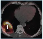
Positron emission tomography/computed tomography imaging shows hypermetabolic involvement.
The pathological results were nonspecific. Because the sputum culture from the patient was positive for M. tuberculosis, this lesion was evaluated as a cavitary lesion due to tuberculosis.
CT plays a crucial role in differentiating between benign and malignant cavitary pulmonary lesions. Radiological features suggestive of malignancy include cavity wall thickness >5 mm, irregular or nodular inner margins, presence of air-fluid levels, and increased metabolic activity on PET-CT1-3.
In cavitary mass lesions with malignant criteria, tuberculosis-related cavitary lesions may also exhibit hypermetabolic uptake on PET-CT4.
ACKNOWLEDGMENTS
We would like to express our sincere gratitude to Atatürk University Research Hospital for providing technical support for the development and implementation of this study.
REFERENCES
- 1 Lal NR, Agarwal GR, Boruah DK. Role of Multidetector Computed Tomography in Differentiation of Benign and Malignant Cavitary Lung Lesions With a Histopathological Correlation: A Retrospective Cross-Sectional Study. Cureus. 2023;15(8):e43005.
- 2 Parkar AP, Kandiah P. Differential Diagnosis of Cavitary Lung Lesions. J Belg Soc Radiol. 2016;100(1):100.
- 3 Naggar A, Laasri K, Berrada K, Kabila B. Differential diagnoses of cavitary lung lesions on computed tomography: a pictorial essay. Egypt J Radiol Nucl Med. 2023;54:149.
- 4 Karaman A, Ucar EY, Kaya S, Araz Ö. Is it Cyst or Neoplasm? The Role of Thorax Magnetic Resonance Imaging. Arch Bronconeumol. 2021;57:(12):769.
Edited by
-
Associated Editor:
Manoel O. da Costa Rocha Orcid: https://orcid.org/0000-0002-4721-1767