 Where is the reward? Floral nectaries in Aspidosperma australe flowers
Abstract
Nectaries are secretory structures that produce a solution rich in sugars, which are involved in animal-plant interactions. Such secretory structures can be externally distinguishable (recognizable), as in glands or discs, or not, as in some gynoecial ones. The absence of floral nectaries has been reported as a characteristic of the genus Aspidosperma (Apocynaceae), except for one species with records of nectariferous tissue in the ovary wall. We observed some ants visiting the flowers of Aspidosperma australe in the field, which could indicate the occurrence of nectaries in these flowers. So, we aimed to verify the presence of nectar and nectaries in the flowers of A. australe in this study, using morphoanatomical analysis. Our results showed a subepidermal differentiated tissue at the base of the ovary, composed of tiny cells with a secretory aspect, rich in starch and polysaccharides. The epidermis cells in this region are smaller, presenting dense content and non-glandular trichomes. We have also achieved positive outcomes through glucose tests conducted on flower nectar. These findings support the presence of an inconspicuous ginoecial nectary at the base of the ovary for A. australe and mark the initial confirmation of nectar secretion for this species.
Key words:
Apocynaceae; Flower; Nectary; Rauvolfioid
Resumo
Nectários são estruturas secretoras que produzem uma solução rica em açúcares envolvida nas interações animais-plantas. Estas estruturas secretoras podem ser distinguíveis externamente, como no caso das glândulas ou discos nectaríferos, ou não, como nos nectários septais ou não septais do gineceu. A ausência de nectários florais tem sido reportada para espécies do gênero Aspidosperma (Apocynaceae), exceto por uma espécie em que se foi registrado tecido nectarífero na parede do ovário. Nós observamos formigas patrulhando/visitando as flores de A. australe no campo, o que poderia indicar a presença de néctar e nectários nessas flores. Assim, objetivamos verificar a presença de nectários nas flores de A. australe nesse estudo, através de uma análise morfoanatômica. Nossos resultados mostram um tecido subepidérmico diferenciado na base do ovário, composto por células diminutas com aspecto secretor, ricas em amido e polissacarídeos. As células da epiderme dessa região são menores, apresentam conteúdo denso e tricomas não-glandulares. Além disso, tivemos resultados positivos através do teste de glicose conduzidos no néctar das flores. Esse conjunto de características corrobora a presença de nectário inconspícuo na base do ovário para A. australe e traz a primeira confirmação da secreção de néctar para essa espécie.
Palavras-chave:
Apocynaceae; Flor; Nectário; Rauvolfioide
Nectaries are secretory structures that produce and secrete a solution rich in sugar called nectar (Roshchina & Roshchina 1993) and are related to animal-plant interactions (Pacini et al. 2003). The definition of nectary is based on the secretory structure function and secretion of the secretory structure, as the morphology and anatomy are quite variable characters (Pacini et al. 2003). There are several types of nectaries in terms of position, origin, structure, and nectar chemical composition (Ray et al. 2017). Such secretory structures can be externally distinguishable (recognizable), as in glands or discs, or not, as in some gynoecial ones (Castro & Machado 2013).
In the Apocynaceae family, ring-shaped nectaries are reported, lobed or not (Gomes 2006), as in Mandevilla Lindl. (Galetto 1997), Secondatia A.DC. (Martins et al. 2013), and Rauvolfia L. (Gomes 2006). A nectariferous zone at the base of the ovary is reported in species of Condylocarpon Desf. (Morokawa et al. 2015), Himatanthus Willd. (Gomes 2006), and Hancornia Gomes (Darrault & Schlindwein 2005). In Aspidosperma Mart. & Zucc., there would be no floral nectaries, according to Marcondes-Ferreira (2005). However, a nectariferous tissue at the base of the ovary of A. quebracho-blanco Schltdl. was reported by Lin & Bernardello (1999). The sporadic presence of a nectary for A. macrocarpon Mart. has also been reported (Gomes & Cavalcanti 2001), and field observations reported ants in flowers of Aspidosperma australe Mull.Arg., which may indicate the occurrence of nectar (Koch I., personal communication), despite the absence of any morphologically distinguishable structure identifiable as a nectary.
Anatomical studies could help to corroborate the hypothesis that there are floral nectaries in Aspidosperma. So, in this study, we aim to verify the presence of nectaries in the flowers of A. australe and describe them.
In order to conduct this study, we collected flowers in pre-anthesis and anthesis of three individuals of Aspidosperma australe Müll.Arg., on the campus of the University of Campinas, in the city of Campinas/SP (Tab. 1). To confirm the presence of sugar release, we conducted a sugar test (ST) using urinalysis test strips. Flowers in anthesis and pre-anthesis buds were collected and dissected to expose the interior of the floral tube. Test strips were placed inside the floral tube and held in contact with the base of the tube, near the ovary region, for approximately 30 seconds. The change in color on the strip indicates the presence of sugar and provides an approximate concentration. We also fixed pre-anthesis buds in FAA 50 (Johansen 1940), stored it in 70% ethanol, dehydrated it in an increasing ethylic series, and included it in historesin following the manufacturer’s protocol. We cut the blocks in a rotary microtome at 8 µm thickness, stained the slides with Toluidine Blue 0.05% pH 4.7 (O’Brien et al. 1964, modified by using acetate buffer), counterstained with ruthenium red, and mounted in water for observation.
We performed histochemical tests using material sectioned in a microtome and subjected to the following reagents and/or dyes: Sudan IV for lipid substances; Lugol for starch; ferric chloride plus sodium carbonate for phenolic compounds (Johansen 1940); bromophenol mercury blue, for proteins (Mazia et al. 1953) and PAS (Schiff reagent/periodic acid) (McManus 1948) for general polysaccharides.
We used light microscopy to analyze the slides and photo-documented the results using an Olympus microscope (BX 53) coupled to a photographic camera (Olympus SC 30).
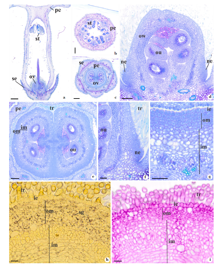
The flower of A. australe is complete (Fig. 1a) and pentamerous (Fig. 1b-c). The calyx is dialyssepalous (Fig. 1c), while the corolla is gamopetalous (Fig. 1a,c). The gynoecium is bicarpelar, with free carpels in the region of the ovaries and a united style, with a single style-head (Fig. 1a). The ST indicated the release of a sugary solution around the ovary, and the coloration of the urinalysis strip indicated a sugar concentration of approximately 250 mg/dL. Anatomically, the ovary is formed by a uniseriate outer epidermis, an ovarian mesophyll divided into two regions, and a uniseriate inner epidermis. At the ovary’s base, a tissue formed by cells with denser content is observed in the region of the external ovarian mesophyll (Fig. 1d-i). This tissue is continuous throughout the base of the ovary, forming a ring (Fig. 1e), which is not visible to the naked eye. In this region, the outer epidermis consists of smaller cells with denser content compared to the rest of the ovary (Fig. 1g), with numerous non-glandular trichomes (Fig. 1f,h-i), and no stomata were observed. The cells of the outer ovarian mesophyll in this region are tiny, with thin walls, a secretory aspect (Fig. 1d-g), many starch grains (Fig. 1h), and polysaccharides (Fig. 1i). No vascular bundle occurs in the external ovarian mesophyll, the bundles being restricted to the region of the internal ovarian mesophyll (Fig. 1g). Based on the results, we conclude that the nectary of A. australe is inconspicuous and located at the base of the ovary.
The Apocynaceae family members are positioned in two paraphyletic grades, rauvolfioids and apocynoids, and tree monophyletic subfamilies, Secamonoideae, Periplocoideae, and Asclepiadoideae (Simões et al. 2007; Endress et al. 2014, 2019). The flowers of these lineages present gradually more complex characteristics (See Fallen 1986). The nectary found in Condylocarpon isthmicum (Vell.) A.DC. is very similar to the nectary of A. australe, with a discreet nectary at the base of the ovary, with small cells and dense cytoplasm, with polysaccharides, starch, and proteins; stomata on the ovary wall through which nectar is secreted during anthesis (Morokawa et al. 2015). Floral nectaries are also reported in another species of rauvolfioid grade (Fallen 1983; Simões et al. 2007, 2016). They can look like a ring at the base of the ovary, as in Allamanda L., Thevetia L., Anechites Griseb. (Fallen 1983), and Rauvolfia (Simões et al. 2016), or lobed as in Vinca L., Catharanthus G.Don., and Laxoplumeria Markgr. (Simões et al. 2007).
In species of the apocynoid grade, the nectary usually presents as a ring fused at the base and divided at the apex into five lobes, with vascular tissue, nectariferous parenchyma, and epidermis with stomata (Galetto 1997; Martins et al. 2013). In Periploca L. species, five nectaries surround the upper part of the ovaries and the styles and are covered by unicellular trichomes (Heneidak & Yougasphree 2015).
In species of Asclepiadoideae, the nectaries are presented as a stigmatic nectariferous chamber, covered by the style-head in its upper part, where the secretory tissue corresponds only to the cells of the epidermis (Monteiro & Demarco 2017.).
Despite nectaries being widely recorded in the rauvolfioid grade, the genus Aspidosperma has been described as lacking nectaries (Marcondes-Ferreira 2005). This idea is reinforced by Gomes & Cavalcanti (2001) results, who analyzing the floral morphology of nine species of the genus, indicated the sporadic presence of nectaries in only one of them (A. macrocarpon), and by Demarco (2005) results, who found no nectariferous tissue in the flowers of A. australe. The only unequivocal record of a nectary in the genus was made by Lin & Bernardello (1999) for A. quebracho-blanco, who emphasizes that it is a non-functional nectary. In this species, the nectary is disc-shaped and located at the base of the ovary, containing a row of stomata on its external surface. Still, they are constantly closed, with no nectar release, and therefore are considered as a non-functional nectary (Lin & Bernardello 1999).
Our results show that the outer epidermis at the base of the ovary is thinner with non-glandular trichomes. These characteristics, besides the presence of sugary solution around the ovary and the presence of ants as floral visitors, indicate that nectaries found in A. australe are functional, at least from the secretory standpoint. We could not observe stomata in the nectary epidermis nor any other obvious structure that could function as a pathway for nectar release. However, we observe small ruptures present in the cuticle wall and the trichomes located around the ovary in A. australe, both of which could serve as potential exit routes for nectar since the presence of trichomes is a significant difference between the nectary structure found in this study and that found in A. quebracho-blanco (Lin & Bernadello 1999). Nectaries that secrete through trichomes have already been documented in previous literature (Evert 2006; Fahn 1988); however, elucidating the nectar exit pathway is not the focus of this study, and future research should be undertaken to address this aspect.
The confirmation of the presence of nectary in the flower of A. australe was possible through anatomical and histochemical analyses that showed the presence of a region in the external ovarian mesophyll with tiny cells, with a thin wall, dense content, and a high presence of starch grains and polysaccharides, differentiating it from the other cells of the mesophyll. These characteristics and its secretory aspect conclude that it is an inconspicuous nectary. We also observed a small protuberance at the base of the ovary with secretory cells in the region. The sporadic presence of these structures, as reported for A. megalocarpon (Gomes & Cavalcanti 2001), can explain why our study found a different result than Demarco’s (2005). Studies with other species could bring more elements to understand the evolution of nectaries in the family.

a-i. Flowers of Aspidosperma australe in longitudinal (a, d, f) and transverse (b-c, e, g-i) sections - a. overview of the flower showing gamopetalous corolla and the superior ovary; b-c. flowers in anthesis cut at the level of the anthers and the ovary, respectively; d-g. details of ovary showing the region nectary in the outer ovarian mesophyll, evidenced by tiny cells with dense content, thin walls, and secretory appearance; h. nectary stained with Lugol’s solution, highlighting the starch grains (in black); i. nectary stained with PAS, highlighting the polysaccharides (in magenta). ie = inner epidermis; im = inner mesophyll; ne = nectary; om = outer mesophyll; ou = ovule; ov = ovary; pe = petal; se = sepal; sg = starch grain; st = stamen; tr = trichome. Scale bars: a = 500 µm; b-c = 250 µm; d-e = 100 µm; f-g = 50 µm; h-i = 20 µm.
Acknowledgements
Authors thank Conselho Nacional de Desenvolvimento Científico e Tecnológico (CNPq, Brazil), processes 315048/2021-2 (research productivity grant of I.Koch).
Data availability statement
In accordance with Open Science communication practices, the authors inform that all data are available within the manuscript.
References
- Castro MM & Machado SR (2013) Células e tecidos secretores. In: Appezzato-da-Glória B & Carmello-Guerreiro SM (eds.) Anatomia vegetal. 3ª ed. Editora UFV, Viçosa. Pp. 169-191.
- Darrault RO & Schlindwein C (2005) Limites fruit production in Hancornia speciosa (Apocynaceae) and pollination by nocturnal and diurnal insects. Biotropica 37: 381-388.
- Demarco D (2005) Estruturas secretoras florais e coléteres foliares em espécies de cerrado de Aspidosperma Mart. e Blepharodon Decne. (Apocynaceae S. L). Dissertação de Mestrado. Universidade Estadual de Campinas, Campinas. 165p.
- Endress ME, Meve U, Middleton DJ & Liede-Schumann S (2019) Apocynaceae. In: Kadereit JW & Bittrich V (eds.) Flowering plants. Eudicots: Apiales, Gentianales (except Rubiaceae): 207-411.
- Endress ME, Liede-Schumann S & Meve U (2014) An updated classification for Apocynaceae. Phytotaxa 159: 175.
- Evert RF (2006) Esau’s plant anatomy: meristems, cells, and tissues of the plant body: their structure, function, and development. John Wiley & Sons, New Jersey. 601p.
- Fallen ME (1983) A systematic revision of Anechites (Apocynaceae). Brittonia 35: 222-231.
- Galetto L (1997) Flower structure and nectar chemical composition in three Argentine Apocynaceae. Flora 192: 197-207.
- Fallen ME (1986) Flora l structure in the Apocynaceae: morphological, functional, and evolutionary aspects. Botanische Jahrbucher fur Systematik, Pflanzengeschichte und Pflanzengeographie 106: 22.
- Fahn A (1988) Secretory tissues in vascular plants. New Phytologist 108: 229-257.
- Gomes SM (2006) Ontogênese floral com ênfase no estudo do gineceu em Apocynaceae s.l Tese de Doutorado. Universidade Estadual de Campinas, Campinas. 267p.
- Gomes SM & Cavalcanti TB (2001) Morfologia floral de Aspidosperma Mart. & Zucc. (Apocynaceae). Acta Botanica Brasilica 15: 73-88.
- Heneidak S & Naidoo Y (2015) Flora l function in relation to floral structure in two Periploca species (Periplocoideae) Apocynaceae. Turkish Journal of Botany 39: 653-663.
- Johansen DA (1940) Plant microtechnique. McGraw-Hill, New York. 523p.
- Lin S & Bernardello G (1999) Flower structure and reproductive biology in Aspidosperma quebraco-blanco (Apocynaceae), a three pollinated by deceit. International Journal of Plant Science 160: 869-878.
- Marcondes-Ferreira W (2005) Aspidosperma Mart. In: Wanderley MGL, Shepherd GJ, Melhem TS & Giulietti AM (eds.) Flora fanerogâmica do estado de São Paulo. Instituto de Botânica, São Paulo. Vol. 4, pp. 39-47.
- Martins FM, Mascarenhas AAS, Macedo TP & Cunha Neto IL (2013) Estruturas secretoras em órgãos vegetativos e florais de Secondatia densiflora A.DC (Apocynaceae-Apocynoideae-Odontadenieae). Revista Brasileira de Plantas Medicinais 15: 13-24.
- Mazia D, Brewer PA & Albert M (1953) The cytochemistry staining and measurement of protein with mercuric bromophenol blue. Biological Bulletin 104: 57-67.
- McManus JFA (1948) Histological and histochemical uses of periodic acid. Stain Tecnology 23: 90-108.
- Monteiro MM & Demarco D (2017) Corona development and floral nectaries of Asclepiadeae (Asclepiadoideae, Apocynaceae). Acta Botanica Brasilica 31: 420-432.
- Morokawa R, Mayer JLS, Simões AO & Kinoshita LS (2015) Flora l development of Condylocarpon isthmicum (Apocynaceae). Botany 93: 769-781.
- O’Brien TP, Feder N & Mccully ME (1964) Polychromatic staining of plant cell walls by toluidine blue O. Protoplasma 59: 368-37.
- Pacini E, Nepi M & Vesprini JL (2003) Nectar biodiversity: a short review. Plant Systematic and Evolution 238: 7-21.
- Ray R, Schmitt AJ, Thomas JB & Carter CJ (2017) Review: nectar biology: from molecules to ecosystems. Plant Science 262: 148-164.
- Roshchina V & Roshchina VD (1993) The excretory function of higher plants. Springer-Verlag, Berlin. 314p.
- Simões AO, Livshultz T, Conti E & Endress ME (2007) Phylogeny and systematics of the Rauvolfioideae (Apocynaceae) based on molecular and morphological evidence. Annals of the Missouri Botanical Garden 94: 268- 297.
- Simões AO, Kinoshita LS, Koch I, Silva MJ & Endress ME (2016) Systematics and character evolution of Vinceae (Apocynaceae). Taxon 65: 99-122.