
 Construction of chitosan-sodium alginate core-shell microcapsules and their dual-functional application in tobacco mildew prevention and flavor preservation
ABSTRACT
Mold contamination and aroma depletion remain critical challenges during tobacco storage. Conventional chemical antifungal agents present residue risks, whereas the direct application of free fragrances is heavily constrained by their rapid volatilization. Herein, we engineered dual-functional core-shell microcapsules via a secondary cross-linking coupled with a vacuum freeze-drying process, encapsulating highly volatile vanillin and citral within a biodegradable chitosan-sodium alginate polyelectrolyte shell. Formulation optimization via an orthogonal array design (L9(34)) identified the optimal parameters as 1.2% chitosan with a 1:2 core-to-wall ratio. Under these conditions, the vanillin and citral microcapsules exhibited high encapsulation efficiencies of 14.23 ± 0.85% and 13.58 ± 0.79%, respectively, alongside highly suppressed 75-day cumulative release rates of 33.8% and 24.8%, respectively. Notably, the citral-loaded microcapsules demonstrated exceptional antifungal efficacy, achieving complete (100.0%) and 96.6 ± 1.1% inhibition against Aspergillus flavus and Aspergillus niger, respectively. Furthermore, during a 28-day accelerated tobacco aging trial, the microcapsule treatment reduced mold proliferation by over two orders of magnitude (>99.8%) and retained >65.0% of key aroma components, substantially enhancing the sensory quality. This study proposes an innovative “distal-proximal” synergistic strategy, providing an efficient, eco-friendly platform for simultaneous microbial control and aroma retention in active preservation materials.
Keywords:
Core-shell microcapsules; Chitosan-sodium alginate; Sustained release of aroma; Mildew prevention; Tobacco storage.
1. INTRODUCTION
The tobacco industry serves as a critical economic pillar globally, yet the quality of its final products is intrinsically linked to the stability of tobacco leaves during post-harvest storage. However, this process generally faces two interrelated core challenges. The first and most critical is microbial contamination, specifically infection by filamentous fungi such as Aspergillus niger and Aspergillus flavus [1, 2]. The second is the rapid volatilization and loss of key aroma substances [3, 4]. Mold contamination not only degrades the tobacco matrix, leading to the deterioration of physical and sensory quality, but more critically, species like A. flavus may produce carcinogenic mycotoxins (e.g., aflatoxins) that pose severe health risks. Recent research using high-precision analysis technologies, such as headspace-gas chromatography-ion mobility spectrometry (HS-GC-IMS), has clearly confirmed this threat. Even mild mildew can fundamentally alter the volatile compound fingerprint of tobacco leaves, leading to an irreversible loss of aroma characteristics [5,6,7]. Meanwhile, the thermal and oxidative degradation of key aroma substances during long-term storage directly results in insufficient product aroma and reduced quality [8, 9]. Therefore, developing innovative strategies that can simultaneously mitigate safety risks and preserve quality is crucial for the sustainable development of the tobacco industry.
To address these challenges, traditional methods have demonstrated significant limitations. Although chemical antifungal agents can inhibit mold in the short term, their potential residual risks are incompatible with increasingly stringent green safety standards. Similarly, the preservation method of directly adding free flavorants has an ephemeral effect. As demonstrated by recent studies on volatile essential oils, direct application suffers from rapid thermodynamic volatilization and susceptibility to environmental stress, rendering free compounds completely incapable of meeting the demands of long-term storage, thus necessitating protective polymeric barriers [10,11,12]. To this end, academia and industry are actively exploring environmentally friendly and durable alternatives. Among them, biological control using plant endophytes has emerged as a frontier direction [13]. However, the industrial application of live microbial preparations is hampered by unstable colonization efficiency in complex storage environments and intricate ecological interactions. These technical bottlenecks severely restrict the commercial viability of this strategy. Consequently, the development of a non-living, standardized functional material system has become an urgent research priority.
In this context, microcapsule technology provides an ideal platform to achieve the dual-functional integration of mildew prevention and fragrance preservation. Among various wall materials, the polyelectrolyte complex formed by chitosan and sodium alginate is particularly favored for its excellent biodegradability, biocompatibility, and recognized food-grade safety, making them highly suitable for bioactive encapsulation, as structurally and functionally validated by the foundational works of ABREU et al., PAIVA FILHO et al. and VALÉRIO et al. [14,15,16]. Chitosan not only possesses excellent biocompatibility but also endows the shell with ‘active’ contact antibacterial potential due to its unique cationic properties (derived from protonated amino groups). Meanwhile, anionic sodium alginate can self-assemble with positively charged chitosan through electrostatic interactions to form a dense shell. This shell acts as an efficient physical barrier for sustained aroma release, with a structural density that can be precisely controlled through cross-linking. Thus, this combination is not a simple blend but an intelligent system with complementary functions, providing an ideal molecular basis for realizing the synergy of sustained release and anti-mold. For instance, chitosan introduced into carboxymethyl cellulose (CMC) hydrogels significantly improved antifungal ability in cigar adhesives [17], while degradable polymer coatings have been successfully employed for slow-release fertilizers in tobacco agriculture [18]. These precedents strongly suggest that building physical barriers to regulate release rates is a viable principle for solving aroma volatilization.
Although progress has been made, previous studies are often limited to simple physical blends [17], single-function applications [18], or complex living systems [13]. A significant gap remains in the functional integration of highly volatile flavorants (core) and bioactive polymers (shell) within a clearly defined structural framework, especially for tobacco preservation. This study aims to bridge this gap. A systematic investigation is conducted on two types of core-shell microcapsules prepared with a solid core material (vanillin) and a liquid core material (citral). The study examines their respective microstructure, encapsulation efficiency (EE), long-term release kinetics, and targeted antibacterial activity. We hypothesize a “Distal and Proximal” synergistic mechanism: the shell serves as a physical barrier for the sustained release of antifungal volatiles (distal vapor-phase inhibition), while the cationic chitosan surface provides contact-killing capability (proximal membrane disruption). To validate this, microcapsules were prepared via a secondary cross-linking and vacuum freeze- drying process. Their physicochemical properties and antimicrobial efficacy were systematically characterized, and their comprehensive application value was finally verified in a tobacco accelerated aging model.
2. MATERIALS AND METHODS
2.1. Materials
High-purity biopolymers were selected to ensure the stability of the microcapsules. Chitosan (deacetylation degree ≥95%, viscosity 200–300 mPa·s) and Citral (purity ≥98%, analytical grade) were purchased from Shanghai Maclean Biochemical Technology Co., Ltd.. Sodium alginate (analytical grade, viscosity 150–250 mPa·s) was obtained from Zhengzhou Pinni Chemical Reagent Factory. Vanillin (purity ≥99%, analytical grade) was supplied by Sain Chemical Technology (Shanghai) Co., Ltd.
Cross-linking agents and other reagents included: Anhydrous calcium chloride (≥99%) and Sodium hydroxide (≥96%) from Tianjin Damao Chemical Reagent Factory; Anhydrous sodium sulfate (≥99%) from Tianjin Jiangtian Chemical Technology Co., Ltd.; Acetic acid (≥99.5%) and Anhydrous ethanol (≥99.7%) from Tianjin Fuyu Fine Chemical Co., Ltd.; Potassium dihydrogen phosphate (≥99%) from Tianjin Rujin Te Chemical Co., Ltd.; and Disodium hydrogen phosphate (≥99%) and Tween 80 (HLB value 15.0) from Tianjin Kemiou Chemical Reagent Co., Ltd.. All reagents were of analytical grade and used as received.
2.2. Preparation of chitosan-sodium alginate core-shell microcapsules
To validate this, microcapsules were prepared via a secondary cross-linking and vacuum freeze-drying process, a robust methodology proven to enhance the biological efficacy of highly volatile terpenes while maintaining structural integrity [19, 20]. The schematic workflow of the microcapsule fabrication is presented in Figure 1.

Schematic illustration of the preparation process for chitosan-sodium alginate core-shell microcapsules.
As depicted in Figure 1, the process involves distinct stages of emulsification, gelation, and coating. First, the primary wall material solutions (chitosan and sodium alginate) are prepared to meet specific viscosity requirements. Using the chitosan solution as the continuous phase, the core material (vanillin or citral) and emulsifier are added. The mixture is subjected to high-speed shearing and ultrasonic treatment to form a stable O/W emulsion. Subsequently, the primary chitosan core particles are formed via ionic cross-linking with anhydrous sodium sulfate. These core particles are then dispersed into the sodium alginate solution, where a secondary cross-linking reaction with calcium chloride forms a dense polyelectrolyte complex shell. Finally, the core-shell microcapsules are purified via centrifugation, pre-frozen, and vacuum freeze-dried to obtain a loose powder with a moisture content of ≤ 3%. Critical processing parameters, including temperature, stirring speed, and dripping rate, are strictly controlled to maximize uniformity and encapsulation efficiency.
2.2.1. Polymer solution preparation
Chitosan powder (12.0 g) was weighed and slowly added to 1,000 mL of a 1% (w/v) glacial acetic acid aqueous solution. The mixture was stirred continuously for 3 h at 25 °C and 800 rpm using a dual-display constant- temperature magnetic stirrer. During this period, the dissolution state was monitored every 30 min to ensure complete dissolution and the formation of a transparent, uniform solution. The pH was then adjusted dropwise to 5.0 using a 1% (w/w) sodium hydroxide solution under continuous stirring. After adjustment, the solution was allowed to stand for 1 h to degas completely before being sealed in a brown reagent bottle for storage.
Separately, sodium alginate powder (10.0 g) was dissolved in 1,000 mL of phosphate buffer solution (pH 7.2). The mixture was stirred for 2 h at 25 °C and 600 rpm. A glass rod was used initially to assist dispersion and prevent agglomeration. The resulting solution was filtered through a 0.45 μm organic membrane to remove undissolved impurities and left to stand for 30 min to eliminate air bubbles. The preparation principle is illustrated in Figure 2.

Schematic illustration of the preparation of chitosan-sodium alginate core-shell microcapsules.
2.2.2. Emulsification and nucleation (primary cross-linking)
The pretreated chitosan solution (100 mL) was placed in a 250 mL three-neck flask, and 10 mL of 1% (w/v) Tween 80 aqueous solution was added. The mixture was stirred at 25 °C and 1,000 rpm for 10 min to disperse the emulsifier. The core material was then added: 20 mL of vanillin ethanol solution for vanillin microcapsules, or 20 mL of citral reagent for citral microcapsules. Stirring continued for 2 h, with sampling every 20 min to ensure emulsion stability. The pre-emulsified liquid was then processed using an ultrasonic processor (JY92-IIN, Ningbo Xinzhi Biotechnology Co., LTD) at 300 W for 10 min. The temperature was strictly controlled using an ice-water bath to prevent volatilization, yielding a stable, milky white emulsion.
Subsequently, this emulsion was transferred to a constant-pressure dropping funnel and dripped at a rate of 1 mL/min into 100 mL of 2% anhydrous sodium sulfate aqueous solution under stirring at 1,200 rpm (25 °C). Stirring was maintained for 30 min to crosslink chitosan amine groups with sulfate ions, forming chitosan core particles. The precipitate was collected by centrifugation (3,500 rpm, 5 min, 25 °C) and washed three times with deionized water.
2.2.3. Shell coating (secondary cross-linking)
The chitosan core particles were dispersed in 100 mL of the pretreated sodium alginate solution within a 250 mL three-necked flask and stirred at 25 °C and 800 rpm for 10 min. The resulting suspension was dripped into 100 mL of 0.1 mol/L calcium chloride aqueous solution at a rate of 0.8 mL/min. Stirring continued for 40 min after dripping to ensure complete cross-linking and prevent agglomeration.
2.2.4. Purification and lyophilization
The core-shell microcapsules were harvested by centrifugation (4,000 rpm, 8 min), and the supernatant was discarded. The particles were washed three times with deionized water containing 0.5% (w/v) Tween 80. The wet particles were spread evenly on a freeze-drying tray (thickness ≤5 mm), pre-frozen at -40 °C for 4 h, and then lyophilized under vacuum (-55 °C, 5 Pa, 24 h) until a loose powder with a moisture content of ≤3% was obtained. The final product was stored in a desiccator.
2.3. Characterization
2.3.1. Morphology (SEM)
Microcapsule samples were subjected to vacuum-drying at 60 °C for 2 h. While this standard protocol is optimal for eliminating residual surface moisture to image the polymer matrix, it inherently subjects the highly volatile liquid core to extreme thermal-vacuum stress. This intrinsic limitation of conventional SEM preparation must be considered when interpreting the morphological integrity of the citral-loaded systems. Dried samples (1 mg) were then mounted on conductive adhesive tape and sputter-coated with gold using a JFC-1600 (JEOL Ltd., Tokyo, Japan). Surface morphology was observed using a scanning electron microscope (SEM, S-3400, Hitachi, Japan) at an acceleration voltage of 3 kV to identify structural features such as protrusions or adhesions.
2.3.2. Chemical structure (FTIR)
FTIR spectra were recorded to verify component compatibility. Samples (1 mg) of chitosan, sodium alginate, core materials, and microcapsules were mixed with 100 mg of KBr powder, ground to ≤ 2 μm, and pressed into 13 mm pellets at 10 MPa. Spectra were acquired using a Nicolet iS 50 FTIR spectrometer (Thermo Fisher Scientific, USA) over a wavenumber range of 400–4000 cm−1 with a resolution of 4 cm−1 (64 scans).
2.3.3. Thermal analysis (TGA & DSC)
Thermal stability was evaluated using a Q600 synchronous thermal analyzer (TA Instruments, USA). Samples (5–10 mg) were heated from 25 °C to 600 °C at 10 °C/min under nitrogen flow (50 mL/min). Thermal transitions were analyzed using a DSC3 differential scanning calorimeter (Mettler-Toledo, Shanghai, China). Samples (3–5 mg) sealed in aluminum crucibles were heated from 25 °C to 250 °C at 10 °C/min under nitrogen (50 mL/min).
2.3.4. X-ray diffraction (XRD)
Crystal structure was analyzed using an Ultima IV XRD instrument (Rigaku Corporation, Japan) with a Cu Kα source (λ = 0.154 nm) at 40 kV and 40 mA. Scans were performed from 5° to 50° (2θ) at a rate of 5°/min.
2.3.5. Zeta potential
Surface charge was measured using a Zetasizer Nano ZS (Malvern Instruments Ltd., UK). Samples (~1 mg/mL) were dispersed in deionized water, ultrasonicated for 5 min, and injected into a disposable capillary cell (DTS1070). Measurements were performed in triplicate at 25 °C, and results are reported as mean ± standard deviation (SD).
2.3.6. Long-term storage stability
Freeze-dried powder was stored in a GDW-225 constant temperature and humidity chamber (Beijing Yashilin Testing Equipment Co., Ltd.) at 40 °C/75% RH for 6 months. Samples taken at high-resolution monthly intervals (0, 1, 2, 3, 4, 5, and 6 months) were characterized for particle size, chemical structure (FTIR), and leakage rate.
2.4 Performance evaluation
2.4.1. Formulation optimization and encapsulation efficiency (EE)
To systematically optimize the encapsulation performance and address the limitations of single-factor experiments, an orthogonal experimental design L9(34) was employed. Three critical process variables were evaluated at three levels: chitosan concentration (0.8%, 1.2%, 1.6%), core-to-wall mass ratio (1:1, 1:2, 1:3), and chitosan-to- alginate mass ratio (1:2, 1:1, 2:1), resulting in nine specific experimental groups.
The EE and fragrance loading were quantified using a UV-visible spectrophotometer (Model UV-5200, Shanghai Yuanxi Instrument Co., LTD). First, absolute ethanol was used to prepare standard solutions of vanillin and citral (0.01–0.1 mg/mL). Absorbance was measured at characteristic wavelengths of 280 nm (vanillin) and 230 nm (citral) to construct standard curves. Microcapsule samples (25 mg) from each group were weighed, placed in centrifuge tubes, and ruptured in 20 mL of absolute ethanol via ultrasonic treatment for 30 min. The supernatant was collected after centrifugation (5,000 rpm, 10 min), diluted appropriately, and absorbance was measured. The mass of the encapsulated core material was calculated using the standard curve. The EE was calculated as follows (performed in triplicate):
where Mencap is the measured mass of the encapsulated core material, and Minitial is the total mass of core material initially added. Based on the range analysis (R) and ANOVA results (presented in Table 1), the optimal formulation (1.2% chitosan, 1:2 core-to-wall ratio, and 2:1 chitosan-to-alginate ratio) was identified and subsequently prepared for all structural and functional characterizations.
Encapsulation Efficiency (EE) of chitosan-sodium alginate core-shell microcapsules under optimized process conditions (L9(34) orthogonal design, n = 3).
2.4.2. In vitro sustained release
Release kinetics were evaluated at room temperature [21]. Equal amounts of free core material (vanillin or citral) and optimized microcapsules were incubated in parallel for 75 days. The samples were stored in a constant temperature and humidity chamber (25 °C, 60% RH) to simulate standard tobacco storage conditions. At 7-day intervals, residual core material was extracted by rupturing the microcapsules in absolute ethanol (ultrasonic treatment, 30 min). The supernatant was analyzed via UV-visible spectrophotometry to calculate the cumulative release rate. Data were fitted to a first-order kinetic equation to verify long-term sustained release performance.
To further elucidate the release mechanism, the Korsmeyer-Peppas model was applied to fit the experimental data (Eq. 2):
where Mt /M∞ represents the fraction of fragrance released at time t, k is the structural and geometric constant, and n is the release exponent characterizing the transport mechanism.
2.4.3. Antibacterial activity
Antibacterial activity against Aspergillus flavus and Aspergillus niger was assessed using the fumigation method. Microcapsule powder (15, 30, and 45 mg) was placed in sterile bottle caps inverted over the center of standard 90 mm diameter Potato Dextrose Agar (PDA) plates inoculated with fungal spores. To accurately assess the vapor-phase inhibition, the plates were tightly sealed with Parafilm immediately after preparation, creating a defined headspace volume of approximately 60 mL to prevent the leakage of volatile active compounds. The sealed plates were then incubated at 28 °C. The diameter of the inhibition zone was measured to calculate the inhibition rate. Empty microcapsules (Blank MCs) without volatile cores were evaluated in parallel as negative/vehicle controls to establish the baseline fungitoxicity of the shell matrix. Due to the spatial incompatibility and lack of volatility of standard contact-dependent agricultural fungicides, a conventional positive control was not included in this specific vapor-phase setup.
2.5. Application experiment of chitosan-sodium alginate core-shell microcapsules in tobacco
The study selected single-material shredded tobacco as the tobacco leaf sample, and established three treatment groups: a blank control group (shredded tobacco without adding any substance), a free spice group (adding the same amount of free spices as the microcapsule core material), and a microcapsule group (adding 0.5% mass fraction of chitosan-sodium alginate core-shell microcapsules). For each group, 15 g of cut tobacco was weighed. The microcapsules and free flavors were diluted with a small amount of deionized water, sprayed evenly on the cut tobacco, and placed in a constant temperature and humidity chamber at 22 °C and 60% RH for 48 h. Subsequently, the three groups of cut tobacco were stored under accelerated aging conditions of 40 °C and 85% RH. Samples were collected regularly (0 d, 7 d, 14 d, 21 d, 28 d) to detect the indicators.
Mold colony counts in shredded tobacco were determined using the plate counting method, and the colonies were counted after culturing on PDA medium. A moisture meter was used to measure the moisture content of cut tobacco. The changes in key aroma components were analyzed by GC-MS. Chromatographic separation was achieved using an HP-5 capillary column (30 m × 0.25 mm × 0.25 μm). The oven temperature program was initiated at 60 °C (held for 2 min), ramped to 250 °C at a rate of 8 °C/min, and maintained for 5 min to ensure complete elution. Helium was used as the carrier gas at a constant flow rate of 1.0 mL/min, with the injector and detector temperatures set at 250 °C and 280 °C, respectively. The peak area of the aroma components was measured and the retention rate was calculated. A highly trained expert panel consisting of seven nationally certified professional smokers, each with over five years of experience, conducted the sensory evaluations. According to the biostatistical guidelines for sensory analysis outlined in ISO 8586:2023 [22] and foundational Quantitative Descriptive Analysis (QDA) methodologies [23], an expert cohort of this size (n = 5–15) is recognized as statistically valid and provides superior reproducibility and discriminative power in ANOVA models compared to larger untrained consumer panels. This specific cohort size (n = 7) was deliberately established following rigorous preliminary psychophysical screening (triangular test methodology) to strictly minimize intra- and inter-assessor variance [24]. This rigorous calibration process ensures the statistical significance and validity of the resultant sensory profiling. The panelists evaluated aroma quality, aroma content, off-gas, irritation, aftertaste, and other indicators, and comprehensively analyzed the effect of microcapsules on regulating tobacco storage quality. To ensure the objectivity and scientificity of the evaluation, all sensory evaluation data were statistically analyzed using SPSS software. Furthermore, the significant differences (p < 0.05) between each treatment group were evaluated through one-way analysis of variance (ANOVA) and Duncan’s multiple range test.
3. RESULTS
3.1. Morphology and structural characterization of chitosan-sodium alginate core-shell microcapsules
The SEM of vanillin-chitosan-sodium alginate core-shell microcapsules and citral-chitosan-sodium alginate core-shell microcapsules is shown in Figure 3.

SEM image of chitosan-sodium alginate core-shell microcapsules with (a) vanillin-chitosan-sodium alginate core-shell microcapsules, (b) citral-chitosan-sodium alginate core-shell microcapsules.
In Figure 3a, vanillin-chitosan-sodium alginate core-shell microcapsules are in regular spherical shape and evenly dispersed. There is only slight local adhesion, no obvious agglomeration, and the particle size is concentrated at 4–8 μm. It meets the morphological requirements of core-shell microcapsules. In Figure 3b, the surface of citral-chitosan-sodium alginate core-shell microcapsules shows fine and rough granular protrusions. This unique morphology is not only a direct reflection of the electrostatic interaction between the amino groups of chitosan and the carboxyl groups of sodium alginate, but is also the key to realizing its dual functions. On the one hand, the dense shell formed by this close packing forms a strong physical barrier, effectively slowing down the diffusion of internal spice molecules. On the other hand, the rough surface increases the specific surface area of the microcapsules, which may facilitate its contact and interaction with the mold cell membrane. It is critical to interpret the morphological topography of the citral-loaded microcapsules (Figure 3b) strictly within the thermodynamic constraints of conventional SEM sample pre-treatment. While standard vacuum-drying (60 °C, 2 h) elegantly preserved the regular spherical architecture of the solid-core vanillin system, subjecting the liquid-core citral system to identical thermal-vacuum stress drastically lowered the boiling point of the sub- surface nanodroplets. This induced rapid, forceful outgassing that physically breached the dehydrating polyelectrolyte network, generating the extreme rough granular artifacts observed [25, 26]. While this stress-induced structural collapse empirically confirms the presence of a highly concentrated, hyper-volatile internal liquid phase, it inherently precludes the definitive visual resolution of the intact core-shell boundary. Therefore, to fundamentally circumvent vapor-liquid phase transition disruptions in future high-resolution morphological enumerations of such liquid-core hydrogel systems, it is imperative to transition from dehydrating techniques to non-destructive, fully hydrated imaging modalities, such as Cryogenic Scanning Electron Microscopy (Cryo-SEM) or Confocal Laser Scanning Microscopy (CLSM) utilizing lipophilic fluorophores.
To confirm the successful compounding of microcapsules from the chemical structure, this study conducts FTIR characterization of all raw materials and final products. As shown in Figure 4, both free vanillin (Figure 4a) and free citral (Figure 4b) exhibit their own unique and sharp characteristic absorption peaks, such as the benzene ring skeleton vibration peak of vanillin at 1,589 cm-1 and the aldehyde group C=O stretching vibration peak of citral at 1,675 cm-1. Chitosan (Figure 4c) and sodium alginate (Figure 4d) shows typical polysaccharide absorption peaks.

FTIR spectrum of chitosan-sodium alginate core-shell microcapsules, including (a) free vanillin, (b) free citral, (c) chitosan, (d) sodium alginate, (e) vanillin microcapsules, and (f) citral microcapsules.
Crucially, the final microcapsule spectrum clearly shows successful complexation of the components. The spectrum of vanillin microcapsules (Figure 4e) shows the broad absorption characteristics of chitosan-sodium alginate wall material as a whole. However, at the same time, it clearly retains the characteristic peak of vanillin at 1,589 cm-1. Similarly, the spectrum of citral microcapsules (Figure 4f) also retains the characteristic peak of citral’s aldehyde group (shown as an identifiable shoulder) at 1,675 cm-1 on the broad peak background of the wall material.
These results provide direct spectroscopic evidence that chitosan is successfully complexed with sodium alginate to form the shell of the microcapsules. Moreover, vanillin and citral are effectively encapsulated inside the core layer, thus successfully constructing the expected core-shell structure.
3.2. Structural and thermodynamic confirmation of microencapsulated state
To further confirm whether microencapsulation is successful, this study conducted systematic characterization from three dimensions: TGA, DSC and XRD.
To verify the core-shell structure of the microcapsules and its protective effect on the core material, TGA is conducted in this study, and the results are shown in Figure 5. Both free flavors show poor thermal stability. The highly volatile free citral begins to lose weight rapidly at about 130 °C, while the weight loss of free vanillin starts at about 180 °C. Pure wall material (chitosan-sodium alginate) itself shows excellent thermal stability, and its main decomposition stage is above 300 °C. Crucially, microencapsulation significantly improves the thermal stability of both core materials. The thermal decomposition onset temperatures of both vanillin microcapsules and citral microcapsules are significantly delayed to about 250–270 °C, which is closer to the thermal stability curve of pure wall materials. This phenomenon clearly shows that the chitosan-sodium alginate shell serves as an effective physical barrier. It greatly inhibits the thermal movement and escape of internal flavor molecules (whether solid or liquid), thus significantly improving its thermal stability.

To further reveal the physical state of the core material inside the microcapsules, DSC analysis is performed in this study (Figure 6). As a crystal, the DSC curve of free vanillin shows a sharp endothermic peak at 82 °C, corresponding to its melting process. In sharp contrast, this characteristic melting peak completely disappears in the curve of vanillin microcapsules. This indicates that the core material is successfully encapsulated and transformed from the crystalline state to the amorphous state. For the citral system, as a liquid, the curve of free citral itself is smooth and has no thermal events. The curve shape of the microcapsules is highly similar to the curves of the wall material and vanillin microcapsules. Only a wide and gentle endothermic valley appears at about 100 °C due to the evaporation of water adsorbed by the wall material. There is no independent thermal event related to citral.

To directly confirm the effect of the microencapsulation process on core materials of different physical forms from the crystal structure level, this study conducts systematic XRD analysis of both systems. For the solid core vanillin system (Figure 7), free vanillin exhibits a series of sharp crystal diffraction peaks (Figure 7a), while the wall material is in a typical amorphous form (Figure 7b). Key control experiments show that the physical mixture of the two (Figure 7c) clearly retains the crystal characteristic peaks of vanillin, proving that simple mixing cannot change its crystal structure. In stark contrast, in the final vanillin microcapsules (Figure 7d), all crystal diffraction peaks completely disappear. The entire map shows an amorphous shape consistent with the wall material. This strongly proves that the microencapsulation process actively converts crystalline vanillin into an amorphous state.

XRD pattern of vanillin system, including (a) free vanillin, (b) wall material, (c) physical mixture, and (d) vanillin microcapsules.
To further verify the universality of this method, this study conducts parallel XRD characterization of the liquid core material citral system (Figure 8). The free citral itself is amorphous and only appears as a broad and gentle bulge of liquid scattering at low angles (Figure 8a). After physically mixing it with amorphous wall material (Figure 8b), the resulting spectrum (Figure 8c) is almost indistinguishable from that of pure wall material. Finally, the XRD spectrum of the citral microcapsules prepared through the microencapsulation process (Figure 8d) also shows a uniform amorphous morphology. This shows that the process is able to stably encapsulate the liquid core material and perfectly maintain its amorphous state.

XRD pattern of citral system, including (a) free citral, (b) wall material, (c) physical mixture, and (d) citral microcapsules.
The comprehensive analysis of TGA, DSC, and XRD clearly shows the powerful universality and dual action mechanism of this packaging technology: First, TGA analysis provides direct evidence for the protective effect of the core-shell structure. It is worth noting that this protective effect is not only reflected in the stabilization of solid vanillin, but also in the effective inhibition of highly volatile liquid citral (Figure 5). This shows that the core-shell structure constructed in this study serves as an effective physical barrier and has good universality and can limit the migration of core material molecules in different physical forms. This is highly consistent with the underlying mechanism of the excellent sustained-release performance (Figure 9) observed subsequently. Second, the joint analysis of DSC and XRD provides solid structural evidence for the successful construction of microcapsules and their stabilizing mechanism for the core material. This mechanism plays a dual role depending on the original physical form of the core material: 1) For solid crystal core materials (such as vanillin), this process plays the role of an “active converter”. XRD and DSC results jointly confirm that the encapsulation process can effectively destroy the original lattice structure of the core material and transform it into a thermodynamically metastable amorphous or molecular-level dispersed state. This state fundamentally inhibits the migration and crystallization of spice molecules, which is the microscopic basis for realizing its thermal stability and long-term sustained release properties. 2) For liquid amorphous core materials (such as citral), this process plays the role of “stability guardian”. XRD and DSC results prove that the encapsulation process uniformly disperses and fixes the liquid spices in the form of nanoscale droplets in the polymer network. Meanwhile, it perfectly maintains the amorphous state of the system without inducing any undesired phase separation or crystallization. In summary, this secondary cross-linking-lyophilization technology is a powerful platform that can effectively stabilize and encapsulate active core materials in different physical forms.

Cumulative release profiles of (a) optimized microcapsules and (b) free flavorants over a 75-day storage period. Solid lines in (a) represent the Korsmeyer-Peppas model fitting; the inset in (a) displays the linear regression of versus used to determine the release exponent (n). Data are presented as mean ± SD (n = 3).
3.3. Analysis of encapsulation performance and sustained release behavior
3.3.1. Optimization of microcapsule formulation and industrial viability
To maximize the payload capacity and structural stability, the formulation was systematically optimized using an orthogonal experimental design. Table 1 presents the Encapsulation Efficiency (EE) of vanillin and citral microcapsules under nine distinct process combinations. The data represent the mean ± standard deviation of triplicate experiments.
Based on the range analysis (R values) presented in Table 1 and the Analysis of Variance (ANOVA), the physicochemical parameters of the polymer solution played a decisive role in encapsulation. The statistical analysis revealed a clear hierarchy of influence on Encapsulation Efficiency (EE): Core-to-Wall Ratio (R = 3.28) > Chitosan Concentration (R = 3.05) > Chitosan-to-Alginate Ratio (R = 0.37). The ANOVA results further confirmed that both the core-to-wall ratio and chitosan concentration had a highly significant impact (p < 0.001), whereas the chitosan-to-alginate ratio showed no statistically significant difference (p > 0.05), indicating that the shell formation is relatively robust across the tested polymer ratios.
Crucially, the effect of chitosan concentration on EE exhibited a distinct non-linear trend, suggesting a kinetic limitation driven by solution viscosity. As indicated by the mean values (K), the EE increased significantly when the concentration rose from 0.8% (K1 = 8.93) to 1.2% (K2 = 11.98) but declined at 1.6% (K3 = 10.32). Mechanistically, while adequate viscosity is essential to stabilize the primary emulsion droplets, excessively high viscosity (at 1.6%) hinders the diffusion of sulfate ions during the rapid cross-linking process. This likely leads to the formation of a heterogeneous or “skin-core” shell structure that fails to effectively retain the core material.
Consequently, for the statistically significant factors, optimal levels were selected based on the maximization of K values (1.2% chitosan and 1:2 core-to-wall ratio). Although the chitosan-to-alginate mass ratio showed no statistical significance regarding EE (p > 0.05), the 2:1 ratio (Group 5) was deliberately selected over the others. Mechanistically, this higher proportion of chitosan ensures a greater density of protonated amino groups on the microcapsule surface, which is fundamentally critical for maximizing the contact-mediated antibacterial efficacy (Proximal Defense) discussed in Section 3.7.2. This optimized formulation achieved the maximum EE of 14.23 ± 0.85% for vanillin and 13.58 ± 0.79% for citral, and was therefore selected for all subsequent structural and functional characterizations.
While Encapsulation Efficiency (EE) values for large, non-volatile macromolecules (e.g., proteins or high-molecular-weight pharmaceuticals) within biopolymer matrices routinely exceed 70% to 90%, the encapsulation of low-molecular-weight, highly volatile organic compounds (VOCs) like vanillin and citral presents a fundamentally different thermodynamic challenge. The optimized EE values obtained in this study (~14.23% for vanillin and ~13.58% for citral) must be fundamentally interpreted through the thermodynamic paradigm of the Selective Diffusivity Theory and the Microregion Entrapment mechanism originally postulated by Thijssen, Rulkens, and Karel [27,28,29,30]. In polymeric drying systems, the retention of VOCs is intrinsically governed by the rate at which the dehydrating matrix crosses a critical moisture threshold to form a vitrified, glassy state. Below this threshold, the matrix becomes selectively permeable to water vapor while acting as an absolute barrier to VOCs. However, because the pre-lyophilized chitosan-alginate polyelectrolyte complex exists as a highly hydrated hydrogel network, the rapid primary sublimation of ice under extreme vacuum (−55 °C, 5 Pa) outpaces the structural vitrification of the polymer micro-regions. Consequently, before the ‘selective diffusion’ barrier can be fully established, the extreme vapor pressure gradients compel the unanchored citral and vanillin molecules to diffuse outward alongside the sublimating water front. Therefore, the ~14% retained fraction does not signify a failure of wall material affinity, but rather represents the thermodynamically stable payload that successfully partitioned into the most rapidly cross-linked, highest-density hydrophobic domains prior to macroscopic vitrification.
Furthermore, despite this seemingly low absolute mass retention, these microcapsules exhibit remarkable functional viability in practical applications. This feasibility is dictated by the inverse relationship between the required operational dosage and the biological potency of the active agents. Both citral and vanillin possess exceptionally low sensory detection thresholds (yielding remarkably high Odor Activity Values, OAVs) and highly potent, broad-spectrum antifungal properties at micro-concentrations. Because their sensory detection thresholds are extraordinarily low, even these residual micro-concentrations inherently dominate the aroma profile and exhibit profound biological efficacy without requiring high absolute mass loadings [31]. As demonstrated in the subsequent in situ application trials (Section 3.6), a minimal application dosage—a mere 0.5% mass fraction of these microcapsules applied to the tobacco—successfully achieves an over 99.8% reduction in mold proliferation over 28 days.
However, it is imperative to distinguish between functional efficacy and manufacturing economics. While the exceptionally high efficacy-per-gram guarantees the robust biological performance of the final product, the initial loss of >85% of the volatile core materials—driven by the aforementioned pre-vitrification sublimation—presents a tangible mass-balance challenge for industrial scale-up. In commercial ton-scale production, such substantial volatilization not only elevates the cost of goods sold (COGS) and risks degrading high-vacuum freeze-drying equipment, but also poses significant environmental compliance challenges regarding volatile organic compound (VOC) emissions. Therefore, the current freeze-dried formulation serves primarily as a highly potent mechanistic proof-of-concept for the “distal-proximal” dual-defense theory. To bridge the gap between this laboratory-scale functional excellence and industrial mass-balance efficiency, future scale-up must modulate the thermodynamic processing pathways of the encapsulation matrix. While transitioning to advanced atmospheric spray drying necessitates the incorporation of continuous-phase film-forming excipients with inherently high glass transition temperatures (Tg) capable of forming a rapid vitrified crust, the elevated inlet temperatures inherently threaten extremely thermolabile terpenes. Alternatively, to circumvent this thermal stress entirely, liquid-phase scaling technologies—such as complex coacervation or spray-chilling/cooling using lipidic matrices—should be actively explored. Operating under moderate or sub-ambient thermal conditions, these methods effectively avert vapor-liquid phase transition disruptions, thereby possessing the potential to drastically elevate the commercial encapsulation efficiency towards >80% without compromising the core-shell architecture.
3.3.2. Sustained release kinetics and mechanism
The protective efficacy of the core-shell structure is quantified by the comparative release profiles shown in Figure 9. As evident in Figure 9b, the free flavorants underwent distinctive volatilization behaviors driven by their physical states. The liquid free citral exhibited an initial burst volatilization, with cumulative loss exceeding 90% within 42 days. In contrast, the solid free vanillin showed a relatively steady sublimation process, reaching 86.4% loss at 75 days. Despite these differences, both free flavorants confirmed their thermodynamic instability in open environments. In sharp contrast, the microencapsulated groups (Figure 9a) demonstrated significantly enhanced retention. The cumulative release was restricted to only 33.8% (vanillin) and 24.8% (citral) over the same period, representing a substantial reduction in volatilization loss.
To further elucidate the underlying release mechanism, the experimental data were fitted to the Korsmeyer- Peppas model (Mt /M∞= k· tn). Given that the maximum cumulative release for both systems remained well below the theoretical boundary of 60% (Mt /M∞≤ 0.6), the application of this model is strictly valid. As shown by the linear regression in the inset of Figure 9a, the release exponent (n) for vanillin microcapsules was 0.564 (R2 = 0.988). This n value (0.43 < n < 0.85) indicates an anomalous transport mechanism (non-Fickian diffusion), suggesting that the release is governed by a synergistic effect of fragrance diffusion across the shell and the slight relaxation/swelling of the hydrophilic chitosan-alginate matrix upon hydration in the storage environment.
Interestingly, the citral microcapsules yielded an n value of 0.492 (R2 = 0.992). Although this value technically falls within the anomalous transport regime (0.43 < n < 0.85), it approaches the theoretical threshold of 0.43 for Fickian diffusion more closely than that of the vanillin system. This proximity suggests that while polymer relaxation (swelling) acts as a minor contributor, the release of the liquid citral core is predominantly governed by diffusion through the dense polyelectrolyte shell. This behavior is physically consistent with the hydrophobic nature of citral, which exhibits lower thermodynamic compatibility with the hydrophilic chitosan-alginate matrix compared to the more polar vanillin. Such hydrophobicity minimizes the localized plasticization or excessive swelling of the shell specifically around the citral droplets, thereby maintaining a tighter network structure. Consequently, the incompatibility increases the interfacial resistance and tortuosity for diffusion, effectively “locking” the citral molecules within the core and resulting in a more sustained release profile. The synergistic integration of these kinetic parameters confirms that the secondary cross-linked shell functions as a high-fidelity barrier, particularly for hydrophobic volatile oils.
3.4. Long-term storage stability of microcapsules
To evaluate the reliability of microcapsules in practical applications, the study conducts a 6-month accelerated aging test on both systems. The results are shown in Figure 10. Both microcapsules showed excellent overall stability.
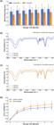
Long-term storage stability of microcapsules under accelerated aging conditions of 40 °C/75%RH, including (a) comparison of the average particle size of vanillin (blue) and citral (orange) microcapsules within 6 months, (b) FTIR spectra of vanillin microcapsules after 0 and 6 months of storage, (c) FTIR spectra of citral microcapsules after 0 and 6 months of storage, and (d) comparison of cumulative leakage rates during storage between vanillin (blue) and citral (orange) core materials. Data are mean ± SD deviation of three measurements.
In terms of physical size (Figure 10a), high-resolution monthly tracking reveals that the average particle size of vanillin and citral microcapsules undergoes a slight and controllable initial expansion during the first 1–2 months, followed by a dynamic equilibrium. Despite minor analytical fluctuations typical of realistic colloidal measurements, the macroscopic dimensions rapidly and strictly stabilize into a long-term plateau. This trend elegantly suggests that the initial small changes in size are primarily determined by the physical moisture uptake and hydration swelling of the hydrophilic shell material, rather than structural degradation or progressive agglomeration.
In terms of chemical structure, FTIR characterization was conducted to monitor the stability of the encapsulated active ingredients. As shown in Figures 10b and 10c, although the spectra of the aged samples (6 months) exhibited a slight baseline drift and peak broadening compared to the initial state, the overall chemical skeletons remained highly consistent. Notably, a synchronous increase in intensity was observed in both the broad O-H stretching band (~3,430 cm-1) and the water bending vibration region (~1,640 cm-1). This spectral evolution provides direct evidence that the minor particle size expansion (Figure 10a) is driven by physical hygroscopic swelling of the polysaccharide shell rather than chemical degradation. Crucially, the characteristic fingerprint peaks of the core materials—specifically the benzene ring skeleton of vanillin at ~1,510 cm-1 and the aldehyde group of citral at ~1,675 cm-1—remained intact and clearly identifiable without the appearance of new decomposition peaks. This confirms that the core-shell structure effectively protects the labile fragrances from oxidation or leakage even under accelerated aging conditions.
Crucially, both microcapsules exhibit excellent anti-leakage capabilities (Figure 10d). Continuous monthly monitoring vividly captures a Fickian-like asymptotic release profile: after an initial minor burst release driven by surface-adsorbed fractions, the cumulative leakage rates sharply decelerate and enter a robust plateau. By the end of the 6-month extreme storage period, the total cumulative leakage for vanillin and citral remains tightly restricted to below 1.3% and 1.5%, respectively. The leakage rate of citral is slightly higher than that of vanillin, which may be attributed to its smaller molecular size and higher mobility. But both show excellent and long-lasting barrier function of the shell.
Taken together, these data strongly demonstrate that the prepared microcapsule systems, whether encapsulating solid or liquid core materials, have the ability to maintain their structural integrity and functional effectiveness under harsh storage conditions. This lays a solid foundation for its commercial application.
3.5. Antibacterial performance and mechanism analysis of microcapsules
The photos of the inhibition zones of chitosan-sodium alginate core-shell microcapsules and the control group are shown in Figure 11.

Photo of inhibition zone of (a) control group and (b) chitosan-sodium alginate core-shell microcapsules group.
In Figure 11a, in the PDA plate of the blank control group, the mold hyphae covers the plate and there is no inhibition zone. In Figure 11b, all molds are inhibited and an inhibition zone is formed, reflecting the strong antibacterial effect of chitosan-sodium alginate core-shell microcapsules. The diameter of the inhibition zone and inhibition rate under different conditions are shown in Table 2.
Quantitative results of antibacterial effects of vanillin microcapsules and citral microcapsules on Aspergillus niger and Aspergillus flavus.
The quantitative antibacterial activity of the microcapsules against Aspergillus flavus and Aspergillus niger is summarized in Table 2. A clear dose-dependent inhibitory effect was observed for both formulations. At the optimal dosage of 45 mg, the citral microcapsules exhibited superior efficacy, achieving a 100% inhibition rate against A. flavus (no visible colony growth) and 96.6% against A. niger (colony diameter: 0.15 cm). While the vanillin microcapsules showed slightly lower activity (94.9% against A. flavus and 88.7% against A. niger), they still maintained effective fungal suppression. Notably, A. flavus appeared more sensitive to both treatments than A. niger.
While this experimental design effectively utilized a negative control to establish the baseline of unimpeded fungal proliferation, we acknowledge that the absence of a standardized volatile positive control (such as commercial vapor-phase prothioconazole or standardized thymol/eugenol vapors) represents a methodological limitation in contextualizing our efficacy against conventional solutions. Traditional contact- dependent synthetic agricultural fungicides are physically incompatible with the spatial separation inherent to the fumigation assay. Furthermore, while utilizing unencapsulated free flavorants as controls might intuitively seem appropriate, they present a fundamentally different thermodynamic limitation: as demonstrated in our sustained-release kinetics (Section 3.3.2), free citral undergoes rapid burst volatilization and rapid exhaustion, precluding its use as a stable long-term continuous-release reference.
Consequently, the primary objective of this fumigation design was not to establish a relative efficacy benchmark against rapidly exhausting free forms, but to validate a critical absolute mechanistic hurdle: demonstrating that the encapsulated system could overcome the internal transport resistance of the dense polyelectrolyte shell to generate and maintain a sufficient vapor concentration for absolute spatial inhibition (achieving 100.0% inhibition across a 60 mL headspace). To precisely quantify the relative spatiotemporal superiority of this chitosan-alginate delivery system over existing interventions, future comparative studies must employ customized, sealed environmental continuous-flow bioassay chambers utilizing standardized, steady-state vapor-phase fungistats.
By utilizing the blank structural carrier as the internal comparative baseline, this study successfully decouples the dual-action mechanism without relying on thermodynamically incompatible external standards. Having functionally established that this baseline “proximal” contact-mediated activity is fundamentally driven by the inherent material characteristics of the shell, the Zeta potential distribution was subsequently measured (Figure 12) to elucidate the electrostatic foundation of this defense mechanism. The vanillin microcapsules displayed a uniform, unimodal distribution centered at +25.8 mV, indicating a homogeneous colloidal state. In contrast, the citral microcapsules revealed an asymmetric bimodal morphology: a dominant main peak at +24.5 mV and a secondary, broader peak at approximately +5.3 mV.

This spectral difference provides internal structural evidence consistent with the SEM observations (Figure 3b). The secondary low-potential peak in the citral system corresponds to a sub-population of particles exhibiting localized surface charge variations. As will be detailed in Section 3.7.1, this phenomenon is driven by the dynamic emulsion interface of the liquid oil core during polyelectrolyte assembly, rather than structural agglomeration or instability. Crucially, the high potential of the dominant peak confirms that the vast majority of the microcapsules retain the strong positive charge required for both robust colloidal stability and the subsequent contact-mediated antibacterial efficacy (to be discussed in Section 3.7.2).
3.6. The application effect of microcapsules in tobacco
To explore the inhibitory effect of chitosan-sodium alginate core-shell microcapsules on mold growth during tobacco storage, the changes in the number of mold colonies in different treatment groups of cut tobacco under accelerated aging conditions were studied. The results are shown in Table 3.
Changes in the number of mold colonies in cut tobacco in different treatment groups under accelerated aging conditions.
In Table 3, as the storage time prolongs, the number of mold colonies in each treatment group shows an increasing trend. However, the fungal load in the microcapsule treatment groups is significantly lower than that in the blank control and free spice groups. After 7 days of storage, the mold counts in the blank control group reached 1.2 × 10³ CFU/g, and that of the free spice group was 8.5 × 10² CFU/g. In contrast, the vanillin and citral microcapsule groups were only 1.5 × 10² CFU/g and 9.0 × 101 CFU/g, respectively, representing reductions of 87.5% and 92.5% compared to the blank control. After 28 days of storage, while the mold counts in the blank control surged to 6.1 × 10⁶ CFU/g, and the free spice group to 2.7 × 10⁶ CFU/g, the vanillin and citral microcapsule groups remained at only 8.9 × 10³ CFU/g and 4.2 × 10³ CFU/g, respectively. This demonstrates that the antibacterial effect becomes more significant with longer storage time. The chitosan-sodium alginate core-shell microcapsules effectively delay mold growth, significantly reducing fungal proliferation and extending the shelf life of tobacco.
Concurrent with antimicrobial evaluation, the retention of key aroma components in the cut tobacco was monitored, as shown in Figure 13.

Relative retention rates of key aroma substances in cut tobacco over a 28-day storage period. Data are presented as mean ± SD (n = 3), illustrating the significant stability improvement provided by microencapsulation compared to free spices (p < 0.01).
As illustrated in Figure 13, the retention rate of the blank control group decreased rapidly to only 12.1% at 28 days, representing a total loss of 87.9%. While the direct addition of free spices slightly improved retention (22.5% at 28 days), it remained insufficient for long-term storage due to the lack of physical protection. In sharp contrast, the vanillin and citral microcapsule groups maintained high retention rates of 68.4% and 65.2% at 28 days, respectively. The statistical analysis confirms that the chitosan-sodium alginate composite shell effectively isolates aroma substances from oxygen and moisture, thereby curtailing oxidative decomposition and volatilization. To systematically evaluate these core functional differences, key performance indicators including EE, kinetic parameters, antibacterial activity, and colloidal properties were measured and summarized in Table 4.
In Table 4, both vanillin and citral microcapsules exhibited excellent encapsulation efficiency and functional synergy. The Korsmeyer-Peppas modeling provided critical insights into the shell’s barrier mechanism: vanillin MCs followed an anomalous transport mechanism (n = 0.564), whereas citral MCs exhibited behavior close to Fickian diffusion (n = 0.492), technically falling within the initial range of anomalous transport, suggesting a release mechanism primarily driven by diffusion with minor polymer relaxation effects. This distinction confirms that the secondary cross-linked shell serves as a high-density matrix, effectively transforming the rapid volatilization of flavorants into a predictable, long-term controlled release.
Regarding biological efficacy, the citral microcapsules demonstrated superior performance, achieving 100.0% and 96.6% inhibition against A. flavus and A. niger, respectively. This enhanced antimicrobial activity is attributed to the synergistic effect of the volatile core release and the ‘active’ contact of the cationic chitosan shell.
Notably, the zeta potential of all functional microcapsules remained stable at +24 to +26 mV. While these values are slightly below the classical DLVO stability threshold (±30 mV), the system demonstrated remarkable colloidal stability without significant aggregation over 6 months. This is explained by a combined electro-steric stabilization mechanism: the electrostatic repulsion from the protonated amino groups of chitosan is augmented by the steric hindrance provided by the hydrophilic polysaccharide chains extending into the aqueous phase. This robust stability ensures consistent performance in the complex tobacco matrix.
Finally, sensory evaluation was conducted via a radar chart for cigarettes prepared from the cut tobacco stored for 28 days (Figure 14). To effectively delineate the statistical variability and the precise significance of the sensory improvements, the absolute sensory attributes, along with their standard deviations and ANOVA results, are comprehensively detailed in Table 5.

As demonstrated in Table 5 and visually supported by Figure 14, the microencapsulation technology induced highly significant (p < 0.01) sensory improvements. For instance, the vanillin microcapsule group achieved an aroma quality score of 6.2 and an aroma content score of 6.3, markedly outperforming the blank control group, which degraded to scores of 4.6 and 4.4, respectively. Although the direct addition of free spices provided a marginal initial benefit, their rapid volatilization resulted in significantly lower retention (p < 0.05) over the 28-day period. This confirms that the chitosan-alginate shell effectively locks in volatile aroma components, compensating for the natural depletion during tobacco storage and yielding a purer, more abundant smoke profile.
Furthermore, the aftertaste score reached 7.2 points, while the harshness/irritation score was significantly reduced to 6.5 points (compared to the highly irritating 7.8 of the aging blank). This statistical divergence proves that the sustained and controlled release of flavorants from the microcapsules can effectively neutralize undesirable oxidative by-products generated during storage. In complex botanical matrices like tobacco, extended storage induces severe lipid peroxidation and the generation of reactive aldehydes, which directly trigger sensory irritation. The encapsulated polyphenolic and terpenoid flavorants serve a dual function: chemically acting as radical scavengers to halt the propagation of these oxidative off-flavor cascades, and neurologically providing competitive sensory masking at the olfactory receptor level [32, 33], thus mitigating harshness and resulting in a cleaner and more comfortable aftertaste. Concurrently, the sweetness of the mainstream smoke in the vanillin microcapsule group reached 6.7, with a harmoniousness score of 6.6. This synergistic improvement demonstrates that the dual-functional microcapsules do not merely act as static flavor reservoirs; they actively optimize the fusion of various smoke components, imparting a distinctly elevated sweetness and a superior, well-coordinated sensory balance to the final tobacco product.
3.7. Comprehensive mechanism discussion
3.7.1. Colloidal stability mechanism: synergistic electro-steric stabilization
The fundamental stability of the microcapsule system, as indicated by the Zeta potential analysis in Section 3.5, is governed by a synergistic electro-steric stabilization mechanism. Although the measured potential of the dominant population (+24.5 to +25.8 mV) is slightly below the classical DLVO threshold for purely electrostatic stability (>30 mV), the system exhibits robust resistance to agglomeration. This is attributed to the specific macromolecular architecture of the shell: the chitosan-alginate polyelectrolyte complex (PEC) forms a dense inner layer, while extended segments of the hydrophilic polysaccharide chains form a hydrated ‘brush’ layer protruding into the aqueous phase. These solvated chains provide significant steric hindrance, creating a physical barrier that prevents particles from approaching the primary minimum of Van der Waals attraction, effectively compensating for the limitation in surface charge magnitude. This theoretical electrosteric stabilization mechanism is empirically validated by the high-resolution temporal tracking of the particle size data during the 6-month accelerated aging test (Figure 10a). By meticulously monitoring the volumetric distribution at continuous monthly intervals (months 0 through 6), the kinetic stability trajectory is fully elucidated. It is highly evident that after a minor initial size expansion during the first 1–2 months (driven by the inevitable hygroscopic equilibration and hydration of the polysaccharide shell), the mean particle size rapidly enters a dynamic plateau. Despite minor analytical fluctuations typical of realistic long-term colloidal measurements, the complete absence of macroscopic exponential growth, multi-modal peaks, or progressive Ostwald ripening over this high-frequency half-year timeline conclusively proves the validity of the extended DLVO (XDLVO) theory in this system. As established in colloidal physics, while pure electrostatic stabilization demands absolute potentials > |30| mV, the dense, hydrated macromolecular brush layers of the alginate-chitosan complex impart formidable osmotic and elastic steric repulsion [34, 35]. This powerful electro-steric synergy successfully neutralizes the sub- 30 mV Zeta potential deficit, effectively establishing a highly elevated energy barrier that prevents irreversible flocculation and arrests Ostwald ripening [36], thereby guaranteeing robust and predictable colloidal dispersion for long-term applications.
Regarding the citral microcapsules, the observed bimodal distribution (with a primary peak at +24.5 mV and a secondary peak at +5.3 mV) provides critical insight into the assembly dynamics. Rather than indicating macroscopic instability, the appearance of the secondary low-potential peak can be attributed to the ‘patchy’ surface charge distribution characteristic of hydrophobic core-loaded polyelectrolyte capsules. Unlike the solid vanillin core, the dynamic oil-water interface of the liquid citral droplets may induce a localized, heterogeneous rearrangement of the chitosan chains during the rapid cross-linking process. However, the dominance of the high-potential peak confirms that the vast majority of microcapsules maintain a strong positive electrostatic barrier. Importantly, the macroscopic stability metrics (i.e., the consistent particle size observed over the 6-month accelerated storage test in Section 3.4) confirm that this microscopic electrostatic heterogeneity is effectively counterbalanced by the robust steric repulsion of the hydrated alginate loops and tails. Crucially, this electro- steric synergy ensures that the minor surface heterogeneity does not propagate into macroscopic agglomeration, maintaining excellent colloidal stability for long-term application.
3.7.2. Synergistic “distal-proximal” dual-defense
Based on the combined experimental data from fumigation assays, the in situ tobacco storage application (Section 3.6), Zeta potential analysis, and kinetic modeling, the core innovation of this system functions on two synergistic levels, as illustrated in Figure 15:

Schematic diagram of the “Distal-Proximal” dual-functional synergy mechanism. (Left) The core-shell microcapsule stabilized by the electro-steric effect, featuring a high cationic surface charge and a hydrated brush layer. (Top Right) The “Distal Defense” is driven by Fickian or anomalous diffusion of volatiles (quantified by n values), creating a vapor-phase inhibition zone against airborne spores. (Bottom Right) The “Proximal Defense” is sustained by the highly concentrated cationic shell; upon direct contact with mold hyphae, the protonated amino groups (NH3+) displace divalent cations (Ca2+, Mg2+), leading to electrostatic disruption, membrane rupture, and cell death.
-
Distal Defense (Vapor-Phase Inhibition) [37]: The fumigation assay (Section 3.5) independently verified the potent spatial antifungal activity of the released volatiles. This mechanism is quantitatively supported by the Korsmeyer-Peppas release exponent (n). For the citral system, the n value of 0.492 approaches the Fickian diffusion limit (0.43), indicating a release mechanism dominated by diffusion through a rigid matrix. This behavior suggests that the hydrophobic citral core minimizes the swelling/relaxation of the hydrophilic shell, creating a “hydrophobic lock” effect. This ensures a slow and steady release of citral molecules, which possess high vapor pressure and strong antifungal volatility, to permeate the gaps between tobacco leaves and inhibit spore germination remotely. In contrast, for vanillin (n = 0.564), the higher affinity with the shell allows for polymer relaxation, resulting in anomalous transport that prevents premature exhaustion and provides long-lasting spatial protection [38, 39].
-
Proximal Defense (Contact-Mediated Inhibition) [2]: While the fumigation assay confirmed the vapor-phase efficacy, the superior mold suppression observed in the tobacco storage experiment (Section 3.6)—where microcapsules were directly sprayed onto the tobacco matrix—highlights a critical synergistic effect. As isolated and quantified by the 32.5% and 28.3% baseline inhibition of the Blank MCs (Table 4), this enhanced in situ efficacy strongly indicates an additional ‘Proximal Defense’ capability. When microcapsules are in direct contact with the tobacco leaf surface, the shell itself becomes a functional unit. As confirmed by the Zeta potential (~+25 mV) and the electro-steric stabilization theory, the shell maintains a stable, highly concentrated cationic surface. According to electrostatic sterilization theory, this positively charged chitosan shell likely acts as a “contact killer” via an electrostatic disruption mechanism. The polycationic amino groups on the shell surface compete with and displace essential divalent cations (such as Ca2+ and Mg2+) that stabilize the fungal cell membrane. This interaction induces immediate structural disruption of the phospholipid bilayer and increased membrane permeability, leading to the leakage of intracellular contents and cell death. The presence of the sodium alginate-chitosan complex layer ensures that this contact-mediated killing remains effective throughout the storage period due to the robust stability of the shell structure.
In summary, this system achieves protection through a synergistic “Space + Surface” strategy. The volatile core provides broad-spectrum spatial protection against airborne spores (Distal, as proven by fumigation), while the cationic shell provides targeted, high-intensity contact sterilization on the tobacco surface (Proximal, as evidenced by the in situ storage trials). This dual-action mechanism effectively explains the superior performance of the citral microcapsules: their optimized diffusion kinetics (approaching the Fickian limit) ensure sustained vapor release, while their stable cationic surface potential maintains effective contact inhibition. This synergy ultimately resulted in a reduction of mold counts by over two orders of magnitude while preserving 65% of key aroma components.
4. SUMMARY
This study successfully designed and constructed a dual-functional microcapsule with vanillin/citral as the core and chitosan-sodium alginate as the shell, and systematically verified its synergistic effect in tobacco mildew prevention and flavor preservation. Research confirmed that the core-shell structure could not only effectively encapsulate solid and liquid flavorants and convert them from a crystalline state into a more stable amorphous form, but also endowed the material with excellent thermal stability, long-term sustained release, and exceptional synergistic antibacterial capabilities. Finally, in application experiments simulating tobacco storage, the microcapsules significantly extended the shelf life of tobacco, demonstrating their great potential as a new generation of smart preservation materials.
SEM observation confirmed the uniform dispersion of the microcapsules. While the solid vanillin microcapsules maintained a regular spherical architecture, the citral microcapsules exhibited rough morphological artifacts under high-vacuum preparation. While representing an artifact of intense thermal-vacuum preparation stress, this distinct structural breach corroborated the heavy presence of the highly volatile liquid core, underscoring the necessity of non-destructive analytical modalities for future characterizations. Moreover, the FTIR spectrum clearly showed the characteristic peaks of chitosan, sodium alginate, and the core materials, proving that the components were effectively compounded. The TGA/DSC and XRD results corroborated each other and jointly confirmed that the core-shell structure not only improved the thermal stability of the core materials in two different physical forms (solid vanillin and liquid citral), but more importantly, it converted the crystalline core material (vanillin) into an amorphous state and stably dispersed the liquid core material (citral) at the molecular level. Under optimized conditions, the highest EEs of vanillin and citral microcapsules reached 14.23 ± 0.85% and 13.58 ± 0.79%, respectively, and the cumulative release rates within 75 days were tightly restricted to only 33.8% and 24.8%, respectively. Compared with free flavorants, the volatilization loss was reduced by up to 73.5% (for citral), achieving reliable long-term retention. Crucially, the core-shell microcapsules exhibited excellent antibacterial activity. The 45 mg citral microcapsules achieved an antibacterial rate of 100.0% against Aspergillus flavus and 96.6% against Aspergillus niger. The microcapsules functioned through a dual synergistic mechanism combining the volatile spatial bacteriostasis of the sustained-release core (“distal defense”) and the contact bacteriostasis of the cationic chitosan shell (“proximal defense”), which effectively inhibited common tobacco molds.
Through structural design innovation and material function synergy, this core-shell microcapsule provides an integrated solution to the two major industry challenges of mildew prevention and flavor preservation in tobacco storage. It exhibits clear industrial application value and robust translational potential. Although this research has achieved highly positive results, this technology platform retains substantial optimization space. Future research should be strategically expanded from the following aspects:
-
Core material library expansion and functional upgrade: The core material library should be systematically expanded. For example, loading natural essential oils with synergistic antibacterial effects (such as thymol or eugenol), or encapsulating key endogenous aroma-causing substances of tobacco, can achieve a functional transition from “broad-spectrum aroma preservation” to “precision aroma supplementation.”
-
Optimization of industrial scale-up operations via thermodynamic and phase-transition engineering: The low absolute encapsulation efficiency inherent to vacuum freeze-drying necessitates a fundamental processing pivot. To bridge the gap between laboratory-scale functional excellence and industrial mass-balance efficiency, future scale-up must circumvent the intense thermal-vacuum stress that triggers VOC outgassing. While modifying spray drying processes via the incorporation of high-Tg excipients can induce rapid vitrification to form an impermeable glassy crust, the associated heat transfer remains a critical risk for thermolabile terpenes. Therefore, transitioning to liquid-phase or sub-ambient manufacturing pathways—such as continuous-flow complex coacervation or spray-cooling/chilling technologies—represents a superior industrial trajectory. By operating well below the boiling points of the core materials, these methods physically halt the selective diffusion and vaporization of highly volatile terpenes, possessing the potential to drastically elevate commercial encapsulation efficiency towards >80%. These processing optimizations are critical for minimizing the cost of goods sold (COGS) and ensuring the environmental compliance of VOC emissions in large-scale commercial markets.
-
In-depth decoupling of synergistic mechanisms: While baseline activities were established herein (e.g., utilizing blank microcapsules), future research should employ advanced molecular tools (such as transcriptomics or in situ ROS fluorescence tracking) to decouple and precisely quantify the molecular contributions of each pathway in the “distal-proximal” defense mechanism. This will provide deeper theoretical guidance for the precise molecular design of customized microcapsules.
-
Commercial application verification and efficacy benchmarking: To comprehensively evaluate the commercial feasibility of this technology, large-scale verification experiments lasting 6–12 months should be conducted within simulated real-world warehouse environments. Crucially, these future trials must incorporate commercially established antifungal agents (positive controls) to quantitatively benchmark the protective efficacy of the microcapsules against conventional industrial solutions. This comparative contextualization represents an integral and critical final step in validating its overarching translational value and market competitiveness.
-
Advanced in situ structural characterization: To fundamentally resolve the thermodynamic outgassing artifacts induced by conventional high-vacuum SEM nearing the triple point of encapsulated volatiles, future morphological analyses must transition from dehydrating techniques to advanced, non-destructive imaging modalities. Specifically, the employment of Cryogenic Scanning Electron Microscopy (Cryo-SEM) and Confocal Laser Scanning Microscopy (CLSM) is imperative. By flash-freezing the colloidal suspension in liquid nitrogen slush, or by utilizing targeted lipophilic fluorophores for optical sectioning, these complementary techniques perfectly preserve the native, fully hydrated topography and undisturbed internal oil-water interfaces. This strategic methodological upgrade effectively bypasses vapor-liquid phase transition disruptions, thereby enabling the precise, high-fidelity visualization of the dynamic core-shell architecture.
More importantly, this research successfully constructs a dual-functional core-shell platform that seamlessly integrates a “physical barrier for long-lasting sustained release” with an “active shell for contact-mediated bacteriostasis.” Coupled with its universal encapsulation capability for both solid and liquid core materials, this study provides a versatile, highly promising technology platform and design strategy for broader fields—such as food preservation, active cosmetics, and functional textiles—that face similar thermodynamic and biological challenges.
5. DATA AVAILABILITY
All data generated or analyzed during this study are included in this published article. Any additional raw data are available from the corresponding author upon reasonable request.
6. BIBLIOGRAPHY
-
[1] WANG, Y., YANG, Q., ZHAO, F., et al., “Synergistic antifungal mechanism of eugenol and citral against Aspergillus niger: Molecular level”, Industrial Crops and Products, v. 213, n. 1, pp. 118435, Jul. 2024. doi: https://doi.org/10.1016/j.indcrop.2024.118435.
» https://doi.org/10.1016/j.indcrop.2024.118435 -
[2] LIU, J., CHEN, H., LV, Y., et al., “Synthesis, characterization, and antifungal activity of benzimidazole-grafted chitosan against Aspergillus flavus”, Journal of Agricultural and Food Chemistry, v. 72, n. 19, pp. 11185–11194, Apr. 2024. doi: https://doi.org/10.1021/acs.jafc.4c01010. PubMed PMID: 38687832.
» https://doi.org/10.1021/acs.jafc.4c01010 -
[3] XIAO, G., WANG, L., PAN, T., et al., “Insights into tobacco leaf quality deterioration under long-term storage by investigating dynamic phytochemical and metabolite profile variations”, BMC Plant Biology, v. 25, n. 1, pp. 611, May. 2025. doi: https://doi.org/10.1186/s12870-025-06375-3. PubMed PMID: 40346464.
» https://doi.org/10.1186/s12870-025-06375-3 -
[4] WU, X., LIU, S., GUO, Z., et al., “A comprehensive map of key aroma-active compounds in cigar tobacco via GC-IMS and GC-O-MS”, Frontiers in Plant Science, v. 17, pp. 1749662, Mar. 2026. doi: https://doi.org/10.3389/fpls.2026.1749662. PubMed PMID: 41929835.
» https://doi.org/10.3389/fpls.2026.1749662 - [5] LIU, X., ZHANG, L., SUN, H., et al., “Discrimination of different degrees of mildewed tobacco based on HS-GC-IMS technique”, Fenxi Ceshi Xuebao, v. 43, n. 9, pp. 1433–1441, Sep. 2024.
-
[6] QIN, G., ZHAO, G., OUYANG, C., et al., “Aroma components of tobacco powder from different producing areas based on gas chromatography ion mobility spectrometry”, Open Chemistry, v. 19, n. 1, pp. 442–450, Jan. 2021. doi: https://doi.org/10.1515/chem-2020-0116.
» https://doi.org/10.1515/chem-2020-0116 -
[7] YAN, T., ZHOU, P., LONG, F., et al., “Unraveling the difference in the composition/content of the aroma compounds in different tobacco leaves: For better use”, Journal of Chemistry, v. 2022, n. 1, pp. 1–10, Dec. 2022. doi: https://doi.org/10.1155/2022/3293899.
» https://doi.org/10.1155/2022/3293899 -
[8] TUREK, C., STINTZING, F.C., “Stability of essential oils: a review”, Comprehensive Reviews in Food Science and Food Safety, v. 12, n. 1, pp. 40–53, Jan. 2013. doi: https://doi.org/10.1111/1541-4337.12006.
» https://doi.org/10.1111/1541-4337.12006 -
[9] ZHAO, L., LI, Y., CAI, J., et al., “Volatile profiles of different tobacco cultivars and their correlation with sensory quality of heated tobacco”, Current Analytical Chemistry, v. 21, n. 4, pp. 345-355, May. 2025. doi: https://doi.org/10.2174/0115734110350070241108054934.
» https://doi.org/10.2174/0115734110350070241108054934 -
[10] ABREU, F.O.M.S., COSTA, E.F., CARDIAL, M.R.L., et al., “Polymeric nanoemulsions enriched with Eucalyptus citriodora essential oil”, Polímeros, v. 30, n. 2, pp. e2020024, 2020. doi: https://doi.org/10.1590/0104-1428.00920.
» https://doi.org/10.1590/0104-1428.00920 -
[11] DONSÌ, F., FERRARI, G., “Essential oil nanoemulsions as antimicrobial agents in food”, Journal of Biotechnology, v. 233, pp. 106–120, Sep. 2016. doi: https://doi.org/10.1016/j.jbiotec.2016.07.005. PubMed PMID: 27416793.
» https://doi.org/10.1016/j.jbiotec.2016.07.005 -
[12] MA, W.L., MOU, C.L., CHEN, S.H., et al., “A mild method for encapsulation of citral in monodispersed alginate microcapsules”, Polymers, v. 14, n. 6, pp. 1165, Mar. 2022. doi: https://doi.org/10.3390/polym14061165. PubMed PMID: 35335496.
» https://doi.org/10.3390/polym14061165 - [13] ZHAO, Q.Y., LI, X.Y., CHEN, H.R., et al., “Research progress on tobacco (Nicotiana tabacum) endophytes and endophyte-based preparations”, Journal of Agricultural Biotechnology, v. 33, n. 9, pp. 2058–2072, Sep. 2025.
-
[14] ABREU, F.O.M.S., FORTE, M.M., KIST, T.B.L., et al., “Effect of the preparation method on the drug loading of alginate-chitosan microspheres”, Express Polymer Letters, v. 4, n. 8, pp. 456–464, 2010. doi: https://doi.org/10.3144/expresspolymlett.2010.58.
» https://doi.org/10.3144/expresspolymlett.2010.58 -
[15] PAIVA FILHO, J.C., MORAIS, S.M.D., NOGUEIRA SOBRINHO, A.C., et al., “Design of chitosan-alginate core-shell nanoparticles loaded with anacardic acid and cardol for drug delivery”, Polímeros, v. 29, n. 4, pp. e2019060, 2019. doi: https://doi.org/10.1590/0104-1428.08118.
» https://doi.org/10.1590/0104-1428.08118 -
[16] VALÉRIO, R.B.R., DA SILVA, N.A., PAIVA JUNIOR, J.R., et al., “Chitosan-based nanoparticles for cardanol-sustained delivery system”, Polymers, v. 14, n. 21, pp. 4695, Nov. 2022. doi: https://doi.org/10.3390/polym14214695. PubMed PMID: 36365690.
» https://doi.org/10.3390/polym14214695 -
[17] FENG, S.R., LIU, F., GUO, Y.S., et al., “Exploring the role of chitosan in affecting the adhesive, rheological and antimicrobial properties of carboxymethyl cellulose composite hydrogels”, International Journal of Biological Macromolecules, v. 190, pp. 554–563, Nov. 2021. doi: https://doi.org/10.1016/j.ijbiomac.2021.08.217. PubMed PMID: 34492250.
» https://doi.org/10.1016/j.ijbiomac.2021.08.217 -
[18] LIAO, J.S., LI, H.X., LINGHU, C., et al., “Preparation and slow-release performance of modified poly (butylene adipate-co-terephthalate) melt-coated urea pellets”, Progress in Organic Coatings, v. 184, pp. 107830, Nov. 2023. doi: https://doi.org/10.1016/j.porgcoat.2023.107830.
» https://doi.org/10.1016/j.porgcoat.2023.107830 -
[19] ANDRÉ, W.P., PAIVA JUNIOR, J.R., CAVALCANTE, G.S., et al., “Chitosan nanoparticles loaded with carvacrol and carvacryl acetate for improved anthelmintic activity”, Journal of the Brazilian Chemical Society, v. 31, pp. 1614–1622, Aug. 2020. doi: https://doi.org/10.21577/0103-5053.20200047.
» https://doi.org/10.21577/0103-5053.20200047 - [20] AHONKHAI, E.I., ARHEWOH, I.M., OKHAMAFE, A.O., “Effect of solvent type and drying method on protein retention in chitosan-alginate microcapsules”, Tropical Journal of Pharmaceutical Research, v. 5, n. 2, pp. 583–588, Dec. 2006.
-
[21] TAO, L., WANG, P., ZHANG, T., et al., “Preparation of multicore millimeter-sized spherical alginate capsules to specifically and sustainedly release fish oil”, Food Science and Human Wellness, v. 12, n. 2, pp. 397–406, Mar. 2023. doi: https://doi.org/10.1016/j.fshw.2022.07.041.
» https://doi.org/10.1016/j.fshw.2022.07.041 - [22] INTERNATIONAL ORGANIZATION FOR STANDARDIZATION, ISO 8586:2023 Sensory Analysis — Selection and Training of Sensory Assessors, Geneva, Switzerland, ISO, 2023.
- [23] STONE, H., SIDEL, J., Sensory evaluation practices, 3 ed., Berkeley, CA, USA, Academic Press, pp. 23–34, 2004.
-
[24] HEYMANN, H., MACHADO, B., TORRI, L., et al., “How many judges should one use for sensory descriptive analysis?”, Journal of Sensory Studies, v. 27, n. 2, pp. 111-122, Feb. 2012. doi: https://doi.org/10.1111/j.1745-459X.2012.00373.x.
» https://doi.org/10.1111/j.1745-459X.2012.00373.x - [25] KALAB, M., “Artefacts in conventional scanning electron microscopy of some milk products”, Food Structure, v. 3, n. 2, pp. 2, Mar. 1984.
-
[26] WANG, M., WANG, S., ZHANG, C., et al., “Microstructure formation and characterization of long-acting injectable microspheres: the gateway to fully controlled drug release pattern”, International Journal of Nanomedicine, v. 19, n. Feb, pp. 1571–1595, 2024. doi: https://doi.org/10.2147/IJN.S445269. PubMed PMID: 38406600.
» https://doi.org/10.2147/IJN.S445269 -
[27] FLINK, J., KAREL, M., “Mechanisms of retention of organic volatiles in freeze-dried systems”, International Journal of Food Science & Technology, v. 7, n. 2, pp. 199–211, Jun. 1972. doi: https://doi.org/10.1111/j.1365-2621.1972.tb01655.x.
» https://doi.org/10.1111/j.1365-2621.1972.tb01655.x -
[28] KUNZ, C., GIESELER, H., “Factors influencing the retention of organic solvents in products freeze-dried from co-solvent systems”, Journal of Pharmaceutical Sciences, v. 107, n. 8, pp. 2005–2012, Aug. 2018. doi: https://doi.org/10.1016/j.xphs.2018.04.001. PubMed PMID: 29649470.
» https://doi.org/10.1016/j.xphs.2018.04.001 -
[29] THIJSSEN, H.A.C., “Flavor retention in drying preconcentrated food liquids”, Journal of Applied Chemistry & Biotechnology, v. 21, n. 12, pp. 372–377, Dec. 1971. doi: https://doi.org/10.1002/jctb.5020211208.
» https://doi.org/10.1002/jctb.5020211208 -
[30] RULKENS, W.H., THIJSSEN, H.A.C., “Retention of volatile compounds in freeze-drying slabs of malto-dextrin”, International Journal of Food Science & Technology, v. 7, n. 1, pp. 79–93, Mar. 1972. doi: https://doi.org/10.1111/j.1365-2621.1972.tb01643.x.
» https://doi.org/10.1111/j.1365-2621.1972.tb01643.x -
[31] PLOTTO, A., MARGARÍA, C.A., GOODNER, K.L., et al., “Odour and flavour thresholds for key aroma components in an orange juice matrix: terpenes and aldehydes”, Flavour and Fragrance Journal, v. 19, n. 6, pp. 491–498, Oct. 2004. doi: https://doi.org/10.1002/ffj.1470.
» https://doi.org/10.1002/ffj.1470 -
[32] SZENTE, L., SOHAJDA, T., FENYVESI, E., “Encapsulation for masking off-flavor and off-tasting in food production”, In: Ho, T.M., Yoshii, Y., Terao, K., Bhandari, B.R. (eds), Functionality of Cyclodextrins in Encapsulation for Food Applications, Cham, Springer International Publishing, pp. 223–253, Oct. 2021. doi: https://doi.org/10.1007/978-3-030-80056-7_12.
» https://doi.org/10.1007/978-3-030-80056-7_12 -
[33] VARDAVAS, C., VARDAVAS, A., PLYTA, Z., et al., “Evaluating tobacco product flavors using a combination of sensory and chemical analysis”, Tobacco Prevention & Cessation, v. 9, pp. A128, Oct. 2023. doi: https://doi.org/10.18332/tpc/172652.
» https://doi.org/10.18332/tpc/172652 -
[34] FRITZ, G., SCHÄDLER, V., WILLENBACHER, N., et al., “Electrosteric stabilization of colloidal dispersions”, Langmuir : The ACS Journal of Surfaces and Colloids, v. 18, n. 16, pp. 6381–6390, Jul. 2002. doi: https://doi.org/10.1021/la015734j.
» https://doi.org/10.1021/la015734j -
[35] MÜLLER, R.H., JACOBS, C., KAYSER, O., “Nanosuspensions as particulate drug formulations in therapy: rationale for development and what we can expect for the future”, Advanced Drug Delivery Reviews, v. 47, n. 1, pp. 3–19, Mar. 2001. doi: https://doi.org/10.1016/S0169-409X(00)00118-6. PubMed PMID: 11251242.
» https://doi.org/10.1016/S0169-409X(00)00118-6 -
[36] TRAN, E., RICHMOND, G.L., “Interfacial steric and molecular bonding effects contributing to the stability of neutrally charged nanoemulsions”, Langmuir : The ACS Journal of Surfaces and Colloids, v. 37, n. 43, pp. 12643–12653, Oct. 2021. doi: https://doi.org/10.1021/acs.langmuir.1c02020. PubMed PMID: 34662126.
» https://doi.org/10.1021/acs.langmuir.1c02020 -
[37] HU, K., WU, S., OU, M., et al., “Citral: A plant-derived compound with excellent potential for sustainable crop protection”, Journal of Agricultural and Food Chemistry, v. 73, n. 46, pp. 29328–29345, Nov. 2025. doi: https://doi.org/10.1021/acs.jafc.5c10997. PubMed PMID: 41190938.
» https://doi.org/10.1021/acs.jafc.5c10997 -
[38] LIN, H.J., HSU, P.H., LIN, T.C., et al., “Solid- and vapor-phase antibacterial activities and mechanisms of essential oils against fish spoilage bacteria”, Antibiotics (Basel, Switzerland), v. 13, n. 12, pp. 1137, Nov. 2024. doi: https://doi.org/10.3390/antibiotics13121137. PubMed PMID: 39766527.
» https://doi.org/10.3390/antibiotics13121137 -
[39] DUAN, X., QIN, D., LI, H., et al., “Study of antimicrobial activity and mechanism of vapor-phase cinnamaldehyde for killing Escherichia coli based on fumigation method”, Frontiers in Nutrition, v. 9, n. 1, pp. 1040152, Oct. 2022. doi: https://doi.org/10.3389/fnut.2022.1040152. PubMed PMID: 36386918.
» https://doi.org/10.3389/fnut.2022.1040152


