 Safety and effectiveness of laparoscopy in patients with blunt abdominal trauma: a 12-year experience in a reference trauma service
ABSTRACT
Background: The benefit of laparoscopy in blunt abdominal trauma (BAT) remains debated. This study analyzed patients with BAT treated by laparoscopy in a tertiary reference trauma service in Brazil.
Methods: A retrospective clinical study of hemodynamically stable patients with BAT treated by laparoscopy over a 12-year period was conducted. Laparoscopies were classified as diagnostic (DL) or therapeutic (TL).
Results: Over a 12-year period, 85 patients with BAT were treated laparoscopically. The median age was 30 years, and most patients were male (80%) and had an ASA of I (72%). Collision was the most frequent mechanism (46%). The main indications for laparoscopy were the presence of free fluid without parenchymal lesions on CT (59%) and abdominal pain/signs of peritoneal irritation (11.5%). The conversion rate was 26%, and these patients were included in the control group (n=22). Among the laparoscopies (n=63), 36% (n=31) were DL, and 38% (n=32) were TL. The seat belt sign was present in 12% of the patients in the total series. The most frequent therapeutic procedure is suturing of the bladder, colon, and diaphragm. There was correspondence between the CT and intraoperative findings, and there were no unnoticed injuries. The surgical time was longer in the CG (p<0.05).
Conclusion: Laparoscopy can be safe and effective for the management of patients who are victims of blunt abdominal trauma, selected preoperatively and hemodynamically stable. Success in this type of approach depends on a tripod: adequate selection of patients, trained surgeons and full-time availability of materials and subsidiary tests.
Keywords:
Laparoscopy; Wounds and Injuries; Abdominal Injuries; Wounds, Nonpenetrating
RESUMO
Introdução: O benefício da laparoscopia no trauma abdominal contuso (TAC) permanece em debate. Este estudo analisou pacientes com TAC tratados por laparoscopia em um serviço terciário de referência em trauma no Brasil.
Métodos: Foi realizado um estudo clínico retrospectivo com pacientes hemodinamicamente estáveis vítimas de TAC tratados por laparoscopia ao longo de um período de 12 anos. As laparoscopias foram classificadas como diagnósticas (LD) ou terapêuticas (LT).
Resultados: Durante o período de 12 anos, 85 pacientes com TAC foram tratados por via laparoscópica. A idade mediana foi de 30 anos, com predomínio do sexo masculino (80%) e pacientes classificados como ASA I (72%). A colisão veicular foi o mecanismo de trauma mais frequente (46%). As principais indicações para laparoscopia foram a presença de líquido livre sem lesão parenquimatosa na tomografia computadorizada (TC) (59%) e dor abdominal/sinais de irritação peritoneal (11,5%). A taxa de conversão foi de 26%, e esses pacientes foram incluídos no grupo controle (n=22). Entre as laparoscopias concluídas (n=63), 36% (n=31) foram diagnósticas e 38% (n=32) terapêuticas. O sinal do cinto de segurança esteve presente em 12% dos pacientes da série total. Os procedimentos terapêuticos mais frequentes foram suturas da bexiga, cólon e diafragma. Houve correspondência entre os achados tomográficos e intraoperatórios, sem identificação de lesões despercebidas. O tempo cirúrgico foi maior no grupo de conversão (p<0,05).
Conclusão: A laparoscopia pode ser segura e eficaz no manejo de pacientes vítimas de trauma abdominal contuso, desde que previamente selecionados e hemodinamicamente estáveis. O sucesso dessa abordagem depende do tripé: seleção adequada dos pacientes, cirurgiões treinados e disponibilidade integral de materiais e exames complementares.
Palavras-chave:
Laparoscopia; Ferimentos e Lesões; Traumatismos Abdominais; Ferimentos Não Penetrantes
INTRODUCTION
Laparoscopy has been safely incorporated worldwide in the management of trauma victims, allowing surgeons not only to diagnose but also to treat intra-abdominal injuries in a minimally invasive way, reducing the use of nontherapeutic laparotomies1-9. The established advantages of laparoscopy, such as a shorter hospital stay, early return to activities and lower infection rates, are also evident in the context of trauma2,4,6,10-16. In penetrating abdominal trauma (PAT) patients, laparoscopy has been shown to have high accuracy and is associated with low morbidity, faster recovery and shorter hospital stays17-19, with sensitivity, specificity and accuracy rates close to 100%20. However, in blunt abdominal trauma (BAT), its real benefit is particularly challenging and still debated.
BAT, considered a high-energy trauma, is often associated with high severity and multiple injuries, both in solid and hollow viscera, with crushing mechanisms and a greater number of neurological injuries. The delay in its management leads to a greater number of second look laparotomies10. Although most solid visceral injuries are identified by imaging tests and can be treated nonoperatively, early diagnosis of hollow visceral injuries is difficult. Furthermore, patients with BAT tend to have higher severity scores and higher morbidity and mortality rates than those with PAT10,21,22.
Currently, laparoscopy in BAT is used in three main situations: stable patients who show free fluid (FF) on abdominal computed tomography (CT) without parenchymal viscera injury; patients who fail nonoperative treatment for solid organ bleeding; and patients whose clinical evaluation is extended when there is diagnostic doubt4,5. For the selection of eligible patients, contrast-enhanced CT is a great tool for assessing vascular and retroperitoneal lesions, in addition to identifying associated lesions7,13,23,24. Although laparotomy reduces the risk of a missed lesion, negative laparotomy carries a considerable risk of complications, including atelectasis, paralytic ileus, pneumonia, wound infection, incisional hernia, and subsequent small bowel obstruction25,26.
Recent studies and systematic reviews have shown that laparoscopy in BAT is feasible and safe, allowing a reduction in nontherapeutic laparotomy, lowering morbidity and mortality, and accelerating postoperative recovery. However, to achieve high success rates, advanced surgical skill is needed, and this technique still has high conversion rates3-6,20. Prospective studies and randomized clinical trials are still scarce, but retrospective observational studies have shown similar and encouraging results in terms of validating the role of laparoscopy in trauma, increasing the use of this approach for therapeutic purposes7,12,16-18,27-29.
Every minute worldwide, nine people die of trauma or violence, with 5.8 million annual deaths of people of all ages and socioeconomic groups due to unintentional injuries or violence. Road traffic collisions are the leading cause of death worldwide, causing more than 1 million deaths a year, in addition to 20 to 50 million temporary and permanent injuries, particularly in developing countries30. In Brazil, traffic accidents were responsible for more than 190 thousand hospitalizations in hospitals of the Unified Health System (SUS) and associated hospitals in 202031. In addition to public policies to prevent such accidents, investment in technologies, such as the use of TL in trauma, can provide treatments that allow for improved efficacy and precision and reduce damage to patients. Therefore, the present study aimed to analyze the use of laparoscopy in the management of patients with BAT over a 12-year period in a tertiary reference service for trauma, detailing selection criteria, indications, procedure classification (DL/TL), conversion rates, and key outcomes.
MATERIALS AND METHODS
This project was submitted to the Ethics Committee for the Analysis of Research Projects (CAPPesq) of the Hospital das Clínicas of the Faculty of Medicine of the University of São Paulo (HCFMUSP), with protocol number 63081122.3.0000.068, and was approved under opinion 5,672,956 in 09/29/2022. This work was carried out at the Emergency Surgery Service of the Division of Clinical Surgery III (SCEDCC-III) of the HCFMUSP.
Study design and eligibility criteria
A retrospective observational clinical study was carried out by analyzing the medical records of patients with BAT treated by laparoscopy at the SCEDCC-III at the HCFMUSP over 12 years (January 2010--December 2022). The inclusion and exclusion criteria are listed in Table 1. The SCEDCC-III at the HCFMUSP is a tertiary trauma center that assists patients brought in by air or land rescue. All the attending surgeons who performed the laparoscopies in this study were experienced in advanced laparoscopy.
Initial assessment of the trauma patient
The patients were assessed, and treatment priorities were established according to the patient’s injuries and vital signs and the mechanism of injury. The rapid primary assessment was performed according to the ABCDE of trauma patient care recommended by the Advanced Trauma Life Support® (ATLS®)30. Patients were classified according to the Revised Trauma Score (RTS)34 and the Injury Severity Score (ISS)35,36.
All patients subsequently underwent full-body CT, according to the institutional protocol of the HCFMUSP Radiology Service. The 1st phase without contrast was performed for the skull, face, neck, chest, abdomen, and pelvis, followed by an arterial phase of the same segments, with a portal phase of the abdominopelvic segments, and the exam ended with an excretory phase. All exams were analyzed together by the radiologists and the trauma medical team, and the indication for laparoscopy was performed by the most experienced trauma surgeon on the team, on the basis of the tripod: clinical history, physical exam, and imaging exam (CT), accounting for the inclusion and exclusion criteria of the study, which are presented in Table I. At the SCEDCC-III, laparoscopy for emergency surgeries was routinely incorporated from 2011 with the availability of full-time laparoscopic instruments for the emergency room37. All surgical procedures were performed by the most experienced trauma surgeon on the staff on duty.
Surgical technique for trauma prevention via laparoscopy
All procedures were performed under general anesthesia, with the patient in the horizontal dorsal decubitus position, with both arms along the body, and depending on the procedure to be performed, the legs were positioned ajar to facilitate access in the event of the need for suturing in the upper abdomen. In the case of lower limb injuries or pelvic fractures, the legs were positioned straight and parallel. The 1st trocar was introduced with an open puncture in the umbilical scar, and pneumoperitoneum was established slowly (between 12-15 mmHg) via a 30° optic. After the abdominal cavity was emptied, the other trocars were placed according to the suspected or known location of the lesion. The cavity examination was carried out systematically, and it was mandatory to inspect the handle in all cases. According to the identified injuries, the surgeon could then decide to proceed with a therapeutic intervention or convert to laparotomy.
Study groups
The laparoscopic procedures were separated into two groups: diagnostic (DL) and therapeutic (TL). Laparoscopy was considered diagnostic when it was performed only to identify abdominal injuries. However, no procedure has been performed to treat these abdominal injuries, and the laparoscopic findings could still be positive or negative depending on whether injuries were identified. Laparoscopy was considered therapeutic when it identified abdominal injuries and when some procedures were necessary to treat these injuries. Washing, aspiration and drainage of the abdominal cavity were not considered therapeutic procedures, and these cases were classified as DL. The cases of conversion to laparotomy were considered the control group, since laparoscopy was performed in all stable patients and with the intention of completing surgical management via the laparoscopic route.
Parameters analyzed
The epidemiological and clinical data collected included age, sex, comorbidities (ASA - American Society of Anesthesiologists, surgical risk classification)38, trauma mechanism, vital signs on admission, clinical findings, seatbelt signs, Glasgow Coma Scale33, severity score (RTS34 and ISS35,36), laboratory and full-body CT results, preoperative diagnosis/surgical indications for laparoscopy, intraoperative findings and procedures performed (in the case of TL), intraoperative complications, surgical time, associated injuries (extra-abdominal), postoperative complications, reoperations and unnoticed injuries, and length of hospital stay and readmissions within 30 days due to surgery-related causes. Postoperative complications were graded according to the trauma classification adapted from Clavien‒Dindo39. Unnoticed injuries were those identified postoperatively by imaging tests or reoperations. Despite the limitations of laparoscopic procedures for evaluating the retroperitoneum, injuries in this area were also considered missed injuries if they were not detected preoperatively or intraoperatively.
Statistical analysis
After the data were obtained, the results were subjected to statistical analysis via the GraphPad Prism 9.1 program (GraphPad Software Inc., CA, USA), and the nature of the studied variables was considered. Data are shown as the median (interquartile range) or mean±SEM. Comparisons were performed with Student’s t test or the Mann‒Whitney test after the normality test. Fisher’s exact test or the chi-square test was performed for comparisons of proportions. The chi-square test for trend was applied in trend comparisons (scales and timelines), setting the null hypothesis rejection level at 0.05 or 5% (p<0.05).
RESULTS
In the period of 12 years (from January 2010 to December 2022), 85 patients with BAT were treated by laparoscopy. Of these, 22 required conversion (26%) and were included in the conversion group. Among the 63 laparoscopies performed, 31 were diagnostic (36%), and 32 were therapeutic (38%). Among the DL cases, 24 were classified as positive (presence of abdominal lesions), and seven were negative (absence of abdominal lesions) (Figure 1).

Distribution of laparoscopies performed on patients suffering from blunt abdominal trauma in a tertiary trauma center from January 2010 to December 2022.
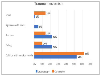
The median age of all patients was 30 years, with a minimum of 15 years and a maximum of 64 years, with the majority being male (80%), ASA I (72%) and Glasgow 15 (58%). The group distributions are detailed in Table 2; all the preoperative epidemiological and clinical data were similar between the groups (p>0.05). In both groups, the most frequent trauma mechanism was motor vehicle collision (53% for the laparoscopy group and 62% for the conversion group), followed by falling and running over for the laparoscopy group (22% each) and running over and crushing for the conversion group (14% each) (Figure 2). The main indications for laparoscopy were the presence of FF without parenchymal injury on abdominal CT (59%), abdominal pain/signs of peritoneal irritation (11.5%) and trauma to the spleen or suspected bladder injury on CT (6% each). (Table 3).

Trauma mechanism of patients suffering from blunt abdominal trauma in a tertiary trauma center. Chi-square test for trend, p<0.0001.
In all patients, there was correspondence between CT findings and intraoperative findings. In the seven patients with negative DLs, the indications for laparoscopy were the presence of FF (in four patients) or abdominal pain/signs of peritoneal irritation (in three patients), and the intraoperative findings were the absence of lesions (in four patients) or a small amount of FF (in three patients). The surgical time was shorter in patients who underwent laparoscopy (p<0.05). However, when the types of laparoscopies were analyzed separately, the surgical time was significantly longer in the TL and Conversion groups than in the DL group (both p<0.05) (Figure 3). The main reasons for conversion were moderate hemoperitoneum with multiple associated lesions (56%), with or without bowel resection (38% and 18%, respectively), and the need for loop suture (14%). Other causes were hemodynamic instability, doubt about the viability of the loop and technical difficulty in presentation (4% for each).

Surgical time (in minutes) of patients who were victims of blunt abdominal trauma, who underwent laparoscopy (diagnostic or therapeutic) and who underwent laparoscopy conversion (conversion) at a tertiary trauma center from January 2010 to December 2022. For the first analysis (blue columns), laparoscopy included both TL and DL, *p<0.05. For the secondary analysis (orange columns), laparoscopies were analyzed separately, in terms of TL or DL, *p<0.05 vs. conversion and TL. Mann-Whitney test. TL: Therapeutic laparoscopy. DL: Diagnostic laparoscopy.
In 29 cases of TL (91%), the therapeutic procedure was performed in a single organ and in two or more organs in three cases (9%). The most frequent therapeutic procedures were enterorrhaphy and bladder repair (Figure 4). We also detail the three combined procedures as follows: Patient 1, diaphragm suture + splenic hemostasis; Patient 2, diaphragm suture + gastric suture; Patient 3, enterorrhaphy + umbilical hernioplasty.

Therapeutic procedures performed individually via laparoscopy in patients suffering from blunt abdominal trauma in a tertiary trauma center from January 2010 to December 2022.
The seat belt sign was present in the physical examination of 10 patients (12% of the total sample). In these cases, the indications for laparoscopy were FF without viscus injury on CT (70%) or persistent abdominal pain/peritoneal irritation (30%). Four cases were converted, while three were DL (one of them being negative), and the other three were TL. The laparoscopic procedures performed were hemostasis with mesentery suturing (2 patients) and bladder suturing (1 patient) (Figure 5). Among the two positive DL cases, the findings were a transverse and sigmoid mesocolon hematoma (one patient) and a colonic serosal lesion with omental hematoma (one patient). In the case of negative DL, CT revealed a minimal amount of FF in the cavity, and the surgical indication was persistent abdominal pain.

Distribution of patients suffering from blunt abdominal trauma with the seat belt sign, according to the intraoperative findings and procedures performed when necessary.
Associated injuries (extra-abdominal) were observed only at laparoscopy, with the thorax being the most frequently affected site. The most frequent number of associated lesions was two (Table 4). Intraoperative complications occurred in two patients in the TL group, both of whom had inadvertent small bowel injuries requiring suturing during the same surgical procedure. In the Conversion group, there was one case of splenic bleeding upon manipulation, which required only local hemostasis. In these three cases, the additional procedures did not necessarily have any effect on patient evolution, nor did they increase the length of hospital stay (Table 5). There were no cases of unnoticed lesions in either group. In the Conversion group, there was one case of reoperation due to septic shock with an abdominal focus, in which the patient had an extensive mesocolon lesion that evolved with suture dehiscence.
Postoperative complications occurred in five patients and are detailed in Table 6. Four patients in the laparoscopy group had complications (two grade II, one grade III and one grade V), and one patient in the conversion group had a grade III complication. The two grade II complications were deep vein thrombosis (DVT); both patients had serious associated injuries - extensive chest fracture and complex pelvic fracture - and underwent surgery by other specialties. Grade III complications occurred in patients who had severe chest trauma-flail chest and hemopneumothorax-requiring chest drainage, one by interventional radiology. The patient who died (grade V) was a positive DL, with a traumatic diaphragmatic hernia and a grade I splenic injury, whose surgical indication was FF without parenchymal injury or traumatic hernia. The cause of death was pulmonary sepsis due to severe thoracic trauma associated with bilateral hemopneumothorax, multiple fractures of the bilateral costal arches and bilateral clavicle fractures.
Hospital discharge was the clinical outcome in 84 patients (with the exception of one patient who died), with no case of readmission within 30 days for causes related to surgery. The length of hospital stay was longer in the Conversion group (12.5 days, IQ 8--17) than in the laparoscopy group, regardless of whether they were diagnostic or therapeutic (8 days for both, IQ 4--16 and 5--14, respectively); however, the difference was not statistically significant (p>0,05) (Table 5).
The distribution of procedures over the years is detailed in Figure 6. In 2010, it was not possible to perform any laparoscopy due to operational reasons that led to the unavailability of the video set for the emergency room. From then on, there was a progressive increase in laparoscopies (p<0.05). In 2020 and 2021, all patients were treated exclusively by laparoscopy (there were no cases of conversion).

Timeline of the distribution of patients suffering from blunt abdominal trauma and treated exclusively by laparoscopy or not in a tertiary trauma center from January 2010 to December 2022. Chi-square test for trend, p<0.0001.
DISCUSSION
This study evaluated 85 hemodynamically stable, conscious, and oriented patients with BAT who underwent surgical treatment via laparoscopy. This, whether diagnostic or therapeutic, avoided unnecessary laparotomies in 74% of the cases analyzed (63 patients treated exclusively by laparoscopy) if the open approach was the first approach option. The already established advantages of laparoscopy for other diseases and for PAT can be incorporated into the context of BAT, and these advantages include faster patient recovery, less postoperative pain and earlier return to activities, in addition to fewer socioemotional and financial requirements, while maintaining safety and effectiveness in assistance7,9,13,29.
The preoperative epidemiological profile revealed that there was a balance between the groups treated exclusively by laparoscopy and those not treated by laparoscopy (conversion cases). Adequate selection of patients for laparoscopic management is essential for the success of this type of approach, and patients must be hemodynamically stable, without neurological impairments that could compromise the abdominal exam (pain referred and verified on physical examination, with signs of acute abdomen). Furthermore, the severity criteria, such as the ASA score and injury severity by trauma severity score (RTS or ISS), were also homogeneously distributed between the groups. Any imbalances in age parameters or severity scores could bias the assessment. All studies are unanimous in reinforcing the importance of adequate selection of hemodynamically stable patients as a key factor in the success of laparoscopic management7,9,13,29,40.
In cases where the seat belt sign was present, 90% of these patients had abdominal injuries, and 80% of these patients needed therapeutic procedures. These data are in line with reports in the literature that the seat belt sign is highly predictive of intra-abdominal injury, and these patients should undergo CT of the abdomen, which is the gold standard of investigation, and remain hospitalized for at least 24 hours for clinical observation when nonoperative management is chosen23.
Our results corroborate reports that CT is the gold standard diagnostic method for BAT because of its high sensitivity and specificity in detecting relevant intra-abdominal injuries9,23,41. In our study, there was a correlation between the tomographic and intraoperative findings in all patients, with no missed injuries or reoperations related to laparoscopic access. The high correlation observed between preoperative diagnosis and indications for laparoscopy vs. intraoperative findings is due to strict compliance with the following steps: 1) selection of hemodynamically stable patients without lowering the level of consciousness; 2) clinical history (mechanism of trauma), combined with the patient’s complaint and a thorough physical examination; and 3) CT findings. The latter directs the surgeon to possible intraoperative findings and possible associated injuries (extra-abdominal), increasing the surgical precision and safety of the laparoscopic approach. In our study, the anamnesis data and physical examination evaluation, although carried out by doctors during training, were always supervised and validated by the most experienced surgeon on duty. The same happens in the evaluation of CT scans by the team of radiologists, in which the findings are discussed with the trauma team.
The main surgical indication was FF on CT, and it was strongly correlated with intraoperative findings, including negative DL findings, in which a small amount of FF was described, and these findings are consistent with prior reports7,42,43. Therefore, it is important to implement a grading/quantification system for this free fluid in future work to further refine its role in CT in the context of BAT. A recent guideline that evaluated stable patients with BAT and FF on CT suggested the following possibilities: 1) patients with moderate to large FF on CT and abdominal pain (with or without abdominal wall contusion), surgical exploration; 2) patients with moderate to large amounts of isolated FF or abdominal pain, decision for surgical exploration on the basis of careful clinical judgment (there is a greater likelihood of surgical intervention); and 3) patients with a small amount of FF and no abdominal pain, strict clinical observation with serial laboratory and imaging tests (the need for surgical intervention is unlikely)24. In patients who fall into option three, serial CT scans at intervals of 8-12 hours may be a viable option to assist in decision-making, avoiding late interventions, in which injuries to the small intestine are the most frequent41,43,44.
We agree that, in cases of doubt, patients with little FF on CT, persistent pain and a seat belt sign, laparoscopy can be a great ally in clarifying doubts because even if it is a DL, it has the benefits of lower morbidity than does laparotomy and a reduction in the risks related to delays in decision-making regarding surgical intervention3,9,44. In addition, improving the quality and standardization of CT findings can contribute to fewer DLs being performed. A recent randomized clinical trial comparing abdominal CT with DL in patients with PAT to the anterior wall who were hemodynamically stable revealed that selective nonoperative management on the basis of CT findings was successful in 93.8% of patients, avoiding negative DL45. Although there is no similar study in cases of CT scans in patients with BAT, this benefit may also be equivalent in these cases.
Most therapeutic procedures performed by laparoscopy in our cohort were performed on a single organ, the most common being loop suturing and bladder suturing. These data reinforce that the success of surgery is more related to cases with damage to a single abdominal organ, in which the treatment can be targeted and specific. Multiple intra-abdominal injuries create a more hostile environment and increase the likelihood of conversion. In our study, extra-abdominal injuries were frequent-mainly thoracic-but did not appear to drive conversion, which was more closely associated with intra-abdominal severity (e.g., moderate hemoperitoneum and need for bowel resection).
Early reports suggested that TL is effective for treating hollow viscus and mesenteric injuries but is less effective for treating splenic trauma13. Laparoscopic splenectomy in trauma patients is still a matter of debate, and its acceptance has been slower than it was in the elective setting, with the literature including only case reports or small series of cases. Although still limited, recent studies have shown the success of laparoscopic splenectomy in the management of patients with BAT who fail nonoperative treatment and continue to experience intermittent bleeding [46], such as our study, in which five procedures were performed uneventfully on the spleen-two splenectomies and three hemostasis procedures.
In our study, the surgical time for TL was the same as that for laparotomies (conversion group), and both were longer than that for DL. These data differ from the reports of most studies showing that laparoscopy in trauma patients has a shorter surgical time5,11,29. However, these studies analyzed different types of trauma (PAT and BAT) and laparoscopies (DL and TD). When we compared only two groups, laparoscopy (DL and TL) and conversion, the surgical time for laparoscopies was significantly shorter than that for converted surgeries. Surgical time varies according to the multiplicity of possible trauma findings and may not be directly related to the team’s learning curve. We had three cases of TL with a surgical time of more than 240 minutes in which more complex procedures, such as pancreatectomy, colon rafia and combined urologic procedures, were performed.
The conversion rate of BAT varies in the literature from 7--23% and depends on adequate patient selection. The indications for conversion differ significantly between centers, as they depend on staff skill and available materials. The main reasons for conversion are massive bleeding, complex injuries, poor safe visibility of the abdominal cavity, deterioration of the patient with hemodynamic instability and equipment failure7,13,17,29. In our study, the conversion rate was 26%, which was due to the hostile environment in trauma/emergency, as 56% of the conversions were associated with moderate hemoperitoneum and multiple injuries. No cases were related to equipment failure, and there were no cases of retroperitoneal injury. The HCFMUSP is a tertiary trauma center that receives patients with high-energy trauma mechanisms, and our data agree with a recent study that evaluated 35 patients with BAT and reported a 23% conversion rate due to continuous abdominal bleeding, which could not be controlled quickly and was followed by multiple complex injuries17.
Three cases of intraoperative complications involved injuries to the small bowel or spleen during manipulation. Although it did not impact the clinical outcome, as they were identified and corrected at the same surgical time, there was a need for additional procedures. To avoid inadvertent injuries, technique standardization, gentle tissue handling (atraumatic graspers, mesenteric manipulation) and the experience of assistants in facilitating the presentation for the surgeon are advisable. No postoperative complications were directly related to the laparoscopic method but rather to the complexity of the associated injuries (thoracic) and the surgeries that other specialties performed (orthopedic). The only case of death in which the procedure was performed was DL, and the cause of death was secondary to lung lesions (pulmonary focus sepsis).
Most studies that report a shorter hospital stay for laparoscopy than for laparotomy do not correlate the type of trauma, whether BAT or PAT5,11,13,16. The systematization of the technique of cavity inventory during laparoscopy, which is used to examine all organs and allow for a detailed inventory of the small bowel, minimizes the possibility of unnoticed injuries. This standardization has already been proposed by our group for patients with PAT8. There were no cases of unnoticed injuries in our study, similar to other reports in the literature17.
Although our institution is a tertiary trauma center and has well-structured protocols, our investigation is subject to all the limitations of retrospective studies, with weaknesses related to surgeons’ preferences, blood transfusion data, lack of subgroup analysis according to severity of injuries and lack of data on the clinical outcome of patients initially managed nonsurgically. Even so, our data are consistent with the global trend of increasing use of laparoscopy in the last decade in stable patients undergoing CT scans safely and effectively for diagnostic and therapeutic purposes, allowing the method to be reproducible in other centers. These results are directly linked to the surgeon’s training and experience in advanced laparoscopic procedures and the structure offered by the service7,13,17,29,40,46.
Using the converted-to-laparotomy subgroup as a “control” has inherent limitations. All patients were initially selected for laparoscopy using the same standardized preoperative criteria (hemodynamically stable, intact mental status, and CT-based indications), so conversion mainly reflects what was found during surgery, not differences at baseline. As a result, differences in operative time, complications, or length of hospital stay likely reflect injury complexity rather than the effect of laparoscopy itself. Nonetheless, comparing completed versus converted cases remains clinically informative because it mirrors real-world “intention-to-laparoscopy” practice, quantifies the drivers of conversion, and reports outcomes within an initially homogeneous eligibility frame (as supported by similar baseline characteristics in Table 2). We therefore present between-group comparisons as exploratory and focus on effect sizes with confidence intervals, avoiding causal claims.
A cost comparison of trauma laparoscopies with open procedures was not performed in this study. The cost of a laparoscopic procedure may be greater than that of laparotomy, as laparoscopy requires special equipment; however, it is possible to use permanent and reusable materials, optimizing costs. Furthermore, the extra expenses associated with possible disposable materials can be offset by the benefits of minimally invasive surgery during the postoperative period, such as less pain and the consequent use of medications, the need for intensive care units, faster recovery, and early hospital discharge. Although limited, the existing data comparing laparoscopy with laparotomy in trauma patients show a significant reduction in operative complications and the perioperative mortality rate, an earlier functional recovery of the intestine, less postoperative pain, a shorter hospital stay and a lower infection rate in the laparoscopy group5,11-13,16.
CONCLUSION
Laparoscopy can be safe and effective for the management of patients suffering from BAT in selected and hemodynamically stable patients. Success in this type of approach depends on the tripod: adequate selection of patients, trained surgeons and full-time availability of materials and complementary exams. Prospective clinical trials are necessary to validate the routine use of this access route in trauma, comparing imaging diagnostic methods with nonoperative therapy.
ACKNOWLEDGMENTS
The authors thank: 1) The teams of the Emergency Surgery Service of the Surgical Clinic Division III and the Radiology Service of the Hospital das Clínicas, Faculdade de Medicina de São Paulo for their support in patient care and in conducting the study, 2) Luciana Lamarão Damous for the suggestions and correction of the final version of the manuscript; 3) Toshiko Oya for assisting with the data collection; and 4) Marcia Kiyome Koike for assisting with the statistical analysis.
REFERENCES
-
1 Damous SHB, Menegozzo CAM, VON-Bahten LC, Utiyama EM. Laparoscopy in Trauma: Can Brazil fit into the global trend? Rev Col Bras Cir. 2023;50:e20233602EDIT01. doi: 10.1590/0100-6991e-20233602EDIT01-en.
» https://doi.org/10.1590/0100-6991e-20233602EDIT01-en -
2 Khubutiya MSh, Yartsev PA, Guliaev AA, Levitsky VD, Tlibekova MA. Laparoscopy in blunt and penetrating abdominal trauma. Surg Laparosc Endosc Percutan Tech. 2013;23(6):507-12. doi: 10.1097/SLE.0b013e3182937c37.
» https://doi.org/10.1097/SLE.0b013e3182937c37 -
3 Johnson JJ, Garwe T, Raines AR, Thurman JB, Carter S, Bender JS, et al. The use of laparoscopy in the diagnosis and treatment of blunt and penetrating abdominal injuries: 10-year experience at a level 1 trauma center. Am J Surg. 2013;205(3):317-20. doi: 10.1016/j.amjsurg.2012.10.021.
» https://doi.org/10.1016/j.amjsurg.2012.10.021 -
4 Hajibandeh S, Hajibandeh S, Gumber AO, Wong CS. Laparoscopy versus laparotomy for the management of penetrating abdominal trauma: A systematic review and meta-analysis. Int J Surg. 2016;34:127-136. doi: 10.1016/j.ijsu.2016.08.524.
» https://doi.org/10.1016/j.ijsu.2016.08.524 -
5 Li Y, Xiang Y, Wu N, Wu L, Yu Z, Zhang M, et al. A Comparison of Laparoscopy and Laparotomy for the Management of Abdominal Trauma: A Systematic Review and Meta-analysis. World J Surg. 2015;39(12):2862-71. doi: 10.1007/s00268-015-3212-4.
» https://doi.org/10.1007/s00268-015-3212-4 -
6 Lee PC, Lo C, Wu JM, Lin KL, Lin HF, Ko WJ. Laparoscopy decreases the laparotomy rate in hemodynamically stable patients with blunt abdominal trauma. Surg Innov. 2014;21(2):155-65. doi: 10.1177/1553350612474496.
» https://doi.org/10.1177/1553350612474496 -
7 Lin HF, Chen YD, Chen SC. Value of diagnostic and therapeutic laparoscopy for patients with blunt abdominal trauma: A 10-year medical center experience. PLoS One. 2018;13(2):e0193379. doi: 10.1371/journal.pone.0193379.
» https://doi.org/10.1371/journal.pone.0193379 -
8 Kawahara NT, Alster C, Fujimura I, Poggetti RS, Birolini D. Standard examination system for laparoscopy in penetrating abdominal trauma. J Trauma. 2009;67(3):589-95. doi: 10.1097/TA.0b013e3181a60593.
» https://doi.org/10.1097/TA.0b013e3181a60593 -
9 Di Saverio S, Birindelli A, Podda M, Segalini E, Piccinini A, Coniglio C, et al. Trauma laparoscopy and the six w's: Why, where, who, when, what, and how? J Trauma Acute Care Surg. 2019;86(2):344-67. doi: 10.1097/TA.0000000000002130.
» https://doi.org/10.1097/TA.0000000000002130 -
10 Adam N, Sorensen V, Skinner R. Not all intestinal traumatic injuries are the same: a comparison of surgically treated blunt vs. penetrating injuries. Injury. 2015;46(1):115-8. doi: 10.1016/j.injury.2014.07.010.
» https://doi.org/10.1016/j.injury.2014.07.010 -
11 Lin HF, Wu JM, Tu CC, Chen HA, Shih HC. Value of diagnostic and therapeutic laparoscopy for abdominal stab wounds. World J Surg. 2010;34(7):1653-62. doi: 10.1007/s00268-010-0485-5.
» https://doi.org/10.1007/s00268-010-0485-5 -
12 Lin HF, Chen YD, Lin KL, Wu MC, Wu CY, Chen SC. Laparoscopy decreases the laparotomy rate for hemodynamically stable patients with blunt hollow viscus and mesenteric injuries. Am J Surg. 2015;210(2):326-33. doi: 10.1016/j.amjsurg.2014.11.009.
» https://doi.org/10.1016/j.amjsurg.2014.11.009 -
13 Lim KH, Chung BS, Kim JY, Kim SS. Laparoscopic surgery in abdominal trauma: a single center review of a 7-year experience. World J Emerg Surg. 2015;10:16. doi: 10.1186/s13017-015-0007-8.
» https://doi.org/10.1186/s13017-015-0007-8 -
14 Chestovich PJ, Browder TD, Morrissey SL, Fraser DR, Ingalls NK, Fildes JJ. Minimally invasive is maximally effective: Diagnostic and therapeutic laparoscopy for penetrating abdominal injuries. J Trauma Acute Care Surg. 2015;78(6):1076-83. doi: 10.1097/TA.0000000000000655.
» https://doi.org/10.1097/TA.0000000000000655 -
15 Wang J, Cheng L, Liu J, Zhang B, Wang W, Zhu W, et al. Laparoscopy vs. Laparotomy for the Management of Abdominal Trauma: A Systematic Review and Meta-Analysis. Front Surg. 2022;9:817134. doi: 10.3389/fsurg.2022.817134.
» https://doi.org/10.3389/fsurg.2022.817134 -
16 Zafar SN, Onwugbufor MT, Hughes K, Greene WR, Cornwell EE 3rd, Fullum TM, et al. Laparoscopic surgery for trauma: the realm of therapeutic management. Am J Surg. 2015;209(4):627-32. doi: 10.1016/j.amjsurg.2014.12.011.
» https://doi.org/10.1016/j.amjsurg.2014.12.011 -
17 Matsevych O, Koto M, Balabyeki M, Aldous C. Trauma laparoscopy: when to start and when to convert? Surg Endosc. 2018;32(3):1344-1352. doi: 10.1007/s00464-017-5812-6.
» https://doi.org/10.1007/s00464-017-5812-6 -
18 Koto MZ, Matsevych OY, Mosai F, Patel S, Aldous C, Balabyeki M. Laparoscopy for blunt abdominal trauma: a challenging endeavor. Scand J Surg. 2019;108(4):273-279. doi: 10.1177/1457496918816927.
» https://doi.org/10.1177/1457496918816927 -
19 Sumislawski JJ, Zarzaur BL, Paulus EM, Sharpe JP, Savage SA, Nawaf CB, et al. Diagnostic laparoscopy after anterior abdominal stab wounds: worth another look? J Trauma Acute Care Surg. 2013 Dec;75(6):1013-7; discussion 1017-8. doi: 10.1097/TA.0b013e3182a1fde8.
» https://doi.org/10.1097/TA.0b013e3182a1fde8 -
20 Coleman JJ, Zarzaur BL. Surgical Management of Abdominal Trauma: Hollow Viscus Injury. Surg Clin North Am. 2017;97(5):1107-1117. doi: 10.1016/j.suc.2017.06.004.
» https://doi.org/10.1016/j.suc.2017.06.004 -
21 Fakhry SM, Brownstein M, Watts DD, Baker CC, Oller D. Relatively short diagnostic delays (<8 hours) produce morbidity and mortality in blunt small bowel injury: an analysis of time to operative intervention in 198 patients from a multicenter experience. J Trauma. 2000 Mar;48(3):408-14; discussion 414-5. doi: 10.1097/00005373-200003000-00007.
» https://doi.org/10.1097/00005373-200003000-00007 -
22 Malinoski DJ, Patel MS, Yakar DO, Green D, Qureshi F, Inaba K, et al. A diagnostic delay of 5 hours increases the risk of death after blunt hollow viscus injury. J Trauma. 2010;69(1):84-7. doi: 10.1097/TA.0b013e3181db37f5.
» https://doi.org/10.1097/TA.0b013e3181db37f5 -
23 Achatz G, Schwabe K, Brill S, Zischek C, Schmidt R, Friemert B, et al. Diagnostic options for blunt abdominal trauma. Eur J Trauma Emerg Surg. 2022;48(5):3575-3589. doi: 10.1007/s00068-020-01405-1. Erratum in: Eur J Trauma Emerg Surg. 2020.
» https://doi.org/10.1007/s00068-020-01405-1 -
24 Gonser-Hafertepen LN, Davis JW, Bilello JF, Ballow SL, Sue LP, Cagle KM, et al. Isolated free fluid on abdominal computed tomography in blunt trauma: watch and wait or operate? J Am Coll Surg. 2014;219(4):599-605. doi: 10.1016/j.jamcollsurg.2014.04.020.
» https://doi.org/10.1016/j.jamcollsurg.2014.04.020 -
25 Renz BM, Feliciano DV. Unnecessary laparotomies for trauma: a prospective study of morbidity. J Trauma. 1995;38(3):350-6. doi: 10.1097/00005373-199503000-00007.
» https://doi.org/10.1097/00005373-199503000-00007 -
26 Morrison JE, Wisner DH, Bodai BI. Complications after negative laparotomy for trauma: long-term follow-up in a health maintenance organization. J Trauma. 1996;41(3):509-13. doi: 10.1097/00005373-199609000-00021.
» https://doi.org/10.1097/00005373-199609000-00021 -
27 Menegozzo CAM, Damous SHB, Sabioni GR, Alves PHF, Rasslan R, de Salles Collet E Silva F, Utiyama EM. Could trauma laparoscopy be the standard of care for hemodynamically stable patients? A retrospective analysis of 165 cases. Surg Endosc. 2023 May 22. doi: 10.1007/s00464-023-10121-x.
» https://doi.org/10.1007/s00464-023-10121-x. -
28 Pau L, Navez J, Cawich SO, Dapri G. Laparoscopic Management of Blunt and Penetrating Abdominal Trauma: A Single-Center Experience and Review of the Literature. J Laparoendosc Adv Surg Tech A. 2021;31(11):1262-1268. doi: 10.1089/lap.2020.0552.
» https://doi.org/10.1089/lap.2020.0552 -
29 Birindelli A, Podda M, Segalini E, Cripps M, Tonini V, Tugnoli G, et al. TraumaLap Study Group. Is the minimally invasive trauma surgeon the next (r)evolution of trauma surgery? Indications and outcomes of diagnostic and therapeutic trauma laparoscopy in a level 1 trauma centre. Updates Surg. 2020;72(2):503-512. doi: 10.1007/s13304-020-00739-0.
» https://doi.org/10.1007/s13304-020-00739-0 - 30 Advanced Trauma Life Support(r) (ATLS(r)) Student Course Manual 10th edition - Chicago: American College of Surgeons, Committee on Trauma, 2018. 391 p. ISBN 0996826238, 9780996826235.
- 31 Ministério da Saúde, Secretaria de Vigilância em Saúde e Ambiente. Boletim Epidemiológico 6, volume 54, 27 de abril de 2023. ISSN 9352-7864.
-
32 Loggers SAI, Koedam TWA, Giannakopoulos GF, Vandewalle E, Erwteman M, Zuidema WP. Definition of hemodynamic stability in blunt trauma patients: a systematic review and assessment amongst Dutch trauma team members. Eur J Trauma Emerg Surg. 2017;43(6):823-833. doi: 10.1007/s00068-016-0744-8.
» https://doi.org/10.1007/s00068-016-0744-8 -
33 Teasdale G, Jennett B. Assessment of coma and impaired consciousness. A practical scale. Lancet. 1974 Jul 13;2(7872):81-4. doi: 10.1016/s0140-6736(74)91639-0.
» https://doi.org/10.1016/s0140-6736(74)91639-0 -
34 Champion HR, Sacco WJ, Copes WS, Gann DS, Gennarelli TA, Flanagan ME. A revision of the Trauma Score. J Trauma. 1989;29(5):623-9. doi: 10.1097/00005373-198905000-00017.
» https://doi.org/10.1097/00005373-198905000-00017 -
35 Baker SP, O'Neill B. The injury severity score: an update. J Trauma. 1976;16(11):882-5. doi: 10.1097/00005373-197611000-00006.
» https://doi.org/10.1097/00005373-197611000-00006 -
36 Copes WS, Champion HR, Sacco WJ, Lawnick MM, Keast SL, Bain LW. The Injury Severity Score revisited. J Trauma. 1988;28(1):69-77. doi: 10.1097/00005373-198801000-00010.
» https://doi.org/10.1097/00005373-198801000-00010 -
37 Damous SHB, Menegozzo CAM, Rocha MC, Collet e Silva F, Utiyama EM. Challenges to implement laparoscopic appendectomy as the first-line treatment for acute appendicitis: a fifteen-year experience in a tertiary hospital in Brazil. Rev Col Bras Cir. 2023; 50:e20233527. DOI: 10.1590/0100-6991e-20233527.
» https://doi.org/10.1590/0100-6991e-20233527 -
38 Mayhew D, Mendonca V, Murthy BVS. A review of ASA physical status - historical perspectives and modern developments. Anaesthesia. 2019;74(3):373-379. doi: 10.1111/anae.14569.
» https://doi.org/10.1111/anae.14569 -
39 Naumann DN, Vincent LE, Pearson N, Beaven A, Smith IM, Smith K, et al. An adapted Clavien-Dindo scoring system in trauma as a clinically meaningful nonmortality endpoint. J Trauma Acute Care Surg. 2017;83(2):241-8. doi: 10.1097/TA.0000000000001517.
» https://doi.org/10.1097/TA.0000000000001517 -
40 Kindel T, Latchana N, Swaroop M, Chaudhry UI, Noria SF, Choron RL, et al. Laparoscopy in trauma: An overview of complications and related topics. Int J Crit Illn Inj Sci. 2015;5(3):196-205. doi: 10.4103/2229-5151.165004.
» https://doi.org/10.4103/2229-5151.165004 -
41 Cinquantini F, Tugnoli G, Piccinini A, Coniglio C, Mannone S, Biscardi A, et al. Educational Review of Predictive Value and Findings of Computed Tomography Scan in Diagnosing Bowel and Mesenteric Injuries After Blunt Trauma: Correlation With Trauma Surgery Findings in 163 Patients. Can Assoc Radiol J. 2017;68(3):276-85. doi: 10.1016/j.carj.2016.07.003.
» https://doi.org/10.1016/j.carj.2016.07.003 - 42 Leow P, Kong V, Rajaretnam N, Ah Yen D, Amey J, Denize B, et al. Contemporary management of isolated free fluid on computed tomography scan in blunt abdominal trauma-experience from a Level 1 trauma centre in New Zealand. N Z Med J. 2022;135(1557):28-37.
-
43 Mahmood I, Tawfek Z, Abdelrahman Y, Siddiuqqi T, Abdelrahman H, El-Menyar A, et al. Significance of computed tomography finding of intra-abdominal free fluid without solid organ injury after blunt abdominal trauma: time for laparotomy on demand. World J Surg. 2014;38(6):1411-5. doi: 10.1007/s00268-013-2427-5.
» https://doi.org/10.1007/s00268-013-2427-5 -
44 Chiu HH, Tee YS, Hsu CP, Hsu TA, Cheng CT, Liao CH, et al. The Role of Diagnostic Laparoscopy in the Evaluation of Abdominal Trauma Patients: A Trauma Quality Improvement Program Study. World J Surg. 2023;47(10):2357-66. doi: 10.1007/s00268-023-07113-2.
» https://doi.org/10.1007/s00268-023-07113-2 -
45 Kaur S, Bagaria D, Kumar A, Priyadarshini P, Choudhary N, Sagar S, et al. Contrast-enhanced computed tomography abdomen versus diagnostic laparoscopy-based management in patients with penetrating abdominal trauma: a randomized controlled trial. Eur J Trauma Emerg Surg. 2023;49(1):1-10. doi: 10.1007/s00068-022-02089-5.
» https://doi.org/10.1007/s00068-022-02089-5 -
46 Huang GS, Chance EA, Hileman BM, Emerick ES, Gianetti EA. Laparoscopic Splenectomy in Hemodynamically Stable Blunt Trauma. JSLS. 2017;21(2):e2017.00013. doi: 10.4293/JSLS.2017.00013
» https://doi.org/10.4293/JSLS.2017.00013
-
Data availability
Datasets related to this article will be available upon request to the corresponding author.
-
Funding source:
Scientific Initiation Scholarship supported by Programa Institucional de Bolsas de Iniciação Científica (PIBIC) 2021/2022, Faculdade de Medicina, Universidade de São Paulo (FMUSP) e Conselho Nacional de Desenvolvimento Científico e Tecnológico (CNPq).
Datasets related to this article will be available upon request to the corresponding author.