 Hepatocellular Expression of Macrophage Migration Inhibitory Factor (MIF) Is Associated with Ischemia-Reperfusion Injury After Liver Transplantation
ABSTRACT
Introduction: Ischemia-reperfusion injury (IRI) is a major determinant of initial graft function after liver transplantation (LT), directly influencing the incidence of early dysfunction and short-term clinical outcomes. Macrophage migration inhibitory factor (MIF), a pleiotropic cytokine involved in inflammatory pathways and mechanisms of cellular adaptation to oxidative stress, may play a modulatory role in IRI, although its immediate tissue behaviour in grafts remains poorly characterised.
Methods: We retrospectively evaluated adult LT recipients who underwent post-reperfusion biopsies suitable for histological and immunohistochemical analysis. Immunohistochemical expression of MIF was quantified using the IHC Profiler plugin (ImageJ), whereas IRI was graded according to validated histopathological criteria.
Results: Among 153 biopsies analysed, 103 met the eligibility criteria, with most cases showing absent or mild IRI (70.9%). Hepatocellular expression of MIF was predominantly weak or moderate. Stronger immunohistochemical staining for MIF was associated with lower IRI severity (p<0.05). MIF expression correlated with pre-transplant laboratory parameters, without association with steatosis or early outcomes, including retransplantation or death within 15 days.
Conclusion: The findings suggest that greater immunohistochemical expression of MIF at reperfusion is associated with lower IRI intensity, indicating a possible adaptive role for this cytokine during this critical phase. MIF emerges as a potential tissue marker complementary to traditional histopathological scoring, with relevance for graft assessment and use in contemporary liver perfusion strategies.
Keywords:
Liver Transplantation; Reperfusion Injury; Immunohistochemistry; Macrophage Migration Inhibitory Factors
RESUMO
Introdução: A lesão de isquemia-reperfusão (LIR) é um evento determinante da função inicial do enxerto após o transplante hepático (TH). O fator inibitório da migração de macrófagos (MIF) é uma citocina pleiotrópica envolvida em vias inflamatórias e em mecanismos de adaptação celular ao estresse oxidativo. Embora possa exercer papel modulador na LIR, sua expressão tecidual imediata em enxertos hepáticos permanece pouco caracterizada.
Métodos: Este estudo retrospectivo avaliou receptores adultos submetidos a transplante hepático, com análise histológica e imuno-histoquímica de biópsias obtidas após a reperfusão. A expressão de MIF foi quantificada por meio do plugin IHC Profiler (ImageJ), enquanto a LIR foi graduada segundo critérios histopatológicos validados.
Resultados: Entre 153 biópsias analisadas, 103 preencheram os critérios de elegibilidade, com predomínio de LIR ausente ou leve (70,9%). A expressão hepatocitária de MIF foi predominantemente fraca ou moderada. Marcações mais intensas de MIF associaram-se a menor gravidade da LIR (p < 0,05). A expressão de MIF correlacionou-se com parâmetros laboratoriais pré-transplante, sem associação com esteatose ou com desfechos precoces, incluindo retransplante ou óbito em até 15 dias.
Conclusão: Esses achados sugerem que maior marcação tecidual de MIF no momento da reperfusão relaciona-se a menor intensidade da LIR, indicando possível papel adaptativo dessa citocina nessa fase crítica. O MIF desponta como potencial marcador tecidual complementar ao escore histopatológico tradicional, com relevância para a avaliação de enxertos e para o desenvolvimento de estratégias contemporâneas de perfusão hepática.
Palavras-chave:
Transplante de Fígado; Lesão por Isquemia-Reperfusão; Fator Inibidor da Migração de Macrófago
INTRODUCTION
Transplantation is the treatment of choice for advanced chronic liver disease and for several forms of acute liver failure, with five-year survival rates exceeding 75%1. Despite improvements in surgical techniques and perioperative support, initial graft function may still be significantly compromised by ischemia-reperfusion injury (IRI), a complex process initiated during organ procurement, sustained throughout cold and warm ischemia, and intensified at the time of graft reperfusion. IRI involves oxidative stress, microvascular dysfunction, Kupffer cell activation, neutrophil recruitment, and the release of damage-associated molecular patterns, resulting in an increased risk of early allograft dysfunction (EAD)2-4.
Histopathological evaluation of post-reperfusion (time-zero) biopsy specimens provides a direct measure of the magnitude of IRI. Alterations such as zonal necrosis, sinusoidal congestion, and hepatocellular degeneration are well-established histological markers of injury associated with ischemic events. Recent studies have reported semiquantitative analyses integrating these findings, demonstrating that different histopathological IRI grades are predictive of unfavourable outcomes, including a higher incidence of EAD5,6.
Macrophage migration inhibitory factor (MIF) is a pleiotropic cytokine and a central mediator of innate immunity, traditionally recognised for its pro-inflammatory effects but now recognised as an important regulator of oxidative stress, cellular survival, and metabolic homeostasis in different organs, including the liver7-11.
MIF is involved in several inflammatory conditions affecting the gastrointestinal tract and liver. In the liver, MIF is expressed by hepatocytes, Kupffer cells, and sinusoidal endothelial cells, where it is involved in both pro-inflammatory pathways and adaptive mechanisms of cellular protection12. Notably, The function of MIF in the liver is highly context-dependent, acting as a deleterious mediator in chronic inflammation while exerting a protective role in acute injury. However, despite its involvement in multiple liver disorders, its role during reperfusion in liver transplantation remains poorly characterised, highlighting the need for further investigation of its expression and potential implications for the severity of hepatocellular injury.
Thus, the primary objective of this study was to evaluate the association between cytoplasmic hepatocellular MIF expression and the histopathological IRI score in post-reperfusion liver graft biopsies.
OBJECTIVES
The primary objective of this study was to evaluate the association between immunohistochemical expression of MIF in hepatocytes and the histopathological IRI score in post-reperfusion liver graft biopsies. Secondary objectives included evaluating the relationship between MIF expression and the occurrence of EAD, as well as its association with short-term clinical outcomes, such as retransplantation or death within the first 15 postoperative days.
METHODS
Study design
This was a retrospective observational study that analysed post-reperfusion (time-zero) liver biopsies archived in the Pathology Department of the Hospital das Clínicas, Universidade Federal de Minas Gerais/EBSERH. Clinical, surgical, laboratory, and histopathological data were obtained from electronic medical records and institutional registries. The study was reported in accordance with the STROBE (STrengthening the Reporting of Observational Studies in Epidemiology) recommendations for observational studies.
Study population and eligibility criteria
The eligible population included adult recipients aged ≥18 years who underwent deceased-donor liver transplantation between 2000 and 2020. As post-reperfusion liver biopsy was not systematically performed during the study period and was obtained only in specific clinical situations or at the transplant team’s discretion, the study was based on a convenience sample of cases with available, adequate histological material for analysis. Patients with biopsies performed immediately after reperfusion, preserved tissue fragments suitable for histopathological and immunohistochemical evaluation, and sufficient clinical and laboratory data to characterise initial graft function were included.
Retransplantation cases, inadequate biopsies (due to insufficient tissue quantity, technical artefacts, or absence of representative parenchyma), cases lacking essential clinical information, and recipients who died intraoperatively were excluded, since correlation between tissue findings and immediate postoperative course would not be possible in these cases.
Variables analyzed
Variables were selected according to their relevance for the assessment of IRI, initial graft function, and early postoperative outcomes.
Regarding recipients, the following variables were included: age, sex, aetiology of liver disease, preoperative clinical conditions, and laboratory tests used to calculate the classical MELD score (bilirubin, INR, and creatinine), according to the allocation system in use during the analysed period13-15. Serum sodium was recorded but not incorporated into the MELD calculation, as MELD-Na was not a prioritisation criterion during the study interval. Length of hospital stay after transplantation and the occurrence of retransplantation or death within the first 15 postoperative days were also recorded.
Regarding donors, the following variables were included: age, sex, clinical characteristics documented during organ procurement, and cold ischemia time.
The following laboratory parameters were analysed up to the seventh postoperative day: total bilirubin, AST, ALT, INR, creatinine, sodium, haemoglobin, leukocyte count, and platelet count. These parameters were used to assess the early postoperative course and define EAD16.
Histopathological assessment of IRI
Post-reperfusion (time-zero) liver biopsies were fixed in 10% buffered formalin, paraffin-embedded, and sectioned into 3-4 μm slices. Slides were stained with hematoxylin-eosin (H&E) for morphological evaluation. Histological assessment was independently and blindly performed by two experienced pathologists following validated morphological criteria described in the literature for classification of ischemia-reperfusion injury17. The evaluated parameters included hepatocellular oedema and swelling, sinusoidal compression and congestion, hepatocellular degeneration (ballooning and cytoplasmic granularity), lobular and sinusoidal leukocytic infiltrate, hepatocellular necrosis predominantly in zone 3, lipopigment deposition, and cellular debris (Table 1).
Immunohistochemistry for MIF
Immunohistochemical analysis aimed to quantify cytoplasmic MIF expression in hepatocytes. Immunohistochemical reactions were performed on post-reperfusion liver biopsies using a polyclonal anti-MIF antibody (ab65869, Abcam, Cambridge, UK) in 4-μm sections of formalin-fixed and paraffin-embedded tissue. Briefly, sections were deparaffinized and hydrated, then antigen-retrieved using the wet-heat method with EDTA buffer (pH 8.0) at 90 °C. Peroxidase blocking (Novolink™, Leica Biosystems) was performed for 10 minutes, followed by protein blocking (Novolink™, Leica Biosystems). Incubation with the primary anti-MIF pAb antibody at a 1:100 dilution was performed for 2 hours, followed by post-primary blocking and polymer detection (Novolink™, Leica Biosystems) for 30 minutes. Finally, sections were treated with the chromogen 3,3-diaminobenzidine (DAB), producing a brown precipitate at the antigenic site. Samples were counterstained with hematoxylin (Novolink™, Leica Biosystems) for 3 minutes and digitally scanned using a slide scanner (3DHistech Ltd., Budapest, Hungary).
Immunohistochemical expression of MIF was quantified using the IHC Profiler plugin of the ImageJ Fiji software (version 1.2; WS Rasband, National Institute of Health, Bethesda, MD)18. Five areas of interest from each slide were selected and photographed by a pathologist and subsequently analysed by the software, which classified marker expression as negative, weak positive, positive, or strongly positive according to an algebraic formula based on the number of pixels within a zone, the zone score, and the total number of pixels in the image. The mode for each case was calculated to standardise expression classification across the five areas of interest. When mode calculation was not possible, the pathologist determined the final classification based on the two most frequent scores. Given the marker’s constitutive expression in hepatocytes, the results were grouped into negative/weakly positive and positive staining to highlight the role of MIF overexpression in IRI.
Outcomes evaluated
The study included a primary and a secondary outcome. The primary outcome was the association between hepatocellular MIF expression and the histopathological IRI score. The secondary outcome was the evolution of serial laboratory parameters over the first 7 postoperative days.
Statistical analysis
Statistical analysis was performed using SPSS Statistics (IBM Corp., version 23.0, Armonk, NY). Continuous variables are expressed as medians and interquartile ranges. Categorical variables are expressed as absolute and relative frequencies. Mann-Whitney, chi-square, or Fisher’s exact tests were used in univariate analysis. A p-value <0.05 was considered statistically significant.
Ethical aspects
This study was conducted in accordance with the ethical principles established by the Declaration of Helsinki and by national guidelines for research involving human subjects. The research protocol was previously submitted to and approved by the Research Ethics Committee of Universidade Federal de Minas Gerais under approval number CAAE 42157120.30000.5149. All stages of the study respected the confidentiality and integrity of clinical patient data, ensuring the privacy of information and strict compliance with current ethical standards for retrospective research involving medical records and archived biological samples.
RESULTS
Sample characteristics and inclusion flow
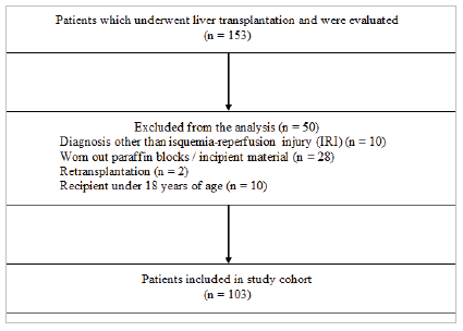
A total of 153 post-reperfusion liver biopsies archived between 2000 and 2020 were evaluated. After exclusion of cases due to insufficient material or incompatible diagnoses, 103 patients were included in the final analysis (Figure 1).

Flowchart of the selection of post-reperfusion biopsies for immunohistochemical analysis and medical record review.
The median recipient age was 51 years, with a predominance of male patients. The main underlying liver diseases were alcoholic cirrhosis (30.1%) and viral hepatitis (29.1%). Data are shown in Table 2.
Most grafts presented absent or mild ischemia-reperfusion injury
Based on histological criteria, 49 biopsies (47.6%) showed no evidence of IRI, 24 biopsies (23.3%) were classified as mild IRI, 25 (24.3%) as moderate, and only 5 (4.9%) as severe (Figures 2D, 2E, and 2F). None of the clinical or epidemiological variables showed a significant association with IRI (data not shown).

Histological classification of IRI in post-reperfusion biopsies. In (A) it is shown the presence of scarce neutrophils in sinusoid capillaries, without hepatocyte necrosis (dotted arrow), corresponding to mild IRI. In (B) it is observed frequent neutrophilic grouping with focal necrosis of hepatocytes (continuous arrow). In (C) it is shown areas of confluence of hepatocyte necrosis (dashed areas), corresponding to severe IRI.
Immunohistochemical expression of MIF in hepatocytes was predominantly weak/moderate
Immunohistochemical analysis demonstrated hepatocellular expression, with weak positive staining in 64 cases (62.1%), moderate positive staining in 34 cases (33%), and negative staining in 5 cases (4.9%). No cases showed strong staining. Figures 2D, 2E, and 2F show cellular localisation and staining intensity, as described above.
Association between MIF immunohistochemical expression and the severity of LIR
Univariate analysis revealed a significant association between positive MIF expression and absent or mild IRI (p=0.024). Table 3 demonstrates the distribution of age, aetiology, MELD, presence of hepatocellular carcinoma, IRI, and steatosis across MIF expression groups. No association was observed between MIF expression and steatosis. These findings are consistent with data from other studies discussed in the Discussion section, demonstrating that MIF is rapidly mobilised and secreted by hepatocytes under stress conditions.
Immunohistochemical expression of MIF correlated with pre-transplant laboratory alterations
Among the 82 patients with complete laboratory data, positive MIF expression was associated with lower pre-transplant serum albumin levels (p=0.013), higher pre-transplant serum AST levels (p=0.010), and higher pre-transplant ALT levels (p=0.018). Table 4 presents the biochemical parameters associated with MIF expression and shows no correlation with post-transplant laboratory tests.
No association was observed between immunohistochemical expression of MIF and early outcomes (retransplantation or death within 15 days)
Ten patients required retransplantation or died within the early postoperative period. None of the variables - including MIF expression, IRI, donor age, MELD score, cold ischemia time, or steatosis - showed a significant association with this outcome (data not shown).
DISCUSSION
In this study, we evaluated hepatocellular MIF expression in biopsies obtained after reperfusion at the end of the liver implantation procedure and explored its relationship with IRI severity. Most grafts showed histopathological findings compatible with absent or mild IRI, and, when detected, immunohistochemical expression of MIF was predominantly hepatocellular and of weak or moderate intensity. We observed that higher levels of hepatocellular MIF expression were associated with lower histopathological grades of IRI. In addition, MIF expression was associated with some pre-transplant laboratory parameters but was not associated with steatosis or early postoperative outcomes. These findings indicate that MIF expression after graft reperfusion may reflect not only intrinsic graft characteristics but, more importantly, its individual initial response to ischemic stress.
The association between greater hepatocellular MIF expression and lower intensity of IRI should be interpreted considering that this cytokine may exert opposite effects in the cellular response to acute stress, depending on the biological context19-23. Although MIF has traditionally been recognised as a pro-inflammatory mediator, its rapid release by hepatocytes in response to hypoxia and reperfusion has also been associated with activation of cytoprotective pathways, modulation of oxidative stress, preservation of cellular viability, and promotion of early parenchymal metabolic recovery. From this perspective, higher levels of MIF at the time of reperfusion may represent an adaptive hepatocellular response to ischemic insult, potentially limiting the extent of immediate histological injury.
Experimental and clinical studies have already demonstrated that MIF participates in different stages of the hepatocellular response to ischemic injury, both by amplifying inflammatory pathways and by modulating repair processes and metabolic adaptation24-26. In models of hypoxia and IRI, early hepatocellular expression of MIF has been associated with mitochondrial preservation, reduction of oxidative stress, and attenuation of zone 3 necrosis, effects compatible with the histological pattern observed in grafts with milder IRI. These data corroborate previous findings describing the heterogeneity of IRI and reinforce the notion that intrinsic graft factors influence the capacity to respond to ischemic insult, as demonstrated in studies linking time-zero histological alterations to initial prognosis after transplantation16,17. In this context, our findings demonstrate that hepatocellular expression of MIF may be part of this spectrum of initial graft responses, possibly serving as a marker of hepatocellular adaptation to reperfusion.
Interpretation of this finding should consider that the dynamics of MIF in the acute context of ischemia and reperfusion remain poorly characterised in human organ transplantation, and no previous descriptions of immediate hepatocellular expression in this setting are available. In light of this gap, a central question emerges: which mechanisms could explain greater MIF expression in grafts presenting milder IRI? In several cellular models, MIF is known to be rapidly released from preformed cytoplasmic stores when cells are exposed to pro-inflammatory, mitogenic, or hormonal stimuli8. This initial release is followed by increased MIF transcription and replenishment of intracellular content, highlighting its role as an early, upstream mediator of the stress response27,28 . In systemic settings of hypoxia and acute inflammation, elevated MIF concentrations have been associated with attenuation of oxidative stress, mitochondrial preservation, and maintenance of cellular viability - mechanisms compatible with the pattern observed in our study29. Furthermore, previous investigations demonstrated increased MIF levels in the hepatic vein and systemic circulation immediately after reperfusion, suggesting that the liver is an important source of this cytokine at this stage 30. Thus, it is plausible that greater hepatocellular MIF expression represents an adaptive response in grafts with greater resilience to ischemic stress, resulting in less histological damage.
The absence of association between hepatocellular MIF expression and variables traditionally related to unfavourable graft or recipient evolution - such as steatosis, preoperative MELD score, donor age, or cold ischemia time - suggests that the initial modulation of this cytokine may reflect phenomena more directly linked to the immediate cellular response to reperfusion than to baseline clinical conditions. Likewise, we observed no correlation between MIF expression and early outcomes, such as retransplantation or death within 15 days. However, these negative findings should be interpreted with caution, since the study was not designed to comprehensively evaluate these outcomes. Case inclusion depended on the availability of post-reperfusion biopsies, resulting in a convenience sample that may not represent the entire spectrum of transplant severity during the analysed period. In addition, other intraoperative and postoperative variables - such as haemodynamic instability, transfusion requirements, anhepatic phase duration, use of vasoactive drugs, and technical complications - were not systematically evaluated, limiting the ability to adjust for potential confounding factors. Therefore, although our results indicate that hepatocellular MIF expression is fundamentally associated with the histological degree of IRI, it is not possible to extrapolate from this study its prognostic role or its relationship with previous or subsequent transplant characteristics.
This study has limitations that should be acknowledged. The retrospective design and inclusion of only cases with available post-reperfusion biopsies resulted in a convenience sample that does not represent the full spectrum of transplantations performed during the study period. The long temporal window analysed (2000-2020) encompasses different phases in the evolution of surgical techniques, preservation methods, and perioperative management, resulting in heterogeneity that could not be fully controlled. Biopsy acquisition did not follow a uniform protocol, and variations in indication, fragment location, and sampling depth may have influenced histological evaluation; even with blinded assessment by experienced pathologists, classification of milder IRI grades remains somewhat subjective. In addition, the analysis focused exclusively on hepatocellular MIF expression and did not include other cellular compartments that may be relevant to the reperfusion response. Intraoperative and postoperative variables - such as haemodynamic instability, transfusions, anhepatic time, or use of vasoactive drugs - were not assessed in a standardised manner, limiting control of potential confounding factors. Finally, the reduced number of early clinical outcomes limited the ability to explore the possible prognostic role of MIF expression. Therefore, although these findings provide an important initial perspective on the dynamics of this cytokine after hepatic reperfusion, they should be interpreted with caution and confirmed in prospective studies with more standardised methodology.
The findings of this study suggest that hepatocellular MIF expression at reperfusion may represent a relevant component of the adaptive response to IRI, adding a pathophysiological dimension that remains poorly explored in liver transplantation. If confirmed in future studies, these findings position MIF as a possible tissue marker complementary to traditional histopathological scoring, with the potential to identify grafts that are better able to withstand ischemic stress. In the context of increasing incorporation of extracorporeal liver perfusion technologies, tissue markers such as MIF may eventually be evaluated during normothermic or hypothermic perfusion itself, assisting decision-making regarding acceptance, reconditioning, or discard of organs from expanded-criteria donors. To achieve this, prospective studies with standardised sampling, dynamic evaluation of MIF expression at different preservation stages, and rigorous control of operative variables will be required.
CONCLUSION
The findings of this study indicate that hepatocellular MIF expression at the time of reperfusion may represent part of the initial adaptive graft response to ischemia-reperfusion injury, since higher levels were associated with lower histological severity of IRI. These results suggest that MIF may function as a tissue marker complementary to traditional histopathological scoring, helping to identify grafts with greater resilience to ischemic stress during reperfusion.
In the current context of expanding extracorporeal liver perfusion technologies, assessing tissue markers such as MIF during normothermic or hypothermic perfusion may enable more precise decision-making regarding the acceptance, reconditioning, or discard of organs from expanded-criteria donors. Prospective studies with dynamic evaluation of MIF during different preservation stages and rigorous control of operative variables will be essential to define its prognostic value and applicability in clinical practice.
ACKNOWLEDGEMENTS
Special thanks to Fernanda Césari, a pathology technician in the Department of Pathological Anatomy and Forensic Medicine.
REFERENCES
-
1 Adam R, Karam V, Delvart V, O'Grady J, Mirza D, Klempnauer J, et al. Evolution of indications and results of liver transplantation in Europe. A report from the European Liver Transplant Registry (ELTR). J Hepatol. 2012;57(3):675-88. doi: 10.1016/j.jhep.2012.04.015.
» https://doi.org/10.1016/j.jhep.2012.04.015 -
2 Zhai Y, Petrowsky H, Hong JC, Busuttil RW, Kupiec-Weglinski JW. Ischaemia-reperfusion injury in liver transplantation-from bench to bedside. Nat Rev Gastroenterol Hepatol. 2013;10(2):79-89. doi: 10.1038/nrgastro.2012.225.
» https://doi.org/10.1038/nrgastro.2012.225 -
3 Hann A, Osei-Bordom DC, Neil DAH, Ronca V, Warner S, Perera MTPR. The Human Immune Response to Cadaveric and Living Donor Liver Allografts. Front Immunol. 2020;11:1227. doi: 10.3389/fimmu.2020.01227.
» https://doi.org/10.3389/fimmu.2020.01227 -
4 Olthoff KM, Kulik L, Samstein B, Kaminski M, Abecassis M, Emond J, et al. Validation of a current definition of early allograft dysfunction in liver transplant recipients and analysis of risk factors. Liver Transpl. 2010;16(8):943-9. doi: 10.1002/lt.22091.
» https://doi.org/10.1002/lt.22091 -
5 Ito T, Naini BV, Markovic D, Aziz A, Younan S, Lu M, et al. Ischemia-reperfusion injury and its relationship with early allograft dysfunction in liver transplant patients. Am J Transplant. 2021;21(2):614-25. doi: 10.1111/ajt.16219.
» https://doi.org/10.1111/ajt.16219 -
6 Fuentes-Valenzuela E, Tejedor-Tejada J, García-Pajares F, Rubiales BM, Nájera-Muñoz R, Maroto-Martín C, et al. Postreperfusion liver biopsy as predictor of early graft dysfunction and survival after orthotopic liver transplantation. J Clin Exp Hepatol. 2022;12(4):1133-41. doi: 10.1016/j.jceh.2021.12.015.
» https://doi.org/10.1016/j.jceh.2021.12.015 -
7 Leng L, Metz CN, Fang Y, Xu J, Donnelly S, Baugh J, et al. MIF Signal transduction initiated by binding to CD74. J Exp Med. 2003;197(11):1467-76. doi: 10.1084/jem.20030286.
» https://doi.org/10.1084/jem.20030286 -
8 Calandra T, Roger T. Macrophage migration inhibitory factor: a regulator of innate immunity. Nat Rev Immunol. 2003;3(10):791-800. doi: 10.1038/nri1200.
» https://doi.org/10.1038/nri1200 -
9 Kang I, Bucala R. The immunobiology of MIF: function, genetics and prospects for precision medicine. Nat Rev Rheumatol. 2019;15(7):427-37. doi: 10.1038/s41584-019-0238-2.
» https://doi.org/10.1038/s41584-019-0238-2 -
10 Sumaiya K, Langford D, Natarajaseenivasan K, Shanmughapriya S. Macrophage migration inhibitory factor (MIF): A multifaceted cytokine regulated by genetic and physiological strategies. Pharmacol Ther. 2022;233:108024. doi: 10.1016/j.pharmthera.2021.108024.
» https://doi.org/10.1016/j.pharmthera.2021.108024 -
11 Herkens L, Droste P, Boor P. The multifaced role of the macrophage migration inhibitory factor family in organ fibrosis. Am J Physiol-Cell Physiol. 2025;329(1):C119-35. doi: 10.1152/ajpcell.00198.2025.
» https://doi.org/10.1152/ajpcell.00198.2025 -
12 Ohkawara T, Nishihira J, Takeda H, Asaka M, Sugiyama T. Pathophysiological roles of macrophage migration inhibitory factor in gastrointestinal, hepatic, and pancreatic disorders. J Gastroenterol. 2005;40(2):117-22. doi: 10.1007/s00535-004-1526-3.
» https://doi.org/10.1007/s00535-004-1526-3 - 13 Ministério da Saúde (Brasil). Portaria no 2.600, de 21 de outubro de 2009. Aprova o Regulamento Técnico do Sistema Nacional de Transplantes. Brasília, DF: Ministério da Saúde; 2009.
-
14 Kamath PS, Wiesner RH, Malinchoc M, Kremers W, Therneau TM, Kosberg CL, et al. A Model to Predict Survival in Patients With End-Stage Liver Disease. Hepatology. 2001;33(2):464-70. doi: 10.1053/jhep.2001.22172.
» https://doi.org/10.1053/jhep.2001.22172 -
15 Wiesner RH, McDiarmid SV, Kamath PS, Edwards EB, Malinchoc M, Kremers WK, et al. MELD and PELD: Application of survival models to liver allocation. Liver Transpl. 2001;7(7):567-80. doi: 10.1053/jlts.2001.25879.
» https://doi.org/10.1053/jlts.2001.25879 -
16 Ali JM, Davies SE, Brais RJ, Randle LV, Klinck JR, Allison MED, et al. Analysis of ischemia/reperfusion injury in time-zero biopsies predicts liver allograft outcomes. Liver Transpl. 2015;21(4):487-99. doi: 10.1002/lt.24072.
» https://doi.org/10.1002/lt.24072 -
17 Miller EJ, Li J, Leng L, McDonald C, Atsumi T, Bucala R, et al. Macrophage migration inhibitory factor stimulates AMP-activated protein kinase in the ischaemic heart. Nature. 2008;451(7178):578-82. doi: 10.1038/nature06504.
» https://doi.org/10.1038/nature06504 -
18 Morrison MC, Kleemann R. Role of macrophage migration inhibitory factor in obesity, insulin resistance, type 2 diabetes, and associated Hepatic co-morbidities: a comprehensive review of human and rodent studies. Front Immunol. 2015;6:308. doi: 10.3389/fimmu.2015.00308.
» https://doi.org/10.3389/fimmu.2015.00308 -
19 Zhang X, Fan L, Wu J, Xu H, Leung WY, Fu K, et al. Macrophage p38a promotes nutritional steatohepatitis through M1 polarization. J Hepatol. 2019;71(1):163-74. doi: 10.1016/j.jhep.2020.06.019.
» https://doi.org/10.1016/j.jhep.2020.06.019 -
20 Ruze A, Chen BD, Liu F, Chen XC, Gai MT, Li XM, et al. Macrophage migration inhibitory factor plays an essential role in ischemic preconditioning-mediated cardioprotection. Clin Sci. 2019;133(5):665-80. doi: 10.1042/CS20181013.
» https://doi.org/10.1042/CS20181013 -
21 El Bounkari O, Zan C, Yang B, Ebert S, Wagner J, Bugar E, et al. An atypical atherogenic chemokine that promotes advanced atherosclerosis and hepatic lipogenesis. Nat Commun. 2025;16(1):2297. doi: 10.1038/s41467-025-57540-z.
» https://doi.org/10.1038/s41467-025-57540-z -
22 Liu A, Fang H, Dirsch O, Jin H, Dahmen U. Early release of macrophage migration inhibitory factor after liver ischemia and reperfusion injury in rats. Cytokine. 2012;57(1):150-7. doi: 10.1016/j.cyto.2011.11.009.
» https://doi.org/10.1016/j.cyto.2011.11.009 -
23 Emontzpohl C, Stoppe C, Theißen A, Beckers C, Neumann UP, Lurje G, et al. The role of macrophage migration inhibitory factor in remote ischemic conditioning induced hepatoprotection in a rodent model of liver transplantation. Shock. 2019;52(5):e124-34. doi: 10.1097/SHK.0000000000001307.
» https://doi.org/10.1097/SHK.0000000000001307 -
24 Chen Z, Zhang J, Zhang L, Liu Y, Zhang T, Sang X, et al. Identification of PANoptosis related biomarkers to predict hepatic ischemia-reperfusion injury after liver transplantation. Sci Rep. 2025;15(1):15437. doi: 10.1038/s41598-025-99264-6.
» https://doi.org/10.1038/s41598-025-99264-6 - 25 Bacher M, Meinhardt A, Lan HY, Mu W, Metz CN, Chesney JA, et al. Migration inhibitory factor expression in experimentally induced endotoxemia. Am J Pathol. 1997;150(1):235-46.
-
26 Fingerle-Rowson G, Koch P, Bikoff R, Lin X, Metz CN, Dhabhar FS, et al. Regulation of macrophage migration inhibitory factor expression by glucocorticoids in vivo. Am J Pathol. 2003;162(1):47-56. doi: 10.1016/S0002-9440(10)63797-2.
» https://doi.org/10.1016/S0002-9440(10)63797-2 -
27 Pohl J, Hendgen-Cotta UR, Rammos C, Luedike P, Mull E, Stoppe C, et al. Targeted intracellular accumulation of macrophage migration inhibitory factor in the reperfused heart mediates cardioprotection. Thromb Haemost. 2016;115(01):200-12. doi: 10.1160/TH15-05-0436.
» https://doi.org/10.1160/TH15-05-0436 -
28 Baron-Stefaniak J, Schiefer J, Miller EJ, Plöchl W, Krenn CG, Berlakovich GA, et al. Graft-derived macrophage migration inhibitory factor correlates with hepatocellular injury in patients undergoing liver transplantation. Clin Transplant. 2017;31(6):e12982. doi: 10.1111/ctr.12982.
» https://doi.org/10.1111/ctr.12982
-
Data availability
Datasets related to this article will be available upon request to the corresponding author.
-
Funding source:
This study was funded by the Coordination for the Improvement of Higher Education Personnel (CAPES), the National Council for Scientific and Technological Development (CNPq), and the Minas Gerais State Research Support Foundation (FAPEMIG).
Datasets related to this article will be available upon request to the corresponding author.