 The K-Punch Method: An Effective Diagnostic Approach for Hirschsprung Disease in Low- and Middle-Income Countries - A Case Series
ABSTRACT
Introduction: Congenital intestinal aganglionosis (Hirschsprung’s disease - HD) is characterized by the absence of intramural parasympathetic ganglion cells in the submucosal and muscular layers of the intestinal wall, predominantly affecting the recto-sigmoid and determining functional intestinal obstruction. Rectal biopsy is the gold standard for diagnosis, and there are several techniques available for obtaining samples. Limitations related to cost, equipment maintenance and access to biopsy instruments have hindered the implementation of minimally invasive aspiration biopsies in Brazil. This study aims to evaluate the performance of the K punch method (MKP) as a minimal invasion and low cost alternative in obtaining submucosal rectal samples for histopathological analysis in the investigation of HD.
Methods: Retrospective analysis of a 25 pediatric patients´ cohort, which has been submitted to histological evaluation for HD using the MKP biopsy method.
Results: The samples were suitable for histological analysis aiming at the diagnosis of HD in 24 cases (96%). One patient needed to repeat the procedure due to the insufficiency of the fragment obtained. A mild complication (self-limited bleeding) was observed, without the need for additional intervention (Clavien-Dindo 1).
Conclusion: The results suggest that the MCK technique can provide adequate rectal submucosal samples for histological analysis towards the diagnosis of HD, and may be applicable in different health care contexts.
Keywords:
Colonic Aganglionosis; Histology, Diagnosis; Biopsy; Intestinal Obstruction; Constipation; Megacolon; Cohort Study
RESUMO
Introdução: A aganglionose intestinal congênita (doença de Hirschsprung - DH) é caracterizada pela ausência de células ganglionares parassimpáticas intramurais nas camadas submucosa e muscular da parede intestinal, afetando predominantemente a região retossigmóide e determinando obstrução intestinal funcional. A biópsia retal é o padrão ouro para o diagnóstico, e existem várias técnicas disponíveis para a obtenção de amostras. Limitações relacionadas ao custo, manutenção dos equipamentos e acessibilidade dos instrumentos de biópsia têm dificultado a implementação de biópsias de aspiração, minimamente invasivas, no Brasil. Este estudo tem como objetivo avaliar o desempenho do método K punch (MKP) como alternativa de mínima invasão e baixo custo na obtenção de amostras retais submucosas para análise histopatológica na investigação da DH.
Métodos: Análise retrospectiva em uma coorte de 25 pacientes pediátricos submetidos à avaliação histológica para DH usando o método de biópsia MKP.
Resultados: As amostras foram adequadas para análise histológica objetivando o diagnóstico de DH em 24 casos (96%). Um paciente necessitou repetir o procedimento devido à insuficiência do fragmento obtido. Foi observada uma complicação leve (sangramento autolimitado), sem necessidade de intervenção adicional (Clavien-Dindo 1).
Conclusão: Os resultados sugerem que a técnica MCK é capaz de prover amostras retais submucosas adequadas para análise histológica para o diagnóstico da DH, podendo ter aplicabilidade em diferentes contextos assistenciais.
Palavras-chave:
Aganglionose Colônica; Histologia, Diagnóstico; Biópsia; Obstrução Intestinal; Constipação; Megacólon; Estudo de Coorte
INTRODUCTION
Hirschsprung disease (HD), also called congenital megacolon and congenital intestinal aganglionosis, is the most common cause of functional intestinal obstruction in newborns (1:5,000 live births), and one of the main organic causes of chronic constipation in childhood1. Its etiopathogenesis is multifactorial and involves several susceptibility genes, among which the RET proto-oncogene, located on the 10q11.2 chromosome, plays a central role2,3.
HD a neurocristopathy resulting from failure in the craniocaudal migration of cells derived from the neural crest during embryonic development2,4, which results in the absence of parasympathetic ganglion cells in the myoenteric and submucous plexuses of the enteric nervous system, most often affecting the recto-sigmoid segment5. The histological demonstration of this aganglionosis is the gold standard for diagnosis4.
Several methods are available to obtain the rectal biopsy specimen. Initially, the diagnosis was based on the resection of a thickness fragment of the rectal wall, under general anesthesia6. The recognition by pathologists that it is possible to diagnose HD by histologically evaluating only the submucosal plexuses allowed the development of less invasive techniques, with biopsies of partial thickness of the rectal wall, as long as the sample includes the submucosa7,8. From the 1960s, rectal biopsy techniques by suction or puncture were introduced and demonstrated to be safe, with good diagnostic accuracy, if performed at the correct anatomical site3,9-13.
More recently, a modification of puncture biopsy techniques was described, using a tube with lateral fenestration to show the fragment to be collected with a punch biopsy forceps (K-punch method - MKP)14. MKP has been proved capable of producing reliable samples, usually larger than those obtained by suction, with low complication rates15,16.
This technique remains underused, but its technical simplicity, the use of widely available instruments and the possibility of performing biopsy in an outpatient environment with light sedation make it a potentially useful and low-cost alternative.
Given this context, the objective of this study was to evaluate the performance of MKP in obtaining adequate submucosal rectal samples for histological analysis and diagnosis of HD.
METHODS
Study Design
Retrospective observational study (case series), based on the review of medical records of pediatric patients with clinical suspicion of HD who underwent a biopsy of the rectal mucosa and submucosa at the Department of Maternal and Child Health of the Antônio Pedro University Hospital (HUAP), in Niterói, Rio de Janeiro, Brazil, between 2003 and 2019.
Inclusion criteria: Pediatric patients (0 to 17 years, 11 months and 29 days) clinically suspected of HD, submitted to rectal mucosa/submucosa biopsy with MKP during hospitalization or in an outpatient environment.
Exclusion criteria: incomplete or unavailable clinical or histological data, patients primarily submitted to full-thick rectal biopsy and cases whose final histological diagnosis was diverse from HD disease.
K-punch method (MKP) - standardization of the technique
-
A circular hole measuring 0.8 cm in the segment with the largest diameter at the distal end of a standard Falcon® tube was made with a cutting instrument (scalpel blade nº 23).
-
The tube was marked at distances of 1 cm, 2 cm and 3 cm from the distal edge with a permanent marker, to guide insertion depth.
-
The patients were sedated according to institutional pediatric protocols (conscious sedation). The procedure was performed under general anesthesia in chosen cases.
-
The patient was placed in dorsal lithotomy position.
-
The modified tube was introduced transanally, with the lateral hole oriented to the posterior rectal wall, and advanced until the desired depth was reached, usually 2 or 3 cm above the pectineal line, based on pre-marked calibration.
-
A firm pressure was applied against the posterior rectal wall, causing the wall to herniate through the tube hole.
-
A sample composed of mucosa and submucosa was then obtained from the protruding segment, using a biopsy forceps introduced through the tube.
-
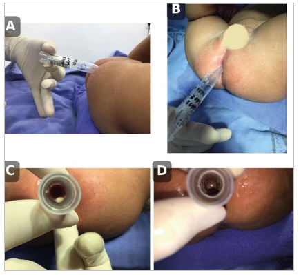
After tissue collection under direct vision, the tube was rotated 180 degrees and kept in position for 2-3 minutes, applying a gentle compression at the biopsy site to facilitate local hemostasis (Figure 1).
Handcrafted device and procedural steps for the MKP in pediatric rectal biopsy. (A) Custom Falcon® tube: 0.8 cm circular window at the widest distal end, with depth markings at 1, 2 and 3 cm from the distal edge. (B) Patient in dorsal lithotomy after pediatric sedation for the procedure. (C) Transanal introduction of the modified tube with the window oriented to the posterior rectal wall. (D) Firm pressure applied against the posterior rectal wall, producing herniation of the mucosa and submucosa through the window, allowing the removal of the tissue. Source: Personal archive.
Variables and Data Analysis
The variables extracted and recorded in a structured eletronic form were included demographic data (age, sex and ethnicity), clinical indication for HD investigation (defecatory dysfunction and associated gastrointestinal symptoms) site of the specimen collection, and information related to the biopsy procedure, including date, environment of performance (surgical center or outpatient) and type of anesthesia (sedation or general anesthesia).
Characteristics of the samples obtained were recorded (fragment size, presence of submucosa and suitability for histological analysis). The samples were considered adequate when they contained enough submucosal tissue for microscopic evaluation by staining with hematoxylin and eosin (HE), allowing the obtaining of multiple histological sections for diagnostic analysis.
The histological data included the dying method (hematoxylin-eosin - HE or other techniques) and the final diagnosis, based on the presence or absence of ganglion cells in the enteric nerve plexuses of the submucosa. Complications related to the procedure were also recorded.
When available, the diagnosis was confirmed by histological findings in surgical specimens obtained in patients undergoing surgery to treat the underlying disease. In patients managed conservatively, the interpretation of histological findings was considered in conjunction with the clinical evolution.
Descriptive statistics were used to summarize clinical and procedural characteristics of the sample. The main outcome was the adequacy of the samples obtained for histological evaluation. As a secondary exploratory analysis, diagnostic metrics were calculated with 95% confidence intervals, based on the correlation between histological findings and patients’ clinical and surgical outcomes.
Ethical considerations
The study was approved by the Research Ethics Committee of the Fluminense Federal University (CAAE Ethical Approval Certificate: 38823120.4.0000.5243 and Opinion No.º 4,559,837).
RESULTS
Between 2003 and 2019, 56 pediatric patients underwent rectal biopsy at the Department of Maternal and Child Health of HUAP. Of these, 31 were excluded from the final analysis: 24 due to the unavailability or incompleteness of medical records, 2 because their histological diagnoses differed from HD and 5 because they had undergone full-thickness surgical rectal biopsies as a primary diagnostic method. Thus, 25 patients remained eligible for evaluation, 15 (60%) males and 10 (40%) females. The demographic characteristics of the cohort are presented in Table 1.
Most patients (56%) had only one clinical symptom at the time of the investigation, mainly abdominal distension and chronic constipation (Table 2).
Among the seven patients over one year of age (4 boys and 3 girls), one had Down syndrome. Five of these children had isolated chronic constipation, one had abdominal distension and one had both symptoms.
The procedure was performed under sedation in 23 patients and under general anesthesia in two cases (8%). Only one patient (28-month-old girl with chronic constipation) presented a complication related to the procedure: transient bleeding and discomfort after biopsy performed under sedation. No other relevant complications were observed.
The samples obtained through the MKP technique were considered adequate in 24 of the 25 cases analyzed (96%). A male newborn (seven days of life), needed to repeat the procedure due to an inadequate specimen (insufficient material). A five-month-old patient, in whom the sample obtained by the MKP was considered adequate, was subsequently submitted to a full-thickness surgical rectal biopsy for diagnostic confirmation (physician´s choice).
Histological analysis identified the presence of ganglion cells in the submucous myoenteric plexus in 15 patients (60% - normal biopsy) and absence of 10 cases (40%, suggesting the diagnosis of HD).
Of the patients diagnosed as HD/absence of ganglion cells in the biopsy specimen 8 were classified as true positives (VP): the diagnosis of HD was confirmed by surgical samples obtained in surgery (colon pull-through). All 15 patients in whom ganglion cells were seen in the biopsy sample were classified as true negatives (NV), and had their symptoms resolved conservatively. Two patients were classified as false positives (FP): in them the initial biopsy indicated the absence of ganglion cells, but the patients presented complete resolution of the symptoms with clinical treatment during the follow-up period and were not operated on. Table 3 presents the clinical and demographic characteristics of patients classified as true and false positives.
Demographic and clinical characteristics of patients with true-positive (TP) and false-positive (FP) biopsy results.
The positive predictive value of MCK in this sample was 80% (8/10; 95%CI ≈ 44-97%) and the negative predictive value of 100% (15/15; 95%CI ≈ 78-100%). The sensitivity was 100% (8/8; 95%CI ≈ 63-100%) and the specificity was 88% (15/17; 95%CI ≈ 64-99%). The overall accuracy observed was 92% (23/25; 95%CI ≈ 74-99%).
The agreement between the biopsy results obtained by the MKP technique and the clinical and surgical outcomes was evaluated using Cohen’s kappa coefficient. The observed agreement (Po) was 0.92 and the expected agreement by chance (Pe) was 0.536, resulting in a kappa coefficient of 0.828, indicating high agreement between histological findings and clinical and surgical outcomes.
DISCUSSION
This study was conducted in a regional reference center in the metropolitan region of Rio de Janeiro. Approximately three pediatric patients per year underwent rectal biopsy for suspected HD17. Considering the estimated incidence of the disease at approximately 1:5,000 live births and about 6,000 annual births in the municipality of Niterói, the expected number of new cases in the city would be close to one per year. The number of cases observed in this sample is similar to that expected based on the estimated incidence for the region, although this observation is descriptive and does not replace formal statistical analyses of population representativeness18.
Female patients predominated in the cohort, including among HD cases, in contrast to the male predominance widely described in the literature (approximate proportion of 4 male cases: 1 female HD)19. This difference may reflect referral bias to tertiary centers, in which atypical or more complex clinical cases tend to be concentrated.
The age distribution was bimodal, with a higher frequency of newborns and preschool children, which is possibly related to the institutional profile, which includes maternity services and provides pediatric surgery for patients referred from other health units or Pediatricians.
Late meconium elimination is described in up to 90% of HD cases20-22, but was recorded in a much smaller proportion in this cohort. This finding possibly reflects limitations inherent in the retrospective collection of clinical data, since incomplete recording of data in medical records is common. Chronic constipation was the most frequent symptom, although less present than in other clinical cohorts20,23, reinforcing the limitations inherent in studies based on medical records.
Contrast enema and anorectal manometry are used for the clinical diagnosis of HD, but have limitations, especially in newborns. Up to 25% of newborns do not show the typical transition zone in the contrast study24,25. Anorectal manometry has restrictions related to the availability of equipment and the need for specialized professionals for its performance and interpretation26,27. No consistent correlation was observed between typical radiological findings and histological confirmation of the disease in this cohort, reiterating the problems found in the use of contrasted enemas as the only method for the diagnosis of HD.
Considering the limitations of radiological and manometric examinations and the need for ablative surgical treatment upon diagnosis of HD, rectal biopsy remains the gold standard for diagnostic confirmation19,28,29,30. Additional dying techniques to HE (histochemistry for acetylcholinesterase and calretinin) increase accuracy, but their availability may be limited, especially in contexts of restricted resources31-33. For this reason, we limited our analysis to samples examined with HE staining, which is available in any histological analysis laboratory and can be done in samples preserved in formaldehyde transferred to external laboratories, allowing, in theory, the execution of the biopsy with simple resources in peripheral health centers for subsequent analysis in reference laboratories, something very important in inner cities.
Full-thickness rectal biopsy continues to be considered the most accurate method for direct evaluation of the myoenteric and submucosal plexuses, but it is the most invasive method of biopsy, requires general anesthesia and has a higher risk of complications. For these reasons, the tendency is to reserve full-thickness biopsies for situations in which less invasive methods produce inconclusive results or doubtful diagnoses19,28.
Comparative studies suggest that suction biopsies may have a higher frequency of inadequate samples, while puncture techniques tend to produce better quality submucosal fragments, although the choice of the method depends on the experience of the physician and availability of specific equipment19,28,30. Our results reinforce the potential of MKP to obtain suitable samples.
An advantage of MKP is its technical simplicity, with a relatively short learning curve and usage of instruments that are widely available in operating rooms. The limited removal of mucosa and submucosa does not change the peri-rectal anatomy and does not compromise subsequent surgical interventions, also allowing the repetition of the biopsy, if necessary.
The only systematic review with meta-analysis published on rectal biopsy techniques for the diagnosis of HD compared suction and puncture methods, demonstrating a higher rate of complications associated with puncture techniques28. Our study had a very small rate of complications, and the results of this meta-analysis should be interpreted with caution, since the included studies showed considerable heterogeneity in relation to the devices used, the operative techniques and the characteristics of the populations evaluated.
This study has important limitations. The retrospective design restricted control over the quality and completeness of clinical information, and the rate of exclusions due to the unavailability or incompleteness of medical records was high, determining a potential selection bias and a cohort with a small number of cases, although applied to a rare disease. In addition, it is a descriptive cohort without a comparative group, capable of determining the feasibility and efficiency of the MKP technique, but not allowing direct performance comparison with other diagnostic methods. The relatively wide confidence intervals observed in diagnostic metrics reflect the small sample size and the limited number of positive cases.
Despite these limitations, the study has some relevant strengths:
-
This is the first evaluation of the MKP method carried out in Brazil and describes the application of the technique in a real clinical practice environment in a public institution (administered by SUS - Brazilian Universal Health Service).
-
The technique proved to be technically viable and suitable for children in general, including newborns.
-
Considering its simplicity, availability of instruments and safety profile, we demonstrate that the MKP technique can be used to obtain rectal biopsies in the investigation of HD in contexts with limited technical resources.
Prospective and multicenter studies with a greater number of patients will be important to confirm these findings and more accurately define the role of MKP in the diagnostic process of HD.
CONCLUSIONS
The MKP technique was able to obtain adequate submucosal rectal samples for histological analysis in the vast majority of children suspected of HD, with few complications, and may represent a useful, easily available and low-cost technical alternative in the diagnostic investigation of HD.
The technique can be performed under conscious sedation, without the need for general anesthesia.
ACKNOWLEDGEMENTS
To the Department of Clinical Pathology of the Fluminense Federal University (UFF), my sincere thanks. This partnership was essential for the development of this research.
REFERENCES
-
1 Gfroerer S, Rolle U. Pediatric intestinal motility disorders. World J Gastroenterol. 2015;21(33):9683-7. doi: 10.3748/wjg.v21.i33.9683.
» https://doi.org/10.3748/wjg.v21.i33.9683 -
2 Brooks AS, Oostra BA, Hofstra RM. Studying the genetics of Hirschsprung's disease: unraveling an oligogenic disorder. Clin Genet. 2005;67(1):6-14. doi: 10.1111/j.1399-0004.2004.00319.x.
» https://doi.org/10.1111/j.1399-0004.2004.00319.x -
3 Moore SW. Chromosomal and related Mendelian syndromes associated with Hirschsprung's disease. Pediatr Surg Int. 2012;28(11):1045-58. doi: 10.1007/s00383-012-3175-6.
» https://doi.org/10.1007/s00383-012-3175-6 - 4 Okamoto E, Ueda T. Embryogenesis of intramural ganglia of the gut and its relation to Hirschsprung's disease. J Ped Surg. 1967;2(5):437-43.
- 5 Kessmann J. Hirschsprung's disease: diagnosis and management. Am Fam Physician. 2006;74(8):1319-22.
- 6 Swenson O, Fisher JH. Hirschsprung's disease in the newborn. Arch Surg. 1959;79:987-93.
- 7 Bodian M, Carter OO. A family study of Hirschsprung's disease. Annals of Human Genetics. 1963;26(3):261-77.
- 8 Gherardi GJ. Pathology of the ganglionic-aganglionic junction in congenital megacolon. Arch Pathol. 1960;69:520-3.
- 9 Dobbins WO, 3rd, Bill AH, Jr. Diagnosis of Hirschsprung's Disease Excluded by Rectal Suction Biopsy. N Engl J Med. 1965;272:990-3.
- 10 Noblett HR. A rectal suction biopsy tube for use in the diagnosis of Hirschsprung's disease. J Pediatr Surg. 1969;4(4):406-9.
- 11 Pease PW, Corkery JJ, Cameron AH. Diagnosis of Hirschsprung's disease by punch biopsy of rectum. Arch Dis Child. 1976;51(7):541-3.
- 12 Shandling B. A new technique in the diagnosis of Hirschsprung's disease. Can J Surg. 1961;4:298-305.
- 13 Shandling B, Auldist AW. Punch biopsy of the rectum for the diagnosis of Hirschsprung's disease. J Pediatr Surg. 1972;7(5):546-52.
- 14 Hirose R, Hirata Y, Yamada T, Kawana T, Taguchi T, Suita S. The simple technique of rectal mucosal biopsy for the diagnosis of Hirschsprung's disease. J Pediatr Surg. 1993;28(7):942-4.
- 15 Kobayashi H, Li Z, Yamataka A, Lane GJ, Miyano T. Rectal biopsy: what is the optimal procedure? Pediatr Surg Int. 2002;18(8):753-6.
- 16 Yoshimaru K, Kinoshita Y, Yanagi Y, Obata S, Jimbo T, Iwanaka T, et al. The evaluation of rectal mucosal punch biopsy in the diagnosis of Hirschsprung's disease: a 30-year experience of 954 patients. Pediatr Surg Int. 2017;33(2):173-9.
- 17 Pratap A, Gupta DK, Tiwari A, Sinha AK, Bhatta N, Singh SN, et al. Application of a plain abdominal radiograph transition zone (PARTZ) in Hirschsprung's disease. BMC Pediatr. 2007;7:5.
-
18 IBGE. 2023 [cited 2024 15 fevereiro 2024]; Available from: https://censo2022.ibge.gov.br/panorama/?utm_source=ibge&utm_medium=home&utm_campaign=portal
» https://censo2022.ibge.gov.br/panorama/?utm_source=ibge&utm_medium=home&utm_campaign=portal -
19 Vervloet G, De Backer A, Heyman S, Leyman P, Van Cauwenberge S, Vanderlinden K, et al. Rectal Biopsy for Hirschsprung's Disease: A Multicentre Study Involving Biopsy Technique, Pathology and Complications. Children (Basel). 2023;10(9). doi: 10.3390/children10091488.
» https://doi.org/10.3390/children10091488. -
20 Dasgupta R, Langer JC. Hirschsprung disease. Curr Probl Surg. 2004;41(12):942-88. doi: 10.1067/j.cpsurg.2004.09.004.
» https://doi.org/10.1067/j.cpsurg.2004.09.004 - 21 Langer JC. Hirschsprung disease. Curr Opin Pediatr. 2013;25(3):368-74.
-
22 Singh SJ, Croaker GD, Manglick P, Wong CL, Athanasakos H, Elliott E, et al. Hirschsprung's disease: the Australian Paediatric Surveillance Unit's experience. Pediatr Surg Int. 2003;19(4):247-50. doi: 10.1007/s00383-002-0842-z.
» https://doi.org/10.1007/s00383-002-0842-z -
23 Martucciello G. Hirschsprung's disease, one of the most difficult diagnoses in pediatric surgery: a review of the problems from clinical practice to the bench. Eur J Pediatr Surg. 2008;18(3):140-9. doi: 10.1055/s-2008-1038625.
» https://doi.org/10.1055/s-2008-1038625 - 24 Holschneider AM, Puri P. Hirschsprung's Disease and Allied Disorders 2008. 1-414 p.
-
25 Peyvasteh M, Askarpour S, Ostadian N, Moghimi MR, Javaherizadeh H. Diagnostic Accuracy of Barium Enema Findings in Hirschsprung's Disease. Arq Bras Cir Dig. 2016;29(3):155-8. doi: 10.1590/0102-6720201600030007.
» https://doi.org/10.1590/0102-6720201600030007 -
26 de Lorijn F, Kremer LC, Reitsma JB, Benninga MA. Diagnostic tests in Hirschsprung disease: a systematic review. J Pediatr Gastroenterol Nutr. 2006;42(5):496-505. doi: 10.1097/01.mpg.0000214164.90939.92.
» https://doi.org/10.1097/01.mpg.0000214164.90939.92 -
27 Pensabene L, Youssef NN, Griffiths JM, Di Lorenzo C. Colonic manometry in children with defecatory disorders. role in diagnosis and management. Am J Gastroenterol. 2003;98(5):1052-7. doi: 10.1111/j.1572-0241.2003.07412.x.
» https://doi.org/10.1111/j.1572-0241.2003.07412.x -
28 Comes GT, Ortolan EVP, de Medeiros Moreira MM, de Oliveira Junior WE, Angelini MC, El Dib R, et al. Rectal Biopsy Technique for the Diagnosis of Hirschsprung Disease in Children: A Systematic Review and Meta-Analysis. J Pediatr Gastroenterol Nutr. 2021;72(4):494-500. doi: 10.1097/MPG.0000000000003041.
» https://doi.org/10.1097/MPG.0000000000003041 -
29 Yoshimaru K, Matsuura T, Yanagi Y, Obata S, Takahashi Y, Kajihara K, et al. Reevaluation of concurrent acetylcholinesterase and hematoxylin and eosin staining for Hirschsprung's disease. Pediatr Int. 2021;63(9):1095-102. doi: 10.1111/ped.14596.
» https://doi.org/10.1111/ped.14596 -
30 Matsukuma K, Gui D, Saadai P. Hirschsprung Disease for the Practicing Surgical Pathologist. Am J Clin Pathol. 2023;159(3):228-41. doi: 10.1093/ajcp/aqac141.
» https://doi.org/10.1093/ajcp/aqac141 -
31 Kapur RP. Calretinin-immunoreactive mucosal innervation in very short-segment Hirschsprung disease: a potentially misleading observation. Pediatr Dev Pathol. 2014;17(1):28-35. doi: 10.2350/13-10-1387-OA.1.
» https://doi.org/10.2350/13-10-1387-OA.1 -
32 Kapur RP, Reed RC, Finn LS, Patterson K, Johanson J, Rutledge JC. Calretinin immunohistochemistry versus acetylcholinesterase histochemistry in the evaluation of suction rectal biopsies for Hirschsprung Disease. Pediatr Dev Pathol. 2009;12(1):6-15. doi: 10.2350/08-02-0424.1.
» https://doi.org/10.2350/08-02-0424.1 -
33 Meier-Ruge W, Lutterbeck PM, Herzog B, Morger R, Moser R, Scharli A. Acetylcholinesterase activity in suction biopsies of the rectum in the diagnosis of Hirschsprung's disease. J Pediatr Surg. 1972;7(1):11-7. doi: 10.1016/0022-3468(72)90394-6.
» https://doi.org/10.1016/0022-3468(72)90394-6