In vitro Regeneration of the 'Piel de Sapo' and 'Cantaloupe' Melon
ABSTRACT
Melon cultivation is of significant socioeconomic importance, especially in the northeast of Brazil. In breeding programs that employ biotechnological techniques, developing efficient methods of in vitro plant regeneration is essential for this species. The aim of this study was to develop a regeneration protocol using cotyledonary explants. The experimental design was completely randomised in a 2 x 5 factorial scheme, with two melon cultivars ('Piel de Sapo' and 'Cantaloupe') x five concentrations of 6- benzylaminopurine (BAP) (0, 4.44, 8.88, 13.32, and 17.75 μM), with five replications of five test tubes, each containing one explant. The percentage of cotyledons that formed calluses and buds, the number of buds formed per callus, and the fresh weight of the callus were evaluated 35 days after the in vitro inoculation of the explants. Data were submitted to analysis of variance, with the mean values compared between cultivars using Tukey’s test, and regression analysis between the BAP doses. No buds were formed in the culture medium containing no BAP, so it was necessary to add BAP to both cultivars. In the culture medium containing 8.88 μM BAP, 68% of the calluses in 'Cantaloupe' formed buds, while for 'Piel de Sapo', buds were formed by 12% of the calluses only. The 'Cantaloupe' cultivar recorded the highest values for fresh callus weight and number of regenerated buds per explant, indicating a better in vitro response than that of 'Piel de Sapo'.
Keywords:
Cucumis melo L; Organogenesis; Plant tissue culture.
RESUMO
A cultura do meloeiro possui relevante importância socioeconômica, especialmente na região Nordeste do Brasil. Nos programas de melhoramento que utilizam técnicas biotecnológicas, nessa espécie, é indispensável o desenvolvimento de métodos eficientes de regeneração in vitro de plantas. Com isso, objetivou-se desenvolver um protocolo de regeneração a partir de explantes cotiledonares. O delineamento experimental foi inteiramente casualizado, em esquema fatorial, duas cultivares de meloeiro (‘Pele de Sapo’ e ‘Cantaloupe’) x cinco concentrações de 6- benzilaminopurina - BAP (0; 4,44; 8,88; 13,32 e 17,75 μM), com cinco repetições de cinco tubos de ensaio contendo um explante cada. Os percentuais de cotilédones que formaram calos e gemas, o número de gemas formadas por calo e a massa fresca do calo foram avaliados aos 35 dias, após a inoculação in vitro dos explantes. Os dados foram submetidos à análise de variância, sendo as médias comparadas pelo teste Tukey entre cultivares, e à análise de regressão entre doses de BAP. No meio de cultivo sem BAP não houve formação de gemas, sendo necessário sua adição, em ambos as cultivares. Em ‘Cantaloupe’ 68% dos calos formaram gemas, no meio de cultivo com 8,88 μM de BAP, em ‘Pele de Sapo’, apenas 12%. A cultivar ‘Cantaloupe’ registrou as maiores massas frescas dos calos e números de gemas regeneradas/explante, indicando ser mais responsiva in vitro do que a ‘Pele de Sapo’.
Palavras-chave:
Cucumis melo L; Organogênese; Cultura de tecidos vegetais.
INTRODUCTION
The melon (Cucumis melo L.) is a fruit-vegetable of family Cucurbitaceae of great economic importance. In 2023, global production was approximately 29.5 million tons, grown on more than 1.09 million hectares (FAO, 2023). That same year, Brazilian production exceeded 862,000 tons, on an area of 30,500 hectares (FAO, 2023), contributing approximately 2.9% of global production. The state of Rio Grande do Norte is the largest melon producer in the northeast of Brazil (IBGE, 2023). Among the most widely grown melon varieties in Brazil are Cucumis melo var. inodorus ('Amarelo' and 'Piel de Sapo') and Cucumis melo var. cantaloupensis ('Cantaloupe') (LANDAU et al., 2020).
In cucurbits, haploid and diploid plants can be obtained in three ways: through haploid parthenogenesis in situ, gynogenesis in vitro, or androgenesis in vitro (NYIRAHABIMANA; SOLMAZ; SARI, 2022). However, in order to apply these biotechnological techniques to genetic improvement programmes, it is essential to develop efficient methods of plant regeneration through tissue culture, especially in the melon (PROBOWATI; DARYONO, 2018).
Regeneration protocols, whether by organogenesis (CAPACI et al., 2025; FAUZAN et al., 2024; MAHDAD et al., 2023; MOHIUDDIN et al., 2022; ULLAH et al., 2023) or somatic embryogenesis (FURQONI; EFENDI, 2018; RAJI et al., 2018) have been described for a significant number of melon varieties and cultivars. However, these studies reveal a large variation in morphogenetic response, showing that the efficiency of the protocols can be affected by various factors, including genotype, source of explant, type and concentration of phyto-regulators, growing conditions and other physical factors. Despite all efforts, manipulating the melon in vitro remains difficult, mainly due to the effect of the genotype (PECHAR, 2022), making it is necessary to develop regeneration protocols for each specific melon variety and cultivar with the aim of optimising the in vitro response.
In regeneration via organogenesis, the most used melon explants are cotyledon (CAPACI et al., 2025; FAUZAN et al., 2024; IVANOVA; GROZEVA; VELKOV, 2017; MOHIUDDIN et al., 2022; NADERI; MAHMOUDI, 2017), hypocotyl (FAUZAN et al., 2024; GROZEVA; VELKOV; IVANOVA, 2019), petiole (FAUZAN et al., 2024; MOHIUDDIN et al., 2022), bud (FAUZAN et al., 2024) and nodal segment (FAUZAN et al., 2024; ULLAH et al., 2023).
In terms of plant regulators, 6-benzylaminopurine (BAP) is the most widely used cytokinin in studies on the in vitro organogenesis of the melon (CAPACI et al., 2025). The beneficial effects of using BAP, both with and without auxins, on shoot regeneration in the melon have been reported by several authors (NADERI; MAHMOUDI, 2017; PROBOWATI; DARYONO, 2018).
The aim of this study was to develop a regeneration protocol using cotyledonary explants for two melon cultivars: 'Cantaloupe' (commercial cultivar 'Cantaloupe Italiano') and 'Piel de Sapo' (commercial cultivar 'Asturia').
MATERIALS AND METHODS
Mature, similar-sized melon seeds (Cucumis melo L.) from two cultivars, 'Cantaloupe' (commercial cultivar 'Cantaloupe Italiano') and 'Piel de Sapo' (commercial cultivar 'Asturia'), were disinfected under aseptic conditions in a laminar flow hood by immersion for one minute in a 70% alcohol solution, then for 7.5 minutes in a 0.1% sodium hypochlorite solution, followed by three one-minute washes with autoclaved distilled water, as per Freire et al. (2020). Once disinfected, the seed coats were removed using sterile tweezers and the seeds were inoculated into a culture medium containing a basic composition of MS medium (MURASHIGE; SKOOG, 1962) with no added growth regulators. The cultures were kept in a growth room at 25 ± 1ºC in the dark for three days. They were then transferred to a 16/8 photoperiod (light/dark) at a light intensity of 30 μmol m-2 s-1, where they remained for a further nine days (FREIRE et al., 2020).
Cotyledon explants were taken from seedlings 12 days after in vitro seed inoculation. Each cotyledon was divided perpendicular to the longitudinal axis into two equal parts under aseptic conditions, and portions, approximately 2.0 mm wide, were removed along each edge, resulting in two segments of approximately 0.8 cm2, which were inoculated with the abaxial side in contact with the culture medium (NADERI; MAHMOUDI, 2017). The cultures were kept in a growth room at 25 ± 1ºC under a 16/8 photoperiod (light/dark) at light intensity of 30 μmol m-2 s-1 for 35 days. The characteristics under evaluation were percentage of explants that formed calluses, percentage of calluses that formed buds, number of buds formed per callus, and fresh weight of the callus. The calluses were assessed for colour and texture, and then classified as friable (calluses that fell apart when touched with tweezers) or compact (calluses that did not fall apart when touched with tweezers).
The experimental design was completely randomised in a 2 x 5 factorial scheme, comprising two melon cultivars (‘Cantaloupe’ and ‘Piel de Sapo’) and five concentrations of BAP (0, 4.44 μM, 8.88 μM, 13.32 μM and 17.75 μM) in five replications of five test tubes, each containing one explant, giving a total of 25 explants per treatment. The results were submitted to analysis of variance (ANOVA) and Tukey's test to compare mean values between cultivars, as well as regression analysis for the BAP doses, at 5% probability.
RESULTS AND DISCUSSION
The analysis of variance showed a significant effect for both the melon cultivar and the BAP concentration added to the culture medium, as well as for the interaction of the two factors on the variables under study: percentage of explants that formed calluses (EC), percentage of calluses that formed buds (CB), number of buds formed per callus (NBC) and fresh weight of the callus (FWC) (Table 1).
Summary of the analysis of variance for the factors melon cultivar (C) and BAP concentration (B), and the C x B interaction on the percentage of explants that formed calluses (EC), percentage of calluses that formed buds (CB), number of buds formed per callus (NBC), and fresh weight of the callus (FWC), after 35 days in vitro culture of cotyledons of the two melon cultivars 'Piel de Sapo' and 'Cantaloupe'.
After 35 days of in vitro culture, calluses formed in the culture medium in 20% and 96% of the cotyledonary explants of the 'Piel de Sapo' and 'Cantaloupe' melon cultivars, respectively, even without the addition of plant growth regulators (Table 2). Similar results were found by Probowati and Daryono (2018) in the F2 line of the Yamatouri x Vankharman cross, recording callus formation in the cotyledons grown in the same culture medium. However, under the same conditions, Naderi and Mahmoudi (2017), studying the Samsoori cultivar, found that no calluses were formed in the cotyledonary explants. Probowati and Daryono (2018) point out that callus formation, even when no growth regulators are added to the culture medium, may indicate that endogenous hormone levels can undergo alterations during the in vitro development of melon cotyledons.
Percentage of explants that formed calluses (EC), percentage of calluses that formed buds (CB), number of buds formed per callus (NBC), and fresh weight of the callus (FWC) in the ‘Piel de Sapo’ and ‘Cantaloupe’ melon cultivars in culture media containing different concentrations of 6-benzylaminopurine (BAP), 35 days after in vitro inoculation of the cotyledons.
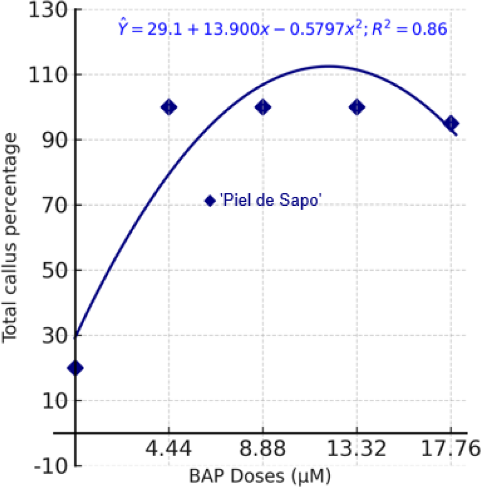
In the culture media with added BAP, calluses formed in each of the explants of both cultivars under study. However, callogenesis was only affected by the presence of the cytokinin in the case of 'Piel de Sapo', showing that for this melon cultivar, it is necessary to add BAP to the culture medium to increase callus formation (Table 2, Figure 1). This confirms that the in vitro response in the melon is genotypedependent (GARCÍA-ALMODÓVAR et al., 2017; SEBASTIANI; FICCADENTI, 2016). Grozeva, Velkov and Ivanova (2019) point out that genotype is the most important factor in determining organogenic and regenerative potential in the melon.
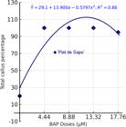
Percentage of explants that formed calluses as a function of the BAP concentration in the culture medium, following 35 days in vitro cultivation of cotyledons of the 'Piel de Sapo' melon cultivar.
In the 'Piel de Sapo' cultivar, increasing the BAP concentration in the culture medium increased the percentage of cotyledons that formed calluses, up to a limit of 11.99 µM, at which point the behaviour was reversed, i.e. there was a reduction in the percentage of cotyledons that formed calluses (Figure 1). It was found that all the explants formed calluses at a BAP concentration of 4.44 µM. Ivanova, Grozeva and Velkov (2017) also found greater callus formation at higher BAP concentrations (8.88 µM) in cotyledonary explants from four melon genotypes.
The calluses formed on the cotyledonary explants of both 'Cantaloupe' and 'Piel de Sapo' were mostly light green in colour. Probowati and Daryono (2018) emphasise that the green colour is related to the presence of BAP in the culture medium, since the cytokinin affects the formation of chloroplasts in the calluses. Despite this, and according to reports in the scientific literature, the colour of the calluses formed on the melon cotyledons can range from white (GROZEVA; VELKOV; IVANOVA, 2019; NADERI; MAHMOUDI, 2017) or cream (PERAITA, 2016) to green (PERAITA, 2016; PROBOWATI; DARYONO, 2018).
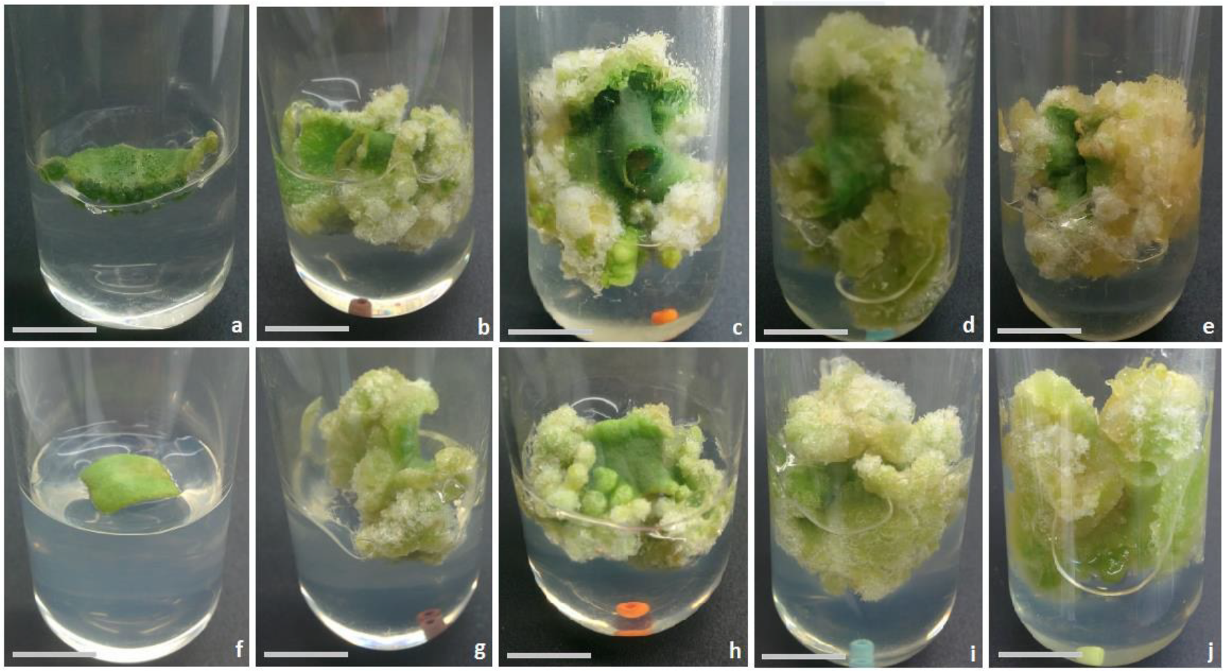
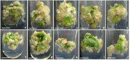
In terms of texture, the ‘Cantaloupe’ calluses obtained in the culture medium with no added BAP were compact (the calluses were rigid and did not fall apart when touched with tweezers), compared to those developed in the culture medium containing the cytokinin, whose texture was friable (the calluses fell apart easily when touched with tweezers). All the ‘Piel de Sapo’ calluses had a compact texture with few friable areas (Figure 2). Peraita (2016) found that the calluses formed on the cotyledons of the Cantaloup Charentais cultivar were of two types: cream-coloured with a friable texture and green-coloured with a compact texture, with buds forming in the latter only. Probowati and Daryono (2018) found the formation of compact green calluses covering the entire surface of the cotyledonary explants of the F2 line from the Yamatouri x Vankharman cross. Grozeva, Velkov and Ivanova (2019) point out that the size, colour and texture of the calluses depend on both the type of growth regulator present in the culture medium and the source of the explants.
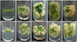
Calluses formed on cotyledon explants of two melon cultivars, ‘Cantaloupe’ (a, b, c, d, e) and ‘Piel de Sapo’ (f, g, h, i, j), grown in MS culture medium containing different concentrations of BAP, following 35 days of in vitro culture: a. f. MS with no growth regulator (control); b. g. BAP 4.44 μM; c. h. BAP 8.88 μM; d. i. BAP 13.32 μM; e. j. BAP 17.75 μM. Bar: 1.0 cm.
In terms of bud formation, it was found that calluses kept in the culture medium with no added BAP did not form any buds after five weeks of in vitro culture in either of the cultivars. Cotyledons of 'Gorgab' (NADERI; ASKARIKHORASGANI; MAHMOUDI, 2016) and the F2 line from the Yamatouri x Vankharman cross (PROBOWATI; DARYONO, 2018) kept in culture medium with no added growth regulator also did not regenerate any buds.
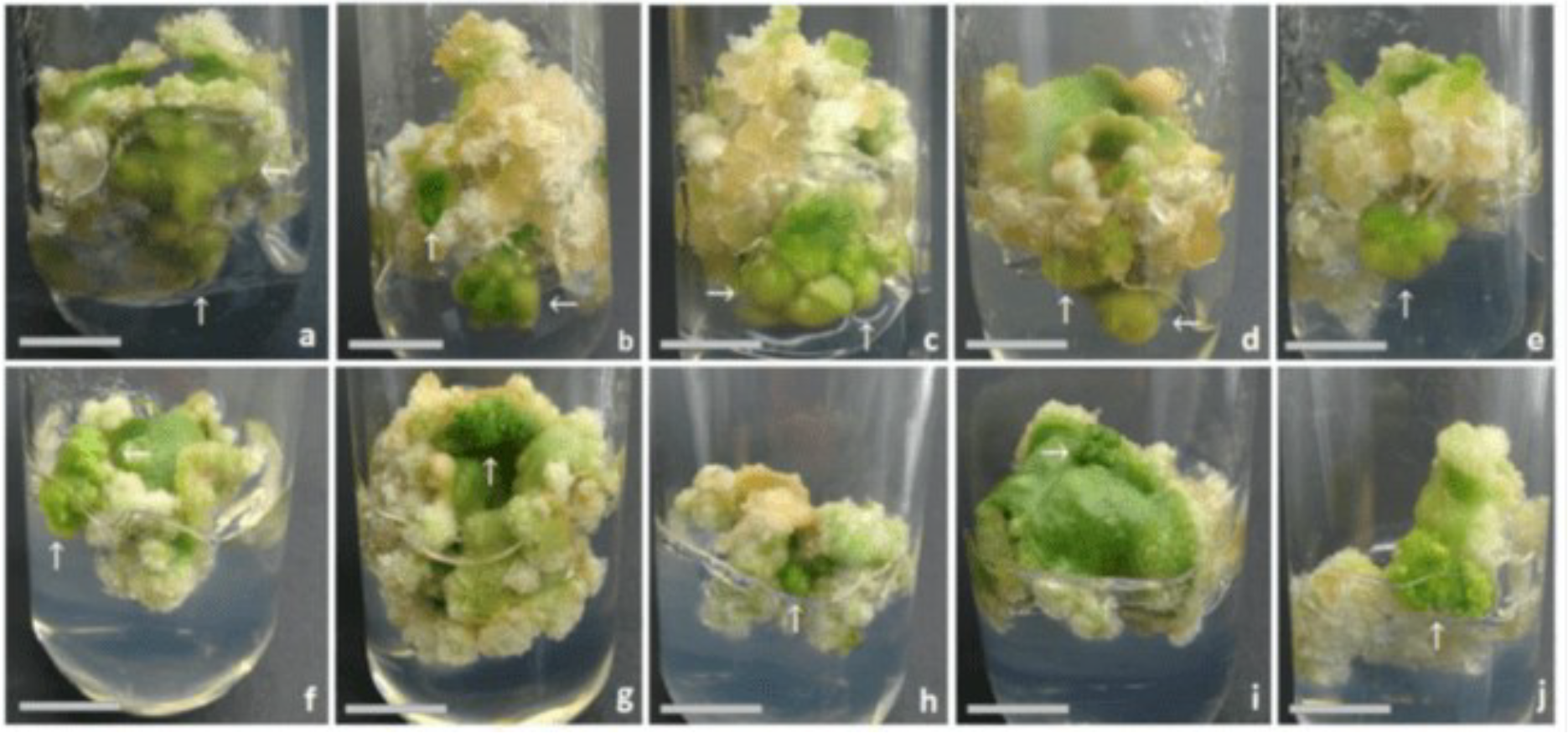
Bud regeneration was seen only in the cotyledons that formed calluses in the culture media with added BAP in both of the cultivars under study (Figure 3). Cytokinins, even when used alone, are able to induce bud formation in the melon (NADERI; MAHMOUDI, 2017). Among the cytokinins, BAP has resulted in higher regeneration rates in the melon compared to 6-furfurylaminopurine (CIN) (IVANOVA; GROZEVA; VELKOK, 2017). These authors point out that the addition of auxins to the culture medium containing cytokinin stimulates callogenesis, but does not affect bud regeneration.
Calluses formed on melon cotyledon explants of the ‘Cantaloupe’ and ‘Piel de Sapo’ cultivars kept in MS culture medium containing different concentrations of BAP, following 35 days of in vitro culture, showing the development of adventitious buds (arrows): a. b. e. ‘Cantaloupe’ calluses in medium with 4.44, 8.88 and 17.75 μM BAP, respectively; c. d. ‘Cantaloupe’ calluses in medium with 13.32 μM BAP; f, g, j. ‘Piel de Sapo’ calluses in medium with 4.44, 8.88 and 17.75 μM BAP, respectively; h. i. ‘Piel de Sapo’ calluses in medium with 13.32 μM BAP. Bars: 1.0 cm
At the lowest concentration (4.44 μM), 12% and 28% of the calluses formed buds in ‘Piel de Sapo’ and ‘Cantaloupe’, respectively (Table 2). Comparing the results with those in the literature, the percentage of cotyledonary explants that formed calluses and subsequently regenerated buds at the same BAP concentration varies significantly. The values range from 12.8% for ‘Charentais’ (GARCÍAALMODÓVAR etal.,2017)to90.0%for ‘Meloncella’ (CAPACI et al., 2025), confirming that the difference in percentage morphogenetic response is probably due to genetic differences between the melon genotypes under study (GARCÍA-ALMODÓVAR et al., 2017; SEBASTIANI; FICCADENTI, 2016). García-Almodóvar et al. (2017) confirm that genotype is the most important factor in determining regenerative potential in the melon.
For the 'Piel de Sapo' cultivar, the percentage of cotyledon explants that formed calluses and subsequently regenerated buds when BAP was added to the culture medium ranged from 12% to 28% regardless of the concentration. The values recorded for the 'Cantaloupe' cultivar were higher, ranging from 28% to 88%. This figure of 88% was obtained at a concentration of 13.32 μM BAP, and showed no statistical difference from the 68% recorded at the lower concentration of 8.88 μM. These results show that, from a concentration of 8.88 μM BAP onwards, the cotyledons of the 'Cantaloupe' cultivar that formed calluses have a higher percentage of bud formation than those of the 'Piel de Sapo' cultivar. At this concentration, only 12% of the 'Piel de Sapo' calluses formed buds, while for 'Cantaloupe', it was 68%. Higher values were recorded by Peraita (2016) for 'Cantaloup Charentais', where 90.62% of cotyledon explants formed buds in a culture medium containing this same concentration of BAP with the addition of 0.28 μM IAA. Probowati and Daryono (2018) point out that in the melon, callus formation with subsequent bud regeneration can occur in culture media containing growth regulator at a wide range of concentrations, including in culture media with no added auxins, as seen in the two cultivars under study.
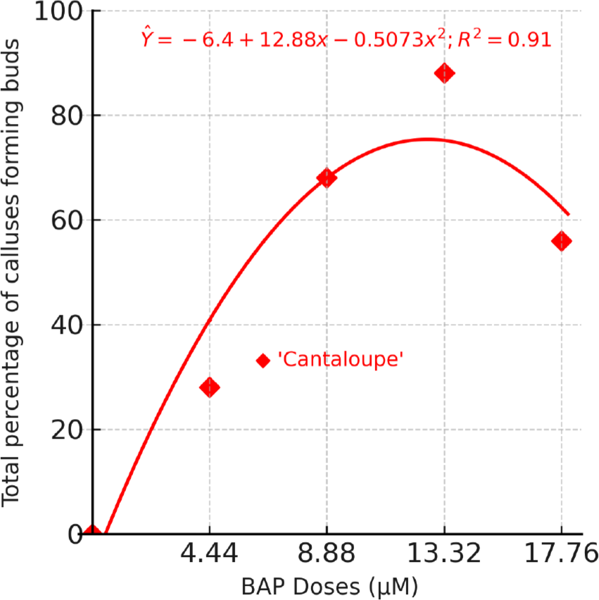
The calluses formed at the different BAP concentrations produced buds, albeit with different percentage values. Values greater than 50% were recorded only for the cotyledons of the 'Cantaloupe' cultivar grown in culture media with added BAP, starting from an estimated concentration of 5.63 μM. In the 'Cantaloupe' cultivar, increasing the BAP concentration in the culture medium increased the percentage of calluses that formed buds, up to 12.69 μM, at which point the behaviour was reversed, indicating a reduction in the number of buds being formed. At a BAP concentration of 12.69 μM, it is estimated that, in the 'Cantaloupe' cultivar, 75.35% of the calluses formed buds (Figure 4).

Percentage of calluses that formed buds as a function of the BAP concentration in the culture medium, following 35 days in vitro cultivation of the cotyledons of the 'Cantaloupe' melon cultivar.
The calluses kept in the culture medium without the addition of BAP showed no morphogenetic response for shoot formation, i.e. buds. When BAP was added to the culture medium, the number of buds formed per callus ranged from 2.63 to 3.78 and from 0.40 to 1.60 for 'Cantaloupe' and 'Piel de Sapo', respectively. When using the culture medium containing 4.44 μM BAP, 1.60 buds were obtained with 'Piel de Sapo' and 2.67 with 'Cantaloupe'. Comparing the results with those in the literature, the number of buds formed per callus at this same BAP concentration varies from 0.5 (SEBASTIANI; FICCADENTI, 2016) to 8.4 (NADERI; ASKARI-KHORASGANI; MAHMOUDI, 2016) for ‘DH-L2’ and ‘Gorgab’, respectively.
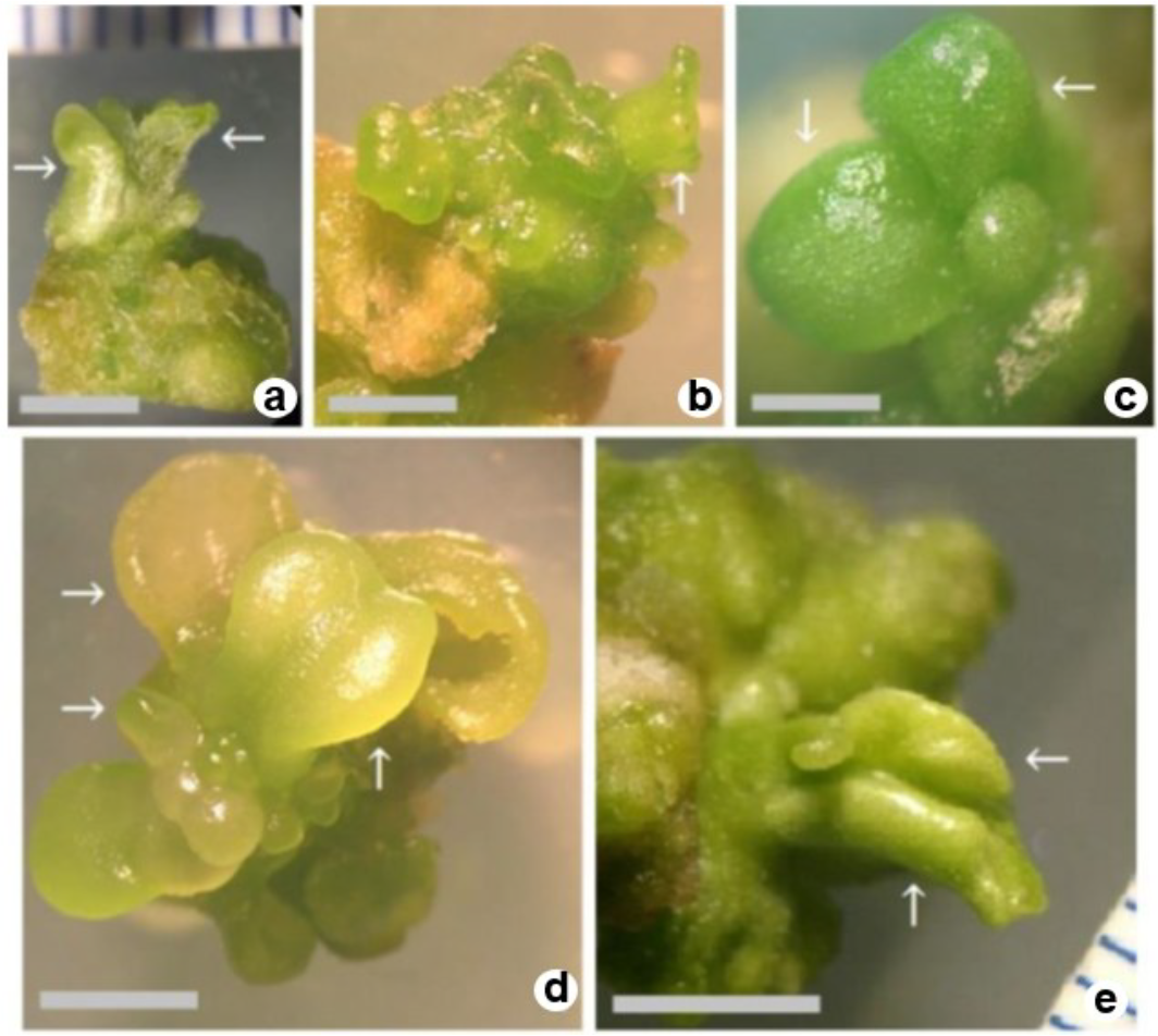
‘Cantaloupe’ formed more buds than did ‘Piel de Sapo’ in the culture media containing 8.88 and 17.75 μM BAP (3.78 and 3.22 buds, respectively). The results show that adding BAP to the medium is necessary to induce bud formation in 'Cantaloupe'. Awatef and Mohamed (2013) recorded 2.92 and 2.50 buds per callus in cotyledons of the ‘Maazoun’ and ‘Beji’ melon cultivars, respectively, at a concentration of 8.88 μM BAP. This wide variation in the number of buds formed per callus from melon cotyledonary explants confirms that genotype and growth regulators play an important role in the morphogenesis of the species (SEBASTIANI; FICCADENTI, 2016). Only buds obtained from the ‘Cantaloupe’ cultivar later developed into shoots (Figure 5).
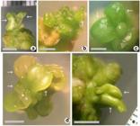
Shoots regenerated from calluses from cotyledonary explants of the ‘Cantaloupe’ melon cultivar showing leaves (arrows), viewed under a magnifying glass: a. shoot formed in the culture medium containing 4.44 μM BAP (Bar: 3 mm); b. containing 8.88 μM BAP (Bar: 3 mm); c. containing 13.32 μM BAP (Bar: 1 mm); d. containing 17.75 μM BAP (Bar: 4 mm); e. containing 13.32 μM BAP (Bar: 4 mm).
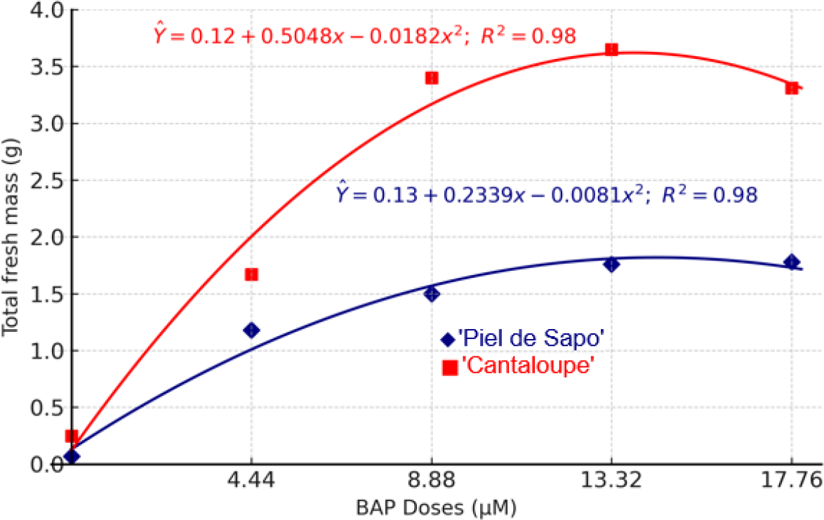
The lowest values for the fresh weight of the callus were recorded in the culture medium with no added plant growth regulator, 0.07 g and 0.25 g for 'Piel de Sapo' and 'Cantaloupe', respectively. Whereas the highest values for fresh weight were obtained at concentrations of 17.75 μM for 'Piel de Sapo' (1.78 g) and 13.32 μM for 'Cantaloupe' (3.65 g), differing statistically from the culture media with no cytokinin, and from the medium containing the lowest concentration, 4.44 μM. Comparing the two melon cultivars, the highest values were obtained in 'Cantaloupe' in the culture media with added BAP. Regardless of the BAP concentration, the fresh weight of the ‘Cantaloupe’ calluses (2.46 g) was 95% greater than that of ‘Piel de Sapo’ (1.26 g). Naderi, Askari-Khorasgani and Mahmoudi (2016) found that the fresh weight of the callus formed on the cotyledons of ‘Gorgab’ was 3.82 g after 21 days in a culture medium containing 4.44 μM BAP, a far higher value than that recorded in the same culture medium for ‘Piel de Sapo’ (1.18 g) and ‘Cantaloupe’ (1.67 g) following 35 days of in vitro culture.
In both melon cultivars, increasing the BAP concentration in the culture medium increased the fresh weight of the callus up to a maximum of 13.32 µM, after which the value decreased. At a concentration of 14.84 µM BAP, the maximum fresh weight of the callus is estimated at 1.82 g in the 'Piel de Sapo' cultivar; for 'Cantaloupe', the maximum is estimated at 3.62 g in a culture medium with the addition of 14.84 µM BAP (Figure 6).

Fresh weight of the callus as a function of the BAP concentration in the culture medium, following 35 days in vitro cultivation of cotyledons from the 'Piel de Sapo' and ‘Cantaloupe’ melon cultivars.
Indirect organogenesis generally consists of two stages: the first is callus induction and the second, shoot regeneration. In the present study, the two morphogenetic responses were achieved in a single step, eliminating the need to transfer the callus to the culture medium for bud regeneration.
According to Sebastiani and Ficadenti (2016), despite all research efforts, the successful application of in vitro tissue culture techniques in the melon remains difficult. This is mainly due to organogenesis being a highly complex process that is strongly affected by small changes made to the protocols, resulting in low regeneration rates (GARCÍAALMODÓVAR et al., 2017).
The percentage of cotyledons that formed calluses and the average number of buds produced per callus varied according to the culture medium and melon genotype (AWATEF; MOHAMMED, 2013). In general, the 'Piel de Sapo' cultivar was less responsive than the 'Cantaloupe' cultivar in both callus formation and bud regeneration. This is in line with observations made by Pinho et al. (2010), that the cantaloupensis variety is more responsive than the inodorus variety. The low regeneration rates of this variety corroborate the results of Stipp et al. (2001), who state that inodorus has difficulty in developing the apical bud meristem. The same authors state that the lack of an apical meristem has been identified as the main reason for low in vitro plant formation in the melon. On the other hand, Grozeva, Velkov and
Ivanova (2019) emphasise that the regeneration rate of the melon depends on the genotype, type of explant and composition of the culture medium.
CONCLUSION
Callogenesis occurs in melon cotyledon explants, even without the addition of a growth regulator to the culture medium. However, the addition of BAP is necessary for increased callus formation in 'Piel de Sapo' explants. The addition of BAP to the culture medium promoted bud induction in the calluses of both cultivars, although the greatest number of buds was recorded for 'Cantaloupe,' with higher fresh weight values than 'Piel de Sapo' in all the culture media with added BAP.
Data Availability:
The data that support the findings of this study can be made available, upon reasonable request, from the corresponding author.
REFERENCES
- AWATEF, R.; MOHAMED, B. Efficient plant regeneration from cotyledonary explants of Tunisian Cucumis melo L. cv. Maazoun and Beji. Journal of Biodiversity and Environamental Sciences, 3: 50-58, 2013.
- CAPACI, P. et al. In vitro shoot regeneration of Cucumis melo ‘Meloncellafasciata’withthecombineduseof6-benzylaminopurine and cefotaxime. Plant Biosystems, 159: 164-172, 2025.
-
FAO. Faostat - Statistics Database 2023. Available at: <https://www.fao.org/faostat/en/#data/QCL>. Access on: Jul. 7, 2025.
» https://www.fao.org/faostat/en/#data/QCL - FAUZAN, F. Q. et al. Plantlet regeneration of Cucumis melo L. Glamour cv. using different types of cytokinin and explants. Journal of Tropical Resources and Sustainable Science, 12: 61-68, 2024.
- FREIRE, F. B. S. et al. Tratamentos pré-germinativos para o estabelecimento in vitro de sementes de meloeiro Cantaloupe. Fortaleza, CE: Embrapa Agroindústria Tropical, 2020. 18 p. (Boletim de Pesquisa e Desenvolvimento, número 202).
- FURQONI, H.; EFENDI, D. Somatic embryogenesis of melon (Cucumis melo L.) as affected by culture media and composition of plant growth regulators. Journal of Tropical Crop Science, 5: 119-125, 2018.
- GARCÍA-ALMODÓVAR, R. C. et al. Production of transgenic diploid Cucumis melo plants. Plant Cell Tissue and Organ Culture, 130: 323-333, 2017.
- GROZEVA, S.; VELKOV, N.; IVANOVA, Z. In vitro plant regeneration of two Cucumis melo L. genotypes using different explant types and culture medium. Ecologia Balkanica, 11: 193-202, 2019.
-
IBGE - Instituto Brasileiro de Geografia e Estatística. Produção Agrícola 2023. Available at: <https://www.ibge.gov.br/explica/producao-agropecuaria/melao/br>. Access on: Mai. 26, 2025.
» https://www.ibge.gov.br/explica/producao-agropecuaria/melao/br - IVANOVA, Z.; GROZEVA, S.; VELKOV, N. Induction of callogenesis and organogenesis of different melon genotypes. Journal of BioScience and Biotechnology, 6: 99-104, 2017.
- LANDAU, E. C. et al. Evolução da produção de melão (Cucumis melo, Cucurbitaceae). In: LANDAU, E. C. et al. (Eds.). Dinâmica da produção agropecuária e da paisagem natural no Brasil nas últimas décadas Brasília, DF: Embrapa, 2020. v. 4, cap. 34, p. 1095-1126.
- MAHDAD, Y. M. et al. Adventitious regeneration from haploid melon (Cucumis melo L.) leaves as an approach to increase the frequency of diploid plants. In vitro Cellular & Developmental Biology - Plant, 59: 167-177, 2023.
- MOHIUDDIN, A. K. M. et al. Relationship between induction of novel somaclonal variants and types of organogenesis in muskmelon (Cucumis melo L.). Journal of Agricultural and Marine Sciences, 27: 90-98, 2022.
- MURASHIGE. T.; SKOOG F. A revised medium for rapid growth and bio assays with tabacco tissues cultures. Journal Plant Physiology, 15: 473-497, 1962.
- NADERI, D.; ASKARI-KHORASGANI, O.; MAHMOUDI, E. Cefotaxime and benzyladenine improve melon regeneration. Iranian Journal of Biotechnology, 14: 56-60, 2016.
- NADERI, D.; MAHMOUDI, E. In vitro regeneration of Iranian melon (Cucumis melo L. ‘Samsoori’) using antibiotic and benzyl adenine micropropagation of Cucumis melo L. ‘Samsoori’. International Journal of Horticultural Science and Technology, 4: 117-126, 2017.
- NYIRAHABIMANA, F.; SOLMAZ, I.; SARI, N. An overview of haploid plant production in Cucurbits. In: BELLİTÜRK, K.; SOLMA, Y (Eds.). Agricultural practices and sustainable management in Turkiye Ancara, Turquia: Iksad Publications, 2022. v. 1, cap. 2, p. 31-56.
- PECHAR, G. S. Impact of melon EIF4E editing on virus resistance and melon fertility 2022. 167 f. Thesis (Doctor of Philosophy - PhD Plant Molecular Biology) - Universidad de Murcia, Murcia, 2022.
- PERAITA, V. A. Sistemas de alto rendimiento en la regeneración in vitro de melón y pepino 2016. 52 f. Dissertação (Mestrado em Biotecnologia Molecular e Celular de Plantas: Área de Concentração em Biotecnologia Molecular e Celular de Plantas) - Universidad Politécnica de Valencia, Valencia, 2016.
- PINHO, D. S. et al. Regeneração in vitro de melão, cv. ‘Gaúcho’. Ciência Rural, 40: 1083-1089, 2010.
- PROBOWATI, W.; DARYONO, B. S. Callus induction and differentiation on melon from in vitro culture with the addition of indole acetic acid and benzyl amino purine growth regulator. Planta Tropika, 6: 15-21, 2018.
- RAJI, M. R. et al. Somatic embryogenesis of muskmelon (Cucumis melo L.) and genetic stability assessment of regenerants using flow cytometry and ISSR markers. Protoplasma, 255: 873-883, 2018.
- SEBASTIANI, M. S.; FICCADENTI, N. In vitro plant regeneration from cotyledonary explants of Cucumis melo L. var. cantalupensis and genetic stability evaluation using RAPD analysis. Plant Cell and Tissue Organ Culture, 124: 69-79, 2016.
- STIPP, L. C. L. et al. In vitro morphogenesis of Cucumis melo var. inodorus Plant Cell, Tissue and Organ Culture, 65: 81-89, 2001.
- ULLAH, A. et al. Establish of protocol for Cucumis melo l.using different hormones through plant tissue culture technique. Pakistan’s Multidisciplinary Journal for Arts & Science, 4: 128-140, 2023.
Edited by
-
Editor in Chief:
Aurélio Paes Barros Júnior
-
Section Editor:
Salvador Barros Torres