
 Protein degradation, oxidation stability, microbiological quality and physicochemical properties in beef muscles subjected to freeze-thawing cycle
ABSTRACT
Freezing is one of the fundamental preservation techniques for increasing the shelf-life of meat. However, the quality of frozen meat is highly dependent on the thawing process. Thus, the current study sought to investigate the effects of freeze-thawing processing on protein degradation, lipid oxidation, microbiological spoilage, and qualitative parameters of two types of beef muscles: Longissimus lumborum and Semitendinosus. Beef muscles were collected from ten steer carcasses of at approximately 15 months of age with a mean body weight of 440 ± 50 kg, at 24 h post-mortem at a commercial slaughterhouse. The muscles were divided into two groups randomly: chilled or frozen-thawed. The freezing and thawing processes resulted in significantly higher degrees of protein and lipid oxidation in meat, resulting in reduced pH, moisture retention ability, and color stability. During the freeze-thawing process, there was a higher desmin degradation but no change in troponin T degradation. Frozen-thawed meat samples exhibited significantly increased growth of total aerobic count, Pseudomonas spp., Enterobacteriaceae, and coliform counts than those from chilled samples. Due to freezing-thawing, Longissimus lumborum muscle was more color-stable and had greater water holding capacity than Semitendinosus muscle (P≤0.05). The data indicate that freeze-thawing consistently caused protein degradation, fat oxidation, color deterioration, and reduced water holding capacity in both beef muscle types. Therefore, to avert quality deterioration during frozen storage, it is imperative to consider the characteristics of each beef muscle.
Keywords
beef muscle; freeze-thawing; meat quality; oxidative stability; proteolysis
1. Introduction
Due to its high content of biologically valuable nutrients, beef is a popular choice among consumers. Compared with similar pieces of mutton, lean beef is considered a typical representation that fits well with the contemporary consumer demand for its favourable nutritional qualities and comparatively low fat and cholesterol content (Liu et al., 2022). Despite its low-fat content, beef is a good source of polyunsaturated fatty acids, including vaccenic acid and conjugated linoleic acids, which have beneficial effects on human health as anti-inflammatory, anti-thrombotic, and atherosclerotic preventatives (Vahmani et al., 2015; Qian et al., 2022). Consequently, the global beef industry has grown and developed to satisfy consumer demand. However, beef is highly susceptible to deterioration due to its high abundance of nutrients (Tian et al., 2022), therefore it is necessary to apply a rigorous methods to ensure its nutritional value. One of the most crucial elements in the processing chain for fresh meat, especially beef, is storage temperature, which is mainly responsible for preserving meat quality.
Chilling and frozen storage are now the most generally used storage methods to extend the shelf-life of beef and beef products without any further treatment (Pan et al., 2021; Liu et al., 2022). Freezing is a traditional meat preservation method that delays biochemical reactions and inhibits the growth and reproduction of microorganisms, thereby slowing the rate of meat spoilage (Leygonie et al., 2012). Although this method has been widely used in commercial production, frozen storage has its disadvantages. For frozen meat, the thawing process is necessary before it is used in subsequent processing. During the thawing process, meat is often accompanied by protein degradation, fat oxidation, color deterioration, and reduced water holding capacity due to the melting of ice crystals (Kim et al., 2017; Teuteberg et al., 2021). To minimize the quality deterioration caused by freeze-thawing, appropriate technologies have been introduced (Gan et al., 2022). However, the thawing process should not be overlooked when evaluating the physical and biochemical quality changes freezing or thawed meat because it may be affected by both the freezing and thawing methods. In the beef industry, marking individual muscles has become common practice (Lang et al., 2017). Beef quality of individual skeletal muscles is highly variable, as they are composed of various kinds of muscle fiber (Cheng et al., 2020). It is expected that the quality of each type of muscle will be affected differently by freeze-thawing susceptibility. For this reason, understanding these inherent variations in characteristics is crucial for the beef industry to develop enhanced freeze-thaw processing technologies and maintain meat quality. The link between the physicochemical quality of frozen-thawed meat and its muscle fiber properties has been the subject of previous investigations (Cheng et al., 2020; Cheng et al., 2021). Nonetheless, scant data exist about the possible impact of freezing and thawing on myofibrillar protein denaturation, microbiological quality, and oxidative stability of beef muscles. Thus, we selected two separate beef muscles for this study: Longissimus lumborum muscle (loin) and Semitendinosus muscle (eye of round), which have diverse muscle fiber compositions and characteristics, to assess the effects of freeze-thawing on their protein degradation, microbiological quality, and oxidative stability of beef muscles compared with their chilled counterparts.
2. Material and methods
The research project was conducted at the Salahaddin University-Erbil, Kurdistan region of Iraq (36.2° N Latitude, 44.0° E Longitude; 420 m above sea level).
2.1. Collection, preparation, and handling of muscle sample
Beef Longissimus lumborum muscle (loin) and Semitendinosus muscle (eye of round) were removed from the left and right sides of 10 Brahman crossbred steer carcasses at approximately 15 months of age with a mean body weight of 440 ± 50 kg, 24 h post-mortem, carcasses at a commercial ruminant slaughterhouse. After being vacuum-packed and kept in ice-filled chest coolers, the beef samples were delivered to the Meat Science Laboratory at Salahaddin University-Erbil. All muscle samples were prepared at the laboratory as whole muscles free of visible connective tissue and fat. Prior to packaging, two equal blocks of 2.5-cm-thick muscle samples were obtained for immunohistochemistry following the removal of around 2.5-cm from the end of each muscle, snap-frozen in liquid nitrogen, and stored at −40 °C for subsequent analysis. Each muscle sample was divided into three equal sections along its length, individually labelled, vacuum packaged, and randomly assigned into two treatment groups (30 samples of each muscle per group): 1) the chilled group was kept at 4 °C for seven days, and 2) the frozen-thawed group was kept at −20 °C for five days before being thawed at 4 °C for two days. A standard freezer set at −20 °C was used for freezing, and all frozen samples were thawed at 4 °C until the internal temperature reached 2 °C. After seven days of storage for each assigned treatment, all beef muscle samples were taken from the packaging and split into pieces for microbiological analysis and meat quality measurements. Samples for lipid-protein oxidation and myofibrillar proteins analyses were snap-frozen in liquid nitrogen, crushed, and vacuum packaged before being stored at −40 °C until analysis.
2.2. Myofibrillar protein preparation
With a slight modification, myofibrillar proteins were extracted using the technique described by Morzel et al. (2006). In duplicate, 2.5 g of liquid nitrogen powdered samples were homogenized for 30 s in 25 mL of ice-cold extraction buffer comprising 150 mM NaCl, 25 mM KCl, 3mM MgCl2, and 4mM ethylenediaminetetraacetic acid (EDTA) at pH 6.5, to which a protease inhibitor (Sigma, Germany) had been added. The homogenate was filtered through 1.0-mm polythelene strainer to remove any remaining collagen. After filtration, the homogenate was incubated at 4 °C for 30 min with continuous shaking. Following the incubation period, the homogenates were centrifuged at 2000 × g for 10 min at 4 °C. The pellet was washed twice, once with 25 mL of a pH 6.4 solution of 50 mM KCl and once with 25 mL of a pH 6 phosphate buffer containing 20 mM sodium phosphate and 0.6 M sodium chloride. After that, the pellet was suspended in the same phosphate buffer and stored at −20 °C until analysis. Using a protein assay kit (Parsazmoon, Iran), the samples’ total protein concentration was calculated using Bradford’s (1976) method. The colorimetric analytical procedure was followed during the assay.
2.3. SDS-PAGE gel electrophoresis and western blot
Myofibrillar proteins were incubated for 4 min at 90 °C in a solution containing 0.05% (w/v) bromophenol blue, 62.5 mM Tris–HCl (pH 6.8), 2.3% (w/v) SDS, 30% (v/v) glycerol, and 5% (v/v) β-mercaptoethanol. The buffer was mixed 1:1 ratio. Using a polyacrylamide gel with dimensions of 8 cm × 5.5 cm (length × width) and 0.8 cm thickness, one-dimensional sodium dodecyl sulfate-polyacrylamide gel electrophoresis (SDS–PAGE) was carried out following the technique outlined by Sabow et al. (2016a). Various sizes can be used for the polyacrylamide gels, however, the mini-gel size used was capable of high-resolution separation of small (low molecular weight) myofibrillar proteins (Sarfo et al., 2003). Additionally, it offers advantages such as faster running times and higher protein band density for optimum resolution (Sobieszek, 1994). A 12% resolving gel and a 5% stacking gel solution were prepared to investigate troponin T and desmin alterations in myofibrillar proteins. A volume of 5 μL of standard protein marker was loaded into the first well, and the conforming 30 μg of proteins from each muscle sample were loaded in duplicate into the wells that were left. In a running buffer comprising 0.025 M Tris base, 0.192 M glycine, and 0.1% SDS at pH 8.3, proteins were separated using a Mini-Protean Tetra Cell system (Bio-Rad, USA) for 90 min at a continuous voltage of 120 V and 400 mA, during which time the tracking dye migrated to the bottom of the gel. Following electrophoresis, the gels were stained with coomassie blue G-250 staining solution (0.05% coomassie blue, 10% acetic acid, and 50% methanol) for 60 min and destained with destaining solution for 45 min. The bands of myofibrillar proteins were visualized using a GS-800 Calibrated Imaging Densitometer (Bio-Rad, USA) (Figure 1). Target protein troponin T and desmin, separated based on their molecular weight through gel electrophoresis, were transferred to polyvinylidene fluoride membranes using the Trans-Blot® SD semi-dry transfer system (Bio-Rad, USA) for 45 min at a constant current of 250 mA per gel and a voltage limit of 25 V at 4 °C. To visualize the target proteins and confirm the electrophoretic transfer, the membranes were placed in a ready-to-use Ponceau S stain for 5 min. After washing the membranes with sufficient deionized water, followed by TBST buffer consisting of 100 mM Tris-HCl, 150 mM NaCl, and 0.05% Tween 20, they were blocked with blocking buffer (5% BSA in TBST buffer) for 3 h at room temperature while being constantly shaken. The membranes were incubated in TBST with continual shaking at 60 rpm on a wave shaker overnight at 4 °C with 1:500 diluted monoclonal anti-desmin primary antibody (D1033, Sigma-Aldrich, Germany) and 1:500 diluted monoclonal anti-troponin T primary antibody (Cat # T6277, Sigma-Aldrich, Germany). The membranes were washed three times in TBST buffer for 5 min at room temperature while continuously shaken at 60 rpm using a wave shaker. Following this, the membranes were incubated with a secondary antibody Goat anti-Mouse IgG H&L (HRP) Cat # ab205719 from Absin, China) diluted to 1:10000 dilution in 3% in TBS-T buffer for 1 h at at room temperature and washed three times again with TBST buffer as described above. Using a DAB substrate kit (Cat # E885, AMRESCO®, Ohio, USA), the bloked mambraners were developed. The membrane was scanned using GS-800 Calibrated Imaging Densitometer (Bio-Rad, USA). The band intensities were quantified by Quantify One® software and then normalized to the intensity of total protein within each lane. The extend of troponin T and desmin degradation was calculated by using the intensity of troponin T and desmin band relative to the intensity of the troponin T and desmin band in a reference sample of 24 h post-mortem to determine the percentage change.

A representative gel showing a pattern of desmin and troponin T proteins of Longissimus lumborum and Semitendinosus muscle in beef affected by freeze-thawing process.
2.4. Analysis of the concentration of free thiols
Thiol oxidation was evaluated using 2, 2-dithiobis (5-nitropyridine) DTNP according to Ellman’s method described by Morzel et al. (2006). Four milligrams of myofibrillar proteins were dissolved in 3 mL of 100 mM phosphate buffer (pH 8.0), with 8 mL of urea was added. After adding approximately 30 µL of the stock solution (10 mM DTNP) in ethanol, the mixture was allowed to incubate at room temperature for 1 h. A spectronic® 20 GENESYSTM spectrophotometer (Spectronic Instruments, USA) was used to measure the absorbance at a wavelength of 386 nm against a protein-free buffer blank. After subtracting the blank’s absorbance, an absorption coefficient of 14 mM1 cm1 was used to calculate the thiol concentration. The final results were presented as nmol free thiol per mg of protein.
2.5. Analysis of the concentration of carbonyl content
The carbonyl content was determined following the manufacturer’s colorimetric protocol using the Protein Carbonyl Assay Kit (Cat # ab126287, BCAM, USA). The final results were expressed as nmol of carbonyl per mg of protein.
2.6. Identification of thiobarbituric acid reactive substances (TBARS)
Following the procedure described by Abdullah et al. (2024) with slight modifications, the lipid oxidation (TBARS) values of the beef muscle samples were determined. Forty-eight milliliters of sterile water and 1.25 mL of 4N HCl were used to homogenize approximately 5 g of muscle samples for 2 min. To get a volume of 25 mL, the liquid was distilled. For 35 min, the distillate and the TBA reagent (15% trichloroacetic acid and 0.375% thiobarbituric acid) were boiled and then chilled under running water for 10 min. Using a spectrophotometer (Beckman, France), the optical density was measured at a wavelength of 538 nm against a blank. The measurements of lipid oxidation were computed by multiplying optical density by 7.843. The TBARS value, which represents the amount of oxidized lipids, was expressed as mg MDA per kg of beef or malondialdehyde equivalent content.
2.7. Determination of peroxide value
Rahman et al. (2015) outlined the peroxide value calculation technique. About 3 g of muscle samples were weighed into a 250 mL glass-stoppered Erlenmeyer flask and heated for 3 min at 60 °C in a water bath (HAAKE P21, Germany) to melt the fat. After that, the flask was gently shaken for 3 min with 30 mL of acetic acid–chloroform solution (3:2, v/v) to dissolve the fat. The suspended particles were then filtered out using Whatman No. 1 filter paper. After filtration, 0.5 mL of saturated potassium iodide solution was added, and starch solution was added as an indicator. Finally, the titration was performed using a standard sodium thiosulfate solution. Following the standard formula, the peroxide value was determined and expressed as milliequivalents (meq) of peroxide per kg of beef:
2.8. Microbiological analysis
To prepare the serial dilutions, 45 mL of 1.5% sterile peptone water (Oxiod, England) was aseptically mixed with 5 g of muscle samples. After homogenizing the sample for 2.5 min in an aseptic food bag, the homogenized solution was designated as the stock solution (dilution 10⁻1). Three dilutions were prepared. The first dilution (10⁻1) (1 mL was transferred to 9 mL of peptone water), and serial decimal dilutions up to 10⁻⁶ were obtained. To count total aerobic count (TAC) on Plate Count Agar (Merck KGaA, Germany), coliform bacteria on MacConkey Agar (HiMedia, India), Enterobacteriaceae on Violet Red Bile Glucose Agar (Merck KGaA, Germany), and Pseudomonas spp. on Cetrimide Agar (Merck KGaA, Germany), tenfold dilutions were spread onto Petri dishes in duplicate. Except for Pseudomonas spp., which were incubated at 25 °C for 72 h, plates were incubated at 32 °C for all bacterial counts. Following the incubation period, the overall microbial population was calculated and expressed as log₁₀ colony-forming units (CFU) per g of beef muscle.
2.9. Determination of meat quality characteristics
After homogenizing approximately 1 g of beef samples in 10 mL of ice-cold distilled water and adding iodoacetate to the solution to halt further degradation, the pH of the homogenates was measured using a portable pH meter (PHS-3C Model, China), previously calibrated at pH 4.0 and 7.0.
Using a Color Flex spectrophotometer (Shenzhen 3nh Technology Co., Ltd, China), meat color of samples with a thickness of approximately 12 mm (King et al., 2023) was measured after 30 min of blooming. The color was determined using the International Commission on Illumination (CIE) L*a*b* color space, including L* (lightness), a* (redness), b* (yellowness), C* (chroma), and H* (hue angle), according to the procedures outlined by Sabow et al. (2021). For each parameter (L*, a*, b*, C*, and H*), the color cup was turned 90° after each measurement, and two measurements were taken, with the mean value calculated for each sample.
Approximately 30 g of meat chops were collected to assess each parameter, including purge and cooking loss. The beef samples’ purge, drip, and cooking losses were measured to calculate the water-holding capacity. Purge or thawing loss was determined by weighing the exudate that leaked from chilled or frozen-thawed beef pieces and was reported as a percentage of the initial weight after the packages were removed (Kim et al., 2018). With some adjustments, the Honikel (1998) technique was used to measure drip loss. Briefly, each piece of beef was weighed separately (about 30 g), placed into plastic bags made of polyethylene, labelled, vacuum-packed, and kept at 4 °C for 24 h in a chiller. After storage, meat cuts were removed from the bags, carefully blotted dry with paper towels, and weighed. To determine the drip loss percentage, the difference between the original weight of the sample and the weight after storage was divided by the initial weight of the sample. To assess the cooking loss, beef muscle samples were weighed, wrapped in vacuum-sealed polyethylene bags, and then transferred to a water bath (HAAKE P21, Germany) preheated to 80 °C. The samples were cooked for 10 min once the samples’ internal temperatures, as measured with a penetration temperature probe, reached 78 °C. Subsequently, the cooked samples were removed from the water bath, cooled to room temperature, gently dried, and weighed again. The following formula was used to estimate the percentage of cooking loss:
Using a texture analyzer (CT3TM, USA) fitted with a Volodkevitch bite jaw, the muscle samples used to calculate cooking loss were prepared to assess shear force values. The device’s height calibration was set at a 10 mm return distance, and the blade speed was adjusted to 10 mm/s. The samples were prepared according to the procedures described by Sabow et al. (2016a). Each sample was divided into two blocks measuring 1 cm in height, 1 cm in width, and 2 cm in length. The blocks were sliced perpendicular to the direction of the muscle fibers. Each block was sheared on a texture analyzer perpendicular to the longitudinal direction of the fibers using the Volodkevitch bite jaw located at the centre. The average peak positive force (kg) of the blocks from each sample was used to report the shear force results.
2.10. Statistical analysis
A mean and standard error were used to present the experiment’s results. The General Linear Model (GLM) procedure of the Statistical Analysis System package (SAS) version 9.1.3 software (SAS Institute Inc., Cary, NC, USA) was used for statistical analysis. The effects of muscle type, Freeze-thawing, and their interaction on the physicochemical characteristics of meat were tested using a two-way ANOVA. Statistical significance was considered at P≤0.05. To determine differences among means, Duncan’s test was used. The following generalized linear model was employed:
in which γijk = dependent variables; μ = mean of all observations; αi = effect of the i-th Freeze-thawing treatment; βj = effect of the j-th muscle type; (αβ)ij = interaction between the i-th Freeze-thawing treatment and j-th muscle type; and εijk = random error.
3. Results
3.1. Degradation of troponin T and desmin
The current study’s Western blot results for the intensity of desmin and troponin T in beef samples indicated that muscle type and Freeze-thawing conditions did not affect the degree of troponin T degradation. At the same time, muscle type significantly affected the degree of desmin degradation (Table 1 and Figures 1 and 2). Regardless of muscle type, frozen-thawed beef samples exhibited greater desmin degradation (lower relative density) than chilled samples.
3.2. Lipid-protein oxidation
Lipid oxidation, as measured by TBARS levels, was only altered by freeze-thawing (P≤0.05), with no effect of muscle type or interaction between the main effects detected (Table 2). Irrespective of the muscle type, frozen-thawed meat had considerably higher TBARS levels than chilled beef samples. Treatments did not affect peroxide value (P>0.05), which was expected given that primary lipid oxidation is halted or occurs at very low rates at freezing temperatures.
The freeze-thawing process had only a minor impact on the oxidative stability of beef proteins, as evidenced by significant increases in carbonyl content (Table 2). The carbonyl concentration of frozen-thawed beef samples was significantly higher than that of chilled meat samples. Based on the quantification of protein thiol groups, the reduction in thiol concentration did not differ between chilled and frozen-thawed treatments.
3.3. Microbial count
The growth of the microbial population did not differ as a function of muscle type, and no interaction was observed between the main effects. However, freeze-thawing processing resulted in significantly higher growth of total aerobic count, Pseudomonas spp., Enterobacteriaceae, and coliforms in samples from different beef muscles (Figure 3).

Effect of freeze-thawing process on the microbial counts in units of beef Longissimus lumborum and Semitendinosus muscle.
3.4. Beef quality properties
Results of beef quality as affected by beef muscle and freeze-thawing are presented in Tables 3 and 4. Irrespective of muscle type, freeze-thawing significantly affected the ultimate pH (Table 3). The frozen-thawed beef had lower pH (5.65) values than chilled beef (5.68).
The water-holding capacity of cattle meat samples was assessed using a variety of tests, including purge loss, drip loss, and cooking loss. Freeze-thawing substantially affected purge loss, and there was an interaction between the main effects (Table 3). Purge loss was higher in the frozen-thawed samples and lower in the chilled samples. Regardless of freeze-thawing, Semitendinosus muscles had significantly higher purge loss values than Longissimus lumborum muscles. Moreover, purge losses of the Longissimus lumborum and Semitendinosus muscles were significantly elevated by freeze-thawing. Freeze-thawing was the only factor that affected drip loss in the beef samples; no interaction between the main effects or muscle type effect was observed (Table 3). The drip loss of frozen-thawed samples was significantly higher than that of chilled samples. For cooking loss, no significant effect was found between chilling and freeze-thawing treatments. Regarding muscle type, cooking loss was higher in the Semitendinosus muscle than in the Longissimus lumborum muscle. An interaction effect between freeze-thawing and muscle type on cooking loss was also observed. Chilled and frozen-thawed Semitendinosus muscle had significantly higher cooking loss values than those observed in Longissimus lumborum muscle (Table 3).
The freeze-thawing process significantly affected the shear force values of beef samples. Shear force values of frozen and thawed beef samples were significantly lower than those of chilled beef samples, regardless of the type of muscle (Table 3). A comparison of the shear force values of beef Longissimus lumborum and Semitendinosus muscle revealed that the latter had higher shear force value. Table 3 also shows a significant interaction (P≤0.05) between the type of muscle and freeze-thawing for shear force values. The chilled Semitendinosus muscle showed significantly higher shear force values than the Longissimus lumborum muscle. After freeze-thawing, shear force values remained higher in the Semitendinosus muscle than in the Longissimus lumborum muscle. In contrast, the Longissimus lumborum muscle’s shear force values are not affected by freezing and thawing.
In the present study, the color of chilled and frozen-thawed beef muscles was compared using lightness (L*), redness (a*), yellowness (b*), chroma, and hue values. Fresh and frozen-thawed meat samples differed significantly in color; chilled beef muscle samples had substantially higher redness, yellowness, hue angle, and chroma values than frozen-thawed beef samples (Table 4). Compared with samples from the Longissimus lumborum muscle, those from the Semitendinosus muscle had significantly higher redness and chroma values. However, the two muscle types had no significant differences in lightness, yellowness, and hue values. In addition, the interaction between muscle type and storage resulted in significant variations in lightness, redness, chroma, and hue values, but not in yellowness. Specifically, compared with the Longissimus lumborum muscle, the freeze-thawed Semitendinosus muscle exhibited significantly higher lightness but lower redness, chroma, and hue values than the freeze-thawed Longissimus lumborum muscle (Table 4). In contrast, under freeze-thawing conditions, the Longissimus lumborum muscle exhibited greater color stability.
4. Discussion
The bands of desmin decreased with the freeze-thawing process, whereas the troponin T band seemed more stable. The reduction in band intensity is a sign of myofibrillar protein degradation in beef during the freeze-thawing process. A similar trend was reported by Grayson et al. (2014) and Setyabrata and Kim (2019), who indicated greater desmin degradation in beef steaks subjected to freeze-thawing. The increased desmin degradation is most likely due to enhanced calpain proteolytic activity caused by calpastatin inactivation upon freezing and thawing (Koohmaraie, 1990; Lu et al., 2020). According to Setyabrata and Kim (2019), compared with intracellular thin filaments (such as troponin T), extracellular ice crystal formation during freezing may cause greater freeze damage to intermediate filaments (such as desmin), which connect adjacent myofibrils and costameric connections. The degradation of this protein is likely the primary factor causing the loss of structural integrity of muscle fibers at the corresponding positions within the sarcomeres in frozen-thawed beef steaks. Likewise, the degradation of desmin was significantly greater in Longissimus lumborum muscle than Semitendinosus. This observation could be attributed to the fact that oxidative muscles contain higher concentrations of free iron than glycolytic muscles, which promotes protein degradation by catalyzing site-specific oxidation (Domínguez et al., 2022; Im et al., 2024).
Since peroxide value, which measures lipid hydroperoxides, and thiobarbituric acid reactive substances (TBARS), which measure lipid oxidation-derived carbonyl compounds, primarily malondialdehyde, are two commonly reported lipid oxidation indices of meat and meat products, they were selected for this study as indicators of primary and secondary lipid oxidation, respectively. There were no significant differences in peroxide value of beef muscles among treatment groups. However, the muscles from the frozen-thawed group resulted in significantly higher TBARS values than those from the chilled group. Several studies have indicated that freezing harms the oxidative stability of meat, with frozen-thawed meat samples exhibiting more lipid oxidation than chilled (never frozen) samples (Xia et al., 2009; Leygonie et al., 2012; Setyabrata and Kim, 2019). The reason for the increase in TBARS values after thawing is attributed to the destruction of cell structure caused by the melting of ice crystals during the freezing and thawing of muscle samples, resulting in the release of numerous oxidative enzymes and pro-oxidant substances into the muscle and accelerating up the oxidation process. This explanation is supported by Soyer et al. (2010) who stated that structural modifications in frozen meat caused by ice crystallization may make the meat more vulnerable to oxidative damage during and following storage. Benjakul and Bauer (2001) also reported that a higher release of heme and non-heme iron following repeated freezing and thawing cycles contributes to an increased degree of lipid oxidation, since they are thought to be the main catalysts for meat oxidation.
Protein oxidation in meat has received immense attention since it significantly affects the physicochemical properties of meat. According to Sabow et al. (2016b), the quantity of protein thiols (the sulfhydryl group (SH) of a cysteine residue) and carbonyl groups is used to measure the degree of meat protein oxidation. Notably, the thiol concentration was found to be independent of Freeze-thawing treatment. It was observed that thiols only oxidized to a certain level, which indicates that not all thiol-containing cysteine residues in the myofibrillar proteins exhibit similar reactivity. This observation suggests that some of the thiol groups are hidden inside the core of the protein, and therefore protected from oxidation (Domínguez et al., 2022). However, the carbonyl content significantly increased in muscle samples subjected to freezen-thawing process, while the values did not differ by muscle type or by the interaction between freeze-thawing and muscle type. In muscle cells, the release of mitochondrial and lysosomal enzymes, heme iron, and other pro-oxidants induced by ultrastructural damage during the freezing and thawing processes may lead to an increase in the extent and degree of protein oxidation. Because protein oxidation is correlated with the dynamics of lipid oxidation, the frozen-thawed group was expected to have a higher number of carbonyls. Protein oxidation in muscle during freeze-thawing treatment can be caused by a variety of factors, such as hydroxyl radicals, myoglobin free radicals, or lipid secondary oxidation products (malondialdehyde) (Leygonie et al., 2012; Wu et al., 2021). Malondialdehyde is one of the substrates that binds to protein derivatives to form carbonyls. Protein and lipid oxidation are thus closely intertwined (Tatiyaborworntham et al., 2022).
Although freeze-thawing processing significantly affected the bacterial counts for all investigated microorganisms, the process resulted in acceptable levels of microbiological quality. According to Ercolini et al. (2006), spoilage happens when the levels of total viable count reach 7 - 8 log CFU/g, and the levels observed were still acceptable in both muscle groups. Microbial spoilage is essentially inhibited by freezing because the microorganisms become dormant; however, they become active again upon thawing. During thawing, microbial activity is restored. Since thawing is a less uniform and slower process than freezing, certain portions of the meat will be exposed to temperatures more suitable for microbial growth. When traditional thawing methods, such as air or water thawing, are used, this is especially concerning. This may be because the time required for thawing is longer, so the meat samples are in contact with air for a longer time, resulting in greater bacterial growth on meat surface. In addition to the risk of exposure to higher temperatures, exudate formation increases the amount of moisture and nutrients available to microorganisms after freezing and thawing. Because the freezing process causes structural disruption, the moisture released upon thawing is rich in proteins, vitamins, and minerals, serving as an ideal medium for microbial development. Therefore, meat intended for freezing and thawing requires even more careful attention to hygiene and handling procedures than meat intended for fresh sale (Leygonie et al., 2012). Vieira et al. (2009) found that microbiological growth did not cause beef to spoil after it was frozen for up to 90 days; however, they reported that thawing beef in a 4 °C chiller for 48 h increased bacterial counts. In pigs, Greer and Murray (1991) observed that frozen or thawed pork had a shorter bacterial growth lag than fresh meat. In addition, the present study did not show any effect of muscle type on bacterial counts for all studied microorganisms, which may be attributed to the lack of variation in pH values between the two muscles. Several studies have shown that factors including pH, glycogen content, and lactic acid play a key role in microbial growth differences between muscles (Abraham et al., 2017; Yu et al., 2019).
Physical quality parameters of beef muscles were affected by freeze-thawing. The pH values decreased after freeze-thawing. According to Cheng et al. (2021), the pH of meat tends to decrease after freezing and thawing, as hydrogen ions are released from the thawed meat due to the denaturation of buffer proteins and/or an increase in solute concentration resulting from exudate loss. A further explanation for this finding may involve the deamination of proteins by microbial or enzymatic action, with the ensuing release of hydrogen atom (Leygonie et al., 2012). This suggests that the amount of moisture loss and subsequent changes in hydrogen ion concentration in the muscle due to the freeze-thawing process could cause the meat’s pH to decrease. These results corroborate those reported by Setyabrata and Kim (2019), who discovered significant variations in the final pH between freeze-thawed and aged-only (non-frozen) beef samples.
The purge loss of beef muscles was significantly affected by freeze-thawing. The higher purge loss observed in frozen-thawed beef samples was expected, as freezing affects muscle structure by forming large extracellular ice crystals in muscle, resulting in enhanced moisture release upon thawing (Hergenreder et al., 2013). Similar to the findings of the present study, Setyabrata and Kim (2019) observed that aged, then frozen-thawed, beef samples had a higher purge loss than aged-only beef samples. Regardless of freeze-thawing, Semitendinosus muscles showed higher purge loss values than Longissimus lumborum muscles. These results could be due to differences in muscle fiber characteristics between beef muscles. It is well known that muscle fiber characteristics, such as size and the relative composition of type I (slow-twitch and oxidative) and type IIA (fast-twitch and oxidative) fibers, are positively correlated with meat quality traits such as water-holding capacity (Cheng et al., 2020; Joo et al., 2013). Moreover, the increase in purge loss after freeze-thawing of Longissimus lumborum and Semitendinosus muscles could be related to the formation of ice crystals during freezing and thawing, which causes protein denaturation and alters protein conformation, exposing hydrophobic aliphatic and aromatic amino acid side chains, reducing the number of protein binding sites for water, and thereby impairing the ability of proteins to retain water in terms of purge loss, as previously mentioned. The drip loss of frozen-thawed samples was significantly higher than that of chilled samples. This may be because meat from the freeze-thawing group lost more water during the initial purge loss, releasing comparatively less drip than samples subjected to chilling. A related explanation was provided by Kim et al. (2018), who linked the considerably higher thaw and purge losses to the lower drip loss of meat from the freeze-thawing group. Overall, cooking loss was greater in the Semitendinosus muscle than in the Longissimus lumborum muscle. According to previous studies, differences in cooking loss between Longissimus lumborum and Semitendinosus muscles have been reported (Setyabrata and Kim, 2019; Grayson et al., 2014). As demonstrated by Purslow (2005), the increased cooking loss observed in Semitendinosus muscle samples in the current investigation is probably caused by variations in connective tissue shrinkage during cooking. The results indicate that cooking loss resulting from freeze-thawing was higher in the Semitendinosus muscle than in the Longissimus lumborum muscle. Muscle fiber characteristics (Song et al., 2020), in combination with greater fiber damage caused by the freeze-thawing process (Qaisar et al., 2016), may explain these differences in cooking loss between muscles.
The shear force values of beef muscle samples decreased during the freeze-thawing process. It was anticipated that beef samples from the freeze-thawing group would exhibit lower shear force values due to structural disruption, including damage to a large portion of myofibrils caused by ice crystal formation (Setyabrata and Kim, 2019), which may be accompanied by increased proteolysis (Crouse and Koohmaraie, 1990). The improved tenderness could also be explained by the degradation of myofibrillar proteins, particularly desmin, and the oxidation of proteins and lipids after thawing. Previous studies have shown that small (low-molecular-weight) myofibrillar structural proteins, such as desmin, in livestock and poultry are closely related to shear force, and their degradation contributes to the development of meat tenderness (Hopkins and Thompson, 2002; Zhang et al., 2013). Although Grayson et al. (2014) similarly showed a decrease in shear force values of beef samples after freezing and thawing, this finding contrasts with the results reported by Setyabrata and Kim (2019). In addition, the Semitendinosus muscle showed higher shear force values than those observed in the Longissimus lumborum muscle. The higher amount of connective tissue in the Semitendinosus muscle compared with the Longissimus lumborum muscle probably accounts for the increased shear force values (Gerrard et al., 1987). Consistent with the findings of the present study, Setyabrata and Kim (2019) observed that the Semitendinosus muscle had higher shear force values than the Longissimus lumborum muscle. Due to differences in muscle fiber size and composition, shear force changes caused by freeze-thawing were not the same between the two beef muscles. The Semitendinosus muscle, after chilling or freeze-thawing, had significantly higher shear force values than those observed in the Longissimus lumborum muscle. The same tendency has also been demonstrated by Renand et al. (2001).
Beef consumers regard color as an essential quality characteristic when purchasing the product (Biraima et al., 2019). Chilled beef muscle samples had substantially higher redness, yellowness, hue angle, and chroma values than frozen-thawed beef samples. These results corroborate the general conclusions of earlier studies, which indicate that frozen-thawed meat has lower color stability (Kim et al., 2018; Setyabrata and Kim, 2019; Cheng et al., 2020; Cheng et al., 2021). Myoglobin denaturation and a disruption myoglobin redox system due to cryo-damage are most likely causes of the loss of color stability induced by freezing. This leads to increased in thawing meat exudates and structural disruption of muscle cells, mainly involving sarcoplasmic proteins (myoglobin included) and metabolites associated with the myoglobin redox system (Leygonie et al., 2012; Zhan et al., 2018). Therefore, compared with the chilled treatment, the decreased color intensity and color stability of frozen-thawed beef muscle samples are likely explained by cryo-induced structural changes and the ensuing thawing loss. The decrease in color stability could also be related to the destruction of muscle tissue and the oxidation of proteins and lipids after thawing. Due to its high myoglobin content, the Semitendinosus muscle had significantly higher redness and chroma values than those of the Longissimus lumborum muscle (Lefaucheur, 2010). Unlike the Semitendinosus muscle, which was significantly affected by the freeze-thawing process, the Longissimus lumborum muscle exhibited the most stable color after freezing and thawing. As previously indicated, the differences observed between frozen-thawed and chilled beef appear to be due to the varying susceptibility of beef muscles to freezing. Furthermore, autoxidation and denaturation of myoglobin may be accelerated during freezing and thawing (Leygonie et al., 2012). Moreover, freezing and thawing promote the release of NADH from mitochondria, a cofactor for the β-hydroxyacyl-CoA dehydrogenase-mediated activation of metmyoglobin reductase, which may hasten meat discoloration (Jeong et al., 2017).
5. Conclusions
According to the current study, freeze-thawing process plays an important role in reducing most chemical properties of meat quality due to lipid-protein oxidation and bacterial development. Water holding capacity decline and discoloration caused by freezing-thawing. The Longissimus lumborum muscle was more stable than the Semitendinosus muscle because freeze-thawing was mainly decreased its ability to hold water, increasing lightness values. As a result, freeze-thawing processing may be an essential element influencing beef quality, and the properties of individual muscles must be considered to avoid quality deterioration during frozen storage. Furthermore, further study on various freeze-thawing processes or alternative preservation strategies for better storage is recommended.

Acknowledgments
The author is grateful to the Laboratories of Department of Animal Resources, College of Agricultural Engineering Sciences, Salahaddin University-Erbil, Kurdistan Region, Iraq, for their technical and scientific support.
References
-
Abdullah, F. A. A.; Dordevic, D. and Kabourkova, E. 2024. Oxidation status and antioxidant activity of analogue meat products in modified atmosphere packaging. Applied Sciences 14:6713. https://doi.org/10.3390/app14156713
» https://doi.org/10.3390/app14156713 - Abraham, A.; Dillwith, J. W.; Mafi, G. G.; VanOverbeke, D. L. and Ramanathan, R. 2017. Metabolite profile differences between beef longissimus and psoas muscles during display. Meat and Muscle Biology 1:18-27.
-
Benjakul, S. and Bauer, F. 2001. Biochemical and physicochemical changes in catfish ( Silurus glanis Linne) muscle as influenced by different freeze-thaw cycles. Food Chemistry 72:207-217. https://doi.org/10.1016/S0308-8146 (00)00222-3
» https://doi.org/10.1016/S0308-8146 (00)00222-3 -
Biraima, A. D. A.; Mohammed, A. M. and Webb, E. C. 2019. Effects of electrical stimulation and age at slaughter on carcass and meat quality of two Sudanese Baggara beef types. South African Journal of Animal Science 49:904-913. https://doi.org/10.4314/sajas.v49i5.14
» https://doi.org/10.4314/sajas.v49i5.14 -
Bradford, M. M. 1976. A rapid and sensitive method for the quantitation of microgram quantities of protein utilizing the principle of protein-dye binding. Analytical Biochemistry 72:248-254. https://doi.org/10.1016/0003-2697 (76)90527-3
» https://doi.org/10.1016/0003-2697 (76)90527-3 -
Cheng, H.; Song, S.; Jung, E. Y.; Jeong, J. Y.; Joo, S. T. and Kim, G. D. 2020. Comparison of beef quality influenced by freeze-thawing among different beef cuts having different muscle fiber characteristics. Meat Science 169:108206. https://doi.org/10.1016/j.meatsci.2020.108206
» https://doi.org/10.1016/j.meatsci.2020.108206 -
Cheng, H.; Song, S. and Kim, G. D. 2021. Frozen/thawed meat quality associated with muscle fiber characteristics of porcine longissimus thoracis et lumborum, psoas major, semimembranosus, and semitendinosus muscles. Scientific Reports 11:13354. https://doi.org/10.1038/s41598-021-92908-3
» https://doi.org/10.1038/s41598-021-92908-3 -
Crouse, J. D. and Koohmaraie, M. 1990. Effect of post-cooking storage conditions on shear-force values of beef steaks. Journal of Food Science 55:858-860. https://doi.org/10.1111/j.1365-2621.1990.tb05248.x
» https://doi.org/10.1111/j.1365-2621.1990.tb05248.x -
Domínguez, R.; Pateiro, M.; Munekata, P. E. S.; Zhang, W.; Garcia-Oliveira, P.; Carpena, M.; Prieto, M. A.; Bohrer, B. and Lorenzo, J. M. 2022. Protein oxidation in muscle foods: A comprehensive review. Antioxidants 11:60. https://doi.org/10.3390/antiox11010060
» https://doi.org/10.3390/antiox11010060 -
Ercolini, D.; Russo, F.; Torrieri, E.; Masi, P. and Villani, F. 2006. Changes in the spoilage-related microbiota of beef during refrigerated storage under different packaging conditions. Applied and Environmental Microbiology 72:4663-4671. https://doi.org/10.1128/AEM.00468-06
» https://doi.org/10.1128/AEM.00468-06 -
Gan, S.; Zhang, M.; Mujumdar, A. S. and Jiang, Q. 2022. Effects of different thawing methods on quality of unfrozen meats. International Journal of Refrigeration 134:168-175. https://doi.org/10.1016/j.ijrefrig.2021.11.030
» https://doi.org/10.1016/j.ijrefrig.2021.11.030 -
Gerrard, D. E.; Jones, S. J.; Aberle, E. D.; Lemenager, R. P.; Diekman, M. A. and Judge, M. D. 1987. Collagen stability, testosterone secretion and meat tenderness in growing bulls and steers. Journal of Animal Science 65:1236-1242. https://doi.org/10.2527/jas1987.6551236x
» https://doi.org/10.2527/jas1987.6551236x -
Grayson, A. L.; King, D. A.; Shackelford, S. D.; Koohmaraie, M. and Wheeler, T. L. 2014. Freezing and thawing or freezing, thawing, and aging effects on beef tenderness. Journal of Animal Science 92:2735-2740. https://doi.org/10.2527/jas.2014-7613
» https://doi.org/10.2527/jas.2014-7613 -
Greer, G. G. and Murray, A. C. 1991. Freezing effects on quality, bacteriology and retail-case life of pork. Journal of Food Science 56:891-894. https://doi.org/10.1111/j.1365-2621.1991.tb14599.x
» https://doi.org/10.1111/j.1365-2621.1991.tb14599.x -
Hergenreder, J. E.; Hosch, J. J.; Varnold, K. A.; Haack, A. L.; Senaratne, L. S.; Pokharel, S.; Beauchamp, C.; Lobaugh, B. and Calkins, C. R. 2013. The effects of freezing and thawing rates on tenderness, sensory quality, and retail display of beef subprimals. Journal of Animal Science 91:483-490. https://doi.org/10.2527/jas.2012-5223
» https://doi.org/10.2527/jas.2012-5223 -
Honikel, K. O. 1998. Reference methods for the assessment of physical characteristics of meat. Meat Science 49:447-457. https://doi.org/10.1016/S0309-1740 (98)00034-5
» https://doi.org/10.1016/S0309-1740 (98)00034-5 -
Hopkins, D. L. and Thompson, J. M. 2002. The degradation of myofibrillar proteins in beef and lamb using denaturing electrophoresis - An overview. Journal of Muscle Foods 13:81-102. https://doi.org/10.1111/j.1745-4573.2002.tb00323.x
» https://doi.org/10.1111/j.1745-4573.2002.tb00323.x -
Im, C.; Song, S.; Cheng, H.; Park, J. and Kim, G. D. 2024. Assessing individual muscle characteristics to enhance frozen-thawed meat quality. Food Science of Animal Resources 44:758-778. https://doi.org/10.5851/kosfa.2024.e39
» https://doi.org/10.5851/kosfa.2024.e39 -
Jeong, J. Y.; Jeong, T. C.; Yang, H. S. and Kim, G. D. 2017. Multivariate analysis of muscle fiber characteristics, intramuscular fat content and fatty acid composition in porcine longissimus thoracis muscle. Livestock Science 202:13-20. https://doi.org/10.1016/j.livsci.2017.05.015
» https://doi.org/10.1016/j.livsci.2017.05.015 -
Joo, S. T.; Kim, G. D.; Hwang, Y. H. and Ryu, Y. C. 2013. Control of fresh meat quality through manipulation of muscle fiber characteristics. Meat Science 95:828-836. https://doi.org/10.1016/j.meatsci.2013.04.044
» https://doi.org/10.1016/j.meatsci.2013.04.044 -
Kim, H. W.; Kim, J. H.; Seo, J. K.; Setyabrata, D. and Kim, Y. H. B. 2018. Effects of aging/freezing sequence and freezing rate on meat quality and oxidative stability of pork loins. Meat Science 139:162-170. https://doi.org/10.1016/j.meatsci.2018.01.024
» https://doi.org/10.1016/j.meatsci.2018.01.024 -
Kim, Y. H. B.; Meyers, B.; Kim, H. W.; Liceaga, A. M. and Lemenager, R. P. 2017. Effects of stepwise dry/wet-aging and freezing on meat quality of beef loins. Meat Science 123:57-63. https://doi.org/10.1016/j.meatsci.2016.09.002
» https://doi.org/10.1016/j.meatsci.2016.09.002 -
King, D. A.; Hunt, M. C.; Barbut, S.; Claus, J. R.; Cornforth, D. P.; Joseph, P.; Kim, Y. H. B.; Lindahl, G.; Mancini, R. A.; Nair, M. N.; Merok, K. J.; Milkowski, A.; Mohan, A.; Pohlman, F.; Ramanathan, R.; Raines, C. R.; Seyfert, M.; Sørheim, O.; Suman, S. P. and Weber, M. 2023. American Meat Science Association guidelines for meat color measurement. Meat and Muscle Biology 6:12473. https://doi.org/10.22175/mmb.12473
» https://doi.org/10.22175/mmb.12473 -
Koohmaraie, M. 1990. Quantification of Ca +2 -dependent protease activities by hydrophobic and ion-exchange chromatography. Journal of Animal Science 68:659-665. https://doi.org/10.2527/1990.683659x
» https://doi.org/10.2527/1990.683659x -
Lang, Y. M.; Wang, Y. F.; Sun, B. Z.; Zhang, S. S.; Xie, P.; Sha, K.; Zhang, L. and Li, H. P. 2017. Myofiber characteristics and eating quality of three major muscles from Chinese Simmental cattle. Canadian Journal of Animal Science 97:101-108. https://doi.org/10.1139/cjas-2015-0182
» https://doi.org/10.1139/cjas-2015-0182 -
Lefaucheur, L. 2010. A second look into fibre typing - Relation to meat quality. Meat Science 84:257-270. https://doi.org/10.1016/j.meatsci.2009.05.004
» https://doi.org/10.1016/j.meatsci.2009.05.004 -
Leygonie, C.; Britz, T. J. and Hoffman, L. C. 2012. Impact of freezing and thawing on the quality of meat: Review. Meat Science 91:93-98. https://doi.org/10.1016/j.meatsci.2012.01.013
» https://doi.org/10.1016/j.meatsci.2012.01.013 -
Liu, J.; Ellies-Oury, M. P.; Stoyanchev, T. and Hocquette, J. F. 2022. Consumer perception of beef quality and how to control, improve and predict it? Focus on eating quality. Foods 11:1732. https://doi.org/10.3390/foods11121732
» https://doi.org/10.3390/foods11121732 -
Lu, X.; Zhang, Y.; Xu, B.; Zhu, L. and Luo, X. 2020. Protein degradation and structure changes of beef muscle during superchilled storage. Meat Science 168:108180. https://doi.org/10.1016/j.meatsci.2020.108180
» https://doi.org/10.1016/j.meatsci.2020.108180 -
Morzel, M.; Gatellier, P.; Sayd, T.; Renerre, M. and Laville, E. 2006. Chemical oxidation decreases proteolytic susceptibility of skeletal muscle myofibrillar proteins. Meat Science 73:536-543. https://doi.org/10.1016/j.meatsci.2006.02.005
» https://doi.org/10.1016/j.meatsci.2006.02.005 -
Pan, N.; Dong, C.; Du, X.; Kong, B.; Sun, J. and Xia, X. 2021. Effect of freeze-thaw cycles on the quality of quick-frozen pork patty with different fat content by consumer assessment and instrument-based detection. Meat Science 172:108313. https://doi.org/10.1016/j.meatsci.2020.108313
» https://doi.org/10.1016/j.meatsci.2020.108313 -
Purslow, P. P. 2005. Intramuscular connective tissue and its role in meat quality. Meat Science 70:435-447. https://doi.org/10.1016/j.meatsci.2004.06.028
» https://doi.org/10.1016/j.meatsci.2004.06.028 -
Qaisar, R.; Bhaskaran, S. and Van Remmen, H. 2016. Muscle fiber type diversification during exercise and regeneration. Free Radical Biology and Medicine 98:56-67. https://doi.org/10.1016/j.freeradbiomed.2016.03.025
» https://doi.org/10.1016/j.freeradbiomed.2016.03.025 -
Qian, S.; Li, X.; Zhang, C. and Blecker, C. 2022. Effects of initial freezing rate on the changes in quality, myofibrillar protein characteristics and myowater status of beef steak during subsequent frozen storage. International Journal of Refrigeration 143:148-156. https://doi.org/10.1016/j.ijrefrig.2022.06.028
» https://doi.org/10.1016/j.ijrefrig.2022.06.028 - Rahman, M. H.; Hossain, M. M.; Rahman, S. M. E.; Amin, M. R. and Oh, D. H. 2015. Evaluation of physicochemical deterioration and lipid oxidation of beef muscle affected by freeze-thaw cycles. Korean Journal for Food Science of Animal Resources 35:772-782.
-
Renand, G.; Picard, B.; Touraille, C.; Berge, P. and Lepetit, J. 2001. Relationships between muscle characteristics and meat quality traits of young Charolais bulls. Meat Science 59:49-60. https://doi.org/10.1016/S0309-1740 (01)00051-1
» https://doi.org/10.1016/S0309-1740 (01)00051-1 -
Sabow, A. B.; Sazili, A. Q.; Aghwan, Z. A.; Zulkifli, I.; Goh, Y. M.; Ab Kadir, M. Z. A.; Nakyinsige, K.; Kaka, U. and Adeyemi, K. D. 2016a. Changes of microbial spoilage, lipid-protein oxidation and physicochemical properties during post mortem refrigerated storage of goat meat. Animal Science Journal 87:816-826. https://doi.org/10.1111/asj.12496
» https://doi.org/10.1111/asj.12496 -
Sabow, A. B.; Zulkifli, I.; Goh, Y. M.; Ab Kadir, M. Z. A.; Kaka, U.; Imlan, J. C.; Abubakar, A. A.; Adeyemi, K. D. and Sazili, A. Q. 2016b. Bleeding efficiency, microbiological quality and oxidative stability of meat from goats subjected to slaughter without stunning in comparison with different methods of pre-slaughter electrical stunning. PLOS ONE 11:e0152661. https://doi.org/10.1371/journal.pone.0152661
» https://doi.org/10.1371/journal.pone.0152661 - Sabow, A.; Abdulla, N. and Ahmed, B. 2021. The carcass and meat quality characteristics of Japanese quail fed a diet supplemented with powdered Lactuca seriola leaves. Potravinarstvo Slovak Journal of Food Sciences 15:521-527.
-
Sarfo, K.; Moorhead, G. B. G. and Turner, R. J. 2003. A novel procedure for separating small peptides on polyacrylamide gels. Letters in Peptide Science 10:127-133. https://doi.org/10.1023/B:LIPS.0000032383.40916.e2
» https://doi.org/10.1023/B:LIPS.0000032383.40916.e2 -
Setyabrata, D. and Kim, Y. H. B. 2019. Impacts of aging/freezing sequence on microstructure, protein degradation and physico-chemical properties of beef muscles. Meat Science 151:64-74. https://doi.org/10.1016/j.meatsci.2019.01.007
» https://doi.org/10.1016/j.meatsci.2019.01.007 -
Sobieszek, A. 1994. Gradient polyacrylamide gel electrophoresis in presence of sodium dodecyl sulfate: A practical approach to muscle contractile and regulatory proteins. Electrophoresis 15:1014-1020. https://doi.org/10.1002/elps.11501501151
» https://doi.org/10.1002/elps.11501501151 -
Song, S.; Ahn, C. H. and Kim, G. D. 2020. Muscle fiber typing in bovine and porcine skeletal muscles using immunofluorescence with monoclonal antibodies specific to myosin heavy chain isoforms. Food Science of Animal Resources 40:132-144. https://doi.org/10.5851/kosfa.2019.e97
» https://doi.org/10.5851/kosfa.2019.e97 -
Soyer, A.; Özalp, B.; Dalmis, Ü. and Bilgin, V. 2010. Effects of freezing temperature and duration of frozen storage on lipid and protein oxidation in chicken meat. Food Chemistry 120:1025-1030. https://doi.org/10.1016/j.foodchem.2009.11.042
» https://doi.org/10.1016/j.foodchem.2009.11.042 -
Tatiyaborworntham, N.; Oz, F.; Richards, M. P. and Wu, H. 2022. Paradoxical effects of lipolysis on the lipid oxidation in meat and meat products. Food Chemistry: X 14:100317. https://doi.org/10.1016/j.fochx.2022.100317
» https://doi.org/10.1016/j.fochx.2022.100317 -
Teuteberg, V.; Kluth, I. K.; Ploetz, M. and Krischek, C. 2021. Effects of duration and temperature of frozen storage on the quality and food safety characteristics of pork after thawing and after storage under modified atmosphere. Meat Science 174:108419. https://doi.org/10.1016/j.meatsci.2020.108419
» https://doi.org/10.1016/j.meatsci.2020.108419 -
Tian, T.; Kang, Y.; Liu, L. and Wang, X. 2022. The effect of super-chilled preservation on shelf life and quality of beef during storage. Food Science and Technology 42:e73222. https://doi.org/10.1590/fst.73222
» https://doi.org/10.1590/fst.73222 -
Vahmani, P.; Mapiye, C.; Prieto, N.; Rolland, D. C.; McAllister, T. A.; Aalhus, J. L. and Dugan, M. E. R. 2015. The scope for manipulating the polyunsaturated fatty acid content of beef: a review. Journal of Animal Science and Biotechnology 6:29. https://doi.org/10.1186/s40104-015-0026-z
» https://doi.org/10.1186/s40104-015-0026-z -
Vieira, C.; Diaz, M. T.; Martínez, B. and García-Cachán, M. D. 2009. Effect of frozen storage conditions (temperature and length of storage) on microbiological and sensory quality of rustic crossbred beef at different states of ageing. Meat Science 83:398-404. https://doi.org/10.1016/j.meatsci.2009.06.013
» https://doi.org/10.1016/j.meatsci.2009.06.013 -
Wu, X. ; Zhang, Z. ; He, Z. ; Wang, Z. ; Qin, F. ; Zeng, M. and Chen, J. 2021. Effect of freeze-thaw cycles on the oxidation of protein and fat and its relationship with the formation of heterocyclic aromatic amines and advanced glycation end products in raw meat. Molecules 26:1264. https://doi.org/10.3390/molecules26051264
» https://doi.org/10.3390/molecules26051264 -
Xia, X.; Kong, B.; Liu, Q. and Liu, J. 2009. Physicochemical change and protein oxidation in porcine longissimus dorsi as influenced by different freeze-thaw cycles. Meat Science 83:239-245. https://doi.org/10.1016/j.meatsci.2009.05.003
» https://doi.org/10.1016/j.meatsci.2009.05.003 -
Yu, Q.; Tian, X.; Shao, L.; Li, X. and Dai, R. 2019. Targeted metabolomics to reveal muscle-specific energy metabolism between bovine longissimus lumborum and psoas major during early postmortem periods. Meat Science 156:166-173. https://doi.org/10.1016/j.meatsci.2019.05.029
» https://doi.org/10.1016/j.meatsci.2019.05.029 -
Zhan, X.; Sun, D. W.; Zhu, Z. and Wang, Q. J. 2018. Improving the quality and safety of frozen muscle foods by emerging freezing technologies: A review. Critical Reviews in Food Science and Nutrition 58:2925-2938. https://doi.org/10.1080/10408398.2017.1345854
» https://doi.org/10.1080/10408398.2017.1345854 -
Zhang, X.; Pan, D.; Cao, J. and Wu, Z. 2013. Changes in the major caspases involved in cytoskeletal degradation of goose muscle during prolonged aging. Food Research International 51:603-610. https://doi.org/10.1016/j.foodres.2013.01.015
» https://doi.org/10.1016/j.foodres.2013.01.015
-
Data availability:
The entire dataset supporting the results of this study was published in the article itself.
Edited by
-
Editor:
Luiz Henrique Pereira Silva
The entire dataset supporting the results of this study was published in the article itself.

 1 - Longissimus lumborum muscle, stored at −20 °C for five days and thawed at 4 °C for two days (freeze-thawed group). 2 - Longissimus lumborum muscle, stored at 4 °C for seven days (chilled group). 3 - A reference sample collected from the Longissimus lumborum muscle 24 h post-mortem. 4 - Semitendinosus muscle, stored at −20 °C for five days and thawed at 4 °C for two days (freeze-thawed group). 5 - Semitendinosus muscle, stored at 4 °C for seven days (chilled group). 6 - A reference sample collected from the Semitendinosus muscle 24 h post-mortem.
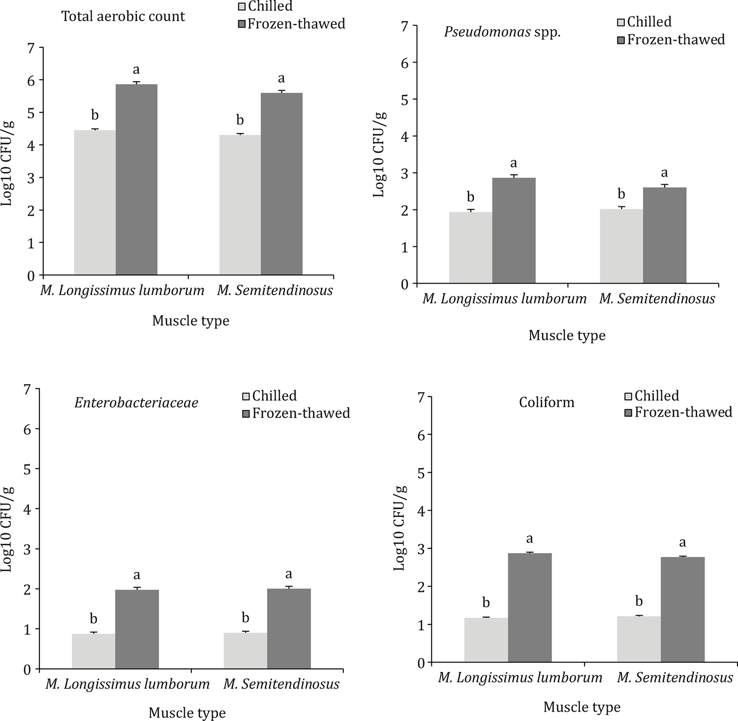 Values are means ± 1 standard error bar. a,b - Means with different letters on the bar indicate significant difference (freeze-thawing effect) at P≤0.05. Muscle type (P = 0.805) and interaction (P = 0.681) were not significantly different.
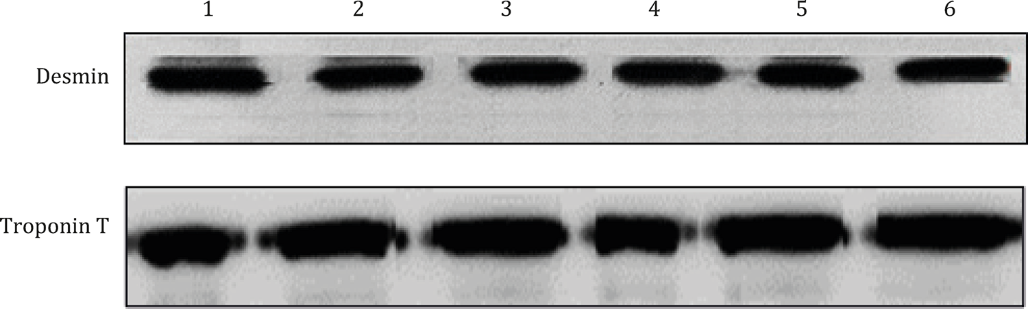 1 - Longissimus lumborum muscle, stored at −20 °C for five days and thawed at 4 °C for two days (frozen-thawed group). 2 - Longissimus lumborum muscle, stored at 4 °C for seven days (chilled group). 3 - A reference sample collected from the Longissimus lumborum muscle 24 h post-mortem. 4 - Semitendinosus muscle, stored at −20 °C for five days and thawed at 4 °C for two days (frozen-thawed group). 5 - Semitendinosus muscle, stored at 4 °C for seven days (chilled group). 6 - A reference sample collected from the Semitendinosus muscle 24 h post-mortem.