 Exploring vitamin A supplementation in beef cattle: Implications for cow-calf systems
ABSTRACT
Vitamin A is listed in the fat-soluble vitamins group, whose absorption, transport, and storage are similar to the lipids. However, the vitamin A functions go beyond those reported in the ocular cells’ metabolism being also described in reproductive functions, growth, and specific tissue maintenance through gene regulation. In ruminants, vitamin A supplementation can be done in different ways. Orally with carotenoids which β-carotene is the main representative, and intramuscularly by injectable applications of synthetic vitamin A. In cows, it is common to perform oral supplementation with β-carotene in the middle and/or final third of gestation to increase plasma retinol concentrations, benefiting the fetus, colostrum synthesis, and reproductive indices. In newborn calves, vitamin A administrated intramuscularly might favor cell differentiation for lipogenesis increase in muscle tissue indicating a higher intramuscular fat degree and resulting in a future improvement in meat quality. Thus, this review aimed to address the vitamin A supplementation dynamic on the effect on gestational/maternal and neonatal metabolism in beef cattle.
Keywords
antioxidant; immunoglobulin; intramuscular fat; pregnancy; retinol
1. Introduction
The micronutrient requirements related to minerals and vitamins must be considered in ruminant nutrition. Given that these nutrients have essential functions in animal organisms acting as cofactors and enzymes, they are related to improving animal metabolism, and subsequently increasing growth and performance (Harris et al., 2018). Regarding fat-soluble vitamins, vitamin A supplementation in the diet can be done by providing plant-originated provitamin A molecules, or by intramuscular injections of molecules artificially synthesized (Maciel et al., 2022).
Until now, cases of vitamin A deficiency in beef cattle produced in pasture systems are rare, once tropical pastures contain adequate β-carotene concentrations (Factor et al., 2024), and its storage in liver tissue ensures long-term use (D’Ambrosio et al., 2011). A reduction in β-carotene concentrations can be observed in pastures during the dry periods of the year, reinforcing the importance of animal supplementation with vitamin A, or its precursors (Reynoso et al., 2004).
Vitamin A recommendations for beef cattle are reported considering the animal physiology and category. According to the NASEM (2016) system for beef cattle, vitamin A requirements can be expressed per international unit in kilograms of dry matter per day (IU/kg DM) or Retinol Equivalents.
Ruminal degradation of vitamin A precursors can affect its bioavailability for animal metabolism. In general, microbial vitamin requirements are limited to water-soluble vitamins, e.g., those of the B complex and vitamin C, to stimulate their cell growth and are associated with biochemical reactions as enzymatic cofactors in microbial metabolism (Nagaraja et al., 1997).
However, in vitro studies conducted by Hino et al. (1993) demonstrated that supplementation with β-carotene and α-tocopherol, whether administered individually or in combination, stimulates the growth of ruminal microorganisms and enhances the digestion of the fibrous fraction of the diet. Therefore, the vitamin A metabolism mechanisms in the rumen are still unclear (Zeoula and Geron, 2006). Vitamin A supplementation is valid when there is an increase in animals’ requirements which depends on the animal’s physiological phase, or when the food quality sources that provide carotenes are poor. Supplementation should be provided for pregnant beef cows, in which the need for macro and micronutrients increases in the peripartum period (Jo et al., 2020). In this case, β-carotene supplementation is indicated and has a positive effect on oxidative stress (Kamiloglu et al., 2005), fetal growth (Marceau et al., 2007; Jo et al., 2020), colostrum synthesis (Nishijima et al., 2017; Ishida et al., 2018), ovulation (Hidalgo et al., 2005; Ay et al., 2012a) and embryonic adherence (Agarwal et al., 2006; Gouvêa et al., 2018). In addition, the vitamin A supplementation in beef cows at late gestation enhances intramuscular adipogenesis in offspring (Dean et al., 2024).
Furthermore, when dealing with the supplementation effect of injectable synthetic vitamin A in newborn calves, it is common to observe a gene action in cell regulation and differentiation through hyperplasia and hypertrophy in intramuscular fat (Harris et al., 2018; Campos et al., 2020; Peng et al., 2020; Maciel et al., 2022; Scapol et al., 2023). These are related to ensuring higher weaning weight (Jo et al., 2020; Peng et al., 2020), and intramuscular fat deposition (Peng, et al., 2020; Maciel, et al., 2022; Scapol et al., 2023) via lipogenesis, providing a higher meat marbling degree during the slaughter period in beef cattle (Daniel et al., 2009; Wellmann et al., 2020; Scapol et al., 2023). Thus, this literature review aimed to analyze the effects of vitamin A supplementation on gestational, maternal, and neonatal metabolism in beef cattle.
2. Vitamin A characterization
Micronutrients, although required in smaller quantities compared to macronutrients, play a crucial role in maintaining health and the proper functioning of the body (Andrès et al., 2024). Among micronutrients, vitamins stand out for their variations and specific functions, which are grouped into two main categories: water-soluble vitamins, including ascorbic acid (vitamin C) and the B-complex vitamins, and fat-soluble vitamins, which include retinol (vitamin A), as well as cholecalciferol (vitamin D), tocopherol/tocotrienol (vitamin E), and phylloquinone/menaquinone (vitamin K) (Andrès et al., 2024).
Vitamin A exhibits a specific affinity for hepatic tissue, where it is primarily stored, although smaller amounts can also be found in the adipose tissue of animals (Peng et al., 2021). In the body, it may bind to plasma transport proteins. Furthermore, vitamin A is present in plant pigments in the form of carotenoids, such as β-carotene, which are generally considered to be abundantly available. Despite structural differences, these carotenoids share an unsaturated isoprenoid chain and perform similar physiological functions in the organism (Carazo et al., 2021).
In grasses and legumes, the reflected light spectrum that results in the green color of the plants indicates proper levels of provitamin A, particularly represented by β-carotene. However, there is a significant decrease in the biodisponibility of β-carotene as the forage matures or undergoes drying or preservation processes (Elgersma et al., 2013; NASEM, 2016; NASEM, 2021). In such conditions, vitamin A supplementation becomes necessary to ensure animals receive an adequate amount of this nutrient.
Vitamin A is biologically active in the forms of retinol, retinal, or retinoic acid, which contain an alcohol group, an aldehyde group, and a carboxylic acid group, respectively (Figure 1) (Polcz and Barbul, 2019). Understanding the different bioforms of vitamin A and its metabolism is crucial for comprehending its role in cellular growth and tissue differentiation in animals.

Schematic of the β-carotene and vitamin A precursors absorption, transport, and storage in the beef cattle intestine.
Although vitamin A is not directly involved in coenzyme-catalyzed reactions, it plays hormonal and regulatory functions in gene expression (Balmer and Blomhoff, 2002). However, both its deficiency and excess can compromise animal metabolism (González and Silva, 2019).
2.1. Bioavailability and functions
The bioavailability of micronutrients refers to the fraction of the ingested nutrient that effectively contributes to meeting the physiological demand of the target tissue (Jackson, 1997). In the case of vitamin A, this availability is essential for the performance of vital functions, such as the regulation of reproductive function, growth, tissue maintenance (Ambrósio et al., 2006), and gene regulation (Balmer and Blomhoff, 2002).
The degradation of vitamin A begins in the rumen, where retinol esters are cleaved by microorganisms, releasing free retinol (Rode et al., 1990), which can be converted into retinal and retinoic acid (Srinivasan and Buys, 2019). In the small intestine, vitamin A esters are hydrolyzed by pancreatic lipases, releasing retinol, which is absorbed and re-esterified in enterocytes, being incorporated into chylomicrons. These chylomicrons are transported to the liver, where vitamin A is esterified and stored in hepatic stellate cells (Grumet et al., 2017). Retinol-binding protein (RBP) carries retinol in circulation, being absorbed by cells expressing Stimulated by Retinoic Acid-6 (STRA6) (Kelly and von Lintig, 2015).
Intracellularly, vitamin A is converted into its active form, retinoic acid, which binds to nuclear receptors such as retinoic acid receptors (RAR) and retinoid X receptors (RXR), regulating the transcription of genes involved in cell differentiation and metabolism (Huang et al., 2014). One of its primary biological roles is to promote cellular differentiation, a process essential for the development and specialization of tissues and organs from embryogenesis to adulthood (Barber et al., 2014). In early life stages—particularly during fetal development, the suckling phase—retinoic acid influences the commitment of mesenchymal stem cells toward the adipogenic lineage, favoring the development of intramuscular adipocytes (Harris et al., 2018). Adequate vitamin A status during these phases, either via maternal colostrum, milk, or forage, is thus critical for proper adipose tissue programming.
Interestingly, the role of vitamin A appears to shift in later stages of production. During the finishing phase, for instance, vitamin A restriction has been associated with increased intramuscular fat deposition or marbling (Kruk et al., 2018). This paradoxical effect may stem from the reduced inhibitory action of retinoic acid on adipocyte differentiation at this stage. Mechanistically, retinoic acid acts as a ligand for RAR, forming heterodimers with RXR (Chawla et al., 2001; de Thé et al., 1990), which bind to retinoic acid response elements in the genome, modulating the expression of key adipogenic and lipogenic genes such as PPARγ (Lefterova et al., 2009). Moreover, there is evidence that retinoic acid influences skeletal muscle metabolism by enhancing fatty acid oxidation, potentially limiting lipid accumulation in muscle tissue (Amengual et al., 2018). Collectively, these findings highlight the stage-specific effects of vitamin A on adipogenesis and energy metabolism, emphasizing the need for targeted nutritional strategies across different phases of cattle development.
2.2. Deficiency and toxicity
In pasture-based production systems, particularly under tropical conditions, clinical cases of vitamin A deficiency or toxicity in beef cattle are uncommon. This is largely attributed to the high availability of β-carotene in fresh green forages, which is efficiently converted into retinol and stored in the liver, ensuring sufficient reserves to maintain vitamin A homeostasis over extended periods (Pickworth et al., 2012).
Fresh tropical forages have been reported to contain vitamin A equivalents ranging from 39,000 to 59,600 IU per kilogram of dry matter (DM), while conserved forages, such as good-quality hay, typically contain much lower concentrations, between 1,400 and 3,600 IU/kg DM. These values are highly susceptible to oxidative degradation during storage, particularly under inadequate drying and preservation conditions (Pickworth et al., 2012). Vitamin A requirements vary according to animal category and physiological status. According to NASEM (2016) and Valadares Filho et al. (2023), beef feedlot cattle require approximately 2,200 IU/kg of dry feed, pregnant heifers around 2,800 IU/kg, and lactating cows or breeding bulls up to 3,900 IU/kg. Under well-managed grazing conditions, cattle typically meet these requirements through voluntary intake of β-carotene from fresh forages. In contrast, animals consuming primarily conserved or low-quality forages are at increased risk of deficiency and may require dietary supplementation to ensure adequate intake and hepatic storage of vitamin A.
If hepatic retinol stores decline, clinical signs of vitamin A deficiency may emerge (NASEM, 2016). According to Speer et al., 2022, the deficiency is characterized by low plasma retinol concentrations (< 300 ng/mL) and deficient liver retinol reserves (< 300 µg/g DM). Clinically, affected cattle exhibit stunted growth, especially in bone development; impaired immunity—marked by reduced phagocytic activity in macrophages and neutrophils, leading to increased susceptibility to infections and, in severe cases, mortality; and reproductive disorders. In females, common manifestations include prolonged estrous cycles, anovulation, reduced conception rates, abortion, and retained placenta. In males, testicular atrophy, degeneration of accessory sex glands, poor sperm quality, and disrupted spermatogenesis have been reported (Zeoula and Geron, 2006).
Regarding hypervitaminosis/toxicity from vitamin A, toxicity in cattle has not been reported under grazing conditions. This may be explained by the ruminal fermentative metabolism and the degradation of carotenoids by the microbiota, which prevents excessive absorption of vitamin A by the intestinal epithelium, ensuring adequate plasma levels for the animal’s metabolic homeostasis (NASEM, 2016; NASEM, 2021; Valadares Filho et al., 2023).
3. Vitamin A metabolism
Such any other nutrient, vitamin A performs specific functions in cellular metabolism. As a result, there is a mechanism for the activation of this biomolecule at both the cytoplasmic and nuclear levels, optimizing the absorption, storage, and transport processes and its use in target tissues (Polcz and Barbul, 2019). The functionality of this regulatory system depends on the lipoproteins association, and proteins and molecular complexes transport, which provide the dynamic for the activation and storage of this molecule for cellular metabolism (Polcz and Barbul, 2019).
3.1. Absorption, storage, and transport
The absorption of vitamin A is directly related to the intake of its precursors, known as provitamin A, represented by carotenoids (D’Ambrosio et al., 2011). The main carotenoids, such as α-carotene, β-carotene, and β-cryptoxanthin (Conaway et al., 2013), are found in the foods provided to animals, meeting the nutritional requirements of this vitamin. After ingestion, these compounds are emulsified in the intestine, forming a micellar aggregate that includes fatty acids, monoacylglycerol, phospholipids, cholesterol, bile salts, and free retinol (Blaner et al., 2016). This emulsification process prepares the carotenoids for hydrolysis, which is carried out by enzymes such as pancreatic lipases. Hydrolysis releases free retinol, which is absorbed by intestinal cells (enterocytes) through facilitated diffusion (Soprano et al., 1994; Bennekum et al., 2000; Reboul et al., 2006).
Since retinol is insoluble in water, it must bind to transport proteins, such as retinol-binding protein I (RBP I) and retinol-binding protein II (RBP II), which facilitate the transport of retinol through intestinal cells (Ong and Chytil, 1978; Conaway et al., 2013; Blaner et al., 2020). Retinol derived from β-carotene can be converted into all-trans retinoic acid (ATRA), also known as tretinoin, or it can remain in the form of retinol. This conversion occurs not only in enterocytes but also in hepatocytes and other cells, where ATRA can be converted back into retinol and released into the bloodstream for use by the body (Carazo et al., 2021).
Retinol-binding proteins are involved in the formation of chylomicrons through the reesterification of compounds absorbed during the digestion of long-chain fatty acids, a process mediated by the enzyme retinyl acyltransferase (Harrison, 2012; Polcz and Barbul, 2019). This results in the formation of a complex that includes retinal esters, carotenoids, and provitamin A, which are esterified with fatty acids, incorporated into chylomicrons, and transported via the lymphatic system for later storage in the liver (Polcz and Barbul, 2019). In this process, as the absorption and transport of retinol depend on the presence of fat, approximately 50% of the consumed provitamin A is absorbed intact by the intestinal mucosa and directed to the portal system, while the other 50% undergo oxidation to be converted into retinol (Conaway et al., 2013). Thus, fat intake is essential to ensure the effective absorption of vitamin A, a fat-soluble compound (Blomhoff et al., 1990). Additionally, some carotenoids, such as β-carotene, are absorbed by enterocytes with the help of membrane proteins, such as the scavenger receptor class B1 (SCARB1) and cluster of differentiation 36 (CD36) (Blomhoff et al., 1990).
After intestinal absorption, vitamin A is incorporated into chylomicrons and transported via the lymphatic system to the liver. In hepatocytes, chylomicron remnants are internalized through receptor-mediated endocytosis, a process involving apolipoprotein E and lipoprotein lipases, which hydrolyze retinyl esters to release free retinol (Harrison, 2012; Cooper, 1997). Once in the liver, retinol may be re-esterified by hepatic cells, primarily as retinyl palmitate, and stored for future use in the absence of immediate metabolic demand (Conaway et al., 2013). It is estimated that approximately 90% of the body’s vitamin A is stored in the liver, with around 40% utilized in metabolic processes and the remainder retained in hepatic reserves. When needed, retinol is mobilized by the action of retinyl ester hydrolase and released into circulation bound to plasma retinol-binding protein (RBP-II) or as complexes with cytoplasmic RBP. These complexes are then transported to storage sites, such as hepatic and adipose tissues (Ambrósio et al., 2006).
The transport of vitamin A in plasma occurs through the esterification of retinyl esters in liver cells, which can be converted into retinol. Retinol then binds to RBP, forming the retinol-RBP complex, which is transported through the bloodstream with the help of membrane transporters (Soprano and Blaner, 1994). In plasma, the retinol-RBP complex requires a carrier protein, which may be transthyretin (TTR), prealbumin, or albumin, to ensure proper transport (Carazo et al., 2021). In addition to transporting the retinol–RBP complex, TTR also carries the thyroid hormones (T3 and T4). Thus, the complex formed by retinyl esters and/or retinol-RBP with TTR helps prevent the excretion of vitamin A through renal filtration (Carazo et al., 2021), protecting circulating vitamin A precursors from depletion or oxidation until they are utilized by specific tissues (Figure 1).
3.2. Molecular mechanisms of retinoid signaling in target tissues
As previously mentioned, the absorption and transport of vitamin A precursors intended for storage depend on specific proteins that convert retinoic acid into retinol, which is subsequently retained in the liver. A portion of this retinol is then mobilized as retinyl esters and converted back into retinoic acid. Thus, when specific tissues require it for metabolism, target cells activate mechanisms to capture these precursors in the bloodstream through membrane receptors. This process directs the precursors to the cell nucleus, where gene expression occurs to benefit cellular metabolism (Figure 2).

Schematic of the cellular uptake and intracellular transport of vitamin A metabolites in target tissues.
When the ATRA complex is available with RBP, or by plasmatic albumin in low concentrations, peripheral cells absorb this compound (Conaway et al., 2013) through the transmembrane receptor Strat6, encoded by retinoic acid 6 stimuli (Kawaguchi et al., 2007; Conaway et al., 2013; Carazo et al., 2021).
In this way, the target tissues’ cellular metabolism for vitamin A precursors, especially retinol, depends on binding proteins and receptors for their signaling pathways, beginning with the retinol oxidation into all-trans retinal acid by cytosolic alcohol dehydrogenase (ADH), being complexed with the cellular retinol receptor protein (CRBP), passing through the oxidative action of the enzyme retinal dehydrogenase (RALDH), providing the active vitamin A metabolite, ATRA, being bound to a cellular retinoic acid receptor protein (CRABP; Conaway et al., 2013; Carazo et al., 2021) at the cytosol level.
Despite the cell nucleus, vitamin A and its precursors are an important factor for gene expression (Balmer and Blomhoff, 2002; Bohn, 2017) in which nuclear receptors interact with transcription, modulating the target gene expression (Carazo et al., 2021). Furthermore, nuclear hormone receptors are responsible for enabling the transport of vitamin A precursors + binding proteins present in the cytosol, namely retinoic acid receptors (RARs) and retinoid X receptors (RXRs). Therefore, RAR receptors form heterodimers with RXR receptors and RXR receptors (e.g., isolated in homodimer form) have transcription factor functions, leaving retinoic acid response elements (RAREs) active in target genes (Bastien and Rochette-Egly, 2004; Conaway et al., 2013; Carazo et al., 2021).
Retinoic acid exerts its biological effects primarily through its interaction with nuclear receptors, particularly the retinoic acid receptors (RARs), which include the isoforms RARα, RARβ, and RARγ (Giguere et al., 1987). Among these, RARα is the most widely expressed isoform in the body, whereas RARβ is predominantly localized in the liver, kidneys, and nervous tissue. The interaction between vitamin A and gene expression is mediated by all-trans retinoic acid (ATRA), which binds to RARs and activates signaling pathways involved in key physiological processes such as cell differentiation and tissue development (Bastien and Rochette-Egly, 2004). Once vitamin A precursors are mobilized from hepatic stores into the circulation—complexed with specific binding proteins—they become available to intracellular receptors. RARs preferentially bind to ATRA, while retinoid X receptors (RXRs) exhibit higher affinity for the 9-cis-retinoic acid isomer (Germain et al., 2002). The functional divergence between RAR and RXR is largely attributed to differences in their ligand-binding domains (LBDs), which define their ligand specificity and determine the nature of the receptor-ligand complex that translocates from the cytosol to the nucleus, ultimately regulating the transcription of target genes (Carazo et al., 2021).
At the molecular level, the inactivated RAR form occurs when there is no binding to retinoids, remaining deprived in the cell nucleus composing the co-repressors complex (Carazo et al., 2021). In this way, there is a structural molecule rearrangement with the specific ligand for the co-activator RAR, generating the co-repressor activation (Chebaro et al., 2017). The transcription machinery of cellular genetic material must be accomplished by the RAR heterodimerization with RXR to carry out transcription, providing the co-activator ligand heterodimer complex for DNA sequences recognition, binding to the promoter region of target genes (e.g., retinoic acid responsive elements, RARE) which affect the genes transcriptional regulation (Carazo et al., 2021).
The RARs present in the cell nucleus also have a binding action to the CRABPII (0) complexes, transporting ATRA to the nucleus (Conaway et al., 2013), which is another active form of vitamin A precursor transport for gene regulation. Another nuclear receptor with a hormonal nature forms heterodimer with RXR and is called peroxisome proliferative activated receptors (PPARs) with isomers α, β, γ, and δ. The PPAR synthesis might be affected by the cellular retinal concentrations in adipose tissue with antagonistic PPAR activity effect, inhibiting adipogenesis and increasing the cell insulin sensitivity (Ziouzenkova et al., 2007). This RXR-PPAR complex called a heterodimer, for example, has transcription factor functions activating specific gene expression responses (Conaway et al., 2013; Carazo et al., 2021). Additionally, to the transporters aforementioned, retinoid transporters might complex with orphan transporters (RORs), which do not form dimers and might regulate gene expression in monomers by binding to RARE (Conaway et al., 2013; Carazo et al., 2021), with α, β, and γ isoforms.
Thus, with the retinol oxidation reducing ATRA, genomic function modifications are regulated by the action of nuclear transporters/receptors related with vitamin A precursors (PPGRs, RARs, RXRs, and RORs) for gene expression or target genes regulation. Considering this, the transcription may not occur when no ligands are available to be associated with the transport complexes present in the nucleus through cellular retinoic acid-binding proteins (CRABP) or fatty acid-binding proteins (FABP), otherwise, transcription is not directed (Carazo et al., 2021).
4. Vitamin A utilization
The vitamin A requirement for beef cattle is expressed in international units (IU) or retinol equivalents (RE) relative to body weight. For ruminants, β-carotene is the recommended precursor of vitamin A to be used in diets and supplements. According to NASEM (2016), the vitamin A requirement varies in accordance to beef cattle category and physiological state, in which at 2,200 IU/kg dry feed for beef feedlot cattle, 2,800 IU/kg dry feed for pregnant beef heifers and 3,900 IU/kg dry feed for lactating cows and breeding bulls.
Regarding the vitamin A requirements for pregnant beef females (e.g., heifers and cows) and pre-weaned calves, there are limitations in data validation for these categories (NASEM, 2016). This makes it essential to understand the role of vitamin A supplementation for performance and metabolism during these specific physiological stages.
4.1. Supplementation forms
The dietary supply of vitamin A in ruminants is influenced by both the type of diet and the rate of ruminal fermentation. Various carotenoid isoforms—such as β-carotene, α-carotene, γ-carotene, and cryptoxanthin—are present in forages and grains and serve as vitamin A precursors (NASEM, 2016). However, the availability of these compounds can fluctuate significantly depending on forage quality, particularly in pasture-based systems. During dry seasons, when forage carotenoid content is reduced, supplementation with provitamin A sources is often required to meet nutritional demands (Pickworth et al., 2012; NASEM, 2016). Although the precise requirements of vitamin A for pregnant ruminants and growing calves have not yet been fully defined, cases of hypervitaminosis A are rare. This low incidence is mainly due to the extensive microbial degradation of vitamin A and its precursors in the rumen, which limits their intestinal absorption and systemic accumulation (Casals and Calsamiglia, 2012; NASEM, 2016; Valadares Filho et al., 2023). In practice, dietary vitamin A supplementation is commonly achieved through the inclusion of β-carotene-rich feedstuffs, vitamin–mineral premixes, or, in some cases, parenteral administration. Injectable vitamin A is typically used in strategic phases such as early life, weaning, or gestation, particularly in situations where dietary intake may be insufficient (Harris et al., 2018; Campos et al., 2020; Peng et al., 2020; Wellmann et al., 2020; Song et al., 2023; Scapol et al., 2023; Dean et al., 2024).
While both supplementation routes are effective in increasing vitamin A status, injectable forms tend to yield more immediate and consistent increases in plasma and hepatic retinol concentrations (Knight and Death, 1999), particularly in neonates and periparturient cows, where gastrointestinal absorption may be limited or dietary intake insufficient. Conversely, long-term maintenance of adequate vitamin A status is more efficiently sustained through dietary supplementation, which better supports gradual accumulation and storage in hepatic tissue (Knight and Death, 1999). Therefore, the choice of supplementation method should consider the animal’s physiological stage, nutritional status, and production system.
4.2. Effects on pregnant beef cows
Metabolic alterations associated with physiological transitions in the pre- and postpartum periods are well-documented in mammals (Redmer et al., 2004). This transition is often characterized by a negative energy balance due to the sudden increase in energy demands for fetal development, parturition, and the onset of lactation. Such an imbalance leads to the mobilization of body reserves, which consequently increases oxidative stress in peripheral tissues as a result of elevated reactive oxygen species (ROS) production at the cellular level (Sordillo and Aitken, 2009). The accumulation of free radicals can disrupt systemic homeostasis (Castillo et al., 2005; Sordillo, 2005), diverting energy toward milk production for the calf and thereby reducing antibody synthesis and reproductive hormonal activity, ultimately compromising maternal health (Tufarelli et al., 2023).
Evaluations of the vitamin A supplementation effects in beef cows at the end of gestation are scarce (Jo et al., 2020; Dean et al., 2024), but hypotheses might be generated by adopting analogies to other ruminant production systems, such as dairy cows or lambs. Jo et al. (2020) identified the vitamin A oral supply interaction for pregnant beef heifers at the end of gestation. It was found that maternal supplementation in the period from 225 days of gestation until the day of calving, with 78,000 IU/day of vitamin A in the total diet leads to a decrease in vitamin A serum levels after calving due to its use for fetal growth, and the female nutritional needs increase during the lactation period. These findings indicate that the supplementation level used should be higher than those suggested by the nutritional requirements of beef cattle nowadays.
It is well established that placental morphology plays a crucial role in the maternal–fetal transfer of nutrients (Fowden et al., 2009). Ruminants, including cattle, possess an epitheliochorial placenta, which consists of multiple cellular layers separating maternal and fetal blood supplies, and therefore significantly limits the passive transfer of vitamin A from the dam to the fetus (Donoghue et al., 1985). Despite this anatomical limitation, vitamin A and its derivatives (retinoids) are critical for embryonic development and fetal tissue differentiation, particularly during late gestation, when adipogenesis is actively occurring.
Recent studies have shown that vitamin A supplementation during late gestation enhances intramuscular adipogenesis in the offspring. Dean et al. (2024) reported increased mRNA expression of retinoic acid receptor β (RARβ) and greater protein abundance of adipogenic markers (DLK1 and PPARγ) in the offspring of beef cows supplemented with vitamin A, which was associated with increased intramuscular fat deposition throughout postnatal development. These findings suggest that maternal retinoid status during late gestation can modulate fetal adipocyte differentiation via retinoid signaling pathways, despite the restrictive placental environment.
In addition to developmental effects, vitamin A and its precursors, such as β-carotene, have recognized antioxidant functions, particularly important during the peripartum period when oxidative stress is heightened due to increased metabolic demands and hormonal fluctuations. Carotenoids can neutralize reactive oxygen species (ROS) through mechanisms involving electron transfer and hydrogen abstraction, contributing to maternal redox balance (Zubova et al., 2021; Mitsuishi and Yayota, 2024). Elevated cortisol levels during labor can exacerbate oxidative stress and modulate immune responses; thus, maintaining adequate antioxidant status through dietary carotenoids may help mitigate these effects (Oliveira, 2014).
Although the transfer of vitamin A across the placenta is limited in ruminants due to the epitheliochorial barrier, studies suggest that neonates are born with detectable hepatic vitamin A stores (Ross and Gardener, 1994; Collins et al., 1994). This indicates that, despite its restrictiveness, the placenta is capable of a regulated and selective retinoid transport. According to Marceau et al. (2007), placental tissues express retinoic acid receptors (RARs) and cellular retinol-binding proteins (CRBPs), which enable the intracellular uptake and trafficking of maternal retinol—carried in the blood bound to retinol-binding protein (RBP)—into placental cells and eventually into the fetal circulation.
Notably, during early organogenesis, the concentration of retinoids in placental tissue can be up to eight times higher than in the embryo, suggesting that the placenta may function as a transient storage site for retinoids. As gestation advances, particularly in the final trimester, fetal retinoid levels gradually surpass those of the placenta, indicating a regulated increase in retinol transfer to support tissue differentiation and accelerated growth (Marceau et al., 2007). However, in ruminants, this transfer remains quantitatively limited due to the structural characteristics of the epitheliochorial placenta, which imposes a greater barrier to fat-soluble vitamin diffusion compared to species with hemochorial placentation (Soares et al., 2018).
Given these physiological constraints, postnatal vitamin A sources become essential to sustain adequate neonatal status. Colostrum, rich in retinol and retinyl esters, plays a crucial role in this regard (Nozière et al., 2006). Nonetheless, in scenarios of poor colostrum intake or maternal deficiency, strategic neonatal supplementation—such as intramuscular vitamin A injection shortly after birth—can be beneficial, promoting immune competence and supporting optimal early development (Harris et al., 2018; Wang et al., 2018).
Jo et al. (2020) observed a marked decline in maternal plasma retinol concentrations during the first week postpartum, attributing this decrease to the high nutritional demands of the fetus during late gestation and the onset of lactation. This finding is supported by other studies in cattle that demonstrate significant prepartum redistribution of vitamin A into fetal and colostral compartments. For instance, Kankofer and Albera (2008) reported that retinol concentrations were significantly higher in the fetal portion of the placenta compared to the maternal portion, and that colostral retinol levels increased substantially within the first 24 hours after birth. Similarly, Speer et al. (2024) estimated that approximately 60% of the vitamin A content in colostrum originates directly from the maternal diet in late gestation, while the remaining 40% is mobilized from hepatic stores. These findings indicate that both dietary intake and liver reserves are strategically mobilized to support colostrum formation at parturition.
Newborn calves typically have reduced hepatic vitamin A stores at birth, and are therefore considered functionally deficient in vitamin A and β-carotene at this stage (Puvogel et al., 2008). Thus, colostrum represents the primary and immediate source of retinol necessary to normalize neonatal vitamin A status. Considering that fat-soluble vitamins, such as vitamin A, do not cross the bovine placenta efficiently, neonatal blood and liver retinol concentrations at birth do not directly reflect maternal levels. In fact, recent data indicate that many calves exhibit low hepatic vitamin A stores at birth even when their dams are vitamin A sufficient.
Taken together, these findings highlight a functional maternal–neonatal axis of vitamin A metabolism in cattle, in which maternal mobilization during late gestation—particularly toward the placenta and colostrum—plays a central role in establishing the neonatal vitamin A status. In this context, it can be concluded that plasma vitamin A concentrations in newborn calves are directly influenced by maternal supplementation during late gestation. This results in elevated vitamin A levels (IU/dL) at birth—even prior to colostrum intake—thereby supporting the hypothesis that placental transfer of vitamin A does occur in beef cattle. In a study with beef cows, Jo et al. (2020) observed that maternal supplementation with 78,000 IU/day of vitamin A during the final third of gestation led to significantly higher plasma retinol concentrations (~120.3 IU/dL) compared to non-supplemented controls (~83.7 IU/dL).
Complementing these findings, Lotfollahzadeh and Golchin (2016) reported that calves born to dams supplemented with 2,000,000 IU of vitamin A via intramuscular injection exhibited pre-colostrum plasma retinol concentrations ranging from 90.6 to 93.1 μg/dL, compared to only 35.5 μg/dL in the control group—representing a more than 2.5-fold increase. Additionally, Kumagai et al. (1994) demonstrated that plasma vitamin A concentrations in calves more than double between birth and five days of age, underscoring the importance of colostrum as a critical source of vitamin A during the neonatal period.
Together, these findings confirm that, although placental transfer of fat-soluble vitamins in ruminants is generally limited (NASEM, 2021), maternal supplementation in late gestation can promote biologically and statistically significant increases in neonatal vitamin A status. Further research is needed to determine the specific vitamin A requirements for pregnant cows—especially considering the increasing fetal demand in late gestation (Jo et al., 2020)—and to better understand how maternal intake and hepatic reserves influence both fetal supply and early calf development.
As described and indicated previously, few considerations are available about vitamin A or its precursors supplementation for beef cows, however, studies conducted with dairy cows indicate a colostrum quality improvement related to higher immunoglobulins (IgG) and fats concentrations when supplementation is performed in prepartum (Nishijima et al., 2017; Ishida et al., 2018). Considering the digestive physiology, gestational physiology, and colostrum production physiology analogies of dairy cattle and beef cattle, we can relate studies carried out with dairy cattle to understand the vitamin A or its precursors’ supplementation effects in colostrum quality.
Colostrum synthesis is influenced by the blood flow and nutrients circulating in the mammary gland, which may alter its composition (Foley and Otterby, 1978). Other factors might alter this synthesis, such as immunity and cow breed. Thus, when cows are supplemented with β-carotene in the final phase of gestation, they provide higher IgG1 and IgA colostral concentrations (Nishijima et al., 2017; Ishida et al., 2018) or not (Kaewlamun et al., 2011; Prom et al., 2022), requiring further investigations into mammary metabolism for colostrum synthesis and immunological quality.
As previously reported, the vitamin A antioxidant action and β-carotene protect the cells from free radicals degenerative effects, produced during oxidative metabolism, in the peripartum period and attenuate oxidative stress by unbalancing the free radical number, also known as reactive oxygen species (ROS) characterized by glutathione, glutathione peroxidase, vitamin A, vitamin E, β-carotene and others (Kamiloglu et al., 2005), and antioxidants.
Considering a temporal scale, during the peripartum period in cattle, the free radicals production is intense due to the metabolic increase resulting from oxidative stress. It is common that in intensive beef cattle production systems, after 60 days of calving, reproductive management occurs with the dams through artificial insemination (whether at a fixed time or not). However, with the higher metabolic rate causing oxidative stress, failures related to estrus may be noted, compromising ovulation (Ay et al., 2012a). In this sense, it is necessary to observe prepartum supplementation with vitamin A or β-carotene, reinforcing the liver storage in the retinoid form idea, or the intramuscular application of commercial products containing vitamin A and β-carotene might ensure adequate blood levels granting benefits in the cows’ reproductive phase in the postpartum period (Mitsuishi and Yayota, 2024). Thus, the lack of antioxidant activity in the presence of synergistic interactions between free radicals and reactive oxygen species (ROS) may impair essential processes such as cell division, primordial cell differentiation, and embryonic development (Agarwal et al., 2006). In addition, oxidative stress can damage the membrane structure of luteal cells, leading to reduced progesterone production and impaired expression of luteinizing hormone receptors, which may ultimately inhibit ovulation (Agarwal et al., 2006).
In this context, in front of situations with a low β-carotene serum concentration in the reproductive period, immediately after parturition, we can witness cases of long estrous periods, low ovulation, and low conception rates. If there is an ideal β-carotene circulation, positive effects are observed regarding a reduction in the uterine involution time, greater progesterone production by the corpus luteum, and a shorter service period (Rakes et al., 1985; Arikan and Rodway, 2000).
Another point that should be addressed regarding the vitamin A effect on the cows’ reproductive period is related to the corpus luteum (CL) metabolism. In general, adequate β-carotene plasma concentrations increase the follicles and CL metabolic activity relative to the estrous cycle and gestation stages (Oliveira, 2014). Regarding CL, when supplementation with β-carotene is used, the follicular fluid and CL become a vitamin A deposit, observing a positive correlation for the CL weight and diameter, proving its functionality in the reproductive period (Haliloglu et al., 2002).
Findings indicate that β-carotene supplementation in the periods preceding the cows’ reproductive phase can positively affect ovarian activity with greater development of mature oocytes (Hidalgo et al., 2005), and can be used as a nutritional strategy for higher pregnancy rates with artificial insemination.
Regarding β-carotene plasmatic levels in cows undergoing reproduction, Michael et al. (1994) suggest a threshold between 300 and 1200 µg/dL, and for vitamin A values between 25 and 80 µg/dL, which should be associated with the pregnant beef cows or heifers’ requirements, equivalent to 2,800 IU/kg of vitamin A in a DM basis (NASEM, 2016; Valadares Filho et al., 2023).
Aguiar-Zalzano et al. (2022) compared dairy cows supplemented with β-carotene via two different routes: a single intramuscular injection administered on day 30 postpartum, and continuous oral supplementation starting 30 days before calving and continuing until 150 days postpartum. Both groups showed similar β-carotene concentrations, all exceeding the reference range (+300 µg/dL). However, intramuscular administration resulted in higher plasma β-carotene levels over shorter time periods following application.
Therefore, to optimize the artificial insemination management of beef cows in intensive production systems, the carotenes or vitamin A application via the intramuscular route can improve ovulation and CL metabolism, as mentioned above. Using this strategy associated with artificial insemination protocols in dairy cows, it was reported that there is an increase in serum β-carotene, causing greater CL functionality and increasing the pregnancy rate (Ay et al., 2012a; Ay et al., 2012b).
Observing the blastocyst interaction after fertilization, the maternal immune system might damage the cell division and differentiation initial phase, due to production of free radicals compromising embryonic and fetal development. However, the damage might be prevented by the vitamin A action with the increase in antioxidant production inactivating the ROS action (Kamiloglu et al., 2005), thus reducing cell degeneration and affecting embryonic development.
Associated with this, supplementation with β-carotene with fat-soluble vitamins (e.g., more specifically vitamins A and E) ensures an increase in the pregnancy rate in beef cows raised on pasture, when they were aroused to the first breeding season using the fixed-time artificial insemination technique (Gouvêa et al., 2018), ensuring more developed calves due to better embryonic cells damage prevention and granting its development (Olson and Seidel, 2000; Gouvêa et al., 2018). This action can be explained by the vitamin A association with β-carotene allowing antioxidant effectiveness ensuring low lipid peroxidation action of embryonic cell membranes, which affects the healthy embryo development.
Studies in the reproduction area have clearly demonstrated the adequate vitamin A or β-carotene applicability to optimize reproductive indices in dairy cows, possibly due to its higher oxidative stress in the peripartum period, reducing the effects of free radicals on ovarian and hormonal action (Khemarach et al., 2021). When compared with beef cows there is little reported about the use of this strategy to optimize reproductive indices, and further research is needed into vitamin A and its precursors use as supplements, when necessary.
4.3. Effects on beef calves
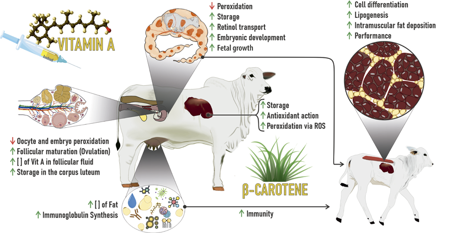
Vitamin A supplementation in neonatal calves has been widely studied from the perspective of muscle development and intramuscular fat accumulation, particularly in early-finishing production systems (Waldner and Uehlinger, 2016; Harris et al., 2018; Jo et al., 2020; Peng et al., 2020; Maciel et al., 2022; Scapol et al., 2023). However, the effects of this fat-soluble vitamin extend far beyond adipogenesis, influencing a range of fundamental physiological processes that begin during gestation and continue throughout the early stages of calf development (Figure 3). Vitamin A acts as an epigenetic and transcriptional modulator, primarily through retinoic acid, which binds to nuclear receptors (RAR and RXR), thereby regulating the expression of genes involved in cellular differentiation (Chawla et al., 2001). In adipose tissue, this signaling pathway is crucial for the formation and proliferation of preadipocytes, with direct implications for intramuscular adipose tissue synthesis and, consequently, meat quality (Peng et al., 2021; Scapol et al., 2023). This process, however, does not occur in isolation. The action of vitamin A on progenitor cell differentiation is also evident in skeletal muscle tissue, promoting the development of muscle fibers and the expansion of satellite cell populations—key elements for muscle hypertrophy and the animal’s future productive performance (Jo et al., 2020; Du et al., 2017). Neonatal vitamin A supplementation has been associated with increased average daily gain, higher weaning weights, and improved early growth, particularly when administered within the first 24 hours of life and, in some protocols, reinforced at 30 days of age (Harris et al., 2018; Maciel et al., 2022). On the other hand, the administration of vitamin A during the growing and finishing phases promotes changes in lipid metabolism, stimulating pathways related to lipid oxidation and the reduction of lipogenesis (Daniel et al., 2009; Kruk et al., 2018; Campos et al., 2020; Wellmann et al., 2020).
Dynamic effects of injectable β-carotene and vitamin A supplementation on the pregnant cows' metabolism, regarding the antioxidant action metabolic responses, reproduction, and gestation; and newborn calves, regarding the metabolic responses in the immune system and cellular differentiation in skeletal muscle tissue.
In addition to its roles in muscle and adipose tissue development, vitamin A plays a pivotal role in the maturation of the immune system. Newborn calves are born with limited hepatic retinol reserves due to the restricted placental transfer of this vitamin. As a result, they rely exclusively on colostrum as their initial source of vitamin A, making them particularly vulnerable to deficiency—especially when born to dams with insufficient vitamin A status. Studies have demonstrated that hypovitaminosis A in the early neonatal period impairs epithelial integrity, weakens mucosal immune responses, and compromises the functional capacity of T lymphocytes and dendritic cells, thereby increasing the incidence of respiratory disease and enteritis (McGill et al., 2019). In this context, intramuscular administration of vitamin A at birth has emerged as a practical and effective strategy to enhance immune competence and reduce the susceptibility of neonatal calves to infectious agents during the critical period of postnatal adaptation (McGill et al., 2019).
It is equally important to recognize that the physiological influence of vitamin A is not confined to the neonatal stage. The maternal-fetal interface plays a central role in shaping the intrauterine environment and, consequently, in the developmental programming of the offspring. Cows supplemented with vitamin A or its precursor β-carotene during the final third of gestation produce colostrum with higher retinol concentrations and are more effective at transferring this vitamin to their calves, resulting in improved immune function and overall performance through weaning (Scapol et al., 2023). This evidence highlights the importance of an integrated approach that accounts for both maternal nutritional management and direct postnatal supplementation to optimize neonatal development.
Within the metabolic–endocrine axis, vitamin A also exerts relevant regulatory effects. The early deposition of adipose tissue, stimulated by neonatal vitamin A administration, may influence circulating levels of leptin—a peptide hormone primarily secreted by mature adipocytes (Tan and Jiang, 2024). Leptin acts as a metabolic signal to the central nervous system, particularly the hypothalamus, modulating appetite, energy homeostasis, and the activation of the reproductive axis (Tan and Jiang, 2024). The differentiation of mesenchymal stem cells into preadipocytes, followed by their maturation into fully functional adipocytes—a process driven by retinoic acid, the bioactive form of vitamin A—leads to an increased pool of leptin-secreting cells. Consequently, early-life vitamin A supplementation may elevate leptin levels during the pre-weaning period. Although the direct causal relationship between this intervention and puberty onset in cattle remains to be fully established, elevated leptin concentrations during early life have been associated with earlier puberty and first estrus in heifers (Fantuz et al., 2024). Furthermore, vitamin A deficiency in both sexes has long been linked to reproductive dysfunction, delayed sexual maturity, and reduced fertility (Shastak and Pelletier, 2024), reinforcing its essential role in gonadal development and neuroendocrine regulation of reproduction.
While the enhancement of intramuscular adipogenesis remains one of the primary advantages associated with neonatal vitamin A supplementation (Cianzio et al., 1985; Li et al., 2023), the systemic effects of this strategy are broad and physiologically interconnected. By modulating cellular differentiation, supporting immune and endocrine maturation, and maintaining epithelial integrity, vitamin A functions as a key regulatory element in early postnatal development. Recognizing the multifactorial roles of this micronutrient enables the design of more comprehensive supplementation protocols that address not only carcass quality, but also calf health, productivity, and reproductive potential throughout the production cycle.
5. Final considerations and future implications
The scientific evidence presented in this review highlights the pivotal role of vitamin A and its precursor, β-carotene, in supporting reproductive performance, fetal development, neonatal growth, and meat quality in beef cattle systems. Although tropical pastures generally meet vitamin A requirements, critical physiological phases such as late gestation, early lactation, and neonatal development demand strategic supplementation to address increased metabolic and antioxidant needs. It is recommended that pregnant beef cows receive elevated levels of vitamin A or β-carotene—approximately 60,000 to 78,000 IU/day of vitamin A via oral supplementation, or 1,000,000 to 2,000,000 IU via a single intramuscular injection during the last 3–4 weeks of gestation—to enhance hepatic retinol stores, optimize colostrum synthesis, reduce oxidative stress, and support fetal tissue development. These levels exceed the standard recommendations of 2,800 IU/kg of dry matter and reflect the increased physiological demands during the peripartum period.
Similarly, a single intramuscular injection of 150,000 to 300,000 IU of vitamin A in newborn calves within the first 24 hours of life has shown potential to promote intramuscular adipogenesis, improve early growth, and enhance meat marbling in early-finishing systems.
Given the current gaps in precise vitamin A requirement values for pregnant beef cows and neonates, further studies are warranted to define optimal dosage, duration, and delivery methods for different production contexts. A better understanding of the long-term effects of maternal and neonatal supplementation on retinoid metabolism, immune competence, reproductive success, and carcass traits will contribute to the development of more efficient and sustainable beef production systems.
Therefore, the strategic use of vitamin A and β-carotene—particularly in key physiological windows—should be considered a valuable nutritional tool to enhance maternal and offspring performance, improve meat quality, and support animal health in modern beef cattle production.
Acknowledgments
The authors would like to thank the Conselho Nacional de Desenvolvimento Científico e Tecnológico (CNPq), the Coordenação de Aperfeiçoamento de Pessoal de Nível Superior (CAPES; #001), the Fundação de Amparo à Pesquisa do Estado de Minas Gerais (FAPEMIG), NSERC (#401862), and the Animal Science Department of the Universidade Federal de Viçosa.
References
-
Agarwal, A.; Gupta, S. and Sikka, S. 2006. The role of free radicals and antioxidants in reproduction. Current Opinion in Obstetrics and Gynecology 18:325-332. https://doi.org/10.1097/01.gco.0000193003.58158.4e
» https://doi.org/10.1097/01.gco.0000193003.58158.4e - Aguiar-Zalzano, E.; Rojas-Bourrillon, A. and Murillo-Barrantes, J. 2022. Efecto de la suplementacipon de ß-caroteno em vacas lecheras sobre concentraciones em sangre y colostro, reproducción y salud de la ubre. Nutrución Animal Tropical 16:53-81.
-
Ambrósio, C. L. B.; Campos, F. A. C. S. and Faro, Z. P. 2006. Carotenóides como alternativa contra a hipovitaminose A. Revista de Nutrição 19:233-243. https://doi.org/10.1590/S1415-52732006000200010
» https://doi.org/10.1590/S1415-52732006000200010 -
Amengual, J.; García-Carrizo, F. J.; Arreguín, A.; Mušinovic, H.; Granados, N.; Palou, A.; Bonet, M. L. and Ribot, J. 2018. Retinoic acid increases fatty acid oxidation and Irisin expression in skeletal muscle cells and impacts Irisin in vivo. Cellular Physiology and Biochemistry 46:187-202. https://doi.org/10.1159/000488422
» https://doi.org/10.1159/000488422 -
Andrès, E.; Lorenzo-Villalba, N.; Terrade, J.-E. and Méndez-Bailon, M. 2024. Fat-soluble vitamins A, D, E, and K: Review of the literature and points of interest for the clinician. Journal of Clinical Medicine 13:3641. https://doi.org/10.3390/jcm13133641
» https://doi.org/10.3390/jcm13133641 -
Arikan, S. and Rodway, R. G. 2000. Effect of cyclodextrin-encapsulated ß-carotene on progesterone production by bovine luteal cells. Animal Reproduction Science 64:149-160. https://doi.org/10.1016/S0378-4320 (00)00202-5
» https://doi.org/10.1016/S0378-4320 (00)00202-5 - Ay, S. S.; Kaya, D.; Kucukaslan, I.; Agaoglu, A. R.; Emre, B.; Handler, J.; Findik, M. and Aslan, S. 2012a. Beneficial effects of beta-carotene injections prior to treatment with PGF 2 a on the fertility of postpartum dairy cows. Revue de Medicine Veterinaire 163:387-392.
- Ay, S. S.; Küçükaslan, I.; Kaya, D.; Mülazimoglu, S. B.; Emre, B.; Kaçar, C.; Kalender, H.; Findik, M.; Bollwein, H.; Riegler, M.; Schäfer-Somi, S.; Scholbach, J. and Aslan, S. 2012b. The change in luteal blood flow and luteal size after beta carotene and GnRH injections in early pregnant dairy cows. Kafkas Üniversitesi Veteriner Fakültesi Dergisi 16:1035-1041.
-
Balmer, J. E. and Blomhoff, R. 2002. Gene expression regulation by retinoic acid. Journal of Lipid Research 43:1773-1808. https://doi.org/10.1194/jlr.r100015-jlr200
» https://doi.org/10.1194/jlr.r100015-jlr200 -
Barber, T.; Esteban-Pretel, G.; Marin, M. P. and Timoneda, J. 2014. Vitamin A deficiency and alterations in the extracellular matrix. Nutrients 6:4984-5017. https://doi.org/10.3390/nu6114984
» https://doi.org/10.3390/nu6114984 -
Bastien, J. and Rochette-Egly, C. 2004. Nuclear retinoid receptors and the transcription of retinol-target genes. Gene 328:1-16. https://doi.org/10.1016/j.gene.2003.12.005
» https://doi.org/10.1016/j.gene.2003.12.005 -
Bennekum, A. M.; Fisher, E. A.; Blaner, W. S. and Harrison, E. H. 2000. Hydrolysis of retinyl esters by pancreatic triglyceride lipase. Biochemistry 39:4900-4906. https://doi.org/10.1021/bi9927235
» https://doi.org/10.1021/bi9927235 - Blaner, W. S.; Li, Y.; Brun, P. J.; Yuen, J. J.; Lee, S. A. and Clugston, R. D. 2016. Vitamin A absorption, storage and metabolization. p.95-125. In: Asson-Batres, M. and Rochette-Egly, C. (eds). The biochemistry of retinoid signaling II. Subcellular biochemistry, v. 81. Springer, Dordrecht.
-
Blaner, W. S.; Brun, P. J.; Calceron, R. M. and Golczak, M. 2020. Retinol-binding protein 2 (RBP2): biology and pathobiology. Critical Reviews in Biochemistry and Molecular Biology 55:197-218. https://doi.org/10.1080/10409238.2020.1768207
» https://doi.org/10.1080/10409238.2020.1768207 -
Blomhoff, R.; Green, M. H.; Berg, T. and Norum, K. R. 1990. Transport and storage of vitamin A. Science 250:399-404. https://doi.org/10.1126/science.2218545
» https://doi.org/10.1126/science.2218545 -
Bohn, T. 2017. Carotenoids, chronic disease prevention and dietary recommendations. International Journal for Vitamin and Nutrition Research 87:121-130. https://doi.org/10.1024/0300-9831/a000525
» https://doi.org/10.1024/0300-9831/a000525 -
Campos, C. F.; Costa, T. C.; Rodrigues, R. T. S.; Guimarães, S. E. F.; Moura, F. H.; Silva, W.; Chizzotti, M. L.; Paulino, P. V. R.; Benedeti, P. D. B.; Silva, F. F. and Duarte, M. S. 2020. Proteomic analysis reveals changes in energy metabolism of skeletal muscle in beef cattle supplemented with vitamin A. Journal of the Science of Food and Agriculture 100:3536-3543. https://doi.org/10.1002/jsfa.10401
» https://doi.org/10.1002/jsfa.10401 -
Carazo, A.; Macáková, K.; Matoušová, K.; Krcmová, L. K.; Protti, M. and Mladenka, P. 2021. Vitamin A update: forms, sources, kinetics, detection, function, deficiency, therapeutic use and toxicity. Nutrients 19:1703. https://doi.org/10.3390/nu13051703
» https://doi.org/10.3390/nu13051703 - Casals, R. and Calsamiglia, S. 2012. Optimum vitamin nutrition in beef cattle. p.309-333. In: Optimum vitamin nutrition in the production of quality animal foods. Linder, J., ed. 5m Publishing Limited, Sheffield, UK.
-
Castillo, C.; Hernandez, J.; Bravo, A.; Lopez-Alonso, M.; Pereira, V. and Benedito, J. L. 2005. Oxidative status during late pregnancy and early lactation in dairy cows. The Veterinary Journal 169:286-292. https://doi.org/10.1016/j.tvjl.2004.02.001
» https://doi.org/10.1016/j.tvjl.2004.02.001 -
Chawla, A.; Repa, J. J.; Evans, R. M. and Mangelsdorf, D. J. 2001. Nuclear receptors and lipid physiology: opening the X-files. Science 294:1866-1870. https://doi.org/10.1126/science.294.5548.1866
» https://doi.org/10.1126/science.294.5548.1866 -
Chebaro, Y.; Sirigu, S.; Amal, I.; Lutzing, R.; Stote, R. H.; Rochette-Egly, C.; Rochel, N. and Dejeagere, A. 2017. Allosteric regulation in the ligand binding domain of retinoic acid receptor?. Plos One 12:e0171043. https://doi.org/10.1371/journal.pone.0171043
» https://doi.org/10.1371/journal.pone.0171043 -
Cianzio, D. S.; Topel, D. G.; Whitehurst, G. B.; Beitz, D. C. and Self, H. L. 1985. Adipose tissue growth and cellularity: Changes in bovine adipocyte size and number. Journal of Animal Science 60:970-976. https://doi.org/10.2527/jas1985.604970x
» https://doi.org/10.2527/jas1985.604970x -
Collins, M. D.; Tzimas, G.; Hummler, H.; Burgin, H. and Nau, H. 1994. Comparative teratology and transplacental pharmacokinetics of all- trans -retinoic acid, 13- cis -retinoic acid, and retinyl palmitate following daily administrations in rats. Toxicology and Applied Pharmacology 127:132-144. https://doi.org/10.1006/taap.1994.1147
» https://doi.org/10.1006/taap.1994.1147 -
Conaway, H. H.; Henning, P. and Lerner, U. H. 2013. Vitamin A metabolism, action, and role in skeletal homeostasis. Endocrine Reviews 34:766-797. https://doi.org/10.1210/er.2012-1071
» https://doi.org/10.1210/er.2012-1071 -
Cooper, A. D. 1997. Hepatic uptake of chylomicron remnants. Journal of Lipid Research 38:2173-2192. https://doi.org/10.1016/S0022-2275 (20)34932-4
» https://doi.org/10.1016/S0022-2275 (20)34932-4 -
D'Ambrosio, D. N.; Clugston, R. D. and Blaner, W. S. 2011. Vitamin A metabolism: an update. Nutrients 3:63-103. https://doi.org/10.3390/nu3010063
» https://doi.org/10.3390/nu3010063 -
Daniel, M. J.; Dikeman, M. E.; Arnett, A. M. and Hunt, M. C. 2009. Effects of dietary vitamin A restriction during finishing on color display life, lipid oxidation, and sensory traits of longissimus and triceps brachii steaks from early and traditionally weaned steers. Meat Science 81:15-21. https://doi.org/10.1016/j.meatsci.2008.07.003
» https://doi.org/10.1016/j.meatsci.2008.07.003 -
de Thé, H.; Vivanco-Ruiz, M. M.; Tiollais, P.; Stunnenberg, H. and Dejean, A. 1990. Identification of a retinoic acid responsive element in the retinoic acid receptor beta gene. Nature 343:177-180. https://doi.org/10.1038/343177a0
» https://doi.org/10.1038/343177a0 -
Dean, S.; Gomes, M.; Silva, W.; Steele, M.; Wood, K.; Du, M.; Costa, T.; Serão, N.; Gionbelli, M. and Duarte, M. 2024. Vitamin A-enriched diet at late gestation affects intramuscular fat deposition in beef offspring. Meat and Muscle Biology 8:17646. https://doi.org/10.22175/mmb.17646
» https://doi.org/10.22175/mmb.17646 -
Donoghue, S.; Richardson, D. W.; Sklan, D. and Kronfeld, D. S. 1985. Placental transport of retinol in ewes fed high intakes of vitamin A. The Journal Nutrition 115:1562-1571. https://doi.org/10.1093/jn/115.12.1562
» https://doi.org/10.1093/jn/115.12.1562 -
Du, M.; Ford, S. P. and Zhu, M. J. 2017. Optimizing livestock production efficiency through maternal nutritional management of fetal developmental programming. Animal Frontiers 7:5-11. https://doi.org/10.2527/af.2017-0122
» https://doi.org/10.2527/af.2017-0122 -
Elgersma, A.; Søegaard, K. and Jensen, S. K. 2013. Fatty acids, a-tocopherol, ß-carotene, and lutein contents in forage legumes, forbs, and grass-clover mixture. Journal of Agricultural and Food Chemistry 61:11913-11920. https://doi.org/10.1021/jf403195v
» https://doi.org/10.1021/jf403195v -
Factor, L.; Vasconcellos, G. S. F. M.; Carvalho, V. V.; Acedo, T.; Cortinhas, C. and Chebel R. C. and Baruselli, P. S. 2024. Effects of supplementation of grazing Nellore cows with ß-carotene and vitamins A + D3 + E + biotin on follicle diameter, oestrus, establishment of pregnancy, and foetal morphometry. Reproduction in Domestic Animals 59:e14660. https://doi.org/10.1111/rda.14660
» https://doi.org/10.1111/rda.14660 -
Fantuz, F.; Fatica, A.; Salimei, E.; Marcantoni, F. and Todini, L. 2024. Nutrition, growth, and age at puberty in heifers. Animals 14:2801. https://doi.org/10.3390/ani14192801
» https://doi.org/10.3390/ani14192801 -
Foley, J. A. and Otterby, D. E. 1978. Availability, storage, composition, and feeding value of surplus colostrum: A review. Journal of Dairy Science 61:1033-1060. https://doi.org/10.3168/jds.S0022-0302 (78)83686-8
» https://doi.org/10.3168/jds.S0022-0302 (78)83686-8 -
Fowden, A. L.; Sferruzzi-Perri, A. N.; Coan, P. M.; Constancia, M. and Burton, G. J. 2009. Placental efficiency and adaptation: endocrine regulation. Journal of Physiology 587:3459-3472. https://doi.org/10.1113/jphysiol.2009.173013
» https://doi.org/10.1113/jphysiol.2009.173013 -
Germain, P.; Iyer, J.; Zechel, C. and Gronemeyer, H. 2002. Co-regulator recruitment and the mechanism of retinoic acid receptor synergy. Nature 415:187-192. https://doi.org/10.1038/415187a
» https://doi.org/10.1038/415187a -
Giguere, V.; Ong, E. S.; Segui, P. and Evans, R. M. 1987. Identification of a receptor for the morphogen retinoic acid. Nature 330:624-629. https://doi.org/10.1038/330624a0
» https://doi.org/10.1038/330624a0 - González, F. H. D. and Silva, S. C. 2019. Minerais e vitaminas no metabolismo animal. Universidade Federal do Rio Grande do Sul, Porto Alegre.
-
Gouvêa, V. N.; Colli, M. H. A.; Gonçales Junior, W. A.; Motta, J. C. L.; Acedo, T. S.; Vasconcellos, G. S. F. M.; Tamassia, L. F. M.; Elliff, F. M.; Miogoti, R. D. and Baruselli, P. S. 2018. The combination of ß-carotene and vitamins improve the pregnancy rate at first fixed-time artificial insemination in grazing beef cows. Livestock Science 217:30-36. https://doi.org/10.1016/j.livsci.2018.09.002
» https://doi.org/10.1016/j.livsci.2018.09.002 -
Grumet, L.; Taschler, U. and Lass, A. 2017. Hepatic retinyl ester hydrolases and the mobilization of retinyl ester stores. Nutrients 9:13. https://doi.org/10.3390/nu9010013
» https://doi.org/10.3390/nu9010013 -
Haliloglu, S.; Baspinar, N.; Serpek, B.; Erdem, H. and Bulut, Z. 2002. Vitamin A and ß-carotene levels in plasma, corpus luteum and follicular fluid of cyclic and pregnant cattle. Reproduction in Domestic Animals 37:96-99. https://doi.org/10.1046/j.1439-0531.2002.00338.x
» https://doi.org/10.1046/j.1439-0531.2002.00338.x -
Harris, C. L.; Wang, B.; Deavila, J. M.; Busboom, J. R.; Maquivar, M.; Parish, S. M.; McCann, B.; Nelson, M. L. and Du, M. 2018. Vitamin A administration at birth promoter calf growth and intramuscular fat development in Angus beef cattle. Journal of Animal Science and Biotechnology 9:55. https://doi.org/10.1186/s40104-018-0268-7
» https://doi.org/10.1186/s40104-018-0268-7 -
Harrison, E. H. 2012. Mechanisms involved in the intestinal absorption of dietary vitamin A and provitamin A carotenoids. Biochimica et Biophysica Acta - Molecular and Cell Biology of Lipids 1821: 70-77. https://doi.org/10.1016/j.bbalip.2011.06.002
» https://doi.org/10.1016/j.bbalip.2011.06.002 -
Hidalgo, C.; Díez, C.; Duque, P.; Prendes, J. M.; Rodríguez, A.; Goyache, F.; Fernández, I.; Facal, N.; Ikeda, S.; Alonso-Montes, C. and Gómez, E. 2005. Oocytes recovered from cows treated with retinal become unviable as blastocysts produced in vitro Reproduction 129:411-421. https://doi.org/10.1530/rep.1.00548
» https://doi.org/10.1530/rep.1.00548 -
Hino, T.; Andoh, N. and Ohgi, H. 1993. Effects of ß-carotene and a-tocopherol on rumen bacteria in the utilization of long-chain fatty acids and cellulose. Journal of Dairy Science 76:600-605. https://doi.org/10.3168/jds.S0022-0302 (93)77380-4
» https://doi.org/10.3168/jds.S0022-0302 (93)77380-4 -
Huang, P.; Chandra, V. and Rastinejad, F. 2014. Retinoic acid actions through mammalian nuclear receptors. Chemical Reviews 114:233-254. https://doi.org/10.1021/cr400161b
» https://doi.org/10.1021/cr400161b -
Ishida, M.; Nishijima, Y.; Ikeda, S.; Yoshitani, K.; Obata, A.; Sugie, Y.; Aold, Y.; Yamaji, T.; Fujita, M.; Nakatsuji, Y. and Kume, S. 2018. Effects of supplemental ß-carotene on colostral immunoglobulin and plasma ß-carotene in immuniglobulin in Japanese Black cow. Animal Science Journal 89:1102-1106. https://doi.org/10.1111/asj.13032
» https://doi.org/10.1111/asj.13032 - Jackson, M. J. 1997. The assessment of bioavailability of micronutrients: introduction. European Journal of Clinical Nutrition 51(Suppl 1):S1-S2.
-
Jo, Y. H.; Peng, D. Q.; Kim, W. S.; Kim, S. J.; Kim, N. Y.; Kim, S. H.; Nejad, J. G.; Lee, J. S. and Lee, H. G. 2020. The effects of vitamin A supplementation during late-stage pregnancy on longissimus dorsi muscle tissue development, birth, traits, and growth performance in postnatal Korean native calves. Asian-Australasian Journal of Animal Sciences 33:742-752. https://doi.org/10.5713/ajas.19.0413
» https://doi.org/10.5713/ajas.19.0413 -
Kaewlamun, W.; Okouyi, M.; Humblot, P.; Techakumphu, M. and Ponter, A. A. 2011. Does supplementing dairy cows with ß-carotene during the dry period affect postpartum ovarian activity, progesterone, and cervical and uterine involution? Theriogenology 75:1029-1038. https://doi.org/10.1016/j.theriogenology.2010.11.010
» https://doi.org/10.1016/j.theriogenology.2010.11.010 - Kamiloglu, N. N.; Beytute, E.; Gürbulak, K. and Ögün, M. 2005. Effects of vitamin A and ß-carotene injection on levels of vitamin E and on glutathione peroxidase activity in pregnant Tuj sheep. Turkish Journal of Veterinary and Animal Sciences 29:1033-1038.
-
Kankofer, M. and Albera, E. 2008. Postpartum relationship of beta carotene and vitamin A between placenta, blood and colostrum in cows and their newborns. Experimental and Clinical Endocrinology & Diabetes 116:409-412. https://doi.org/10.1055/s-2008-1081214
» https://doi.org/10.1055/s-2008-1081214 -
Kawaguchi, R.; Yu, J.; Honda, J.; Hu, J.; Whitelegge, J.; Ping, P.; Wiita, P.; Bok, D. and Sun, H. 2007. A membrane receptor for retinol binding protein mediated cellular uptake of vitamin A. Science 315:820-825. https://doi.org/10.1126/science.1136244
» https://doi.org/10.1126/science.1136244 -
Kelly, M. and von Lintig, J. 2015. STRA6: Role in cellular retinol uptake and efflux. Hepatobiliary Surgery and Nutrition 4:229-242. https://doi.org/10.3978/j.issn.2304-3881.2015.01.12
» https://doi.org/10.3978/j.issn.2304-3881.2015.01.12 -
Khemarach, S.; Yammuen-art, S.; Punyapornwithaya, V.; Nithithanasilp, S.; Jaipolsaen, N. and Sangsritavong, S. 2021. Improved reproductive performance achieved in tropical dairy cows by dietary beta-carotene supplementation. Scientific Reports 11:23171. https://doi.org/10.1038/s41598-021-02655-8
» https://doi.org/10.1038/s41598-021-02655-8 -
Knight, T. W. and Death, A. F. 1999. Effects of oral and injected vitamin A (retinol) supplements on liver vitamin A and plasma carotenoid and cholesterol concentrations in cattle. Animal Science 69:607-612. https://doi.org/10.1017/S1357729800051468
» https://doi.org/10.1017/S1357729800051468 -
Kruk, Z. A.; Bottema, M. J.; Reyes-Veliz, L.; Forder, R. E. A.; Pitchford, W. S. and Bottema, C. D. K. 2018. Vitamin A and marbling attributes: intramuscular fat hyperplasia effects in cattle. Meat Science 137:139-146. https://doi.org/10.1016/j.meatsci.2017.11.024
» https://doi.org/10.1016/j.meatsci.2017.11.024 - Kumagai, H.; Ikeda, K. and Mitani, K. 1994. Changes of vitamin A and E status in newborn calves. Proceedings of the Society of Nutrition Physiology 3:302.
-
Lefterova, M. I. and Lazar, M. A. 2009. New developments in adipogenesis. Trends Endocrinology and Metabolism 20:107-114. https://doi.org/10.1016/j.tem.2008.11.005
» https://doi.org/10.1016/j.tem.2008.11.005 -
Li, W.; Wang, F.; Sun, F.; Qu, Y.; Liu, C.; Han, Y.; Wang, H.; Jiang, B.; Zhong, P.; Wang, J.; Song, X.; Huang, M. and Ding, D. 2023. Effects of vitamin A on intramuscular fat developments in beef cattle: A meta-analysis. Frontiers in Veterinary Science 10:1105754. https://doi.org/10.3389/fvets.2023.1105754
» https://doi.org/10.3389/fvets.2023.1105754 - Lotfollahzadeh, S. and Golchin, V. 2016. Effects of intramuscular injection of vitamin A on serum vitamin A concentrations in neonatal calves. Iranian Journal of Veterinary Research 17:199-204.
-
Marceau, G.; Gallot, D.; Lemery, D. and Sapin, V. 2007. Metabolism of retinol during mammalian placental and embryonic development. Vitamins and Hormones 75:97-115. https://doi.org/10.1016/S0083-6729 (06)75004-X
» https://doi.org/10.1016/S0083-6729 (06)75004-X -
Maciel, F. C.; Machado Neto, O. R.; Duarte, M. S.; Du. M.; Lage, J. F.; Teixeira, P. D.; Martins, C. L.; Domingues, E. H. R.; Fogaça, L. A. and Ladeira, M. M. 2022. Effect of vitamin A injection at birth on intramuscular fat development and meat quality in beef cattle. Meat Science 182:108676. https://doi.org/10.1016/j.meatsci.2021.108676
» https://doi.org/10.1016/j.meatsci.2021.108676 -
McGill, J. L.; Kelly, S. M.; Guerra-Maupome, M.; Winkley, E.; Henningson, J.; Narasimhan, B. and Sacco, R. E. 2019. Vitamin A deficiency impairs the immune response to intranasal vaccination and RSV infection in neonatal calves. Scientific Reports 9:15157. https://doi.org/10.1038/s41598-019-51684-x
» https://doi.org/10.1038/s41598-019-51684-x -
Michael, J. J.; Heirman, L. R.; Wong, T. S.; Chew, B. P.; Frigg, M. and Valker, L. 1994. Modulatory effects of dietary beta-carotene on blood and mammary leukocyte function in periparturied dairy cows. Journal of Dairy Science 77:1408-1421. https://doi.org/10.3168/jds.S0022-0302 (94)77079-X
» https://doi.org/10.3168/jds.S0022-0302 (94)77079-X -
Mitsuishi, H. and Yayota, M. 2024. The efficacy of ß-carotene in cow reproduction: a review. Animals 14:2133. https://doi.org/10.3390/ani14142133
» https://doi.org/10.3390/ani14142133 - Nagaraja, T. G.; Newbold, C. J.; Van Nevel, C. J. and Demeyer, D. I. 1997. Manipulation of ruminal fermentation. p.523-632. In: The rumen microbial ecosystem. Hobson, P. N. and Stewart, C. S., eds. Chapman & Hall, London.
- NASEM - National Academies of Science, Engineering and Medicine. 2016. Nutrient requirements of beef cattle. 8th ed. The National Academies Press, Washington, DC.
- NASEM - National Academies of Science, Engineering and Medicine. 2021. Nutrient requirements of dairy cattle. 8th ed. National Academy Press, Washington, DC.
-
Nishijima, Y.; Taniguchi, S.; Ikeda, S.; Yoshitani, K.; Hamano, T.; Tani, H.; Fujita, M.; Murakami, K.; Kogusa, K.; Sato, K.; Sugimoto, M. and Kume, S. 2017. Effects of ß-carotene-enriched dry carrots on ß-carotene status and colostral immunoglobulin in ß-carotene-deficient Japanese Black cows. Animal Science Journal 88:653-658. https://doi.org/10.1111/asj.12693
» https://doi.org/10.1111/asj.12693 -
Nozière, P.; Graulet, B.; Lucas, A.; Martin, B.; Grolier, P. and Doreau, M. 2006. Carotenoids for ruminants: From forages to dairy products. Animal Feed Science and Technology 131:418-450. https://doi.org/10.1016/j.anifeedsci.2006.06.018
» https://doi.org/10.1016/j.anifeedsci.2006.06.018 - Oliveira, R. C. 2014. Suplementação de vacas leiteiras em final de gestação com betacaroteno. Dissertação (M.Sc). Universidade Federal de Lavras, Lavras.
-
Olson, S. E. and Seidel, G. E. 2000. Culture of in-vitro produced bovine embryos with vitamin E improves development in vitro and after transfer to recipients. Biology of Reproduction 62:248-252. https://doi.org/10.1095/biolreprod62.2.248
» https://doi.org/10.1095/biolreprod62.2.248 -
Ong, D. E. and Chytil, F. 1978. Cellular retinol-binding protein from rat liver. Purification and characterization. Journal of Biological Chemistry 253:828-832. https://doi.org/10.1016/S0021-9258 (17)38178-4
» https://doi.org/10.1016/S0021-9258 (17)38178-4 -
Peng, D. Q.; Jo, Y. H.; Kim, S. J.; Kim, N. Y.; Nejad, J. G. and Lee, H. G. 2020. Oral vitamin A supplementation during neonatal stage enhances growth, pre-adipocyte and muscle development in Korean native calves. Animal Feed Science and Technology 268:114609. https://doi.org/10.1016/j.anifeedsci.2020.114609
» https://doi.org/10.1016/j.anifeedsci.2020.114609 -
Peng, D. Q.; Smith, S. B. and Lee, H. G. 2021. Vitamin A regulates intramuscular adipose tissue and muscle development: promoting high-quality beef production. Journal of Animal Science and Biotechnology 12:34. https://doi.org/10.1186/s40104-021-00558-2
» https://doi.org/10.1186/s40104-021-00558-2 -
Pickworth, C. L.; Loerch, S. C.; Kopec, R. E.; Schwartz, S. J. and Fluharty, F. L. 2012. Concentration of pro-vitamin A carotenoids in common beef cattle feedstuffs. Journal of Animal Science 90:1553-1561. https://doi.org/10.2527/jas.2011-4217
» https://doi.org/10.2527/jas.2011-4217 -
Polcz, M. E. and Barbul, A. 2019. The role of vitamin A in wound healing. Nutritional in Clinical Practice 34:695-700. https://doi.org/10.1002/ncp.10376
» https://doi.org/10.1002/ncp.10376 -
Prom, C. M.; Engstrom, M. A. and Drackley, J. K. 2022. Effects of prepartum supplementation of ß-carotene on colostrum and calves. Journal of Dairy Science 11:8839-8849. https://doi.org/10.3168/jds.2022-22210
» https://doi.org/10.3168/jds.2022-22210 -
Puvogel, G.; Baumrucker, C. and Blum, J. W. 2008. Plasma vitamin A status in calves fed colostrum from cows that were fed vitamin A during late pregnancy. Journal of Animal Physiology and Animal Nutrition 92:614-620. https://doi.org/10.1111/j.1439-0396.2007.00757.x
» https://doi.org/10.1111/j.1439-0396.2007.00757.x -
Rakes, A. H.; Owens, M. P.; Britt, J. H. and Whitlow, L. W. 1985. Effects of adding beta-carotene to rations of lactating cows consuming different forages. Journal of Dairy Science 69:1732-1737. https://doi.org/10.3168/jds.S0022-0302 (85)81019-5
» https://doi.org/10.3168/jds.S0022-0302 (85)81019-5 -
Reboul, E.; Berton, A.; Moussa, M.; Kreuzer, C.; Crenon, I. and Borel, P. 2006. Pancreatic lipase and pancreatic lipase-related protein 2, but not pancreatic lipase-related protein 1, hydrolyze retinyl palmitate in physiological conditions. Biochimica et Biophysica Acta - Molecular and Cell Biology of Lipids 1761:4-10. https://doi.org/10.1016/j.bbalip.2005.12.013
» https://doi.org/10.1016/j.bbalip.2005.12.013 -
Redmer, D. A.; Wallace, J. M. and Reynolds, L. P. 2004. Effect of nutrient intake during pregnancy on fetal and placental growth and vascular development. Domestic Animal Endocrinology 27:199-217. https://doi.org/10.1016/j.domaniend.2004.06.006
» https://doi.org/10.1016/j.domaniend.2004.06.006 -
Reynoso, C. R.; Mora, O.; Nieves. V.; Shimada, A. and González de Meji´a, E. 2004. ß-Carotene and lutein in forage and bovine adipose tissue in two tropical regions of Mexico. Animal Feed Science and Technology 113:183-190. https://doi.org/10.1016/j.anifeedsci.2003.11.007
» https://doi.org/10.1016/j.anifeedsci.2003.11.007 -
Rode, L. M.; McAllister, T. A. and Cheng, K. J. 1990. Microbial degradation of vitamin A in rumen fluid from steers fed concentrate, hay or straw diets. Canadian Journal of Animal Science 70:227-233. https://doi.org/10.4141/cjas90-026
» https://doi.org/10.4141/cjas90-026 -
Ross, A. C. and Gardner, E. M. 1994. The function of vitamin A in cellular growth and differentiation, and its roles during pregnancy and lactation. p.187-200. In: Allen, L.; King, J. and Lönnerdal, B. (eds). Nutrient regulation during pregnancy, lactation, and infant growth. Advances in Experimental Medicine and Biology, v. 352. Springer, Boston. https://doi.org/10.1007/978-1-4899-2575-6_15
» https://doi.org/10.1007/978-1-4899-2575-6_15 -
Scapol, R. S.; Baldassini, W. A.; Gagaoua, M.; Ramírez-Zamudio, G. D.; Ladeira, M. M.; Poleti, M. D.; Ferraz, J. B. S.; Torres, R. N. S.; Torricilhas, J. A.; Pereira, G. L.; Machado-Neto, O. R.; Curi, R. A. and Chardulo, L. A. L. 2023. Muscle proteome of crossbred cattle that received vitamin A at birth: impacts on meat quality traits. Livestock Science 275:105316. https://doi.org/10.1016/j.livsci.2023.105316
» https://doi.org/10.1016/j.livsci.2023.105316 -
Shastak, Y. and Pelletier, W. 2024. Review of liquid vitamin A and E formulations in veterinary and livestock production: Applications and perspectives. Veterinary Sciences 11:421. https://doi.org/10.3390/vetsci11090421
» https://doi.org/10.3390/vetsci11090421 -
Soares, M. J.; Varberg, K. M. and Iqbal, K. 2018. Hemochorial placentation: development, function, and adaptations. Biology of Reproduction 99:196-211. https://doi.org/10.1093/biolre/ioy049
» https://doi.org/10.1093/biolre/ioy049 -
Song, P.; Chen, X.; Zhao, J.; Li, Q.; Li, X.; Wang, Y.; Wang, B. and Zhao, J. 2023. Vitamin A injection at birth improves muscle growth in lamb. Animal Nutrition 14:204-212. https://doi.org/10.1016/j.aninu.2023.05.011
» https://doi.org/10.1016/j.aninu.2023.05.011 - Soprano, D. R. and Blaner, W. S. 1994. Plasma retinol-binding protein. p.257-282. In: The retinoids, biology, chemistry and medicine. Sporn, M. B.; Roberts, A. B. and Goodman, S. D., eds. Raven Press, New York.
-
Sordillo, L. M. 2005. Factors affecting mammary gland immunity and mastitis susceptibility. Livestock Production Science 98:89-99. https://doi.org/10.1016/j.livprodsci.2005.10.017
» https://doi.org/10.1016/j.livprodsci.2005.10.017 -
Sordillo, L. M. and Aitken, S. L. 2009. Impact of oxidative stress on the health and immune function of dairy cattle. Veterinary Immunology and Immunopathology 128:104-109. https://doi.org/10.1016/j.vetimm.2008.10.305
» https://doi.org/10.1016/j.vetimm.2008.10.305 -
Speer, H.; Freetly, H. and Drewnoski, M. E. 2022. 11 Evaluating relationships between plasma and liver retinol concentrations in the beef cow and calf. Journal of Animal Science 100(Suppl 4):6. https://doi.org/10.1093/jas/skac313.007
» https://doi.org/10.1093/jas/skac313.007 -
Speer, H. F.; Wilke, K. H. and Drewnoski, M. E. 2024. Effects of vitamin A supplementation on liver retinol concentrations of beef cows and their calves managed in confinement. Applied Animal Science 40:619-626. https://doi.org/10.15232/aas.2024-02564
» https://doi.org/10.15232/aas.2024-02564 -
Srinivasan, K. and Buys E. M. 2019. Insights into the role of bacteria in vitamin A biosynthesis: Future research opportunities. Critical Reviews in Food Science and Nutrition 59:3211-3226. https://doi.org/10.1080/10408398.2018.1546670
» https://doi.org/10.1080/10408398.2018.1546670 -
Tan, Z. and Jiang, H. 2024. Molecular and cellular mechanisms of intramuscular fat development and growth in cattle.International Journal of Molecular Sciences 25:2520. https://doi.org/10.3390/ijms25052520
» https://doi.org/10.3390/ijms25052520 -
Tufarelli, V.; Colonna, M. A.; Losacco, C. and Puvaca, N. 2023. Biological health markers associated with oxidative stress in dairy cows during lactation period. Metabolites 13:405. https://doi.org/10.3390/metabo13030405
» https://doi.org/10.3390/metabo13030405 - Valadares Filho, S. C.; Andrade, D. R.; Silva, J. T.; Silva, F. A. S.; Rennó, L. N. and Paulino, M. F. 2023. Vitaminas para bovinos de corte. p.361-388. In: Exigências nutricionais de zebuínos puros e cruzados. 4.ed. Valadares Campos, S. C.; Saraiva, D. T.; Benedeti, P. B.; Silva, F. A. S. and Chizzotti, M. L., eds. Suprema, Visconde do Rio Branco.
-
Waldner, C. L. and Uehlinger, F. D. 2016. Factors associated with serum vitamin A and E concentrations in beef calves from Alberta and Saskatchewan and the relationship between vitamin concentration and calf health outcomes. Canadian Journal of Animal Science 97:65-82. https://doi.org/10.1139/cjas-2016-0055
» https://doi.org/10.1139/cjas-2016-0055 -
Wang, B.; Nie, W.; Fu, X.; Avila, J. M.; Ma, Y.; Zhu, M. J.; Maquivar, M.; Parish, S. M.; Busboom, J. R.; Nelson, M. L. and Du, M. 2018. Neonatal vitamin A injection promotes cattle muscle growth and increases oxidative muscle fibers. Journal of Animal Science and Biotechnology 9:82. https://doi.org/10.1186/s40104-018-0296-3
» https://doi.org/10.1186/s40104-018-0296-3 -
Wellmann, K. B.; Kim, J.; Urso, P. M.; Smith, Z. K. and Johnson, B. J. 2020. Evaluation of the dietary vitamin A requirement of finishing steers via systematic depletion and repletion, and its effects on performance and carcass characteristics. Journal of Animal Science 98:skaa266. https://doi.org/10.1093/jas/skaa266
» https://doi.org/10.1093/jas/skaa266 - Zeoula, L. M. and Geron, L. J. V. 2006. Vitaminas. p.355-395. In: Nutrição de ruminantes. Berchielli T. T.; Pires, A. V. and Oliveira S. G., eds. Funep, Jaboticabal.
-
Ziouzenkova, O.; Orasanu, G.; Sharlach, M.; Akiyma, T. E.; Berger, J. P.; Viereck, J.; Hamilton, J. A.; Tang, G.; Dolnikowski, G. G.; Vogel, S.; Duester, G. and Plutzky, J. 2007. Retinaldehyde represses adipogenesis and diet-induced obesity. Nature Medicine 13:695-702. https://doi.org/10.1038/nm1587
» https://doi.org/10.1038/nm1587 -
Zubova, T. V.; Pleshkov, V. A.; Smolovskaya, O. V.; Mironov, A. N. and Korobeynikova, L. N. 2021. The use of carotene-containing preparation in cows for the prevention of postpartum complications. Veterinary World 14:1059-1066. https://doi.org/10.14202/vetworld.2021.1059-1066
» https://doi.org/10.14202/vetworld.2021.1059-1066
-
Data availability:
Data sharing is not applicable to this article as no data were created or analyzed in this study.
Edited by
-
Editors:
Mateus Pies GionbelliAna Clara Baião Menezes
Data sharing is not applicable to this article as no data were created or analyzed in this study.

 1) Part of the β-carotene contained in the diet, related to the forage content and/or in vitamin and mineral supplements, undergoes the enzymatic action of 15-15’ β-carotene decarboxylase enzyme (from β-carotene to retinal) and retinaldehyde reductase (from retinal to retinyl and/or retinyl esters) to form the micelle for absorption by facilitated diffusion, cleaving the β-carotene molecule into free retinol and/or retinyl esters; some β-carotene fraction or other carotenoids can be absorbed by membrane receptors without necessarily undergoing enzymatic action. As in lipid digestion, vitamin A precursors are re-esterified and grouped into chylomicrons by the retinol transport proteins (RBPs) action. 2) After re-esterification and chylomicron formation, the aggregate containing lipids, fat-soluble vitamins, and lipoproteins is directed to the lymphatic system, once this system caliber supports the transporting lipids and their aggregates function, with the liver as the final destination in the remaining chylomicrons form. In the liver tissue, retinol and retinol esters are transported to the liver stellate cells, where they are stored in the retinyl palmitate form (the most stable vitamin A form). 3) When the vitamin A need in tissue is triggered, the vitamin A stored in the liver is converted by enzymatic action into free retinol or retinol esters, which are transported to the hepatic portal system with the cytoplasmic RBPs help, and to the plasma system by type II RBPs in addition to albumin and transthyretin (TTR), which direct the provitamin A molecules to the target tissue. Some of the provitamin A molecules are converted into all-trans retinoic acid (ATRA), being absorbed by the target cell.
 After transport to the target tissue, at the cellular level, membrane receptors allow the retinyl esters, retinol, and all-trans retinoid acid (ATRA) entry into the cytoplasm. This results in a retinyl ester and retinol to ATRA conversion, which will be oxidized by cytosolic alcohol dehydrogenase (ADH) and the enzyme retinal dehydrogenase (RALDH), enabling the molecule to have bioactivity as vitamin A. The ATRA + CRBP complex associates with RAR molecules, which have a regulatory function in protein translation in the cytoplasmic environment. Some transport proteins found in the cytosol have an affinity for ATRA and have the function of transferring the molecule to the cell nucleus, with this function being commonly performed by cellular retinoic acid-binding protein (CRABP) and fatty acid transport protein (FABP). Thus, in a nuclear environment when bioactivated, ATRA associates with the co-activators + nuclear receptors complex, represented by RAR, RXR, PPAR, and RARE, activating genomic functions through the target genes transcription or their repression, when necessary.