Current evidence-based treatments for diabetic retinopathy: a comprehensive review for ophthalmologists
ABSTRACT
Diabetic retinopathy and diabetic macular edema are leading causes of preventable vision loss, with a rising global impact due to the increasing prevalence of diabetes mellitus. This review article synthesizes current evidence regarding the management of diabetic retinopathy, addressing pathophysiological mechanisms and evidence-based therapeutic recommendations from major clinical trials. Key topics include screening strategies, updated classification systems, systemic control, and ocular therapies such as anti-vascular endothelial growth factor (anti-VEGF) agents, corticosteroids, laser photocoagulation, and vitrectomy. Emerging medication approaches, sustained-release drug delivery systems, gene therapy, Artificial Intelligence (AI) and telemedicine are also discussed. Special considerations are highlighted for specific populations, including pregnant women and young individuals with type 1 diabetes. The review highlights the importance of individualized, multidisciplinary and evidence-based management to optimize visual outcomes and the quality of life for patients with diabetic retinopathy.
Keywords:
Diabetic retinopathy; Vitrectomy; Macular edema; Blindness; Diabetes mellitus
RESUMO
A retinopatia diabética e o edema macular diabético constituem importantes causas de perda visual potencialmente evitável, com impacto global crescente em decorrência da elevação da prevalência do diabetes mellitus. Este capítulo revisa criticamente as evidências atuais relacionadas ao manejo da retinopatia diabética, abordando os mecanismos fisiopatológicos envolvidos e as principais recomendações terapêuticas baseadas em estudos clínicos de grande porte. São discutidos tópicos fundamentais como estratégias de rastreamento, atualizações nos sistemas de classificação, controle sistêmico rigoroso e terapias oculares, incluindo agentes antifator de crescimento endotelial vascular (anti-VEGF), corticosteroides, fotocoagulação a laser e vitrectomia. Abordam-se ainda abordagens terapêuticas emergentes, sistemas de liberação prolongada de fármacos, terapia gênica, inteligência artificial e telemedicina. Considerações específicas são destacadas para subgrupos populacionais como gestantes e indivíduos jovens com diabetes tipo 1. Reforça-se, ao final, a relevância de uma abordagem individualizada, multidisciplinar e fundamentada em evidências para otimização dos desfechos visuais e da qualidade de vida de pacientes com retinopatia diabética.
Descritores:
Retinopatia diabética; Vitrectomia; Edema macula; Cegueira; Diabetes mellitus
INTRODUCTION
Epidemiology and socioeconomic impact of diabetic retinopathy in Brazil and worldwide
Diabetic retinopathy (DR) is one of the most prevalent and disabling complications of diabetes mellitus (DM), representing an important cause of avoidable blindness in the working-age population. Globally, recent estimates suggest that approximately 22.3% of individuals with DM have DR, totaling around 103 million people in 2020, with projections reaching 160.5 million by 2045.(1) This increasing global burden reflects population aging, increased life expectancy among people with diabetes, and lifestyle changes associated with urbanization. Ocular complications of diabetes account for up to 2.6% of global blindness cases and about 4.8% of moderate to severe visual impairment, according to the Global Burden of Disease.(2) This burden is disproportionately higher in low- and middle-income countries, where screening and treatment programs are often structurally deficient.
In Brazil, the scenario is equally concerning: the prevalence of DM is estimated at 9.4%, with significant underreporting (42.5%) and low frequency of ophthalmic exams. Only 41% of diagnosed individuals underwent a fundus examination in the past year, and approximately 20% have never had one. These gaps in screening and disease control lead to increased morbidity, hospitalizations, and healthcare costs, in addition to significant socioeconomic losses related to visual disability.(3)
Pathophysiology of diabetic retinopathy: key mechanisms
The pathophysiology of DR is multifactorial and complex, involving interactions between metabolic, inflammatory, oxidative, and angiogenic pathways, primarily triggered by chronic hyperglycemia. This condition initiates a cascade of cellular and molecular changes, resulting in dysfunction and damage to the retinal microvasculature, which progressively compromises retinal integrity.
Hyperglycemia is the primary factor that promotes oxidative stress, mitochondrial damage, and the activation of inflammatory and apoptotic mediators. It also induces basement membrane thickening and pericyte loss, events that lead to microaneurysm formation, increased vascular permeability, and breakdown of the blood-retinal barrier.(4-6)
Among the angiogenic mediators, vascular endothelial growth factor (VEGF) plays a prominent role. It is upregulated by retinal hypoxia and promotes increased capillary permeability and pathological neovascularization. Other mediators, such as angiopoietin-2 and interleukin 6, are also involved in vascular destabilization and act synergistically with VEGF in the progression to proliferative DR and diabetic macular edema (DME). The study of these mediators has expanded as targets for new DME treatments.(5,6)
More recently, retinal neurodegeneration has been recognized as an early event in DR, even preceding microvascular alterations. The ganglion cell loss and thinning of the retinal nerve fiber layer have been observed even in the absence of overt clinical lesions, suggesting that DR also constitutes a sensory neuropathy associated with diabetes.(6,7)
Updated classification of diabetic retinopathy
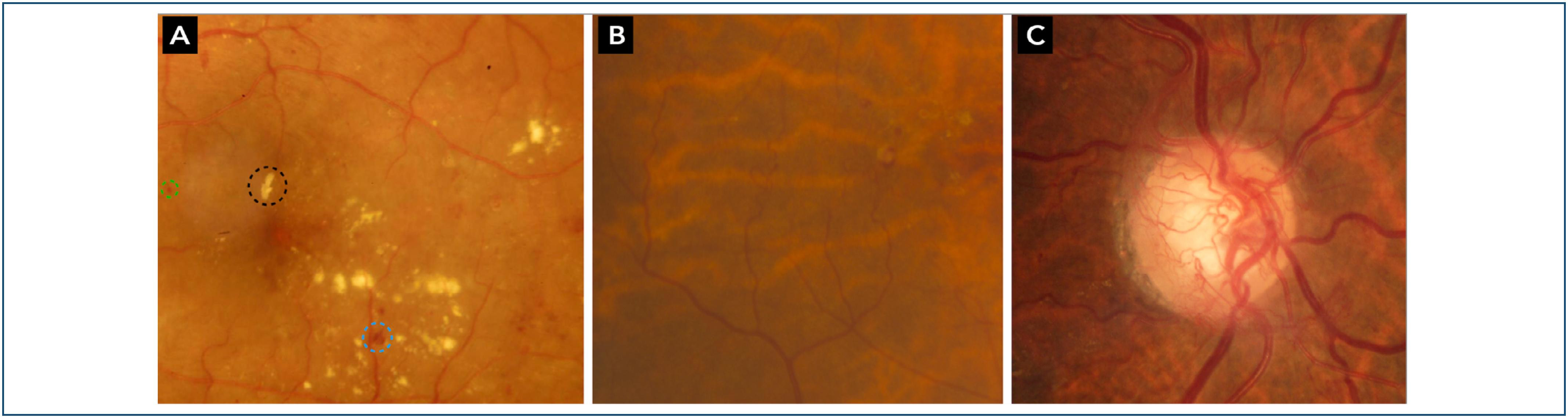
The current classification of DR is based on clinical and imaging findings (Figure 1), with the International Clinical Diabetic Retinopathy (ICDR) system being the gold standard in clinical research (Table 1). This system evaluates seven standard retinal photographic fields to provide a detailed assessment.(8,9)

Color fundus photographs illustrating key features of diabetic retinopathy. (A) shows microaneurysms (green circle), intraretinal hemorrhages (blue circle) and hard exudates (black circle); (B) highlights intraretinal microvascular abnormalities; (C) displays neovascularization of the optic disc.
Clinical classification of diabetic retinopathy according to severity. The progression from mild non-proliferative to proliferative diabetic retinopathy reflects increasing microvascular damage and risk of vision loss. Criteria are based on fundoscopic findings and guide the timing of referral and therapeutic intervention
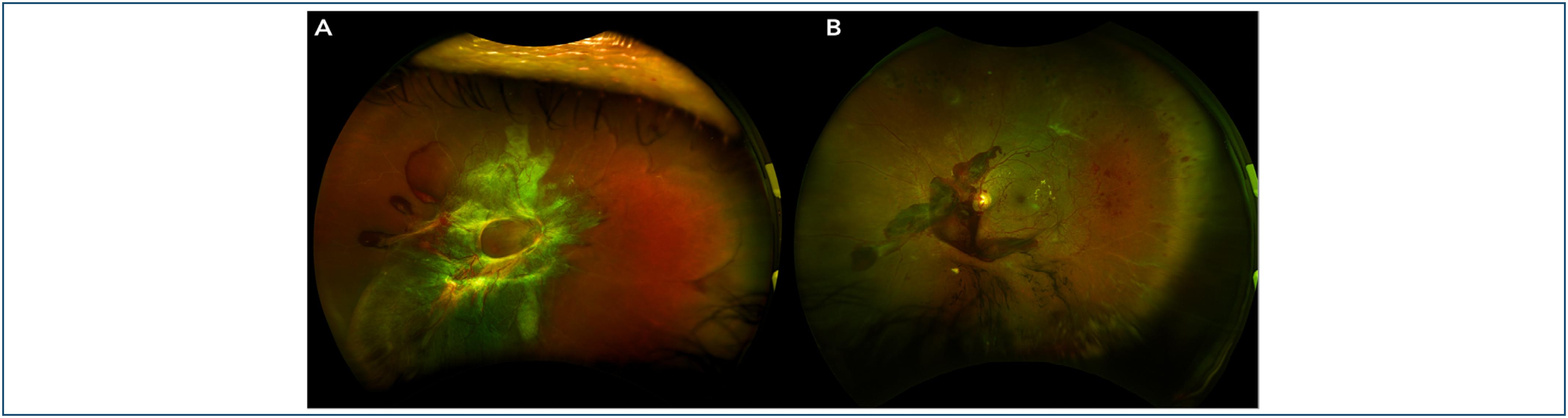
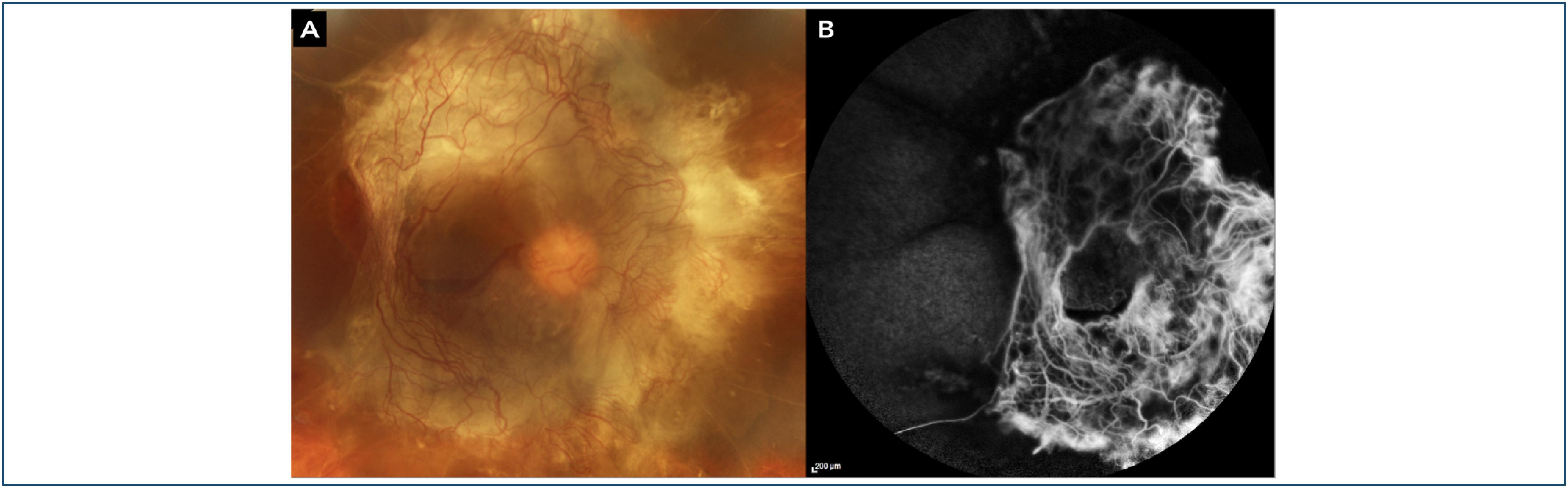
The advent of ultra-widefield (UWF) imaging has challenged the limitations of the ETDRS seven-field assessment (Figure 2). Comparative studies have shown that, although discrepancies in severity grading occur in less than 6% of cases, UWF imaging can reveal vision-threatening lesions such as neovascularization and pre-retinal hemorrhages located in the peripheral retina, beyond the ETDRS fields.(10)

Ultra-widefield fundus images illustrating bilateral high-risk proliferative diabetic retinopathy. The right eye (A) presents a complex tractional retinal detachment, while the left eye (B) shows a pre-retinal hemorrhage.
Diabetic macular edema: pathophysiology, classification, and relationship with diabetic retinopathy stages
Diabetic macular edema is the leading cause of vision loss in patients with DM and may occur at any stage of DR, whether non-proliferative or proliferative. The pathophysiology of DME is multifactorial, involving breakdown of the blood-retinal barrier, oxidative stress, chronic inflammation, vascular dysfunction, and neurodegeneration.(11,12)
Diabetic macular edema can be classified according to different criteria. Morphologically, it is described as focal when localized leakage areas are associated with microaneurysms, or diffuse when the macular thickening is more extensive and uniform. Another classification is based on central involvement, dividing DME into center-involving and non-center-involving types, as determined by optical coherence tomography (OCT) imaging. Additionally, DME may be described as cystoid when intraretinal fluid filling spaces are observed on OCT.(12)
The development of DME is closely related to DR progression. Although it can occur at any stage, its prevalence and severity increase with DR severity. Macular involvement, particularly when central, is associated with more significant visual loss.(11,12)
Critical importance of systematic screening and early diagnosis: current recommendations
Systematic DR screening is a key public health strategy to reduce the incidence of preventable blindness in people with DM. Organized screening programs allow for early identification of retinal lesions, risk stratification, and timely therapeutic interventions. According to the guidelines of the Sociedade Brasileira de Diabetes (SBD), ophthalmological examinations should begin five years after the diagnosis of type 1 diabetes and at the time of diagnosis for patients with type 2 diabetes, due to the possibility that DR may already be present in this population due to the frequent absence of precise onset of the disease.
Screening can be performed by clinical examination with pupillary dilation or through imaging methods such as fundus photography. The latter has gained prominence, especially when associated with telemedicine, allowing trained technicians to capture retinal images for remote analysis by ophthalmologists in specialized centers. Fundus photography combined with automated AI algorithms has proven effective and feasible, even in regions with a shortage of specialists. Artificial Intelligence use is in an advanced validation stage and has already shown high diagnostic performance in international studies.(9)
The recommended screening interval is based on the DR stage. For patients with no DR or mild NPDR, annual exams are sufficient. For moderate or advanced cases, the interval should be reduced to six months or less, as determined by clinical judgment. Patients with PDR or center-involving DME should be referred immediately to a specialist for treatment initiation.(9)
MANAGEMENT OF NON-PROLIFERATIVE DIABETIC RETINOPATHY
Optimized systemic control as a therapeutic foundation: glycemic, blood pressure, and lipid targets
Optimized systemic control is a cornerstone in managing non-proliferative diabetic retinopathy (NPDR), as demonstrated by landmark clinical trials such as DCCT/EDIC, UKPDS, and ACCORD-Eye. Intensive glycemic control, particularly maintaining a glycated hemoglobin (HbA1c) below 7%, significantly reduces the risk of DR onset and progression. In the DCCT and its long-term follow-up EDIC, patients with type 1 diabetes who received intensive glycemic treatment had up to 76% reduction in DR progression over 6.5 years, with sustained benefits despite later convergence of HbA1c levels—an effect known as "metabolic memory". Likewise, the UKPDS demonstrated that 0.9% reduction in HbA1c among individuals with type 2 diabetes led to 25% decrease in microvascular complications, including DR. Furthermore, tight blood pressure control led to 37% reduction in the risk of vision-threatening events.(13,14)
However, rapid and intensive glycemic control—particularly in patients with poor baseline metabolic control or advanced retinopathy—has been associated with early worsening of DR. This paradoxical effect, first observed in the DCCT and confirmed in subsequent studies such as ACCORD-Eye, is thought to result from abrupt changes in retinal blood flow, oxygenation, and osmotic gradients, potentially exacerbating ischemic and inflammatory responses in the retina. Therefore, glycemic targets should be approached gradually in high-risk individuals, with close ophthalmologic monitoring to mitigate the risk of early retinal deterioration during initial metabolic adjustment.(13,15)
In addition to glycemia and blood pressure, lipid control has emerged as an important modifiable factor in DR progression. Fenofibrate, a peroxisome proliferator-activated receptor-alpha (PPAR-α) agonist with lipid-modulating and anti-inflammatory effects, has shown consistent retinal protective effects across multiple trials. The FIELD study (16) demonstrated a reduction in the need for laser therapy for both proliferative DR and macular edema in patients with type 2 diabetes treated with fenofibrate, regardless of baseline lipid levels. Similarly, the ACCORD-Eye study found that fenofibrate added to statin therapy reduced the risk of DR progression by 40% over four years, supporting its retinal benefit.
Most recently, the LENS trial (2024) further substantiated the protective role of fenofibrate in a real-world population enrolled in the Scottish Diabetic Eye Screening program. Among 1,151 participants with early, non-referable DR or maculopathy, those randomized to receive 145 mg of fenofibrate daily experienced a 27% reduction in the composite endpoint of progression to referable disease or need for ocular treatment (hazard ratio [HR]: 0.73; 95% of confidence interval [95%CI] 0.58-0.91). The drug also halved the risk of developing macular edema and showed favorable effects on overall DR progression, without significant impact on visual acuity or quality of life. These findings reinforce the role of fenofibrate as a systemic adjunct in the early stages of DR, especially in patients not yet requiring ophthalmologic intervention.(17)
Mild to moderate non-proliferative diabetic retinopathy: strategy of active surveillance and systemic optimization
The management of mild to moderate NPDR primarily involves active surveillance paired with rigorous systemic control. At these early stages, patients are generally asymptomatic, and the main recommendation is periodic monitoring to detect early progression.
This conservative strategy is supported by evidence indicating that early invasive or pharmacological interventions, such as anti-VEGF injections, are not indicated for mild to moderate NPDR, except when center-involving DME is present.(6,12,18)
Severe and very severe non-proliferative diabetic retinopathy
Panretinal photocoagulation: indications based on ETDRS, optimal timing (early versus deferred), controversies, and current role
Panretinal photocoagulation (PRP) is traditionally reserved for PDR but plays a role in severe and very severe NPDR, although this remains controversial. The ETDRS showed that early PRP in eyes with very severe NPDR reduced the risk of progression to high-risk PDR, particularly in cases with poor follow-up reliability or imminent loss to follow-up. However, it also demonstrated that early PRP in NPDR did not confer a significant visual benefit over deferred treatment and was associated with adverse effects, including reduced visual field and impaired dark adaptation.(4,18,19)
Thus, treatment decisions should be individualized, especially in public health systems with limited access and adherence.
Intravitreal anti-vascular endothelial growth factor therapy: evidence for prevention of progression to proliferative diabetic retinopathy and development of diabetic macular edema
Intravitreal anti-VEGF therapy has been studied as a means of preventing progression from NPDR to PDR and the onset of DME, even in the absence of visual symptoms. Studies such as PANORAMA, Protocol W, and Protocol V(20, 21, 22) have provided strong evidence.
The PANORAMA trial evaluated aflibercept in patients with severe to very severe NPDR, showing 79% reduction in progression to PDR or DME compared to observation, with sustained benefit up to 100 weeks.(20) Protocol W, conducted by the DRCR Retina Network, demonstrated that early treatment with aflibercept in patients with moderate to severe NPDR without baseline DME significantly reduced the risk of vision-threatening complications, including progression to proliferative DR and the need for vitrectomy. These findings support the role of anti-VEGF therapy as a preventive strategy in carefully selected patients at high risk of disease progression.(21)
In contrast, Protocol V evaluated patients with center-involving DME and good baseline visual acuity. The study found no meaningful advantage of immediate anti-VEGF treatment over observation with deferred therapy. These results emphasize the importance of individualized treatment decisions, suggesting that close monitoring may be a safe and effective approach for patients with preserved vision at presentation.(22)
Comparative analysis: panretinal photocoagulation versus anti-vascular endothelial growth factor in severe non-proliferative diabetic retinopathy
In severe NPDR, the choice between PRP and anti-VEGF must balance efficacy, safety, cost-effectiveness, and patient adherence. Anti-VEGF requires frequent, costly injections and visits, while PRP, though less protective of central vision and peripheral field, often requires only one or a few sessions, favoring adherence and lower cost.(18,21,23)
MANAGEMENT OF PROLIFERATIVE DIABETIC RETINOPATHY
Panretinal photocoagulation
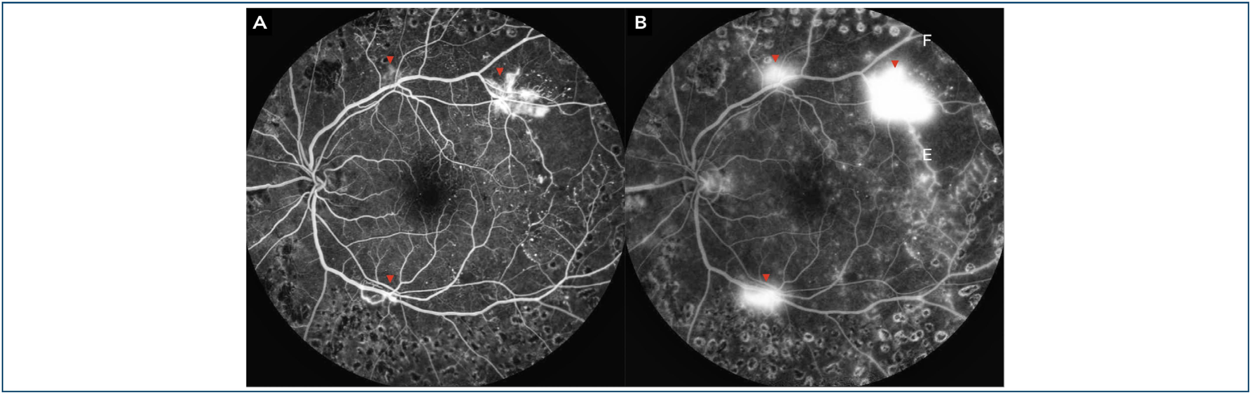
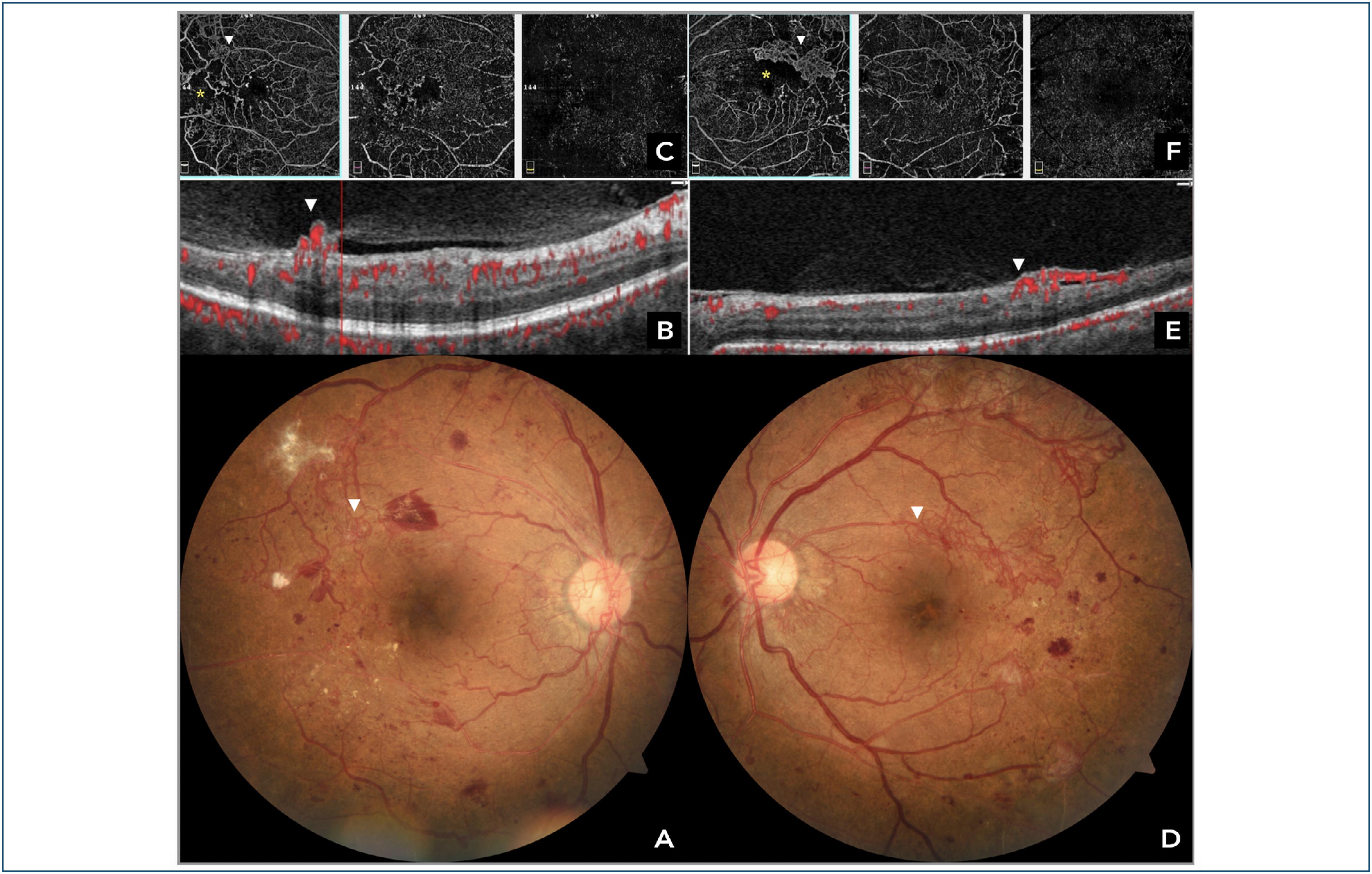
PRP remains a mainstay treatment for PDR (Figures 3 and 4) and has been considered the historical standard since the Diabetic Retinopathy Study (DRS), which demonstrated a 50% reduction in the risk of severe vision loss in eyes treated with PRP compared to observation.(24) The ETDRS further supported early PRP benefits in high-risk PDR cases. The technique involves applying laser burns to the peripheral ischemic retina to reduce the angiogenic stimulus. Despite its proven efficacy, PRP is associated with adverse effects, including peripheral visual field loss, impaired night vision, and the potential exacerbation of macular edema.(4)
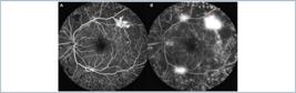
Fluorescein angiography showing mid-phase (A) and late-phase (B) images, highlighting characteristic leakage from neovascularization elsewhere (red triangle).
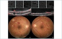
Color fundus photographs (A, D) showing proliferative diabetic retinopathy with neovascularization elsewhere (white triangle). Corresponding optical coherence tomography angiography B-scans (B, E) demonstrate the decorrelation signal and internal limiting membrane disruption caused by the NVE. Structural optical coherence tomography-angiography images (C, F) depict the NVE and associated areas of capillary nonperfusion (yellow asterisk).
Nevertheless, PRP continues to play an essential role, particularly in patients with poor adherence to continuous intravitreal therapies.
Intravitreal anti-vascular endothelial growth factor therapy as primary or adjunctive option
Anti-vascular endothelial growth factor monotherapy
Intravitreal anti-VEGF agents have emerged as effective alternatives to PRP for PDR. The DRCR.net Protocol S showed that ranibizumab was non-inferior to PRP in terms of visual acuity over two years, with a lower risk of developing DME and better preservation of visual field.(25) The CLARITY study confirmed these findings, demonstrating superior efficacy of aflibercept compared to PRP for visual improvement and neovascular regression.(26)
Treatment regimens vary and include monthly loading doses followed by PRN (pro re nata) or Treat-and-Extend maintenance. Among the available agents, bevacizumab, ranibizumab, and aflibercept are all effective, with aflibercept offering superior outcomes in patients with worse baseline visual acuity.(27)
Combination therapy (anti-vascular endothelial growth factor + panretinal photocoagulation)
Combining anti-VEGF with PRP has shown advantages in specific subgroups, particularly those at risk of treatment discontinuation. Initial anti-VEGF therapy facilitates rapid neovascular regression, allowing for more targeted and less aggressive PRP. This strategy is especially beneficial in cases with vitreous hemorrhage or optic disc neovascularization, where prompt angiogenic suppression is required.(6)
Advantages and disadvantages: anti-vascular endothelial growth factor versus panretinal photocoagulation
Anti-VEGF agents have emerged as a frontline treatment option in proliferative diabetic retinopathy (PDR), offering not only superior anatomical outcomes but also improved visual acuity and preservation of peripheral vision when compared to traditional laser therapy. These benefits are particularly evident in patients with concurrent DME, where anti-VEGF therapy addresses both situations simultaneously. Additionally, studies have shown a lower incidence of vitrectomy and neovascular complications with anti-VEGF treatment, reinforcing its efficacy as a disease-modifying intervention.(25,26)
However, the success of anti-VEGF therapy is highly dependent on patient adherence to a regimen of frequent intravitreal injections and long-term follow-up, which may pose challenges in real-world settings. The cumulative financial burden—both for healthcare systems and for patients—can be substantial, particularly in countries with limited access to biologics or reimbursement barriers.
On the other hand, PRP, a time-tested and widely available intervention, requires significantly fewer clinical visits and has a lower overall cost. While PRP does not typically improve central visual acuity and may result in peripheral visual field loss, night vision impairment, and exacerbation of DME in some cases, it remains an effective strategy for inducing regression of retinal neovascularization and reducing the risk of severe vision loss. In healthcare settings where patient compliance, access to specialized care, or logistical support are limited, PRP continues to play a critical role in managing PDR.(25,26)
Thus, the choice between anti-VEGF therapy and PRP should be individualized, balancing the patient's clinical profile, visual demands, systemic comorbidities, and the capacity for sustained treatment adherence within the healthcare system.
Pars plana vitrectomy for complicated proliferative diabetic retinopathy
Classical and Contemporary Indications
Pars plana vitrectomy (PPV) is indicated for complicated PDR cases, such as dense and persistent vitreous hemorrhage, tractional retinal detachment with or without rhegmatogenous component, tractional macular edema, and extensive fibrovascular membranes. The Diabetic Retinopathy Vitrectomy Study (DRVS) demonstrated the visual benefits of early PPV in eyes with recent vitreous hemorrhage, especially in patients with type 1 diabetes.(28)
Timing of surgical intervention
The optimal timing for PPV depends on vitreous opacity severity and risk of permanent retinal damage. DRVS findings showed that early PPV (within six months) resulted in better final visual acuity compared to that of patients with bilateral vitreous hemorrhage and no useful vision.(28) Factors such as glycemic control, cardiovascular stability, and tractional detachment extent including or close to the macula influence the surgical decision.
Advanced surgical techniques
Advances in surgical technology have improved PPV safety and efficacy using smaller-gauge instruments (23G, 25G, 27G), reducing operating time and postoperative inflammation. Dyes assist in identifying epiretinal membranes (ERM) and the internal limiting membrane. Intraoperative moderate intensity endolaser is widely used, and preoperative anti-VEGF injections decrease active neovascularization, facilitating dissection and minimizing intraoperative bleeding.(6)
The development of 3D electronic visualization screen systems, per operative OCT, high-speed cutters (up to 30,000 cpm), and efficient fluidics has expanded surgical indications, including previously inoperable cases, and supports safer and faster visual recovery.(29)
Visual and anatomic outcomes, complications, and postoperative management
Visual outcomes after PPV in complicated PDR are generally favorable when performed before macular involvement. Complications include recurrent hemorrhage, secondary rhegmatogenous retinal detachment, and ocular hypertension. Postoperative care requires close monitoring of intraocular pressure, retinal integrity, and adjunctive treatments (anti-VEGF or PRP) when indicated. Visual rehabilitation may be prolonged, especially in cases with residual edema, retinal atrophy or significative macular damage.
MANAGEMENT OF DIABETIC MACULAR EDEMA
Diagnostic assessment and monitoring
The diagnostic assessment of DME is primarily based on OCT, the gold standard for measuring central macular thickness and identifying structural abnormalities such as intraretinal cystoid spaces, neurosensory detachment, and vitreomacular traction (VMT). Fluorescein angiography (FA) remains useful for evaluating capillary perfusion and identifying leakage sites, although its use has been partially replaced by OCT-angiography (OCT-A), a non-invasive technique that provides high-resolution images of the retinal vascular plexuses. Technological advancements have made OCT-A increasingly integrated into routine clinical practice.
The combination of these tools allows for precise characterization of DME phenotypes and longitudinal therapeutic response monitoring. The presence of macular ischemia, often identified by enlargement of the foveal avascular zone (FAZ) on FA or OCT-A, serves as a prognostic marker for poor functional outcomes.(30)
First-line anti-vascular endothelial growth factor therapy
Intravitreal anti-VEGF agents (bevacizumab, ranibizumab, aflibercept and faricimab) are the first-line treatment for center-involving DME with decreased visual acuity. The RISE and RIDE studies demonstrated the efficacy and safety of ranibizumab, showing significant improvements in visual acuity and macular thickness.(31) Similarly, the VIVID and VISTA trials validated the use of aflibercept, particularly in patients with poorer baseline vision.(32)
DRCR.net Protocol T compared all three agents and found that aflibercept offered superior outcomes in patients with baseline vision of 20/50 or worse, while the agents were comparable in other subgroups.(27) Treatment regimens include monthly loading doses followed by PRN or Treat-and-Extend approaches.
Intravitreal corticosteroids
Intravitreal corticosteroids are indicated in cases that are refractory to anti-VEGF, pseudophakic patients, or when an inflammatory component is prominent. The dexamethasone implant (Ozurdex®) showed visual benefits and edema control lasting 4 to 6 months in the MEAD study.(33) The fluocinolone acetonide implant (Iluvien) was evaluated in the FAME study and provided sustained efficacy for up to 36 months in chronic cases.(34) Triamcinolone acetonide, although effective, is off-label and associated with a higher risk of intraocular pressure elevation and cataract formation. DRCR.net Protocol U showed that combining anti-VEGF with dexamethasone improves anatomy but not visual outcomes.(35)
Focal/grid laser photocoagulation
Laser photocoagulation was a mainstay treatment for DME before the anti-VEGF era. The ETDRS demonstrated reduced risk of visual loss using focal laser in clinically significant macular edema. Today, laser plays an adjunctive role, indicated for non-center-involving DME, isolated leaking microaneurysms, or as a complement to other therapies. DRCR.net Protocol I showed that combining laser with ranibizumab offered no additional benefit over anti-VEGF monotherapy, supporting its selective use.(24)
Diabetic macular edema without center involvement or with good visual acuity
In patients with non-center-involving DME and good visual acuity, observation may be a safe and effective option. DRCR.net Protocol V demonstrated that active monitoring without immediate treatment did not result in significant visual decline over two years, representing a valid alternative to early intervention.(36) Treatment decisions should consider anatomical features, follow-up feasibility, and progression risk.
Surgical approach in diabetic macular edema
Pars plana vitrectomy plays an important role in the management of selected cases of DME, particularly when there is a prominent mechanical component contributing to retinal thickening. Surgical intervention is primarily indicated in the presence of VMT, taut or thickened ERM, or in cases refractory to optimized pharmacologic therapy, including repeated anti-VEGF or corticosteroid injections. In such scenarios, PPV can alleviate the anteroposterior or tangential tractional forces exerted on the macula, promoting retinal reattachment and improved oxygenation of the inner retina.(37)
Several studies have shown that the surgical release of vitreomacular adhesion and peeling of ERM can lead to significant anatomical improvement and, in select cases, meaningful functional recovery—particularly in patients where traction is the predominant pathogenic mechanism. However, visual prognosis depends on multiple factors, including the chronicity of the edema, the degree of photoreceptor damage, and integrity of the ellipsoid zone on spectral-domain OCT (SD-OCT). Therefore, surgical indication should be individualized and based on detailed multimodal imaging, with OCT findings playing a central role in evaluating the presence and extent of traction, foveal architecture, and surgical accessibility. The decision to proceed with PPV must also consider systemic factors such as glycemic control, ocular comorbidities, and the patient's overall visual potential.(37)
In summary, while PPV is not the first-line therapy for most cases of DME, it remains a valuable option in cases with tractional pathology or treatment-resistant edema, offering anatomical stability and, in selected patients, functional improvement when pharmacologic options are insufficient.
Biomarkers and predictive factors for treatment response
Optical coherence tomography-derived biomarkers, such as large intraretinal cysts, neurosensory detachment, and intraretinal hyperreflective foci, have been associated with a suboptimal anti-VEGF response.(38) Additionally, thickening of the inner plexiform layer and loss of the ellipsoid zone are correlated with a poorer visual prognosis. Emerging studies suggest that intraocular inflammatory profiles and cytokine levels, including VEGF, may help tailor personalized treatment strategies.
Treatment algorithm and management of refractory or suboptimal cases
Managing refractory DME requires an individualized approach. Patients unresponsive after 3 to 6 anti-VEGF injections may benefit from switching agents or adding corticosteroids.(35) Anatomical assessment with OCT and identification of tractional or structural abnormalities guide the decision for surgical intervention. An effective treatment algorithm should consider DME phenotype, systemic comorbidities, and treatment adherence, integrating pharmacologic, surgical, and intensive follow-up strategies.(12)
EMERGING THERAPIES AND PERSPECTIVES
Novel pharmacological agents
Several novel pharmacological agents are being developed to improve the durability and efficacy of DR and DME treatment. Faricimab stands out as a bispecific antibody targeting both VEGF-A and angiopoietin-2 (Ang-2), approved for intravitreal use. Clinical trials such as YOSEMITE and RHINE demonstrated their non-inferiority to aflibercept with longer dosing intervals, offering promise to reduce treatment burden.(39)
Sustained drug delivery systems
Sustained-release systems aim to reduce the frequency of intravitreal injections. The Port Delivery System (PDS), a surgically implanted, refillable reservoir delivering ranibizumab, showed non-inferior results to monthly injections in the Archway trial.(40) Additionally, technologies involving nanoparticles and hydrogels are under investigation for controlled, less invasive drug release over weeks or months.
Non-invasive or minimally invasive therapeutic approaches
Non-invasive and minimally invasive therapies, such as eye drops and oral medications, are in development for DR and DME. Oral tyrosine kinase inhibitors and PPAR agonists have demonstrated effects on inflammatory and vascular pathways in preclinical and early human studies.(41) However, phase III trials are needed to confirm efficacy and safety.
Gene therapy
Gene therapy represents an innovative strategy to treat retinal diseases by enabling prolonged expression of therapeutic proteins following a single administration. Approaches under investigation involve subretinal or intravitreal delivery of viral vectors for sustained anti-VEGF expression. Early studies with RGX-314 and ADVM-022 have shown safety and durable protein expression, though challenges remain regarding immunogenicity, response variability, and target tissue access.(42)
Artificial Intelligence and telemedicine
The integration of AI into teleophthalmology has emerged as a promising strategy to expand DR screening, particularly in regions with limited access to ophthalmic care. AI-assisted systems can automate the analysis of retinal fundus images, identifying referable DR and vision-threatening disease with high sensitivity and specificity. In conjunction with telemedicine, these tools enable the remote capture, triage, and interpretation of retinal images, reducing the need for in-person evaluation and increasing screening coverage (Figure 5). Food and Drug Administration (FDA)-approved systems such as IDx-DR, EyeArt, and AEYE demonstrate that AI-based screening can be effectively implemented in clinical settings, with performance comparable to human graders when detecting more-than-mild DR.
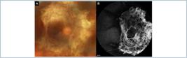
Color fundus image (A) acquired using a portable handheld camera (Eyer, Phelcom, Brazil), and corresponding Artificial Intelligence interpretation with a map overlay (B).
Despite these advancements, significant challenges remain in the deployment of AI-assisted telemedicine programs. Many studies reviewed focus on high-income countries, with limited representation from low- and middle-income countries (LMICs), where the need for scalable screening tools is greatest. Moreover, the lack of transparency regarding dataset composition, model architecture, and bias mitigation strategies raises concerns about generalizability and fairness. Biases in training data can result in inequitable performance across demographic groups, and the absence of post-deployment monitoring may allow these disparities to persist. Effective implementation requires not only technical validation but also ethical oversight, ongoing recalibration, and integration with existing healthcare workflows.(43)
Economic evaluations suggest that AI-assisted DR screening can reduce costs compared to traditional human-based approaches, particularly when using semi-automated models. However, the cost-effectiveness of these systems depends heavily on contextual factors such as greater salaries, IT infrastructure, and screening uptake. Moreover, the success of implementation relies on adequate internet access, training of local personnel, and patient engagement with digital platforms—factors that may vary widely across healthcare systems. Therefore, broader adoption of AI in telemedicine must be guided by robust evidence, inclusive development practices, and policies that promote equitable access to screening and treatment.(44)
SPECIAL CONSIDERATIONS IN CLINICAL PRACTICE
Management of diabetic retinopathy and diabetic macular edema during pregnancy
Pregnancy increases the risk of DR progression in women with preexisting diabetes, making ophthalmologic screening in the first trimester essential, followed by quarterly or severity-based monitoring. Although tight glycemic control is beneficial for the fetus, it may transiently worsen DR, requiring specialized vigilance.(45) The use of anti-VEGF agents is generally contraindicated because of limited safety data for the fetus. In severe cases, corticosteroids may be cautiously considered.
Specific aspects of diabetic retinopathy in pediatric and young adult patients (type 1 diabetes)
Young patients with type 1 diabetes are at cumulative risk of developing DR over their lifetime, typically after five years of disease duration. Strict glycemic control from childhood, as demonstrated in the DCCT/EDIC study, is crucial to delay DR onset and progression.(46) However, adherence to ophthalmologic follow-up in this population can be challenging and incorporating psychosocial and educational support is key to effective care.
Interaction between cataract surgery and diabetic retinopathy/ diabetic macular edema
Cataract surgery in patients with DR or DME requires careful planning due to the risk of postoperative retinal decompensation. Phacoemulsification-induced inflammation may exacerbate DME, particularly in moderate to severe NPDR. Studies indicate that prophylactic intravitreal anti-VEGF or corticosteroids in the perioperative period can reduce this risk and improve both anatomical and visual outcomes.(47)
Importance of a multidisciplinary approach
The management of DR and DME extends beyond the domain of ophthalmology and requires coordinated, multidisciplinary care. Optimal control of disease progression is closely tied to systemic metabolic regulation, which underscores the essential role of endocrinologists in achieving and maintaining glycemic control, as well as in adjusting antidiabetic therapies based on disease severity and comorbidities. In patients with concurrent diabetic nephropathy, nephrologists are crucial for managing fluid balance, blood pressure, and renal function—factors that significantly influence retinal vascular homeostasis. Cardiologists contribute by addressing systemic hypertension, dyslipidemia, and macrovascular complications that are known risk factors for the progression of DR. Furthermore, primary care physicians and diabetes educators play a central role in coordinating patient follow-up, reinforcing lifestyle modifications, and ensuring long-term adherence to treatment plans.(14,15,48)
Growing evidence supports that interprofessional collaboration improves not only visual outcomes but also systemic disease markers, enhances patient satisfaction, and increases adherence to both ophthalmologic and systemic treatments. Studies have shown that patients managed within integrated care networks—where communication among specialties is standardized—are more likely to receive timely referrals, benefit from early detection of retinopathy, and demonstrate improved control of HbA1c, blood pressure, and lipid profiles. Therefore, the implementation of structured care pathways in diabetes centers, involving ophthalmologists as part of a broader chronic disease management team, is essential to address the multifactorial nature of DR and DME. This holistic approach is particularly important in preventing irreversible vision loss and reducing the overall burden of diabetes-related complications.(14,15,48)
CONCLUSION
Summary of current evidence-based recommendations for the management of diabetic retinopathy and diabetic macular edema
Current recommendations for managing DR and DME are supported by evidence from major clinical trials. For mild to moderate NPDR, close surveillance with strict systemic control is recommended. In cases of proliferative DR, both PRP and anti-VEGF therapies are effective, with the choice depending on individual patient profiles. These therapies may also be indicated for patients with severe or very severe NPDR. For center-involving DME, anti-VEGF agents are the first-line treatment, while corticosteroids are reserved for refractory cases or when anti-VEGF is contraindicated.
Major ongoing challenges
Despite therapeutic advances, several challenges remain. These include low adherence to treatment, limited access to intravitreal therapies in underserved regions, and high medication costs. Additionally, approximately 40% of DME patients do not respond satisfactorily to initial anti-VEGF therapy, requiring alternative approaches.(27) Educational initiatives, population-based screening strategies, and the incorporation of technologies such as AI may help overcome these barriers.
Future directions to optimize visual outcomes and quality of life in diabetic retinopathy
The future of DR and DME management is moving toward longer-lasting, personalized, and less invasive therapies, along with the integration of AI to predict risk and treatment response. Gene therapy and long-acting delivery systems promise to reduce treatment burden, while multidisciplinary, patient-centered care approaches can improve adherence and functional outcomes.(38) Investment in public health policies and equitable access to treatment will also be essential.
REFERENCES
- 1 Teo ZL, Tham YC, Yu M, Chee ML, Rim TH, Cheung N, et al. Global prevalence of diabetic retinopathy and projection of Burden through 2045: Systematic review and meta-analysis. Ophthalmology. 2021;128(11):1580-91.
- 2 Curran K, Peto T, Jonas JB, Friedman D, Kim JE, Leasher J, et al. Global estimates on the number of people blind or visually impaired by diabetic retinopathy: a meta-analysis from 2000 to 2020. Eye. 2024;38(11):2047-57.
- 3 Muzy J, Campos MR, Emmerick I, da Silva RS, de Andrade Schramm JM. Prevalence of diabetes mellitus and its complications and characterization of healthcare gaps based on triangulation of studies. Cad Saude Publica. 2021;37(5).
- 4 Cheung N, Mitchell P, Wong TY. Diabetic retinopathy. Lancet. 2010;376(9735):124-36.
- 5 Whitehead M, Wickremasinghe S, Osborne A, Van Wijngaarden P, Martin KR. Diabetic retinopathy: a complex pathophysiology requiring novel therapeutic strategies. Expert Opin Biol Ther. 2018;18(12):1257-70.
- 6 Wang W, Lo AC. Diabetic retinopathy: Pathophysiology and treatments. Int J Mol Sci. 2018;19(6):1816.
- 7 Tan TE, Wong TY. Diabetic retinopathy: Looking forward to 2030. Front Endocrinol (Lausanne). 2023;13:1077669.
- 8 Aiello LM. Perspectives on diabetic retinopathy. Am J Ophthalmol. 2003;136(1):122-35.
-
9 Malerbi F, Andrade R, Morales P, Travassos S, Rodacki M, Bertoluci M. Manejo da retinopatia diabética. Diretriz Oficial da Sociedade Brasileira de Diabetes; 2023. DOI: 10.29327/557753.2022-17
» https://doi.org/10.29327/557753.2022-17 - 10 Attiku Y, Nittala MG, Velaga SB, Ramachandra C, Bhat S, Solanki K, et al. Comparison of diabetic retinopathy severity grading on ETDRS 7-field versus ultrawide-field assessment. Eye (Basingstoke). 2023;37(14):2946-9.
- 11 Das A, McGuire PG, Rangasamy S. Diabetic Macular edema: pathophysiology and novel therapeutic targets. Ophthalmology. 2015;122(7):1375-94.
- 12 Kuroiwa DAK, Malerbi FK, Regatieri CVS. New insights in resistant diabetic macular edema. Ophthalmologica. 2021;244(6):485-94.
- 13 Genuth S. The UKPDS and its global impact. Diabet Med. 2008;25 Suppl 2:57-62.
- 14 Gubitosi-Klug RA, Lachin JM, Backlund JY, Lorenzi GM, Brillon DJ, Orchard TJ. Intensive diabetes treatment and cardiovascular outcomes in type1 diabetes: The DCCT/EDIC study 30-year follow-up. Diabetes Care. 2016;39(5):686-93.
- 15 Frank RN. Systemic therapies for diabetic retinopathy: the ACCORD eye study. Ophthalmology. 2014;121(12):2295-6.
- 16 Keech A, Simes RJ, Barter P, Best J, Scott R, Taskinen MR, Forder P, Pillai A, Davis T, Glasziou P, Drury P, Kesäniemi YA, Sullivan D, Hunt D, Colman P, d’Emden M, Whiting M, Ehnholm C, Laakso M; FIELD study investigators. Effects of long-term fenofibrate therapy on cardiovascular events in 9795 people with type 2 diabetes mellitus (the FIELD study): randomised controlled trial. Lancet. 2005;366(9500):1849-61. Erratum in: Lancet. 2006;368(9545):1420. Erratum in: Lancet. 2006;368(9545):1415.
- 17 Bain SC, Klufas MA, Ho A, Matthews DR. Worsening of diabetic retinopathy with rapid improvement in systemic glucose control: A review. Diabetes Obes Metab. 2019;21(3):454-66.
- 18 Danek D, Larsen B, Anderson-Nelson S. Non-proliferative diabetic retinopathy. Disease-a-Month. 2021;67(5).
- 19 Early photocoagulation for diabetic retinopathy. ETDRS report number 9. Ophthalmology. 1991;98(5):766-85.
- 20 Brown DM, Wykoff CC, Boyer D, Heier JS, Clark WL, Emanuelli A, et al. Evaluation of intravitreal aflibercept for the treatment of severe nonproliferative diabetic retinopathy. JAMA Ophthalmol. 2021;139(9):946.
- 21 Maturi RK, Glassman AR, Josic K, Antoszyk AN, Blodi BA, Jampol LM, et al. Effect of intravitreous anti-vascular endothelial growth factor vs sham treatment for prevention of vision-threatening complications of diabetic retinopathy. JAMA Ophthalmol. 2021;139(7):701.
- 22 Chew EY. Patients with good vision and diabetic macular edema involving the center of the macula. JAMA. 2019;321(19):1873.
- 23 Stevens GA, White RA, Flaxman SR, Price H, Jonas JB, Keeffe J, et al. Global prevalence of vision impairment and blindness. Ophthalmology. 2013;120(12):2377-84.
- 24 Photocoagulation for diabetic macular edema. Archives of Ophthalmology. 1985;103(12):1796.
- 25 Gross JG, Glassman AR, Jampol LM, Inusah S, Aiello LP, Antoszyk AN, et al. Panretinal photocoagulation vs intravitreous ranibizumab for proliferative diabetic retinopathy. JAMA. 2015;314(20):2137.
- 26 Sivaprasad S, Prevost AT, Vasconcelos JC, Riddell A, Murphy C, Kelly J, et al. Clinical efficacy of intravitreal aflibercept versus panretinal photocoagulation for best corrected visual acuity in patients with proliferative diabetic retinopathy at 52 weeks (CLARITY): a multicentre, single-blinded, randomised, controlled, phase 2b, non-inferiority trial. Lancet. 2017;389(10085):2193-203.
- 27 Diabetic Retinopathy Clinical Research Network; Wells JA, Glassman AR, Ayala AR, Jampol LM, Aiello LP, Antoszyk AN, Arnold-Bush B, Baker CW, Bressler NM, Browning DJ, Elman MJ, Ferris FL, Friedman SM, Melia M, Pieramici DJ, Sun JK, Beck RW. Aflibercept, bevacizumab, or ranibizumab for diabetic macular edema. N Engl J Med. 2015;372(13):1193-203.
- 28 Flynn HW, Chew EY, Simons BD, Barton FB, Remaley NA, Ferris FL. Pars plana vitrectomy in the early treatment Diabetic Retinopathy Study. Ophthalmology. 1992;99(9):1351-7.
- 29 Kantor P, Matonti F, Varenne F, Sentis V, Pagot-Mathis V, Fournié P, et al. Use of the heads-up NGENUITY 3D Visualization System for vitreoretinal surgery: a retrospective evaluation of outcomes in a French tertiary center. Sci Rep. 2021;11(1):10031.
- 30 Gill A, Cole ED, Novais EA, Louzada RN, de Carlo T, Duker JS, et al. Visualization of changes in the foveal avascular zone in both observed and treated diabetic macular edema using optical coherence tomography angiography. Int J Retina Vitreous. 2017;3(1):19.
- 31 Nguyen QD, Brown DM, Marcus DM, Boyer DS, Patel S, Feiner L, et al. Ranibizumab for diabetic macular edema. Ophthalmology. 2012;119(4):789-801.
- 32 Shaw J, Kerrison J. Intravitreal aflibercept for diabetic macular edema. Ophthalmology. 2014;121(7):A4.
- 33 Boyer DS, Yoon YH, Belfort R, Bandello F, Maturi RK, Augustin AJ, et al. Three-year, randomized, sham-controlled trial of dexamethasone intravitreal implant in patients with diabetic macular edema. Ophthalmology. 2014;121(10):1904-14.
- 34 Campochiaro PA, Brown DM, Pearson A, Chen S, Boyer D, Ruiz-Moreno J, et al. Sustained delivery fluocinolone acetonide vitreous inserts provide benefit for at least 3 years in patients with diabetic macular edema. Ophthalmology. 2012;119(10):2125-32.
- 35 Maturi RK, Glassman AR, Liu D, Beck RW, Bhavsar AR, Bressler NM, et al. Effect of adding dexamethasone to continued ranibizumab treatment in patients with persistent diabetic macular edema. JAMA Ophthalmol. 2018;136(1):29.
- 36 Baker CW, Glassman AR, Beaulieu WT, Antoszyk AN, Browning DJ, Chalam KV, et al. Effect of initial management with aflibercept vs laser photocoagulation vs observation on vision loss among patients with diabetic macular edema involving the center of the macula and good visual acuity. JAMA. 2019;321(19):1880.
- 37 Simunovic MP, Hunyor AP, Ho IV. Vitrectomy for diabetic macular edema: a systematic review and meta-analysis. Can J Ophthalmol. 2014;49(2):188-95.
- 38 Hwang HS, Chae JB, Kim JY, Kim DY. Association between hyperreflective dots on spectral-domain optical coherence tomography in macular edema and response to treatment. Invest Ophthalmol Vis Sci. 2017;58(13):5958-67.
- 39 Wykoff CC, Abreu F, Adamis AP, Basu K, Eichenbaum DA, Haskova Z, et al. Efficacy, durability, and safety of intravitreal faricimab with extended dosing up to every 16 weeks in patients with diabetic macular oedema (YOSEMITE and RHINE): two randomised, double-masked, phase 3 trials. The Lancet. 2022;399(10326):741-55.
- 40 Holekamp NM, Campochiaro PA, Chang MA, Miller D, Pieramici D, Adamis AP, et al. Archway randomized phase 3 trial of the port delivery system with ranibizumab for neovascular age-related macular degeneration. Ophthalmology. 2022;129(3):295-307.
- 41 Chandra S, Tan EY, Empeslidis T, Sivaprasad S. Tyrosine kinase Inhibitors and their role in treating neovascular age-related macular degeneration and diabetic macular oedema. Eye. 2023;37(18):3725-33.
- 42 Odio-Herrera M, Orozco-Loaiza G, Wu L. Gene therapy in diabetic retinopathy and diabetic macular edema: an update. J Clin Med. 2025;14(9):3205.
- 43 Abràmoff MD, Lavin PT, Birch M, Shah N, Folk JC. Pivotal trial of an autonomous AI-based diagnostic system for detection of diabetic retinopathy in primary care offices. NPJ Digit Med. 2018;1(1):39.
- 44 Nakayama LF, Zago Ribeiro L, Novaes F, Miyawaki IA, Miyawaki AE, de Oliveira JAE, et al. Artificial intelligence for telemedicine diabetic retinopathy screening: a review. Ann Med. 2023;55(2).
- 45 Chew EY, Mills JL, Metzger BE, Remaley NA, Jovanovic-Peterson L, Knopp RH, et al. Metabolic control and progression of retinopathy: the diabetes in early pregnancy study. Diabetes Care. 1995;18(5):631-7.
- 46 Diabetes Control and Complications Trial/Epidemiology of Diabetes Interventions and Complications Research Group; Lachin JM, Genuth S, Cleary P, Davis MD, Nathan DM. Retinopathy and nephropathy in patients with type 1 diabetes four years after a trial of intensive therapy. N Engl J Med. 2000 Feb 10;342(6):381-9. Erratum in: N Engl J Med 2000;342(18):1376.
- 47 Mohamed Q, Gillies MC, Wong TY. Management of diabetic retinopathy. JAMA. 2007;298(8):902.
- 48 Chous AP, Richer SP, Gerson JD, Kowluru RA. The Diabetes Visual Function Supplement Study (DiVFuSS). Br J Ophthalmol. 2016;100(2):227-34.