Efficacy of isolated treatment with Intense Pulsed Light applied directly to the eyelids of patients with Meibomian gland dysfunction: a retrospective cohort
ABSTRACT
Objective: To describe the clinical benefits of intense pulsed light applied directly to the eyelids of patients with Meibomian gland dysfunction and without glandular expression.
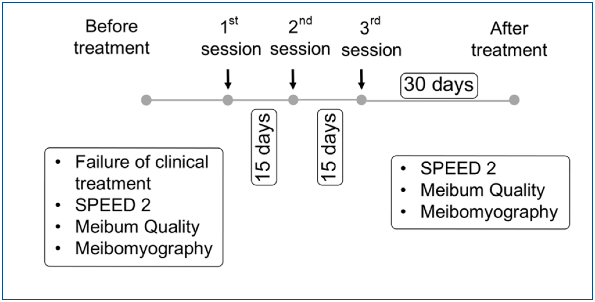
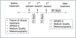
Methods: A paired, retrospective cohort involving 16 patients diagnosed with symptomatic Meibomian gland dysfunction according to the criteria of the 2011 International Workshop on Meibomian gland dysfunction and treated with Intense Pulsed Light. The patients received three biweekly sessions on the eyelids and with the Lumenis OPT M22™ equipment. Glandular expression was not performed and after 30 days, clinical follow-up was performed with the evaluation including SPEED 2 questionnaire, meibomigraphy and glandular assessment.
Results: SPEED 2 significantly improved in 75% (p < 0.001) and Meibum quality improved in 84.7% (p < 0.001) of patients. Glandular loss decreased from 27.1% to 22.5%, without statistical significance (p = 0.129), but with a moderate effect size (d = 0.452).
Conclusion: Our findings suggest that Intense Pulsed Light applied directly to the eyelids and without glandular expression is effective in the treatment of Meibomian gland dysfunction.
Keywords:
Intense pulsed light therapy; Meibomian gland; Dry eye syndromes; Treatment expectations
RESUMO
Objetivo: Descrever os benefícios clínicos da luz intensa pulsada (LIP) aplicada diretamente nas pálpebras de pacientes com disfunção da glândula meibomiana (MGD) e sem expressão glandular.
Métodos: Coorte retrospectiva pareada envolvendo 16 pacientes diagnosticados com MGD sintomática de acordo com os critérios do International Workshop on MGD de 2011 e tratados com LIP. Os pacientes receberam 03 sessões quinzenais nas pálpebras e com o equipamento Lumenis OPT M22. A expressão glandular não foi realizada e após 30 dias foi realizado acompanhamento clínico com o questionário SPEED 2, meibomigrafia e expressão glandular.
Resultados: O SPEED 2 melhorou significativamente em 75% (p<0,001) e a qualidade do Meibum em 84,7% (p<0,001). A perda glandular diminuiu de 27,1% para 22,5%, sem importância estatística (p=0,129), mas com tamanho de efeito moderado (d =0,452).
Conclusão: Nossos achados sugerem que a LIP aplicada diretamente nas pálpebras e sem expressão glandular é eficaz no tratamento da MGD.
Descritores:
Terapia de luz pulsada intensa; Glândulas tarsais; Síndromes do olho seco; Expectativas do tratamento
INTRODUCTION
Meibomian gland dysfunction (MGD) is a chronic, diffuse condition caused by obstruction of glandular orifices and alterations in meibum quantity and/or quality.(1) It is the primary cause of evaporative dry eye (EDE) disease, with prevalence varying depending on diagnostic criteria and region. In 2024, Chen et al. identified an MGD prevalence of 86% in patients with dry eye disease.(2,3)
Patients report symptoms such as burning, tearing, a sensation of a foreign body, and intermittent visual blurriness. Severe cases may lead to difficulties in everyday tasks, including reading, watching television, driving, and computer work, and may negatively affect quality of life and work performance.(4)
Current MGD treatments are divided into non-thermal (gland probing, topical/oral antibiotics, Manuka honey, emollient drops, therapeutic expression) and thermal (warm compresses, pulsed thermotherapy, and Intense Pulsed Light [IPL]).(5)
Intense Pulsed Light, initially used in dermatology for treating photoaging, rosacea, pigmented lesions, and telangiectasias, was found in 2002 by Toyos et al. to relieve symptoms of MGD in rosacea patients.(6-8) Studies confirm its efficacy in treating DED and MGD, typically applied around the eyelids in conjunction with gland expression.(9-11)
This study's primary objective is to describe the clinical benefits of IPL applied directly to the eyelids of patients with Meibomian gland dysfunction and without glandular expression.
METHODS
This non-concurrent, paired cohort study included all 46 MGD patients treated with IPL at Santa Luzia Eye Center in Guarabira between August 2019 and December 2020. It adhered to the Helsinki Declaration guidelines, and informed consent was obtained from all participants. The study was approved by the Research Ethics Committee of the Clementino Fraga Filho University Hospital and the Faculty of Medicine of the Universidade Federal do Rio de Janeiro on December 22, 2023 (approval no. 76359123.4.0000.5257).
Data extraction
All the data used were collected from information recorded in medical charts, and the information was entered into a clinical form specifically designed for the study.
The medical charts contained the following information: age, sex, meibography of the lower eyelids, quality of Meibomian secretion from 15 glands of the lower eyelid, results of the SPEED 2 questionnaire, NITBUT, and TMH. After applying the inclusion and exclusion criteria, 16 patients participated in the study.
To reduce confounding bias due to age, stratification by age group was employed (20 to 40 years, 41 to 60 years, 61 to 80 years, and ≥ 81 years).
Inclusion criteria
Patients were unresponsive to conservative treatment (lubricant eye drops, heat, and eyelid hygiene, omega-3 fatty acid supplementation) with symptomatic evaporative DED and MGD (SPEED 2 >2; NITBUT < 10 s; TMH >0.25 mm; gland loss > 25% on meibography or meibum quality classification ≥ 1, below 20 years of age and ≥ 2, above 20 years of age).
Exclusion criteria
Aqueous deficient or mixed DED (DEWS II, 2017), patients < 18 years, ongoing glaucoma drops, ocular surgeries within six months, eyelid disorders, prior DED treatments (IPL, glandular probing, pulsed thermotherapy), Fitzpatrick phototype > IV.
Procedure
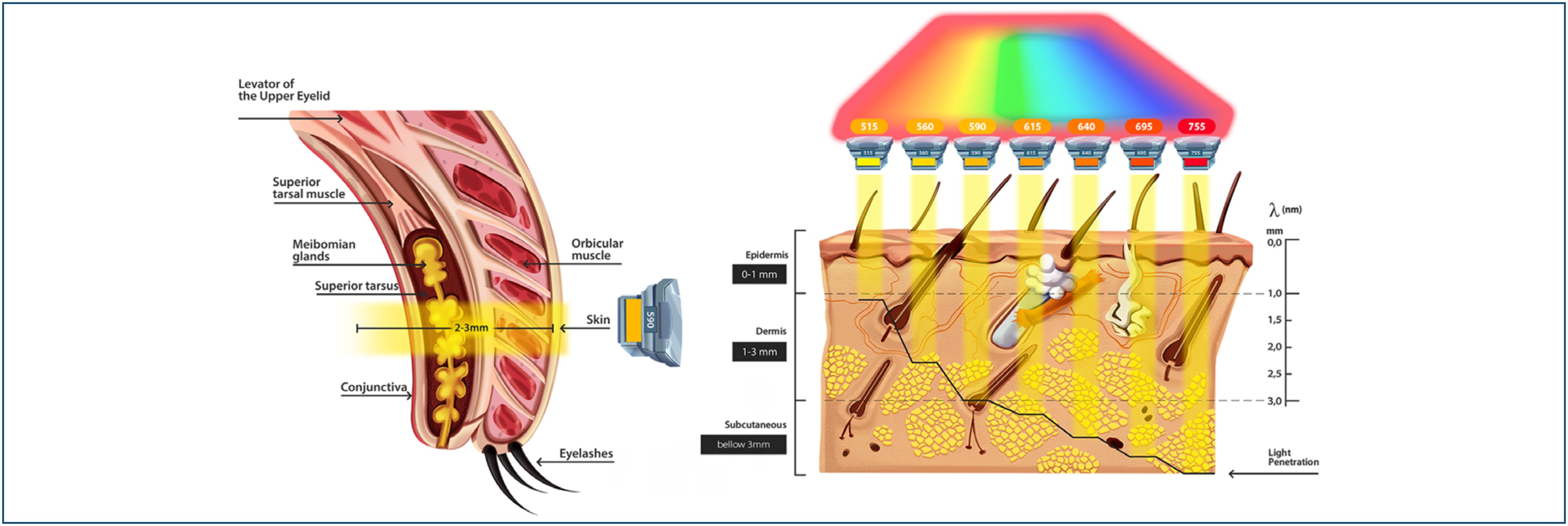
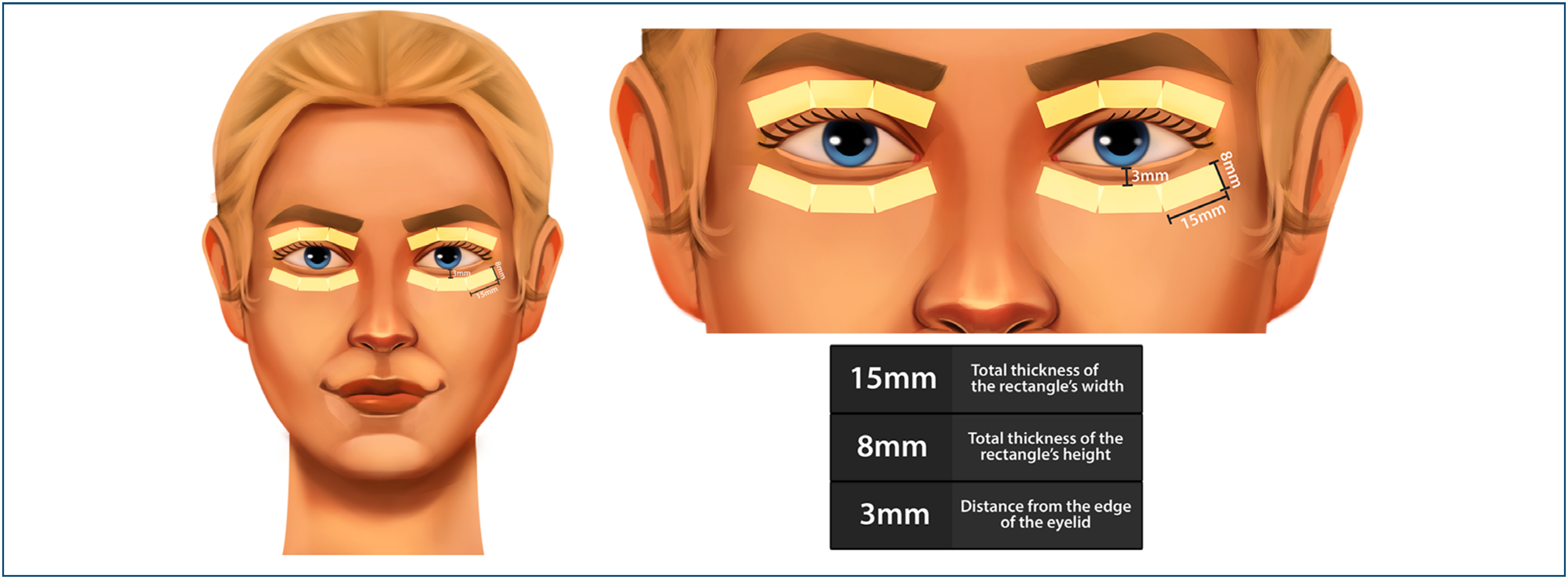
IPL was administered by a single physician using the Lumenis OPT M22™ device (590 nm filter, triple pulse, 6 milliseconds duration, 50 milliseconds interval) with initial fluence of 10 J/cm², increasing by 1 J/cm² per session (Figure 1). Treatments included three sessions every 15 days with a cooled 8 × 15 mm sapphire tip handpiece applied directly to the upper and lower eyelids (Figure 2). Six (06) flashes were delivered (2 passes of 3 flashes) per eyelid (Figure 3).


Before the flashes, both eyes were anesthetized with one anesthetic eye drop (oxybuprocaine hydrochloride 0.4%, Latinofarma), and ocular shields (Surgical Steel Medium, Laser shield) were placed on each eye. The periocular skin was cleaned with micellar water (Neutrogena®). Subsequently, cleaning was completed with 2% aqueous chlorhexidine (Bioquímica). Ultrasound gel (Carbogel) was applied to both eyes’ upper and lower eyelids.
After the flashes were delivered, the ultrasound gel and protective shields were removed. One drop of dexamethasone with ciprofloxacin 3 mg/mL eye drop (Latinofarma) was instilled in each eye. The eyelids were cleaned with micellar water (Neutrogena) and protected with sunscreen SPF 50 (Mantecorp).
As post-session care, patients were advised to avoid sun exposure for 14 days, avoid local heat for 24 hours, and use daily sunscreen protection. They were also instructed to continue using lubricating eye drops, oral omega-3 supplementation, applying warm compresses to the eyelids, and maintaining eyelid hygiene. Gland expression was not performed.
Assessments
The start time of observation for each patient (To) was defined immediately after the study inclusion criteria were fulfilled.
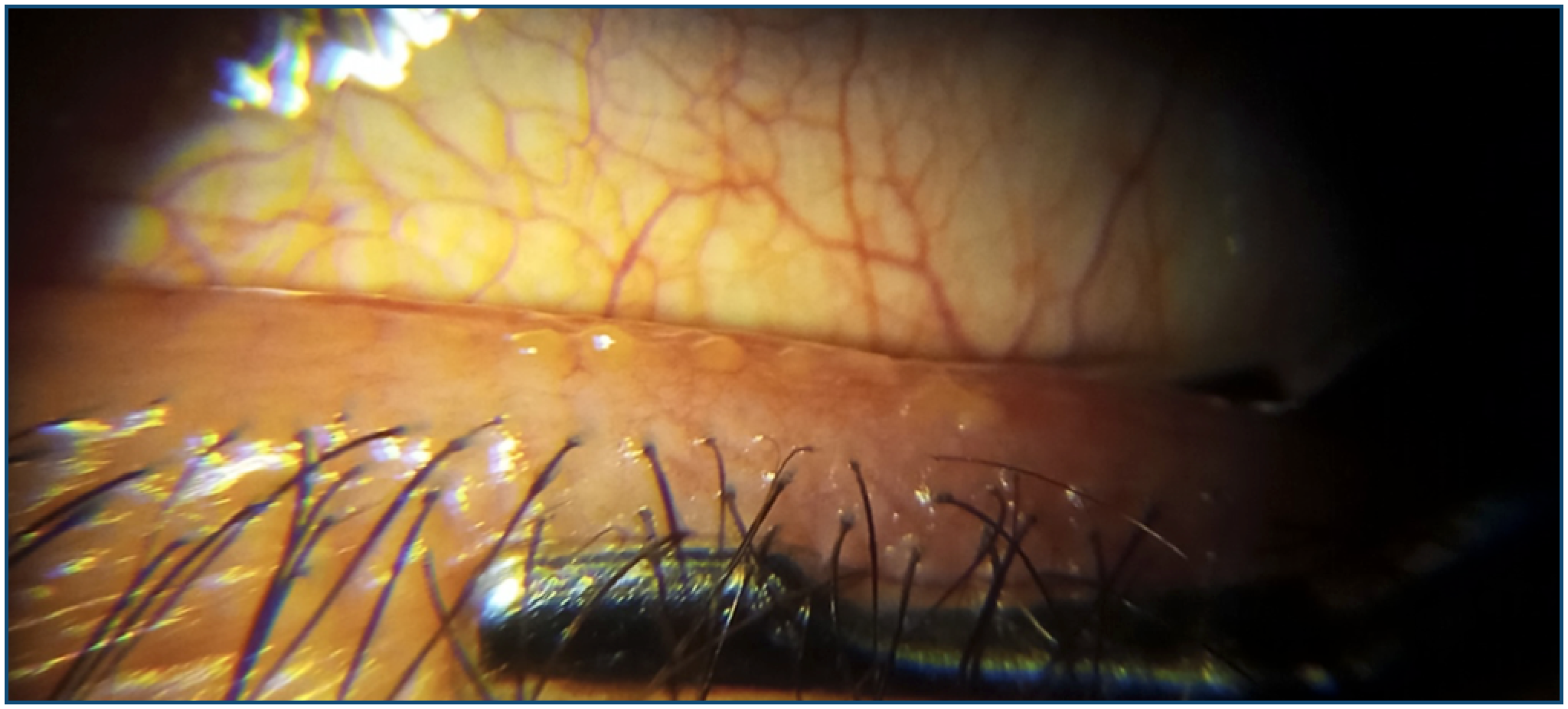
Approximately 30 days after the last session, all patients completed the dry eye evaluation questionnaire (SPEED 2), and the intensity of symptoms was graded on a scale for statistical analysis (Table 1). The lower eyelids were used for performing meibography and expressing the Meibomian glands (Figure 4) and the meibography (Figura 5). The CA800 equipment from Topcon was used to perform non-contact infrared meibography.


The lower eyelid was everted with a tongue depressor, images of the glands were captured, and gland dropout analysis was performed using the device software. The Pult and Nichols grading system was chosen for intensity evaluation, where a loss > 25% in patients over 20 years old and any loss in patients under 20 years old was considered positive (diseased) for MGD.
The CA800 Topcon equipment was used to quantify NITBUT in seconds and TMH in mm. The patient was examined in a dimly lit room with the air conditioning unit turned off. They were asked to blink, and the TMH was immediately measured at the lower corneal level (6 hours). For the NITBUT, the patient was asked to blink three times, and three images were captured; the equipment's software then chose the average of the images.
Finally, 15 Meibomian glands in the lower eyelids were expressed using Arita forceps (Figure 4). Obstruction or poorer meibum quality was then analyzed. A patient was considered having the disease with a grade ≥ 1 for those under 20 years old or ≥ 2 for those over 20 years old (Table 2).
Statistical analysis
The right eye (RE) was selected for statistical analysis, which was performed using the Jamovi software, version 2.3.28.0 (www.jamovi.org). Data normality was verified using the Shapiro-Wilk test. The non-parametric Wilcoxon test was used to compare meibum quality, while the paired Student's t-test was employed to evaluate SPEED 2 results and gland dropout in meibography. A p-value less than 0.05 was considered statistically significant.
All analyses were conducted before and after a 4-week period following the last session of IPL treatment.
A univariate logistic analysis was performed to identify predictors that could affect symptom improvement and be included in the initial model of the multivariate logistic analysis. A p-value less than 0.20 (univariate logistic analysis) and/or a p-value less than 0.05 (multivariate logistic regression) was considered statistically significant.
RESULTS
Among all patients with MGD and treated with IPL at the Santa Luzia Eye Center during the study period, 30 patients were not included in the analysis: 6 patients were asymptomatic, 4 had a NITBUT > 10 seconds, 1 patient was younger than 20 years, 9 patients had TMH < 0.25 mm, 2 patients had meibomyography < 25% and 8 patients answered a different questionnaire (Ocular Surface Disease Index [OSDI]).
This study included 16 eyes from 16 patients (10 women) with symptomatic MGD. The mean age was 60.4 ± 18.4 years, ranging from 27 to 85 (Table 3).
Demographic characteristics and clinical examinations of 16 patients undergoing Intense Pulsed Light treatment
Primary outcome
The treated patients showed a statistically significant improvement in their symptoms, with 75% becoming asymptomatic and/or reporting mild symptoms (change 7.31; 95% of confidence interval [95%CI] 4.19-9.18; p < 0.001) (Figure 6A).

(A) Behavior of SPEED 2 in 16 patients treated with Intense Pulsed Light. (B) Behavior of meibum quality in 16 patients treated with Intense Pulsed Light. (C) Behavior of gland dropout in 16 patients treated with Intense Pulsed Light.
None of the analyzed predictors (sex, p = 0.374; age, p = 0.980; meibum quality, p = 0.726; meibography, p = 0.764) influenced the response to treatment.
Secondary outcomes
The quality of the meibum showed a statistically significant improvement with treatment (Mdn 0.00; IQ 0-2; p < 0.001), with 68.8% of patients presenting oily or yellowish secretion without granules (Figure 6B).
A reduction in gland dropout was observed but did not reach statistical significance (change 4.85; 95%CI -1.63-11.3; p = 0.129), with the mean dropout decreasing from 27.1% to 22.5%, with a moderate effect size (d = 0.452) (Figure 6C).
DISCUSSION
This study was the first to demonstrate the efficacy of standalone IPL therapy applied directly to the eyelids in improving symptoms and glandular function in patients with MGD. In 2020, Xue et al., when treating 87 patients with IPL without eyelid massage in the tragus region, achieved improvement in symptoms (OSDI) mean: 21 ± 17 and SPEED mean: 9 ± 5; p < 0.001), but without statistically significant differences in meibum quality (Mdn: 2; IQR: 1-2; p = 0.28) or meibography (Mdn: 1; IQR: 0-2; p = 0.29), with results inferior to this study.(10)
In 2021, Shin et al. (12) conducted a comparative study of patients treated with standalone IPL versus those with therapeutic expression, concluding that expression alone did not improve symptoms (p = 0.84) or Meibomian gland quality (p = 0.731). In 2022, Martínez-Hergueta et al. applied IPL directly to the eyelids, combined with glandular expression, and reported improvement in meibography (M: 1.40 ± 0.74; p = 0.024).(13) Toyos et al. confirmed that IPL combined with glandular expression improved meibum quality (p < 0.0001).(14)
Meibomian gland dysfunction is characterized by obstruction and alterations in secretion quality, often accompanied by telangiectasias and secondary Demodex infestations. Treatments targeting these alterations, including IPL, are effective.(5,15)
Intense Pulsed Light is a non-coherent, scattered, pulsed light beam with wavelengths between 400 and 1200 nm. Its tissue action is based on selective photothermolysis, targeting chromophores (hemoglobin, melanin, or water) using specific filters (Figure 7).(16)

In 2015, dermatologist Rolando Toyos et al. observed that his facial rosacea patients treated with IPL experienced improvement in dry eye symptoms. Since then, IPL has gained prominence in the treatment of dry eye.(8,14)
The likely mechanisms of action of IPL therapy involve several processes. First, the local heat generated by the IPL flash raises tissue temperature to approximately 50ºC, sufficient to liquefy glandular secretions and facilitate drainage, thereby reducing gland dropout. However, many IPL devices lack small tips designed for direct eyelid application, resulting in protocols being applied to the face, as seen in rosacea treatment, even in patients with MGD alone. Some researchers have noted that IPL may not sufficiently elevate eyelid temperature to melt glandular secretions effectively.(14)
Another mechanism is the occlusion of small vessels at the eyelid margin. Meibomian glands function optimally in low-oxygen environments, and small ships at the eyelid margin increase oxygen delivery, impairing gland function. Intense Pulsed Light coagulates hemoglobin in these vessels, creating the anaerobic environment essential for gland efficiency.(17)
Intense Pulsed Light also aids in the elimination of Demodex folliculorum and Demodex brevis, which contribute to the inflammatory cycle in dry eye syndrome by releasing toxins on the ocular surface. The IPL treatment increases porphyrin production in the mitochondria of Demodex mites, leading to their destruction.(18)
Furthermore, IPL reduces pro-inflammatory mediators such as interleukin (IL) 17A, IL-6, and prostaglandin E2 while combating blepharitis.(19) It also induces photobiomodulation by altering intracellular gene and protein expression, which improves cellular function. IPL's effect on fibroblasts enhances collagen synthesis, improving eyelid tone and elasticity. This results in more efficient blinking, improved Meibomian gland expression, reduced tear evaporation, enhanced ductal gland architecture, and activation of germinative cells, thereby improving secretion quality.(20)
Lastly, IPL exerts neurological effects by stimulating two branches of the parasympathetic nerve in the lower eyelid, improving the function of the Meibomian and lacrimal glands. These combined mechanisms contribute to the therapeutic efficacy of IPL in treating MGD and associated conditions.(21)
In this study, IPL treatment was performed on the lower eyelid, as in most studies, and on the upper eyelid.(8,13,21-23) Meibomian gland dysfunction is not exclusive to the lower eyelid. Unfortunately, the initial indication for this therapy was for dermatological patients with dry eyes and facial rosacea. The initial machines had large tips, which made it impossible to treat the upper eyelid without causing harmful effects, leading many doctors to avoid treating it.
The upper eyelid has an average of 30 Meibomian glands, while the lower has 25. These glands produce an average of 26 and 13 µL of secretion, respectively.(24) Thus, the upper eyelid's reservoir of meibum is significant and should not be neglected.
Unfortunately, most IPL machines have large tips, typically 35 mm x 15 mm, which are unsuitable for eyelid dimensions. This mismatch can cause burns, loss of eyebrows and eyelashes, uveitis, and iris transillumination defects.(7,13,15,25,26)
Despite treatment on the upper and lower eyelids, meibomography and meibum evaluation were performed only on the lower eyelid due to technical limitations, as they already represent the disease.
Direct IPL treatment on the eyelids did not increase gland dropout percentages, a significant concern for proponents of pulsed thermotherapy, given that eyelid temperature can exceed 50°C.(13,26,27) Improvements in meibography averages (Figure 6C) may be attributed to the photobiomodulatory effects of IPL on the acinar units of Meibomian glands, which liquefy meibum, facilitate drainage, reduce ductal pressure, and slow down acinar apoptosis and gland dropout.(13,20)
The therapy resulted in symptom improvement, with 75% of patients becoming asymptomatic or reporting mild symptoms, achieving statistical significance (p<0.001) and a substantial effect size (d = 1.44). In 2024, Elbakary et al. treated 15 patients with moderate-to-severe EDE using IPL and achieved 73% of asymptomatic and mild cases, which is similar to the values we found. A significant improvement in patients’ quality of life (QOL) was noticed in all patients treated with IPL, with a reduction of the mean OSDI score from 56.9 ± 11.1(moderate-to-severe) to 22.9 ± 16.4(asymptomatic-to-mild).(28)
Additionally, 68.8% exhibited clear or yellowish meibum, with an important effect size (rpb 0.889). These results demonstrate clinical relevance and justify the standalone use of IPL for treating patients with refractory MGD.
Neither sex nor age influenced treatment response as they did not meet the assumptions for the initial multiple linear regression model. This was unexpected, given that age and sex are significant risk factors for MGD, likely due to glandular loss with age and hormonal changes.(29-31)
This study employed a retrospective cohort design, with data collected from medical records. This led to information loss. Another limitation was the transformation of numerical data into ordinal variables for statistical analysis, which reduced the study's power.
The treatment period coincided with the COVID-19 pandemic, which discouraged patient follow-up. Several patients reported significant symptom improvement but did not attend the post-therapy control examinations, affecting the sample size. Adverse effects included eyelash loss in two patients and localized burning sensation for a few hours.(7,31,32)
The sample size was small, and follow-up was short, only 30 days post-treatment. Longer follow-up would be necessary to assess the duration of the therapy's effects on glandular secretion, symptom improvement, and glandular loss.
An interesting observation was made in patients using prostaglandin analogs for the management of glaucoma. Long-term use of these medications leads to orbital fat atrophy, eyelid stiffening, and darkening. Following IPL sessions, skin lightening and improved eyelid tone were observed, thereby enhancing periocular aesthetics.(6,33)
CONCLUSION
Our findings suggest that concomitant treatment of the upper and lower eyelids was associated with statistically and clinically significant improvements in symptoms, demonstrating the possible irrelevance of associating glandular expression with Intense Pulsed Light. The quality of meibum secretion also enhanced, with no worsening of gland dropout in meibography, thereby, contributing to the reduction of the disease's progression.
Financial Support:
Institution:
Data availability statement:
The datasets generated and/or analyzed during the current study are included in the manuscript.
REFERENCES
- 1 Nichols KK, Foulks GN, Bron AJ, Glasgow BJ, Dogru M, Tsubota K, et al. The international workshop on Meibomian gland dysfunction: executive summary. Invest Ophthalmol Vis Sci. 2011;52(4):1922-9.
- 2 Chen H, McCann P, Lien T, Xiao M, Abraham AG, Gregory DG, et al. Prevalence of dry eye and Meibomian gland dysfunction in Central and South America: a systematic review and meta-analysis. BMC Ophthalmol. 2024;24(1):50.
- 3 Daniel E, Pistilli M, Ying GS, Bunya VY, Massaro-Giordano M, Asbell PA, et al. Association of Meibomian gland morphology with symptoms and signs of dry eye disease in the Dry Eye Assessment and Management (DREAM) study. Ocul Surf. Oct 2020;18(4):761-9.
- 4 Lin CW, Lin MY, Huang JW, Wang TJ, Lin IC. Impact of dry eye disease treatment on patient quality of life. Front Med (Lausanne). 2024;11:1305579.
- 5 Lam PY, Shih KC, Fong PY, Chan TCY, Ng AL, Jhanji V, et al. A review on evidence-based treatments for Meibomian gland dysfunction. Eye Contact Lens. 2020;46(1):3-16.
- 6 Kalil C, Reinehr C, Milman L. Intense Pulsed Light: Review of clinical indications. Surgical & Cosmetic Dermatology. 2017;9(1):9-17.
- 7 Husein-ElAhmed H, Steinhoff M. Laser and light-based therapies in the management of rosacea: an updated systematic review. Lasers Med Sci. 2021;36(6):1151-60.
- 8 Toyos R, McGill W, Briscoe D. Intense pulsed light treatment for dry eye disease due to Meibomian gland dysfunction; a 3-year retrospective study. Photomed Laser Surg. 2015;33(1):41-6.
- 9 Karaca EE, Evren Kemer Ö, Özek D. Intense regulated pulse light for the Meibomian gland dysfunction. Eur J Ophthalmol. 2020;30(2):289-92.
- 10 Xue AL, Wang MTM, Ormonde SE, Craig JP. Randomised double-masked placebo-controlled trial of the cumulative treatment efficacy profile of intense pulsed light therapy for meibomian gland dysfunction. Ocul Surf. 2020;18(2):286-97.
- 11 Vergés C, Salgado-Borges J, Ribot FM. Prospective evaluation of a new Intense Pulsed Light, thermaeye plus, in the treatment of dry eye disease due to Meibomian gland dysfunction. J Optom. 2021;14(2):103-13.
- 12 Shin KY, Lim DH, Moon CH, Kim BJ, Chung TY. Intense pulsed light plus Meibomian gland expression versus Intense Pulsed Light alone for Meibomian gland dysfunction: A randomized crossover study. PLoS One. 2021;16(3):e0246245.
- 13 Martínez-Hergueta MC, Alió Del Barrio JL, Canto-Cerdan M, Amesty MA. Efficacy and safety of Intense Pulsed Light direct eyelid application. Sci Rep. 2022;12(1):15592.
- 14 Toyos R, Desai NR, Toyos M, Dell SJ. Intense pulsed light improves signs and symptoms of dry eye disease due to Meibomian gland dysfunction: A randomized controlled study. PLoS One. 2022;17(6):e0270268.
- 15 Craig JP, Nelson JD, Azar DT, Belmonte C, Bron AJ, Chauhan SK, et al. TFOS DEWS II report executive summary. Ocul Surf. 2017;15(4):802-12.
- 16 Vigo L, Giannaccare G, Sebastiani S, Pellegrini M, Carones F. Intense pulsed light for the treatment of dry eye owing to Meibomian gland dysfunction. J Vis Exp. 2019;(146).
- 17 Liu Y, Wang J, Chen D, Kam WR, Sullivan DA. The role of hypoxia-inducible factor 1α in the regulation of human Meibomian gland epithelial cells. Invest Ophthalmol Vis Sci. 2020;61(3):1.
- 18 Rhee MK, Yeu E, Barnett M, et al. Demodex blepharitis: a comprehensive review of the disease, current management, and emerging therapies. Eye Contact Lens. 2023;49(8):311-8.
- 19 Suwal A, Hao J-l, Zhou D-d, Liu X-f, Suwal R, Lu C-w. Use of Intense Pulsed Light to Mitigate Meibomian Gland Dysfunction for Dry Eye Disease. Int J Med Sci. 2020;17:1385-92.
- 20 Fineide F, Magnø MS, Khan AZ, Chen X, Vehof J, Utheim TP. Intense pulsed light treatment in Meibomian gland dysfunction: Past, present, and future. Acta Ophthalmol. 2024;102(4):e414-42.
- 21 Jiang X, Yuan H, Zhang M, Lv H, Chou Y, Yang J, et al. The efficacy and safety of new-generation Intense Pulsed Light in the treatment of Meibomian gland dysfunction-related dry eye: a multicenter, randomized, patients-blind, parallel-control, non-inferiority clinical trial. Ophthalmol Ther. 2022;11(5):1895-912.
- 22 Toyos R, Toyos M, Willcox J, Mulliniks H, Hoover J. Evaluation of the safety and efficacy of Intense Pulsed Light treatment with Meibomian gland expression of the upper eyelids for dry eye disease. Photobiomodul Photomed Laser Surg. 2019;37(9):527-31.
- 23 Tashbayev B, Yazdani M, Arita R, Fineide F, Utheim TP. Intense pulsed light treatment in Meibomian gland dysfunction: A concise review. Ocul Surf. Oct 2020;18(4):583-94.
- 24 Knop E, Knop N, Millar T, Obata H, Sullivan DA. The international workshop on Meibomian gland dysfunction: report of the subcommittee on anatomy, physiology, and pathophysiology of the Meibomian gland. Invest Ophthalmol Vis Sci. 2011;52(4):1938-78.
- 25 Lee WW, Murdock J, Albini TA, O'brien TP, Levine ML. Ocular damage secondary to intense pulse light therapy to the face. Ophthalmic Plast Reconstr Surg. 2011;27(4):263-5.
- 26 Zhang-Nunes S, Guo S, Lee D, Chang J, Nguyen A. Safety and efficacy of an augmented intense pulse light protocol for dry eye syndrome and blepharitis. Photobiomodul Photomed Laser Surg. 2021;39(3):178-84.
- 27 Borchman D. The optimum temperature for the heat therapy for Meibomian gland dysfunction. Ocul Surf. 2019;17(2):360-4.
- 28 Elbakary MA, Shalaby OE, Allam WA, Alagorie AR, Shafik HM. Quality of life improvement in dry eye patients after intense pulsed light therapy compared to punctal plugs. Oman J Ophthalmol. 2024;17(1):108-12.
- 29 Beining MW, Magnø MS, Moschowits E, Olafsson J, Vehof J, Dartt DA, et al. In-office thermal systems for the treatment of dry eye disease. Surv Ophthalmol. 2022;67(5):1405-18.
- 30 Huang B, Fei F, Wen H, Zhu Y, Wang Z, Zhang S, et al. Impacts of gender and age on Meibomian gland in aged people using artificial intelligence. Front Cell Dev Biol. 2023;11:1199440.
- 31 Machalińska A, Zakrzewska A, Safranow K, Wiszniewska B, Machaliński B. Risk factors and symptoms of Meibomian gland loss in a healthy Population. J Ophthalmol. 2016;2016:7526120.
- 32 Stapleton F, Alves M, Bunya VY, albert I, Lekhanont K, Malet F, et al. TFOS DEWS II Epidemiology Report. Ocul Surf. 2017;15(3):334-65.
- 33 Goldberg DJ. Current trends in Intense Pulsed Light. J Clin Aesthet Dermatol. 2012;5(6):45-53.