Knowledge and skills for extraocular foreign body removal: a study with physicians, medical students and ophthalmologists
ABSTRACT
Objective: To identify the knowledge and skills of generalist physicians and medical interns, as well as the perception of ophthalmologists regarding academic training for extraocular foreign body removal.
Methods: This was a descriptive cross-sectional study with a qualitative and quantitative approach. The study included physicians on duty in the emergency department of a university hospital and an Emergency Care Unit.
Results: The total number of participants was 87, including 18 generalist physicians on duty, 59 medical students, and 10 ophthalmologists. Three different semi-structured questionnaires were used. 11.11% of the on-duty physicians and 3.39% of the students considered the knowledge acquired about extraocular foreign body removal to be good. The majority of the on-duty physicians (58.82%) and students (76.27%) did not consider themselves capable of performing the procedure. Among ophthalmologists, 90% considered that foreign body removal should not be performed by non-ophthalmologists.
Conclusion: There is a need to develop teaching-learning strategies that can minimize the knowledge and skill deficiencies of on-duty physicians and medical students for the removal of extraocular foreign bodies due to the relative frequency of this occurrence in general emergency care services.
Keywords:
Extraocular foreign body; Medical education; Education, medical, continuing
RESUMO
Objetivo: Identificar o conhecimento e as habilidades de médicos plantonistas generalistas, internos de medicina e percepção dos oftalmologistas sobre a formação acadêmica para a retirada de corpo estranho extraocular.
Métodos: Estudo transversal descritivo de abordagem qualiquantitativa. Foram incluídos médicos plantonistas do Setor de Emergência de um hospital universitário e uma unidade de pronto atendimento.
Resultados: O total de participantes foi de 87, sendo 18 médicos plantonistas generalistas, 59 estudantes de medicina e 10 oftalmologistas. Foram utilizados três distintos questionários. Consideraram bom o conhecimento adquirido sobre retirada de corpo estranho extraocular 11,11% dos médicos plantonistas e 3,39% dos estudantes. A maioria dos plantonistas (58,82%) e dos estudantes (76,27%) não se considerava apta para o procedimento. Entre os oftalmologistas, 90% consideraram que a retirada de corpo estranho não deve ser realizada por não oftalmologistas.
Conclusão: Conclui-se pela necessidade do desenvolvimento de estratégias de ensino-aprendizagem que possam minimizar as deficiências de conhecimento e as habilidades dos médicos plantonistas e acadêmicos de medicina para a retirada de corpo estranho extraocular em razão da relativa frequência dessa ocorrência em serviços gerais de pronto atendimento.
Descritores:
Corpo estranho extraocular; Educação médica; Educação médica continuada
INTRODUCTION
The occurrence of eye accidents, estimated at 55 million per year, depending on the severity of the trauma, impacts society in different ways and at different levels.(1) The search to treat acute eye diseases, defined as those that have been developing for up to 15 days and present a risk of loss of ocular and visual integrity, comprise around 3% of visits to general emergency care, but can vary and reach rates of up to 20%, depending on the context of the emergency care.(2–4)
Most ocular traumas are caused by superficial foreign bodies that mainly affect young male adults, who are individuals in the productive age group, generating economic, psychological, and social problems, as well as compromising at least 1 workday.(5–9) Occupational accidents are among the main causes and could be avoided with the use of personal protective equipment (PPE).(10,11)
Foreign bodies usually involve the cornea through the projection of pieces of rust, wood, glass, plastic, fiberglass, or plant material; corneal injuries account for 8% to 13% of ocular presentations, depending on the setting in which patients are seen, such as primary care offices, general Emergency Departments, or ocular Emergency Departments.(3)
The most common etiologies are mechanical (blunt or penetrating), chemical (acid or caustic), thermal or radiation (ultraviolet, solar radiation, laser, etc.) traumas.(4) Among children, closed traumas resulting from aggression and accidents with pencils, pens and balls, generally occurring at home, are the most common.(12)
Regarding location, foreign bodies present in the orbital cavity can be classified as: ocular, adnexal and mixed, with ocular foreign bodies being subdivided into intraocular and extraocular.(13) Extraocular foreign bodies in the conjunctival cul-de-sac and palpebral tarsi are easily removable by everting the eyelid, but those lodged in the cornea may require the participation of a specialist.(4)
Regarding emergency ophthalmological care, it is worth noting that general practitioners and family physicians are the only opportunities for ophthalmological care for the Brazilian population, since 45 million people live in municipalities that do not have ophthalmologists.(14) Thus, the difficulties in providing the most appropriate treatment are enormous, either due to the limited number of specialized professionals for medical care or due to a lack of health equipment.
The factors that have negatively impacted ophthalmological care in Emergency Departments are the deficiency of services, the regulatory process and the lack of knowledge of health professionals.(1) Improving the resolution of care avoids unnecessary expenses and brings benefits to patients.(15)
Foreign bodies are removed by careful removal, with the help of suitable material, avoiding the dispersion of toxins, residues or extension of the wounds.(4) The visual prognosis, in addition to the severity of the trauma, depends on the quality and speed of patient care. Therefore, it is important to develop research that allows us to understand how ophthalmological emergency care is being provided in emergency care units to develop care and preventive policies for the population susceptible to this condition.(9)
Medical education presupposes a set of practices and processes included in the curricular structure of the courses, followed by respective evaluation.(16) However, during undergraduate studies, ophthalmology teaching has been shown to be deficient, with reduced workload in the curricular matrix, presenting a lack of practical preparation of students and general practitioners both for the diagnosis and management of problems in emergency care situations and for the removal of extraocular foreign bodies.(14,17)
In this context, it is important to identify these deficiencies and seek ways to minimize them. Thus, the guiding question of this research is: what is the perception of medical students, general practitioners on duty and ophthalmologists regarding their knowledge and ability to remove extraocular foreign bodies?
In view of the above, the general objective of the present study was to identify the knowledge and skills of generalist physicians and medical interns, as well as the perception of ophthalmologists regarding academic training for extraocular foreign body removal.
METHODS
This is a descriptive cross-sectional study with a qualitative and quantitative approach. The following were invited as a sample for this research: on-duty generalist physicians from the emergency room of the Hospital Universitário Santa Terezinha (HUST), in the Emergency Care Unit (UPA) of Herval d’Oeste; ophthalmologists who worked in the Midwest of Santa Catarina; and medical internship students at the Medical School of the Universidade do Oeste de Santa Catarina (Unoesc). The total number of participants was 87, of which 18 were on-duty generalist physicians, 10 were ophthalmologists, and 59 were medical internship students.
Three semi-structured questionnaires previously prepared by the researchers and applied from March to August 2023 through the Google Forms platform were used to collect data, as well as, alternatively, applied through interviews in the participants’ work or study environment.
Additional information was collected, such as age, gender, professional factors such as time since graduation, place of specialization, as well as information on contact with extraocular foreign body removal and its frequency in hospital and clinical practice. In addition, the use, availability, and type of equipment used in extraocular foreign body removal were analyzed. The questions focused on identifying knowledge about foreign body removal equipment, the perception of the quality of practical knowledge received during graduation, as well as the aptitude to perform the procedure.
The responses were tabulated in Microsoft Excel® software and presented in the form of graphs and tables, representing the data contained in the questionnaire, and were analyzed quantitatively through descriptive statistics.
The bibliographic material was also used to prepare instructive pedagogical material with guidelines on the most appropriate procedures and equipment for removing extraocular foreign bodies.
RESULTS
The total number of participants in this research was 87, and the majority were women (72.41%). There were 59 students, with a mean age of 23.19 ± 0.66 years; 18 general practitioners, with a mean age of 32.61 ± 8.09 years; and 10 ophthalmologists, all with more than 10 years of experience.
Regarding the course phase of the students interviewed, the majority were in the 9th phase of the medical course (47%), followed by the 10th phase (26%), 11th phase (16%), 12th phase (7%), and 8th phase (4%). Regarding the on-duty physicians, 17% had graduated less than 1 year ago, 39% had graduated less than 5 years ago, 22% had graduated more than 5 years ago and less than 10 years ago; 22% had graduated more than 15 years ago. Additionally, it is worth noting that only 28% of the on-duty physicians had specializations, which were: internal medicine, dermatology, occupational medicine, and general surgery. The ophthalmologists presented the following distribution of time since graduation: 30% had graduated more than 10 years ago and less than 15; 20% had graduated more than 15 years ago and less than 20; 30% over 20 years and under 30, 20% over 30 years.
The interviewees were initially asked about the equipment and/or materials used or known to be used to remove extraocular foreign bodies. Among the on-duty physicians, the cotton swab was the most commonly used (42%), followed by irrigation with saline solution (35%) and needle (23%). Among the students, the most frequent answer was cotton swab (64%), followed by needle (22%) and spatula (7%). Among the ophthalmologists, the most commonly used instruments were needles (40%), drills (20%) and both (40%), as shown in table 1.
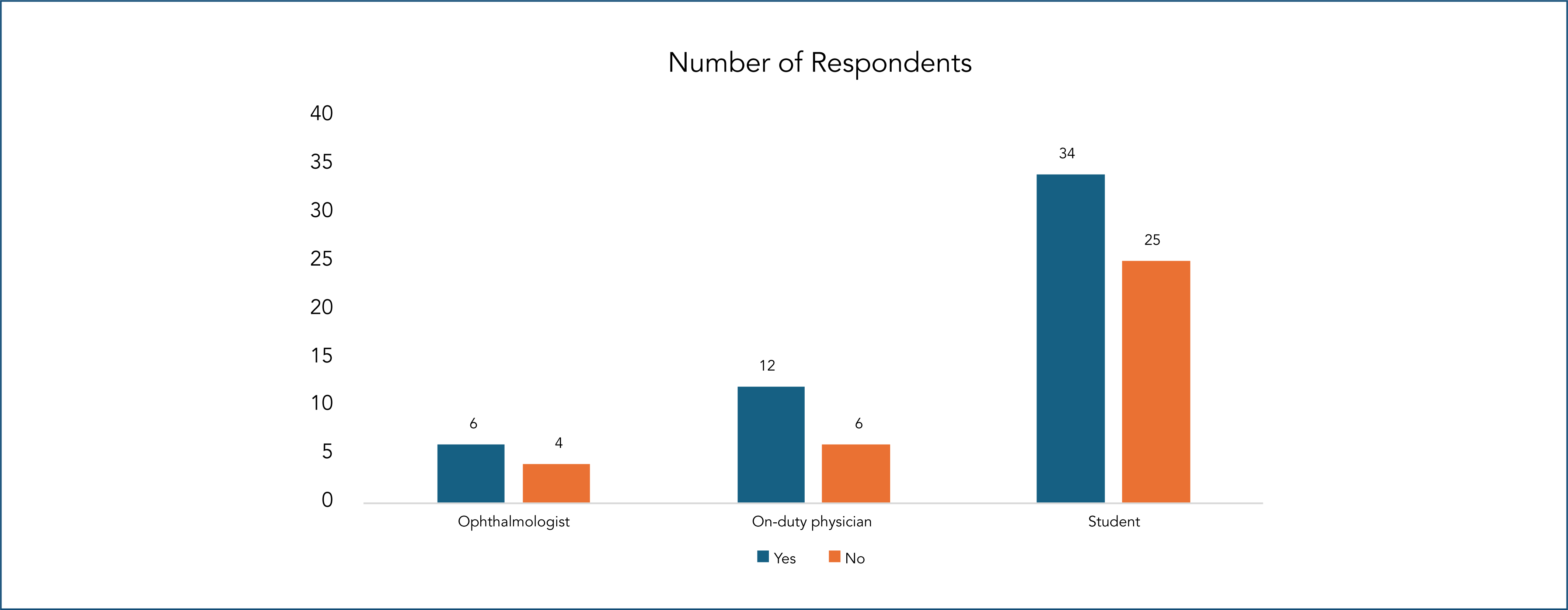
The practical instruction received during undergraduate studies for the removal of foreign bodies from the cornea showed significant differences between the groups (Figure 1). Among the ophthalmologists, 60% (n = 6) reported having received practical instruction, while 40% (n = 4) stated that they had not received it. In the case of on-duty physicians, the proportion was similar, with 66.7% (n = 12) indicating that they had received practical training and 33.3% (n = 6) that they had not. Among the students, 57.6% (n = 34) had received practical instruction, while 42.4% (n = 25) had not.
Training or practice of on-duty physicians and students to use ocular foreign body removal equipment.
When asked about the level of knowledge acquired about extraocular foreign body removal during their undergraduate studies, 64.4% (n = 38) of the students classified it as acceptable, while 32.2% (n = 19) considered it poor and only 3.4% (n = 2) considered it good. In the case of on-duty staff, 50% (9) evaluated the knowledge as acceptable, while 38.9% (n = 7) considered it poor and 11.1% (n = 2) as good.
The on-duty staff and students also evaluated the practical knowledge offered in their undergraduate course and perceived the need for this content to be deepened. Thus, 50% of the on-duty staff and 64.41% of the students evaluated the acquired knowledge as acceptable; it was evaluated as good for 11.1% of the on-duty staff and for 3.39% of the students and poor for 38.89% of the on-duty staff and for 32.2% of the students. The need for deepening knowledge to remove foreign body was reported by 93% of the students and 39% of the on-duty staff (p < 0.001).
Of the 10 ophthalmologists interviewed, 8 (80%) reported treating patients who suffered iatrogenesis resulting from inadequate interventions during the initial management for the removal of extraocular foreign bodies, such as melting (n = 2), fragment remains (n = 2), deep corneal injury (n = 2), corneal clouding (n = 1) and iris hernia (n = 1). Regarding the removal of foreign bodies by non-ophthalmologists, 9 (90%) considered that it should not be performed, mainly due to the lack of adequate equipment and the risk of ocular perforation.
Perception of practical knowledge and aptitude
The aim was to better understand the need for practical experience to perform the procedure. Thus, the following question was asked to the ophthalmologist: "Do you consider any of the procedures presented in this questionnaire or others not mentioned to be an inappropriate procedure for physicians who do not have a specialty in ophthalmology?"
Thus, analyzing the content of the ophthalmologists’ responses, we observed a consensus that the extraction equipment mentioned is adequate; however, the most qualified professional for such a procedure would be the ophthalmologist. One of the interviewees answered:
The removal of a foreign body from the eye is a medical procedure, and for this purpose, correct handling of the patient is necessary, using biomicroscopy through a slit lamp, equipment that require a lot of training time to reach the level of excellence that the equipment offers for the correct handling of the ophthalmological patient. (Interviewee 6)
In turn, the perception of on-duty staff and students regarding their knowledge and their ability to perform extraocular foreign body removal procedures was also questioned. Regarding knowledge, most students (n = 43; 72.88%) perceived it as regular, but among the on-duty staff, the perception of knowledge was divided between good (n = 10; 55.56%) and regular (n = 8; 44.44%) (Table 2). Regarding the perception of aptitude in removing foreign bodies, 76.27% (n = 45) of the students and 58.82% (n = 10) of the on-duty staff did not consider themselves capable of performing this procedure.
Table 3 presents the self-perception of students and on-duty physicians regarding ophthalmological complications. For chemical burns, 38.9% of on-duty physicians (n = 7) and 15.3% of students (n = 9) considered their knowledge good, while 50% (n = 9) and 62.7% (n = 37), respectively, rated it as regular. 22% of students (n = 13) and 11.1% of on-duty physicians (n = 2) rated it as poor. The difference was significant (p = 0.002).
Regarding ocular trauma, 16.7% of on-duty physicians (n = 3) and 8.5% of students (n = 5) reported good knowledge, while 61.1% (n = 11) and 66.1% (n = 39), respectively, evaluated it as regular. The percentage of "poor" responses was 22.2% (n = 4) among on-duty physicians and 25.4% (n = 15) among students. The difference was significant (p < 0.001).
Regarding ocular perforation, 11.1% of the on-duty staff (n = 2) and 8.5% of the students (n = 5) considered their knowledge to be good, while 61.1% (n = 11) and 37.3% (n = 22) classified it as regular. The "poor" responses were, respectively, 27.8% of the students (n = 32) and 27.8% of the on-duty staff (n = 5). There was no statistical difference (p = 3.488).
In the management of extraocular foreign bodies, 55.6% of the on-duty physicians (n = 10) and 13.6% of the students (n = 8) rated their knowledge as good, while, respectively, 44.4% (n = 8) and 72.9% (n = 43) considered it regular. No on-duty physician rated it as poor, against 13.6% of the students (n = 8; p < 0.001).
The students were also asked about the conduct they considered most appropriate for treating patients with extraocular foreign bodies (Table 4). Removal with a cotton swab before referral to an ophthalmologist was the most frequent practice (n = 49; 45.79%), followed by ocular irrigation (n = 29; 27.1%) and immediate referral to an ophthalmologist (n = 23; 21.5%). More invasive methods, such as removal with a needle or spatula, were rare (n = 1; 0.93%), as was the use of a drill (n = 5; 4.67%).
DISCUSSION
Extraocular foreign bodies cause damage without penetrating the eyeball and usually cause acute damage that can develop into chronic complications, either through chemical action or through opportunistic infections of the cornea, conjunctiva, and eyelid. As for the origin, they can come from animals (hair, nails, parts and stingers), plants (small seeds, sap, fragments of stem and leaves) and non-biological (metals, glass, including contact lenses that move to the conjunctival cul-de-sac region).(4)
Although ophthalmologists have more training and better instruments to assist patients with extraocular foreign bodies, emergency care is often provided in emergency rooms and general emergency services and removal could be performed by an emergency room physician, as long as they are qualified to perform such a procedure.(4)
Regarding the instruments used, the students and physicians on duty first chose the least invasive option, which is the use of cotton swabs and irrigation, since, they mostly did not consider themselves capable of removing extraocular foreign bodies, in agreement with what was found in other studies.(18,19) In Australia, the removal of a foreign body without the use of a slit lamp is in the guidelines of The Royal Australian College of General Practitioners, which recommends irrigation, cotton swab, and needle removal using good lighting and magnifying lenses.(20) In this regard, research has shown that irrigation with saline solution has some advantages, especially reducing the scale of pain and the number of retained foreign bodies, reducing the need for referral for specialized ophthalmological examination.(21)
Foreign body removal requires the acquisition of specialized skills for problem-solving in a real-life problem situation.(22) However, approximately one-third of the students and on-duty staff in this study considered that their practical instruction for extraocular foreign body removal was deficient ("poor"), highlighting the need for strategies to deepen knowledge, or the procedure will be compromised. For the removal of extraocular foreign bodies, according to the opinion of the ophthalmologists in this research, the specialist is better qualified to perform this procedure, which normally involves careful removal, with the aid of special instruments.
Most students and on-duty staff were not prepared for extraocular foreign body removal due to a lack of practical opportunities, a teaching deficiency that could be compensated for through simulated training that would prepare them to provide emergency care services. In this sense, there are models already tested by researchers proposed for the teaching and learning of this training.
One of these models, which appears to be quite viable, uses an agar plate with iron filings added and sprayed with 2.1% saline solution to create the rust halo; after waiting 48 hours for the agar to acquire consistency, the plate is fixed to a slit lamp where participants carry out training using a 2.5 m syringe with a 30G needle.(23) In another more realistic model for the reality of our teaching and lack of slit lamps in emergency services, it consists of using a table tennis ball to mold the agar into a rounded shape and better simulate the shape of an eye for removing a foreign body with a 27G needle.(24)
Other resources can be used. Wood's lamp is a portable device with a magnifying lens and ultraviolet light, which highlights the fluorescein staining during ocular inspection and stands out for its practicality and simplicity of operation, being especially useful for cases of more obvious ocular injuries, such as foreign bodies.(25) This feature makes Wood's lamp a low-cost and easily available device, a promising alternative to reduce the deficiency in eye emergency care, previously identified.
Results from other studies show the need to improve teaching and learning in undergraduate courses. A study conducted with medical students showed that, although they demonstrated knowledge about important eye diseases, they did not have the necessary skills and experience to perform the role of general practitioners in emergency rooms.(19)
The results of this research are also in agreement with other authors, who also found a deficiency in ophthalmology teaching in training undergraduates to work with ophthalmological problems, even the simplest ones, both among students and among general practitioners.(14,17,22,26) Furthermore, the assessment of theoretical knowledge through a test does not constitute a guarantee of acquisition of knowledge or practical skills to deal with common problems in ophthalmology, such as those encountered by a general practitioner in an emergency or in a primary health care setting.(27)
Although most on-duty physicians have more knowledge about treating ophthalmologic emergencies, most ophthalmologists reported complications due to previous foreign body removal in Emergency Departments and consider this to be a specialized procedure to be performed with a slit lamp. However, a study conducted in Ireland compared patients with corneal foreign bodies who were treated by on-duty physicians in a general emergency department, whose removal was performed with and without a slit lamp; patients treated without the use of a slit lamp had a higher pain scale, but visual acuity, satisfaction rate, and complication rates were similar.(28)
From another perspective, a broad qualitative and quantitative analysis of medical students who had already taken the Ophthalmology course in 42% of the states in all Brazilian regions were interested in acquiring more knowledge on ophthalmology as preparation for general practitioners.(29) Academic leagues in Ophthalmology have been present in medical training since 1977 and are important initiatives that contribute to improving the "curriculum" in Ophthalmology and allow for the implementation of new active methodologies in the educational system, with students as protagonists of their own learning.(30,31) In the league activities, simulated removal of corneal foreign bodies could be improved through practice on more easily produced models.(23,24)
Finally, it is worth highlighting that the main limitation of this study was the small size of the sample and institutions, which may not reflect the realities of other emergency care services.
CONCLUSION
Approximately half of the on-duty physicians and medical students reported having received sufficient guidance on removal, but the majority did not consider themselves qualified to perform the procedure. On the other hand, the majority of ophthalmologists considered removal by non-specialists to be inappropriate.
Among on-duty physicians and medical students, the use of a "cotton swab", followed by irrigation and use of a needle, are the most commonly used methods for removing extraocular foreign bodies, while the needle, followed by the drill, are the preferred methods for ophthalmologists.
The results allow us to conclude that there are weaknesses in the teaching and learning of this procedure during the undergraduate course of the participants that negatively impact the training of general practitioners for activities in general emergency care. The care of patients with extraocular foreign bodies requires specific practical skills, followed by opportunities, even simulated ones, for students to carry out practical activities during the undergraduate course and acquire skills necessary for on-duty physicians. Academic ophthalmology leagues provide an opportunity to expand this knowledge.
Finally, it is necessary to develop strategies that can minimize deficiencies in knowledge and skills related to ophthalmological care due to its relative frequency in general emergency services. More comprehensive studies in other settings on the preparation of professionals to provide care for the removal of extraocular foreign bodies in emergency services are necessary for a more thorough analysis of this intriguing topic.
Financial support:
Institution:
Data availability statement:
The datasets generated and/or analyzed during the current study are included in the manuscript.
REFERENCES
- 1 Bemerguy CE, Silva Martins IC, Guerreiro JF. Regulação de acesso da urgência e emergência oftalmológica em um hospital universitário. Rev Atenção Saúde. 2021;19(70):47-55.
- 2 Espinosa PG, Knob HH, Marchetti MP, Stock RA, Rieger A, Bonamigo EL. Atendimento às urgências oftalmológicas em unidade de pronto atendimento. Arq Catarin Med. 2020;49(1):78-90.
- 3 Garcia GC, Rosário SR, Alves VT, Mendonça GA, Corgozinho RC, Gildin F, et al. As manifestações clínicas e o diagnóstico de abrasões da córnea. Braz J Surg Clin Res. 2021;35(1):98-103.
- 4 Cardoso Filho AP, Costa LC, Rocha EM. Urgências oftalmológicas: o que todo médico precisa saber. Medicina (Ribeirão Preto). 2022;55(2):62-8.
- 5 Ferreira FQ, Nascimento MF, Meneguim RL, Padovani CR, Schellini SA. Trauma ocular na Faculdade de Medicina de Botucatu. Rev Bras Oftalmol. 2016;75(3):185-9.
- 6 Jayme BC, Silva LV, Rezende LC, Silva MF, Moreira TC, Oliveira TM, et al. Principais ocorrências na emergência oftalmológica com enfoque em traumas oculares: uma revisão integrativa. Res Soc Develop. 2023;12(1):e14912139765.
- 7 Rassi AJ, Nascimento JL, Duarte LC, Freitas LP, Di Filice LC, Morais LT, et al. Epidemiologia das urgências e emergências oftalmológicas em um Hospital Universitário Terciário. Rev Bras Oftalmol. 2020;79(4):227-30.
- 8 Campos GM, Brum IV, Brum IV. Epidemiological profile of visits in a public ophthalmic emergency service. Rev Bras Oftalmol. 2019;78(5):297-9.
- 9 Almeida HG, Fernandes VB, Peixoto e Lucena AC, Kara-Junior N. Avaliação das urgências oftalmológicas em um hospital público de referência em Pernambuco. Rev Bras Oftalmol. 2016;75(1):18-20.
- 10 Costa EP, Gomes TM, Mendes TA, Campos MA, Bertrand RH, Pinto LM, et al. Epidemiological profile and visual outcome of ocular trauma in a referral center in northeastern Brazil. Rev Bras Oftalmol. 2019;78(5):310-4.
- 11 Milanez M, Saraiva PG, Barcellos NN, Saraiva FP. Epidemiological and occupational profile of eye trauma at a referral center in Espírito Santo, Brazil. Rev Bras Oftalmol. 2017;76(1):7-10.
- 12 Matos AG, Cavalcante RG, Figueiredo TF, Chaves MF, Souza FA. Perfil do trauma ocular infantil em unidade de emergência oftalmológica. Rev Bras Oftalmol. 2018;77(3):124-7.
- 13 Shukla B. New classification of ocular foreign bodies Chin J Traumatol. 2016;19(6):319-21.
- 14 Furtado JM, Oliveira IP. Simpósio: Oftalmologia para a graduação em Medicina e o Médico Generalista. Medicina (Ribeirão Preto). 2022;55(2):e-20015.
- 15 Farias Filho ER. Prevenção dos principais traumas oculares em combate [monografia]. Rio de Janeiro. 2019.
- 16 Machado CD, Wuo A, Heinzle M. Educação médica no Brasil: uma análise histórica sobre a formação acadêmica e pedagógica. Rev Bras Educ Méd. 2018;42(4):66-73.
- 17 Succar T, Grigg J, Beaver HA, Lee AG. A systematic review of best practices in teaching ophthalmology to medical students. Surv Ophthalmol. 2016;61(1):83-94.
- 18 Portes AL. Perfil Multicêntrico do Acadêmico de Medicina e suas Perspectivas sobre o Ensino da Oftalmologia. Rev Bras Oftalmol. 2021;79(6):427.
- 19 Abreu AM, Abreu EB, Paulino IC, Pierre NB. Conhecimento dos Alunos de Medicina sobre oftalmologia. Rev Bras Educ Méd. 2019;43(3):100-9.
- 20 Fraenkel A, Lee LR, Lee GA. Managing corneal foreign bodies in office-based general practice. Australian Fam Phys. 2017:46(3):89-93.
- 21 Chou HD, Chen KJ, Kang EY, Lin JY, Yeh PH, Chen YT, et al. Eye irrigation as a first-line treatment and diagnostic method for emergency department patients who complain of ocular foreign bodies. Sci Rep. 2021;11:23386.
- 22 De Biase CL. Atenção Primária à Saúde-Oftalmologia: Percepção discente sobre competências na formação médica. Rev Portal: Saúde e Sociedade. 2018;3(2):750-66.
- 23 Maguire MJ, Bader T. A simple and effective simulation model for corneal foreign body and rust ring removal. Can J Ophthalmol. 2024;59(6):e871-e873.
- 24 Lin J, Chua MT. A low cost surrogate eye model for corneal foreign body removal. BMC Ophthalmol. 2020;20(1):48.
- 25 Hamou S, Ghiaee S, Chung C, Lloyd M, Khem K, Zhang XC. Emergency Department Slit Lamp Interdisciplinary Training Via Longitudinal Assessment in Medical Practice. West J Emerg Med. 2024;25(5):725-34.
- 26 Damasceno MB, Ferreira JL, Lopes LS, Ricca GE, Girão LS, Canuto IF, et al. Avaliação do conhecimento em Oftalmologia na Graduação Médica. RMMG Rev Med Minas Gerais. 2021;31:e31115.
- 27 Mendes Júnior ES. Avaliação do conhecimento de estudantes de medicina e do ensino curricular em oftalmologia em três escolas médicas do estado da Bahia [tese]. Escola Bahiana de Medicina e Saúde Pública; 2023.
- 28 Quirke M, Mullarkey C, Askoorum S, Coffey N, Binchy J. A prospective observational study of techniques to remove corneal foreign body in the emergency department. Emerg Med J. 2014;31(6):463-6.
- 29 Ferreira MA, Gameiro GR, Cordeiro FM, Santos TV. Perfil multicêntrico do acadêmico de Medicina e suas perspectivas sobre o ensino da Oftalmologia. Rev Bras Oftalmol. 2019;78(5):315-20.
- 30 Gameiro GR, Gameiro GR. Ophthalmology Academic Leagues: making an impact on Medical Education. Arq Bras Oftalmol. 2020;83(6):V-VI.
- 31 Araújo RF, Cotta BS, Silva BD, Resende LS, Alvarenga RP, Magalhães LL, et al. A contribuição na disseminação do conhecimento de Oftalmologia da primeira Liga Acadêmica de Belo Horizonte: um relato de experiência. Medicina (Ribeirão Preto). 2019;52(4):329-37.