Manual small incision cataract surgery versus phacoemulsification on mature cataract: a systematic review and meta-analysis
ABSTRACT
This systematic review and meta-analysis compared Manual Small Incision Cataract Surgery (MSICS) with phacoemulsification for the management of mature cataracts, evaluating outcomes such as visual acuity, surgical complications, endothelial cell loss, central corneal thickness, surgery duration, and cost-effectiveness. A comprehensive search was conducted across PubMed®, Web of Science, Embase, and the Cochrane Library through November 23, 2023, without language restrictions. Of 163 articles initially retrieved, five studies met the inclusion criteria, encompassing a total of 577 eyes. Pooled results showed no significant difference in best-corrected visual acuity between MSICS and phacoemulsification groups (OR 0.75; p = 0.71; I2 = 0%). However, MSICS was associated with significantly lower postoperative endothelial cell loss (mean difference of −141.66; p = 0.003; I2 = 61%) and shorter surgery time (mean difference of -3.35; p < 0.00001; I2 = 0%). No significant differences were observed regarding central corneal thickness (mean difference of -5.77; p = 0.07; I2 = 0%) or complication rates. These findings support MSICS as a safe, effective, and economically favorable surgical approach for mature cataracts, particularly in settings where resources or surgical infrastructure are limited.
PROSPERO registry: #CRD42023484137
Keywords:
Phacoemulsification; Cataract; Visual acuity
RESUMO
Esta revisão sistemática e metanálise comparou a cirurgia de catarata por pequena incisão manual (Manual Small Incision Cataract Surgery – MSICS) e a facoemulsificação no tratamento da catarata madura, avaliando desfechos como acuidade visual, complicações cirúrgicas, perda de células endoteliais, espessura corneana central, tempo cirúrgico e custo-efetividade. Foi realizada uma busca abrangente nas bases de dados PubMed®, Web of Science, Embase e Cochrane Library até 23 de novembro de 2023, sem restrição de idioma. Dos 163 artigos inicialmente identificados, cinco estudos preencheram os critérios de inclusão, totalizando 577 olhos avaliados. A análise agrupada não demonstrou diferença significativa na acuidade visual corrigida entre os grupos MSICS e facoemulsificação (OR 0,75; p = 0,71; I2 = 0%). Entretanto, a MSICS esteve associada a menor perda endotelial pós-operatória (diferença média de −141,66; p = 0,003; I2 = 61%) e menor tempo cirúrgico (diferença média de -3,35; p < 0,00001; I2 = 0%). Não foram observadas diferenças significativas quanto à espessura corneana central (diferença média de -5,77; p = 0,07; I2 = 0%) nem quanto às taxas de complicações. Esses achados sustentam a MSICS como uma abordagem cirúrgica segura e eficaz para o tratamento da catarata madura, especialmente em contextos com recursos ou infraestrutura cirúrgica limitados.
Registro PROSPERO: #CRD42023484137
Descritores:
Facoemulsificação; Catarata; Acuidade visual
INTRODUCTION
Cataract is the leading cause of reversible blindness and visual impairment worldwide. Blindness due to cataracts is more prevalent in populations with low socioeconomic status and in developing countries compared to developed countries. The sole treatment for cataracts is surgery.(1,2)
The primary method for treating cataracts involves surgical removal of the lens, which is then replaced with a permanent artificial intraocular lens (IOL). Successful cataract surgery universally enhances vision and quality of life.(1)
It is widely recognized that phacoemulsification, a technique involving the ultrasonic emulsification of the lens contents, is considered the gold standard for surgery in developed countries. However, it is important to note that manual small incision cataract surgery (MSICS) with a self-sealing and sutureless incision is still performed in various developing countries with limited resources due to cost constraints. Both phacoemulsification and MSICS deliver excellent visual outcomes.(1-4)
Nonetheless, even in developed countries, when surgeons encounter mature cataracts, a discussion ensues about the most suitable technique, as several studies recommend the manual technique as the preferred approach in such cases.( ) The literature reports good visual outcomes and comparatively lower complication rates when MSICS is employed for challenging cases, including brunescent cataracts, white cataracts, and cataracts causing phacolytic and phacomorphic glaucoma.(8-12)
Thus, based on the literature, the authors conducted a systematic review and meta-analysis to compare MSICS with phaco for mature cataracts.
METHODS
Protocol and registration
The protocol for this meta-analysis followed the Cochrane Handbook for Systematic Reviews of Interventions and Preferred Reporting Items for Systematic Reviews and Meta-Analysis (PRISMA) method.(13) This study was registered in the International Prospective Register of Systematic Reviews (PROSPERO; CRD42023484137).
Search strategy and data extraction
We searched PubMed® (MEDLINE), Web of Science, Embase (Elsevier) and the Cochrane library from inception to November 23, 2023, without language restrictions. The terms "("Manual Small-Incision Cataract Surgery" OR MSICS OR MSCS OR "manual small incision cataract surgery" OR SICS OR "manual small-incision") AND (Phacoemulsification OR phaco OR "phaco-emulsification" OR phako OR phakoemulsification) AND ("mature cataract" OR "brunescent cataract" OR "white cataract" OR "advanced cataract" OR "complete cataract" OR "hypermature cataract" OR "hard nucleus" OR "hard cataract" OR "LOCS" OR "morgagnian cataract")" were used for the search. The references from all studies included, previous systematic reviews, and meta-analyses were also searched manually for any additional studies. Two authors independently extracted the data, adhering to predefined search criteria and quality assessment guidelines. Two additional authors subsequently reviewed the extracted data to ensure accuracy and consistency. Discrepancies were resolved and discussed by consensus.
Eligibility criteria
This comprehensive meta-analysis compared Phacoemulsification and MSICS for surgical mature cataract outcomes. The inclusion criteria of this study were as follows:
-
Participants: individuals with mature cataracts, additionally denominated as mature, hypermature, advanced, brunescent, complete, or morgagnian cataracts, as well as those manifesting a cataract nuclear grade of III or higher according to the Lens Opacities Classification System III.
-
Intervention: phacoemulsification.
-
Comparison to MSICS.
-
At least one or more clinical outcomes: visual acuity, endothelial cell loss (ECL), central corneal thickness (CCT), time of surgery and complications.
-
Type of study: randomized clinical trials (RCTs) and observational studies, including prospective or retrospective.
The exclusion criteria were as follows: noncomparative single-arm studies, case reports or series (with cases < 10 patients), and animal studies; abstracts, editorials, letters, and conference proceedings without efficient data; research papers that did not provide clear data on whether they were reporting mature cataracts or not to enhance the quality and relevance of the study. This exclusion was implemented to ensure that only high-quality studies were included in the analysis.
Statistical analysis
This systematic review was conducted in accordance with the Cochrane Collaboration and PRISMA guidelines. Odds ratios (OR) with 95% confidence intervals were used to compare treatment effects for categorical endpoints. Continuous outcomes were compared using mean differences (MD). Heterogeneity across studies was evaluated with Cochran's Q test, I2 test, and τ2 test. An I2 value greater than 45% indicated high statistical heterogeneity, for which a random-effects model was applied. Otherwise, a fixed-effect model was used. Review Manager 5.3 (Cochrane Centre, The Cochrane Collaboration, Denmark) was utilized for statistical analysis.
RESULTS
Study selection
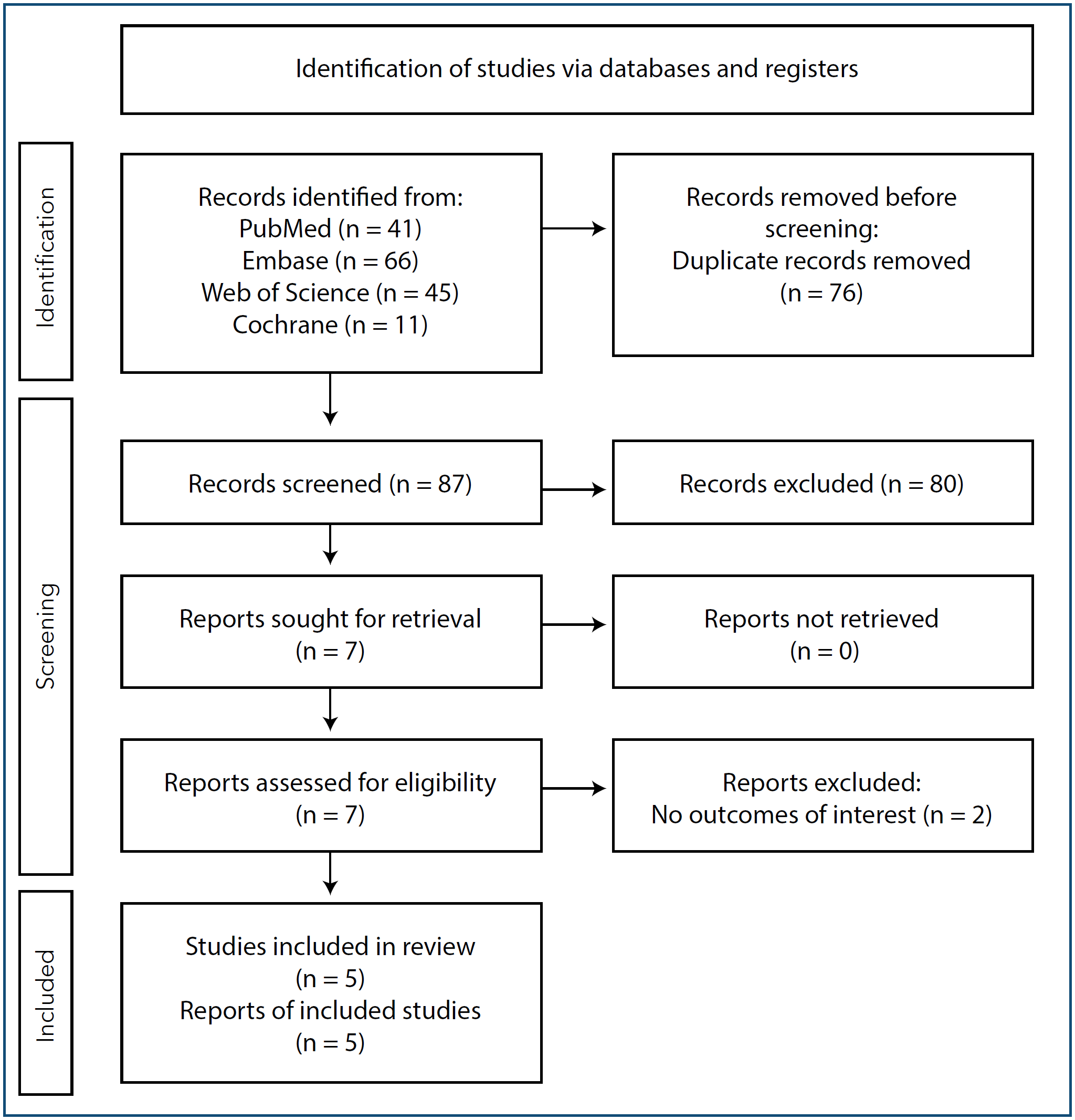
A comprehensive search of the multiple databases yielded a total of 163 articles. After removing duplicates, 87 unique citations were screened. Following a thorough review of titles and abstracts, 80 articles were excluded. Of the remaining articles, seven were selected for full-text review based on their abstracts. Subsequently, another two articles were excluded after the full-text screening and data extraction process. Finally, five of seven studies met the inclusion criteria and were included in the final analysis, two RCTs(8,15) and three prospective studies.(5,16,17) A detailed overview of the search process can be found in figure 1.

Baseline characteristics of included studies
A total of 577 eyes (337 assigned to the phacoemulsification group and 240 assigned to the MSICS group) were pooled from the five studies conducted between 2010 and 2022, with the sample size varying from 42 to 270 across studies. The distribution of patients by sex showed no significant difference, with 274 males (47.4%) and 303 females (52.6%) included in the studies. The duration of follow-up ranged from 6 weeks to 6 months. Table 1 summarizes the main characteristics of patients in each study.
Best-corrected visual acuity
Among the included studies, two publications reported the proportion of patients achieving best-corrected visual acuity (BCVA) of 20/60 or better at the end of follow-up after treatment.(8,16) There was no evidence of heterogeneity (I2 = 0%), and a fixed effect was used. The comparison between the phacoemulsification and MSICS groups did not show a significant difference in the proportion of participants with a BCVA of 20/60 or better after treatment (OR 0.75; CI95% 0.16-3.51; p = 0.71; Figure 2). Similarly, no significant difference was observed between the groups for best corrected visual acuity of less than 20/60.

Endothelial cell count loss
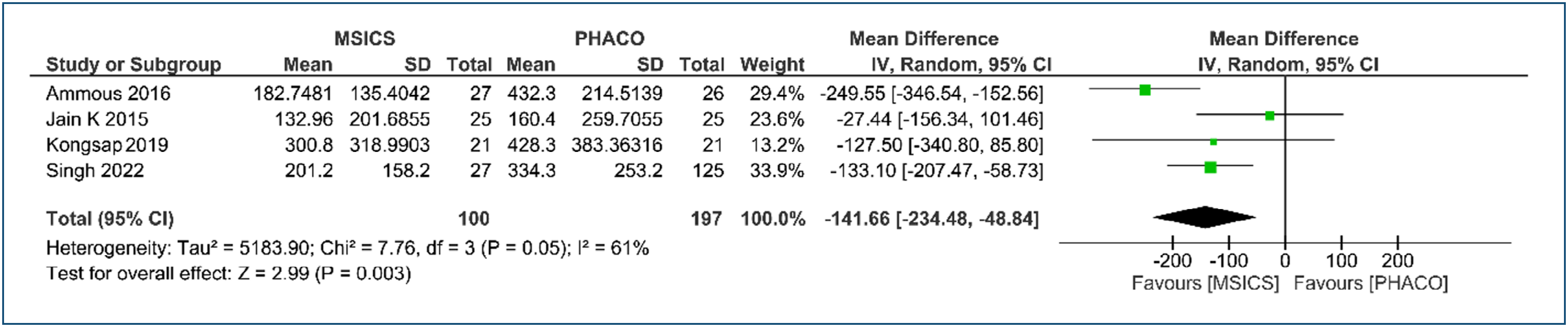
Four of the included studies reported data about mean endothelial cell loss after surgery, and the results showed a significant difference in postoperative endothelial cell loss between the phacoemulsification and MSICS groups (MD −141.66; CI95% -234.48--48.84; p = 0.003; I2=61%; Figure 3).

Central corneal thickness
Among the included studies, three studies(15-17) reported the data about mean CCT after surgery, and the results showed that there was no significant difference in postoperative CCT between the phacoemulsification and MSICS groups (MD -5.77; CI95% −12.05--0.51; p = 0.07; I2 = 0%; Figure 4).

Time of surgery
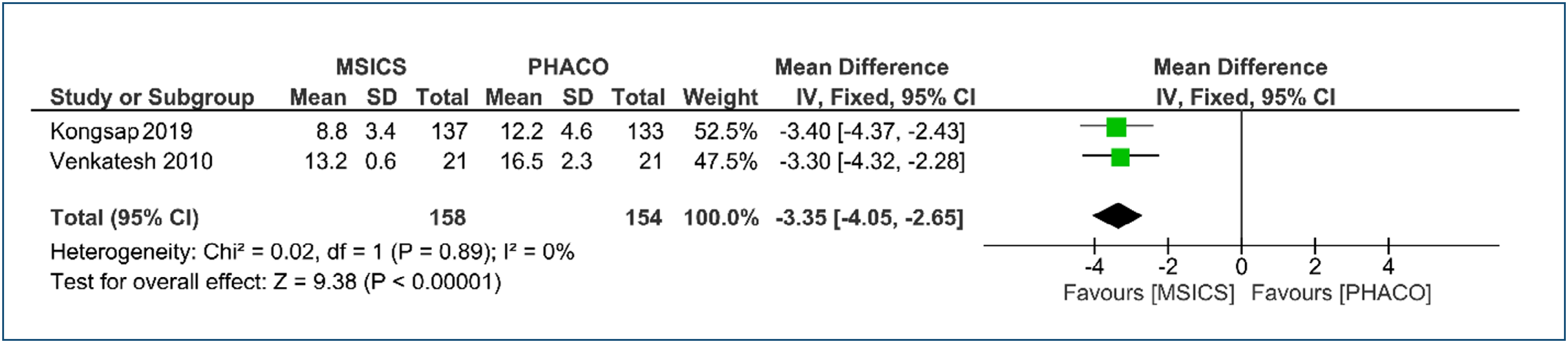
Two studies compared the time of surgery during phacoemulsification and MSICS.(8,16) These data analyses showed that the MSICS group surgery time was significantly smaller than phaco group surgery (MD -3.35; CI95% -4.05--2.65; p < 0.00001; I2 = 0%; Figure 5).

Surgically induced stigmatism
One study compared surgically induced astigmatism after phacoemulsification and MSICS in 270 eyes, finding no statistically significant difference.8
Complications
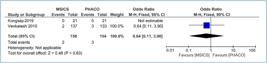
Out of all the analyzed studies, three excluded patients who experienced surgical complications during or after the surgery.(5,15,17) On the other hand, two studies reported certain complications, such as posterior capsule rupture during the surgery and dropping of the nucleus after the surgery.(8,16) However, there was no significant difference observed between the two groups for intraoperative complications (represented by posterior capsule rupture) (OR 0.64; 95%CI 0.11-3.90; p = 0.63; Figure 6) or postoperative complications (OR 0.32; 95%CI 0.01-8.26; p = 0.49; Figure 7). Venkatesh et al. found that the incidence of corneal edema was lower in the MSICS group (10.2%) compared to the phacoemulsification group (18.7%).(8)

DISCUSSION
In 1987, Blumenthal et al. were the first to describe the use of an anterior chamber maintainer (ACM) in extracapsular cataract extraction (ECCE) while reducing the incision size. The classic ‘mininuc’ MSICS procedure, as outlined by Blumenthal, involves extensive use of the ACM to perform nearly all steps under positive irrigation pressure. The ACM cannula is self-retained, a side port is created, and a capsulotomy is executed. The scleral tunnel incision is made, and the hydro procedures are initiated. The nucleus is maneuvered out of the eye using a glide, aided by the positive pressure generated by the ACM. Cortex aspiration is carried out through a side port aspirating cannula, while the ACM ensures continuous irrigation. The ACM is only removed after the IOL has been inserted and the incision's integrity is confirmed to be watertight.(2,18)
Another significant modification to the MSICS technique was introduced later by Ruit et al. They created a 6.5- to 7-mm temporal scleral tunnel with a straight incision, starting 2 mm posterior to the limbus. A side-port incision was made to facilitate further intraocular manipulation. A V-shaped capsulotomy and hydro dissection were performed. Viscoelastic material was injected above and behind the nucleus, subsequently prolapsing into the anterior chamber. An irrigating Simcoe cannula with a serrated surface was inserted below the nucleus, facilitating its extraction through the scleral tunnel. The remaining cortex was manually removed using the same Simcoe irrigation-aspiration cannula. After implanting a polymethyl methacrylate (PMMA) lens into the capsular bag, the unsutured scleral pocket incision was confirmed to be watertight.(2,19)
As described in the literature, substantial modifications to the MSICS technique include various aspects, such as changes to the incision procedure and advancements in nucleus delivery methods.
Regarding incision variations, Kratz was the first surgeon to move the cataract incision posteriorly from the limbus to the sclera, aiming to enhance wound healing and reduce astigmatism.(2) Subsequently, Girard et al. coined the term ‘scleral tunnel’ incision.(20) Singer described the ‘frown incision,’ which involved a modified scleral pocket incision curved in opposition to the natural limbal contour. This frown-shaped configuration was designed to minimize wound-induced astigmatism.(21)
Lam et al. developed the sutureless large-incision manual cataract extraction technique, a modification of the manual ECCE technique. This approach was specifically crafted to enable less experienced surgeons in developing countries to safely extract the nucleus through a self-sealing temporal scleral pocket incision. Notable features of this technique include a medium to large scleral pocket incision (8-mm linear length) for safe and efficient nucleus expression, a long sclerocorneal tunnel (4 mm) for a self-sealing, sutureless wound, a posterior incision location (2 mm posterior to the limbus), and a frown-shaped wound configuration designed for astigmatic neutrality. The use of an ACM facilitates the delivery of the nucleus.(22)
MSICS employs diverse nucleus delivery methods, including hyperexpression and vis coexpression,(23) the sandwich technique,(24) the modified fish hook technique,(25) the use of an ACM,(18) an irrigating cannula,(26) nucleus trisection,(27) nuclear management by snare technique,(28) and the Sinskey hook method.(29)
On the other hand, phacoemulsification was first developed by Charles Kelman in the late 1960s. This technique utilizes ultrasonic energy to vibrate a titanium needle at high frequencies, fragmenting the rigid lens nucleus. The resulting emulsate is simultaneously aspirated from the eye.(30)
One of the primary advantages of phacoemulsification over purely manual methods is its ability to extract a large nucleus through a small incision of ≤ 3.0 mm. Subsequently, foldable IOLs are implanted through this small incision, which generally can be left unsutured. Advances in technology now allow surgery through mini incisions of 2.2 mm or micro-incisions of ≤1.8 mm. The benefits of small incisions are numerous: topical anesthesia can be used, especially if the incision is made in the peripheral cornea; the surgeon gains better control of the intraocular environment, ensuring greater safety in case the patient moves; structural integrity of the incision is quickly reestablished, requiring fewer physical restrictions after surgery; and smaller incisions minimize alterations of the corneal shape, reducing the risk of astigmatism.(1)
Phacoemulsification has been considered the best surgical technique for cataract surgery in developed countries for over two decades. This technique has several advantages such as reducing astigmatism, enabling faster recovery, and allowing for smaller incisions.
However, the effectiveness of MSICS versus phacoemulsification for mature cataracts remains unclear. When determining the optimal surgical technique for cataract surgery, several factors must be considered, including improvement in BCVA, surgical complications, CCT, and surgical duration. These factors are influenced by individual characteristics, including the size and chronicity of the cataract. This systematic review compares MSICS and phacoemulsification as surgical techniques for cataract surgery. By analyzing and evaluating this approach, a better understanding of the advantages and disadvantages of each technique can be achieved, leading to a more comprehensive discussion of the various options available for cataract surgery.
We conducted a systematic review and meta-analysis of 5 studies and 577 eyes to compare the effectiveness of MSICS and phacoemulsification for mature cataract surgery. We found that the mean age of patients in both groups was similar, with the majority being 55 years or older when the research studies began. The results showed that the MSICS group had lower rates of ECL during surgery and a shorter surgery time compared to the phacoemulsification group. However, there was no statistically significant difference in BCVA, CCT, and complications between the two groups.
The analysis showed no significant difference between groups in BCVA better than 20/60 in the postoperative period, consistent with results from three randomized, prospective studies conducted in developing countries comparing phacoemulsification with MSICS. These studies demonstrated that MSICS is comparably effective to phacoemulsification, achieving excellent visual outcomes. These findings indicate that both techniques yielded similar BCVA outcomes in the postoperative period.(2-4,8)
The analysis indicates a significant difference between the groups with a better outcome for ECL in the MSICS group. This finding may be related to the absence of ultrasonic energy and reduced intraocular turbulence during MSICS. However, this outcome differs from previous studies that focused on non-restrictive types of cataracts and included only softer grades or other types of cataracts, resulting in negligible corneal endothelial alteration.(31-33)
Nevertheless, the CCT remained stable in both groups. This suggests that corneal endothelial function was unaffected. This supports the notion that both MSICS and phacoemulsification, with proper technique and viscoelastic use, did not cause significant harm to the endothelium, which could have resulted in postoperative corneal edema due to a marked impairment of the corneal endothelial pump. Our study's findings align with previous research that noted a return to normal CCT values in the postoperative period.(15,16,34)
Notably, the analysis revealed a more favorable outcome of surgery time for the group of MISCS, as indicated by the MD, when compared to the phacoemulsification group. According to the literature, MSICS has the potential of achieving outstanding results with lower costs and shorter surgical times when compared to phacoemulsification. In addition to being fast and affordable, MSICS is easier for less experienced surgeons to learn and is considered safer for managing advanced mature cataracts.(1,2,35-37)
When considering complications, our findings demonstrated that the overall rates of complications were not significantly different between the two groups. This is reassuring, as it suggests that the use of MSICS or phacoemulsification does not inherently introduce a greater risk of complications, as previously observed in other studies. Notably, the incidence of a severe complication, such as dropped nuclei, which involves the dislocation of the nucleus onto the retina, is rare in MSICS. Thus, both MSICS and phacoemulsification are deemed safe procedures.(1,2)
According to previous studies, the incidence of anterior chamber contamination is similar between eyes undergoing MSICS and phacoemulsification.(38) Furthermore, both phacoemulsification and MSICS resulted in significant and similar reductions in intraocular pressure (IOP) 6 months after surgery. Additionally, both surgeries produced similar changes in anterior chamber and angle parameters.(39)
Only one study reported surgically induced astigmatism, suggesting better results in the phacoemulsification group.(8) Similarly, many studies conclude that phacoemulsification reduces surgically induced astigmatism at a 6-week follow-up compared to MSICS for all types of cataract.(3,8,33,40,41)
In a study by Gokhale and Sawhney, they compared surgically induced astigmatism with various scleral incision locations for MSICS. Their findings revealed that surgically induced astigmatism was lower with temporal and superotemporal incisions than with superior incisions.(42)
It is important to note that our analysis shows a low level of variation, with an I2 value of 0% in most of the analysis, except for the ECL that utilized random-effect models. This indicates a significant level of consistency among the studies included in the analysis.
Furthermore, patients with pseudoexfoliation syndrome have a high prevalence of mature cataracts. Naik et al. reported that, with careful pre-operative assessment, intraoperative modifications, and surgical expertise, both PHACO and MSICS appear to be safe procedures for those with pseudoexfoliation syndrome.(43) However, according to Subudhi et al., in moderately hard nuclear cataracts with pseudoexfoliation syndrome grades II and III (LOCS II grading system), phacoemulsification provides better unaided visual outcomes with less endothelial dysfunction than MSICS in the immediate and subsequent postoperative periods.(44)
The stage of cataract in a population significantly affects surgical outcomes. Along with CCT and ECL, BCVA and complications are also important factors. Advanced and complicated cataracts are more common among impoverished populations. As a result, the MSICS technique emerges as a more fitting option for developing nations due to its minimal capital and consumable expenses, particularly regarding fluid and tubing. This makes MSICS an economically viable choice, addressing the resource constraints often faced by developing countries in comparison to phacoemulsification surgery, which requires expensive equipment. The cost-effectiveness of MSICS renders it a pragmatic and sustainable solution for cataract surgery in these regions, ensuring that essential eye care services can be efficiently provided without straining limited financial resources.(45-51)
Limitations
Although the results provided significant insights into the safety and efficacy of MISCS as an alternative to phacoemulsification for treating mature cataracts, it is crucial to consider the limitations of this study. Our analysis included a relatively small number of studies and patients due to the limited research on phacoemulsification versus MSICS in mature cataracts. The definition of mature cataracts varies significantly among studies, leading to a lack of consensus. The use of random-effects models was necessary due to the variability in endothelial cell loss, indicating differences in treatment responses across studies. This variability could be influenced by factors such as variations in surgical protocols, patient populations, and study methodologies. Many studies included in this analysis reported follow-up periods of ≥ 1.5 months. However, long-term outcomes beyond the follow-up period were not assessed. It is crucial to evaluate the long-term stability of surgical effects. The analysis of intraoperative complications was somewhat limited, as some studies only included patients without complications. Additionally, mean surgically induced astigmatism and IOP-related events were not reported in the studies, and only one study provided the average pre- and postoperative BCVA analysis data. The included studies were conducted in three countries, with three in India, which could introduce variations in patient demographics, surgical techniques, and follow-up protocols, thereby limiting the generalizability of the findings to a broader population. Furthermore, there is a potential for publication bias, as studies with positive or significant outcomes are more likely to be published, while those with negative or non-significant findings may not be. The meta-analysis relied on aggregated data from published studies, and individual patient data was not available for analysis, limiting a more detailed understanding of the treatment effects and predictors of response.
CONCLUSIONS
In conclusion, we found manual small incision cataract surgery to be a safe and effective alternative to phacoemulsification for mature cataracts, showing no noteworthy disparities in complications or outcomes related to best-corrected visual acuity and central corneal thickness, while also demonstrating a reduction in endothelial cell loss. Manual small incision cataract surgery has a notable advantage because it does not rely on advanced technology, making it a faster and more affordable surgical choice. These characteristics position manual small incision cataract surgery as a viable procedure, particularly for mature cataracts and challenging cases, including brunescent cataracts, white cataracts, and cataracts causing phacolytic and phacomorphic glaucoma, and in regions where access to high-cost phacoemulsification techniques remains limited.
Data Availability Statement:
The datasets generated and/or analyzed during the current study are included in the manuscript.
REFERENCES
- 1 Lam D, Rao SK, Ratra V, Liu Y, Mitchell P, King J, Tassignon MJ, Jonas J, Pang CP, Chang DF. Cataract. Nat Rev Dis Primers. 2015;1:15014.
- 2 Venkatesh R, Chang DF, Muralikrishnan R, Hemal K, Gogate P, Sengupta S. Manual small incision cataract surgery: a review. Asia Pac J Ophthalmol (Phila). 2012;1(2):113-9.
- 3 Gogate PM, Kulkarni SR, Krishnaiah S, Deshpande RD, Joshi SA, Palimkar A, et al. Safety and efficacy of phacoemulsification compared with manual small-incision cataract surgery by a randomized controlled clinical trial: six-week results. Ophthalmology. 2005 May;112(5):869-74.
- 4 Ruit S, Tabin G, Chang D, Bajracharya L, Kline DC, Richheimer W, et al. A prospective randomized clinical trial of phacoemulsification vs manual sutureless small-incision extracapsular cataract surgery in Nepal. Am J Ophthalmol. 2007;143(1):32-38.
- 5 Ammous I, Bouayed E, Mabrouk S, Boukari M, Erraies K, Zhioua R. [Phacoemulsification versus manual small incision cataract surgery: Anatomic and functional results]. J Fr Ophtalmol. 2017;40(6):460-6. French.
- 6 van Zyl L, Kahawita S, Goggin M. Manual small incision extracapsular cataract surgery in Australia. Clin Exp Ophthalmol. 2014;42(8):729-33.
- 7 Bernhisel A, Pettey J. Manual small incision cataract surgery. Curr Opin Ophthalmol. 2020;31(1):74-9.
- 8 Venkatesh R, Tan CS, Sengupta S, Ravindran RD, Krishnan KT, Chang DF. Phacoemulsification versus manual small-incision cataract surgery for white cataract. J Cataract Refract Surg. 2010;36(11):1849-54.
- 9 Gogate PM, Deshpande M, Wormald RP, Deshpande R, Kulkarni SR. Extracapsular cataract surgery compared with manual small incision cataract surgery in community eye care setting in western India: a randomised controlled trial. Br J Ophthalmol. Jun 2003;87(6):667-72.
- 10 Venkatesh R, Das M, Prashanth S, Muralikrishnan R. Manual small incision cataract surgery in eyes with white cataracts. Indian J Ophthalmol. 2005;53(3):173-6.
- 11 Venkatesh R, Tan CS, Kumar TT, Ravindran RD. Safety and efficacy of manual small incision cataract surgery for phacolytic glaucoma. Br J Ophthalmol. 2007;91(3):279-81.
- 12 Ramakrishanan R, Maheshwari D, Kader MA, Singh R, Pawar N, Bharathi MJ. Visual prognosis, intraocular pressure control and complications in phacomorphic glaucoma following manual small incision cataract surgery. Indian J Ophthalmol. 2010;58(4):303-6.
- 13 Shamseer L, Moher D, Clarke M, Ghersi D, Liberati A, Petticrew M, et al. Preferred reporting items for systematic review and meta-analysis protocols (PRISMA-P) 2015: elaboration and explanation. BMJ. 2015;350:g7647.
- 14 Page MJ, McKenzie JE, Bossuyt PM, Boutron I, Hoffmann TC, Mulrow CD, Shamseer L, et al. The PRISMA 2020 statement: an updated guideline for reporting systematic reviews. Syst Rev. 2021;10(1):89.
- 15 Singh R, Sharma AK, Katiyar V, Kumar G, Gupta SK. Corneal endothelial changes following cataract surgery in hard nuclear cataract: Randomized trial comparing phacoemulsification to manual small-incision cataract surgery. Indian J Ophthalmol. 2022;70(11):3904-9.
- 16 Kongsap P. Central corneal thickness changes following manual small incision cataract surgery versus phacoemulsification for white cataract. Rom J Ophthalmol. 2019;63(1):61-7.
- 17 Jain K, Mallik KP, Gupta S. Corneal status following modified Blumenthal technique of manual small incision cataract surgery (MSICS) compared to phacoemulsification in treatment of grade III or more nuclear sclerosis-cohort study. Nepal J Ophthalmol. 2015;7(1):47-51.
- 18 Blumenthal M, Moisseiev J. Anterior chamber maintainer for extracapsular cataract extraction and intraocular lens implantation. J Cataract Refract Surg. 1987;13(2):204-6.
- 19 Ruit S, Paudyal G, Gurung R, Tabin G, Moran D, Brian G. An innovation in developing world cataract surgery: sutureless extracapsular cataract extraction with intraocular lens implantation. Clin Exp Ophthalmol. 2000;28(4):274-9.
- 20 Girard LJ, Rodriguez J, Mailman ML. Reducing surgically induced astigmatism by using a scleral tunnel. Am J Ophthalmol. 1984;97(4):450-6.
- 21 Singer JA. Frown incision for minimizing induced astigmatism after small incision cataract surgery with rigid optic intraocular lens implantation. J Cataract Refract Surg. 1991;17 Suppl:677-88.
- 22 Lam DS, Rao SK, Fan AH, Congdon NG, Wong V, Liu Y, et al. Endothelial cell loss and surgically induced astigmatism after sutureless large-incision manual cataract extraction (SLIMCE). Arch Ophthalmol. 2009;127(10):1284-9.
- 23 Corydon L, Thim K. Continuous circular capsulorhexis and nucleus delivery in planned extracapsular cataract extraction. Journal of Cataract & Refractive Surgery. 1991;17(5):628-32.
- 24 Bayramlar H, Çekiç O, Totan Y. Manual tunnel incision extracapsular cataract extraction using the sandwich technique. Journal of Cataract & Refractive Surgery. 1999;25(3):312-315.
- 25 Hennig A, Kumar J, Yorston D, Foster A. Sutureless cataract surgery with nucleus extraction: outcome of a prospective study in Nepal. Br J Ophthalmol. 2003;87(3):266-70.
- 26 Nishi O. A new type of irrigating cannula for lens nucleus delivery by extracapsular extraction. Ophthalmic Surg. 1986;17(1):47-9.
- 27 Kansas PG, Sax R. Small incision cataract extraction and implantation surgery using a manual phacofragmentation technique. J Cataract Refract Surg. 1988;14(3):328-30.
- 28 Keener GT. The nucleus division technique for small incision cataract extraction. In: Rozakis GW, editor. Cataract surgery: Alternative small incision Techniques. New Delhi: Jaypee Brothers; 1995. p. 163–91.
- 29 Rao SK, Lam DS. A simple technique for nucleus extraction from the capsular bag in manual small incision cataract surgery. Indian J Ophthalmol. 2005;53(3):214-5.
- 30 Linebarger EJ, Hardten DR, Shah GK, Lindstrom RL. Phacoemulsification and modern cataract surgery. Surv Ophthalmol. 1999;44(2):123-47.
- 31 Zhang JY, Feng YF, Cai JQ. Phacoemulsification versus manual small-incision cataract surgery for age-related cataract: meta-analysis of randomized controlled trials. Clin Exp Ophthalmol. 2013;41(4):379-86.
- 32 Bhargava R, Sharma SK, Chandra M, Kumar P, Arora Y. Comparison of endothelial cell loss and complications between phacoemulsification and manual small incision cataract surgery (SICS) in uveitic cataract. Nepal J Ophthalmol. 2015;7(14):124-34.
- 33 George R, Rupauliha P, Sripriya AV, Rajesh PS, Vahan PV, Praveen S. Comparison of endothelial cell loss and surgically induced astigmatism following conventional extracapsular cataract surgery, manual small-incision surgery and phacoemulsification. Ophthalmic Epidemiol. 2005;12(5):293-7.
- 34 Ventura AC, Wälti R, Böhnke M. Corneal thickness and endothelial density before and after cataract surgery. Br J Ophthalmol. 2001;85(1):18-20.
- 35 Bali J, Bali O, Sahu A, Boramani J, Deori N. Health economics and manual small-incision cataract surgery: An illustrative mini review. Indian J Ophthalmol. 2022;70(11):3765-770.
- 36 Muralikrishnan R, Venkatesh R, Prajna NV, Frick KD. Economic cost of cataract surgery procedures in an established eye care centre in Southern India. Ophthalmic Epidemiol. 2004;11(5):369-80.
- 37 Khan A, Amitava AK, Rizvi SA, Siddiqui Z, Kumari N, Grover S. Cost-effectiveness analysis should continually assess competing health care options especially in high volume environments like cataract surgery. Indian J Ophthalmol. 2015;63(6):496-500.
- 38 Parmar P, Salman A, Kaliamurthy J, Prasanth DA, Thomas PA, Jesudasan CA. Anterior chamber contamination during phacoemulsification and manual small-incision cataract surgery. Am J Ophthalmol. 2006;141(6):1160-1.
- 39 Sengupta S, Venkatesh R, Krishnamurthy P, Nath M, Mashruwala A, Ramulu PY, et al. Intraocular Pressure Reduction after Phacoemulsification versus Manual Small-Incision Cataract Surgery: A Randomized Controlled Trial. Ophthalmology. 2016;123(8):1695-703.
- 40 Kimura H, Kuroda S, Mizoguchi N, Terauchi H, Matsumura M, Nagata M. Extracapsular cataract extraction with a sutureless incision for dense cataracts. J Cataract Refract Surg. Sep 1999;25(9):1275-9.
- 41 Singh K, Misbah A, Saluja P, Singh AK. Review of manual small-incision cataract surgery. Indian J Ophthalmol. 2017;65(12):1281-8.
- 42 Gokhale NS, Sawhney S. Reduction in astigmatism in manual small incision cataract surgery through change of incision site. Indian J Ophthalmol. 2005;53(3):201-3. d
- 43 Naik AU, Gadewar SB. Visual outcome of phacoemulsification versus small incision cataract surgery in pseudoexfoliation syndrome - a pilot study. J Clin Diagn Res. 2017;11(1):NC05-NC08.
- 44 Subudhi P, Khan Z, Subudhi BN, Sitaram S, Patro S. Phacoemulsification vs manual small incision cataract surgery in eyes affected by pseudo exfoliation syndrome with grade II and III cataracts. Saudi J Ophthalmol. 2020;34(1):18-24. d
- 45 Jaggernath J, Gogate P, Moodley V, Naidoo KS. Comparison of cataract surgery techniques: safety, efficacy, and cost-effectiveness. Eur J Ophthalmol. 2014;24(4):520-6.
- 46 Jongsareejit A, Wiriyaluppa C, Kongsap P, Phumipan S. Cost-effectiveness analysis of manual small incision cataract surgery (MSICS) and phacoemulsification (PE). J Med Assoc Thai. 2012;95(2):212-20.
-
47 Amaral DC, Cheidde L, Ferreira BFA, Cheidde L, Júnior PPL, Menezes I, et al. Cataract in HIV patients: a systematic review and meta-analysis. Cureus. 2024;16(10):e72370. doi:10.7759/cureus.72370.
» https://doi.org/10.7759/cureus.72370 -
48 Amaral DC, Magalhães PLM, Sá AG, Neto ABC, de Medeiros FMT, Alves MR, et al. Functional optical balance in cataract surgery: a review. Optics. 2025;6(3):36. doi:10.3390/opt6030036.
» https://doi.org/10.3390/opt6030036 -
49 Barroso FVC, Amaral DC, Pereira SM, Figueiredo ÂSDF, Gadelha JG, Lopes Filho HF, et al. Comparison between Ringer's lactate and balanced salt solution on postoperative outcomes after phacoemulsification: a systematic review and meta-analysis of randomized controlled trials. Einstein (Sao Paulo). 2025;23:eRW1569. doi:10.31744/einstein_journal/2025RW1569.
» https://doi.org/10.31744/einstein_journal/2025RW1569 -
50 Amaral DC, Cheidde L, Mora-Paez DJ, Haddad EH, Magalhães PLM, Nascimento LM, et al. Digital versus manual workflows for cataract surgery: a systematic review and meta-analysis. Einstein (Sao Paulo). 2025;23:eRW1478. doi:10.31744/einstein_journal/2025RW1478.
» https://doi.org/10.31744/einstein_journal/2025RW1478 -
51 Silva LL, Cunha Amaral D, Kunigami da Silva K, Viegas Mendonça VP, Braz LD, Rios-Garcia W, et al. Risk of cataracts in patients using typical and atypical antipsychotics: a systematic review. Expert Rev Ophthalmol. 2026;21(1):41-49. doi:10.1080/17469899.2026.2623680.
» https://doi.org/10.1080/17469899.2026.2623680
Edited by
-
Associate editor:
Ricardo Augusto Paletta Guedes Universidade Federal de Juiz de Fora, Juiz de Fora, MG, Brazil. https://orcid.org/0000-0002-9451-738X